appendix a oblique projection operation - home - springer978-1-4614-1821-4/1.pdf · appendix a...
TRANSCRIPT
Appendix AOblique Projection Operation
This appendix introduces an oblique projection operation, which underpins thesubspace system identification algorithms discussed in Chap. 7. In addition, a fewfigures are drawn to illustrate parts of the subspace identification procedures.
A.1 Methodology
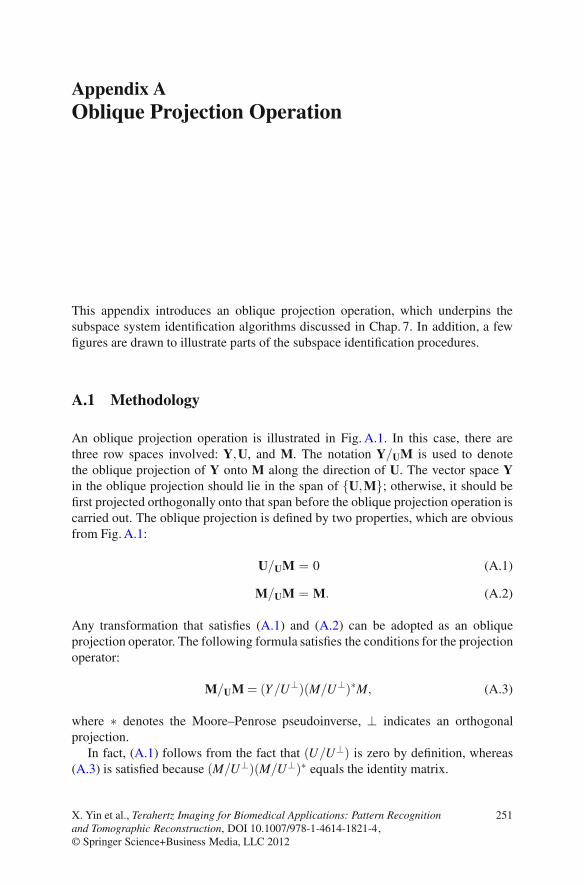
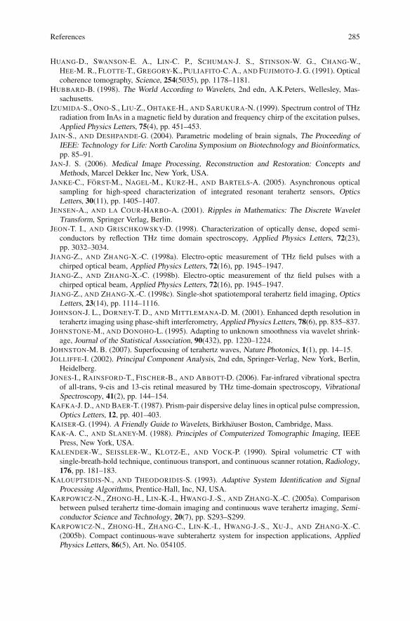
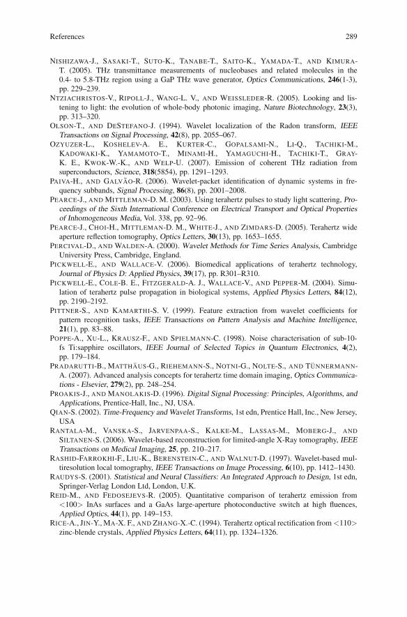
An oblique projection operation is illustrated in Fig. A.1. In this case, there arethree row spaces involved: Y,U, and M. The notation Y/UM is used to denotethe oblique projection of Y onto M along the direction of U. The vector space Yin the oblique projection should lie in the span of {U,M}; otherwise, it should befirst projected orthogonally onto that span before the oblique projection operation iscarried out. The oblique projection is defined by two properties, which are obviousfrom Fig. A.1:
U/UM = 0 (A.1)
M/UM = M. (A.2)
Any transformation that satisfies (A.1) and (A.2) can be adopted as an obliqueprojection operator. The following formula satisfies the conditions for the projectionoperator:
M/UM = (Y/U⊥)(M/U⊥)∗M, (A.3)
where ∗ denotes the Moore–Penrose pseudoinverse, ⊥ indicates an orthogonalprojection.
In fact, (A.1) follows from the fact that (U/U⊥) is zero by definition, whereas(A.3) is satisfied because (M/U⊥)(M/U⊥)∗ equals the identity matrix.
X. Yin et al., Terahertz Imaging for Biomedical Applications: Pattern Recognitionand Tomographic Reconstruction, DOI 10.1007/978-1-4614-1821-4,© Springer Science+Business Media, LLC 2012
251
252 A Oblique Projection Operation
Y
U
Y/U M M
Fig. A.1 Schematic ofoblique projection. Obliqueprojection of Y onto M alongthe direction of U
WiUf
Oi = Xf
Yf
Wp
Wi-1Uf-
Oi-1 = i Xi+1
Yf-
Wp+
a b
i
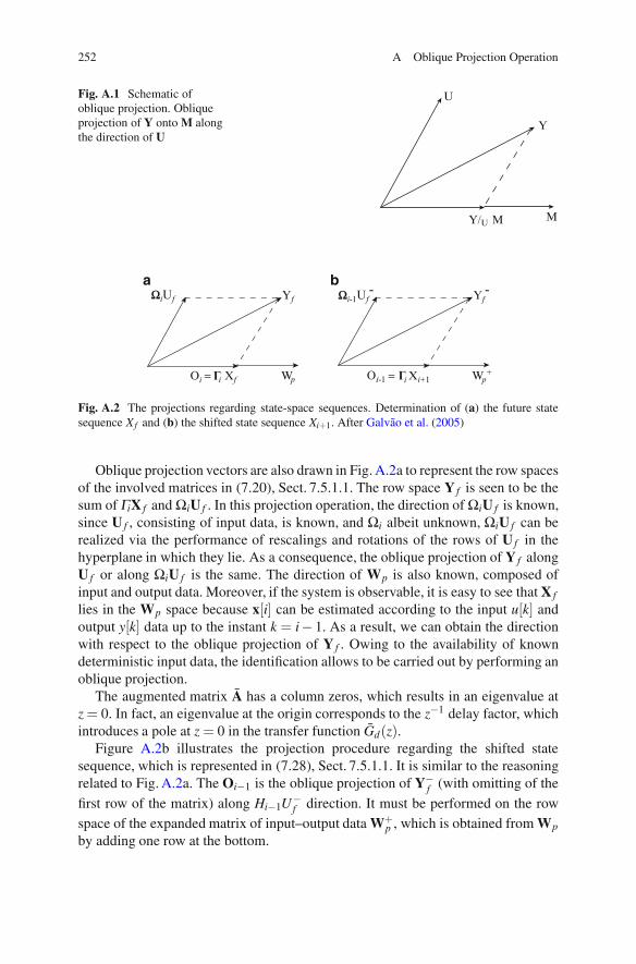
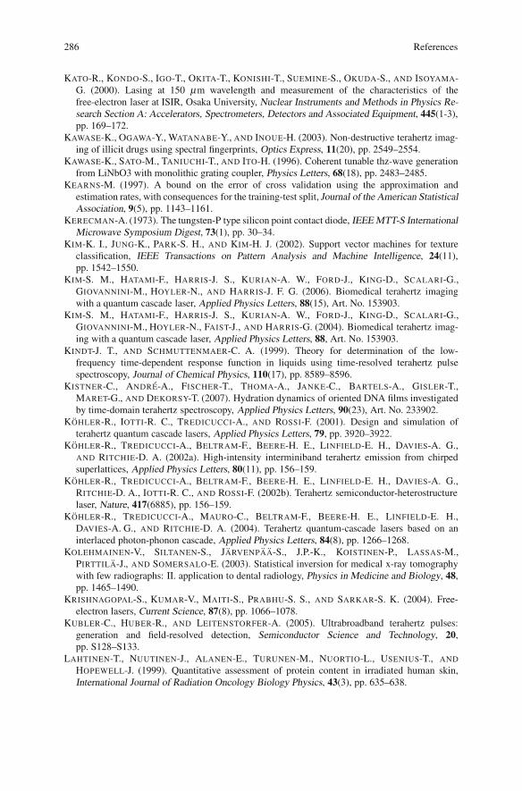
Fig. A.2 The projections regarding state-space sequences. Determination of (a) the future statesequence Xf and (b) the shifted state sequence Xi+1. After Galvao et al. (2005)
Oblique projection vectors are also drawn in Fig. A.2a to represent the row spacesof the involved matrices in (7.20), Sect. 7.5.1.1. The row space Y f is seen to be thesum of ΓiX f and ΩiU f . In this projection operation, the direction of ΩiU f is known,since U f , consisting of input data, is known, and Ωi albeit unknown, ΩiU f can berealized via the performance of rescalings and rotations of the rows of U f in thehyperplane in which they lie. As a consequence, the oblique projection of Y f alongU f or along ΩiU f is the same. The direction of Wp is also known, composed ofinput and output data. Moreover, if the system is observable, it is easy to see that X f
lies in the Wp space because x[i] can be estimated according to the input u[k] andoutput y[k] data up to the instant k = i− 1. As a result, we can obtain the directionwith respect to the oblique projection of Y f . Owing to the availability of knowndeterministic input data, the identification allows to be carried out by performing anoblique projection.
The augmented matrix A has a column zeros, which results in an eigenvalue atz = 0. In fact, an eigenvalue at the origin corresponds to the z−1 delay factor, whichintroduces a pole at z = 0 in the transfer function Gd(z).
Figure A.2b illustrates the projection procedure regarding the shifted statesequence, which is represented in (7.28), Sect. 7.5.1.1. It is similar to the reasoningrelated to Fig. A.2a. The Oi−1 is the oblique projection of Y−
f (with omitting of the
first row of the matrix) along Hi−1U−f direction. It must be performed on the row
space of the expanded matrix of input–output data W+p , which is obtained from Wp
by adding one row at the bottom.
Appendix BBack-Projection Algorithms
This appendix provides further details about back-projection algorithms. This is aspecific supplement made for Chap. 10 in respect of CT reconstruction.
B.1 Theory
The back projection is represented via parallel beam projections. Recalling theformula for the inverse Fourier transform, the object function, f (x,y), can beexpressed as
f (x,y) =∫ ∞
−∞
∫ ∞
−∞F(u,v)ej2π(ux+vy)dudv. (B.1)
Exchanging the rectangular coordinate system in the frequency domain, (u,v), for apolar coordinate system, (w,θ ), by making the substitutions
u = wcosθ (B.2)
v = wsin θ (B.3)
and changing the differentials by using
dudv = wdwdθ , (B.4)
we can write the inverse Fourier transform of a polar function as
f (x,y) =∫ 2π
0
∫ ∞
0F(w,θ )ej2πw(xcosθ+ysinθ)wdwdθ . (B.5)
X. Yin et al., Terahertz Imaging for Biomedical Applications: Pattern Recognitionand Tomographic Reconstruction, DOI 10.1007/978-1-4614-1821-4,© Springer Science+Business Media, LLC 2012
253
254 B Back-Projection Algorithms
This integral can be split into two by considering θ from 0◦ to 180◦ and then from180◦ to 360◦,
f (x,y) =∫ π
0
∫ ∞
0F(w,θ )ej2πw(xcosθ+ysinθ)wdwdθ
+
∫ π
0
∫ ∞
0F(w,θ )ej2πw[xcos(θ+180◦)+ysin(θ+180◦)]wdwdθ (B.6)
and then using the property
F(w,θ + 180◦) = F(−w,θ ) (B.7)
the above expression for f (x,y) may be written as
f (x,y) =∫ π
0
[∫ ∞
−∞F(w,θ )|w|ej2πwtdw
]dθ . (B.8)
Here, we have simplified the expression by setting
t = xcosθ + ysinθ . (B.9)
If we substitute the Fourier transform of the projection at angle θ , sθ (w), for thetwo-dimensioinal Fourier transform F(w,θ ), we get
f (x,y) =∫ π
0
[∫ ∞
−∞sθ (w)|w|ej2πwt dw
]dθ . (B.10)
This integral in (B.10) may be expressed as
f (x,y) =∫ π
0Qθ (xcosθ + ysinθ )dθ (B.11)
where
Qθ =∫ ∞
−∞Sθ (w)|w|ej2πwtdw. (B.12)
Equation (B.11) represents a filtering operation, where the frequency response of thefilter is given by |w|; therefore, Qθ (w) is called a “filtered projection.” The resultingprojections for different angles θ are then back-projected to form the estimate off (x,y).
We relabel Sθ (w) to S(θ ,β ), t to ξ , then we rewrite (B.11) to yield,
I(x,y) =∫ π
0
[∫ ∞
−∞S(θ ,β )|β |exp[i2πβ ξ ]dβ
]dθ , (B.13)
where (B.13) is the same as (10.2) that we use in Sect. 10.2 for THz reconstruction.
Appendix CError Analysis Regarding Wavelet-BasedLocal Reconstruction
This appendix provides further details about error analysis with respect towavelet-based local reconstruction. This is a specific supplement made forChap. 12 to validate local CT via wavelet transforms.
C.1 Methodology
Radon transform error is not negligible because of the nonlocal property of thederivative Hilbert transform (the impulse response of the filter |β |). In this case,even a small local ROI can be reconstructed by considering some data outside theROI for a negligible reconstruction error. In terms of the amount of nonlocal dataapplied in the reconstruction, an upper bound of the reconstruction error can becalculated. The comparison is made between the wavelet-based reconstruction andthe traditional reconstruction algorithm for the local tomography image recovery.
In the current algorithm, the ROI and the ROE are assumed to be centered at thecenter of an image. The support of a completed image is a disk of radius R pixelscentered at the origin. Disks of radius ri pixels and re pixels centered at the originare used to denote the ROI and ROE, respectively. Consider (C.5), the traditionalFBP algorithm, which is shifted to the time domain scheme:
Ir(x,y) =∫ π
0s(θ ,ξ )hθ (xcosθ + ysinθ )dθ . (C.1)
The reconstructed function Ir(x,y) is an approximation of the function I(x,y) if hθis the angle dependent impulse response of the ramp filter, θ ∈ [0,2π), and is anapproximation of the wavelet and scaling coefficients if wavelet and scaling filtersare substituted for |β |.
X. Yin et al., Terahertz Imaging for Biomedical Applications: Pattern Recognitionand Tomographic Reconstruction, DOI 10.1007/978-1-4614-1821-4,© Springer Science+Business Media, LLC 2012
255
256 C Error Analysis Regarding Wavelet-Based Local Reconstruction
The discrete version is expressed as follows:
Ir(x,y) =πk
k
∑k=1
1R
R
∑n=−R
sθk (n)hθk(m− n), (C.2)
where m = (xcosθ + ysinθ ) ∈ ROE, K is the number of the measured projectionangles, sθk indicates the projection at kth angle and θk = k(π/k). The completedimage based on global data consists of two parts: the ROE and its complementROE:
Ir(x,y) =πk
k
∑k=1
1R ∑
|n|≤re
sθk(n)hθk(m−n)+πk
k
∑k=1
1R ∑
|n|>re
sθk (n)hθk(m−n). (C.3)
Therefore, the magnitude of error regarding the ROE can be calculated asfollows:
|ε(x,y)| =∣∣∣∣∣πk
k
∑k=1
1R ∑
|n|>re
sθk(n)hθk(m− n)
∣∣∣∣∣. (C.4)
The Cauchy–Schwartz inequality is used to achieve an upper bound of the errorcalculation:
|ε(x,y)| =
∣∣∣∣∣πk
k
∑k=1
1R ∑
|n|>re
sθk(n)hθk(m− n)
∣∣∣∣∣
≤ πk
k
∑k=1
1R ∑
|n|>re
|sθk (n)hθk(m− n)|
≤ πk
k
∑k=1
1R
(
∑|n|>re
|sθk (n)|2)1/2(
∑|n|>re
|hθk(m− n)|2)1/2
. (C.5)
There exists such an approximation that |sθk | ≤ 2max |I(x,y)|:
|ε(x,y)| = 2√
2πk
max |I(x,y)|√
R− re
R·
K
∑k=1
(
∑|n|>re
|hθk(m− n)|2)1/2
. (C.6)
The relative error is defined as
|εrel(x,y)| = |ε(x,y)|/max |I(x,y)|
=2√
2πk
√R− re
R·
K
∑k=1
(
∑|n|>re
|hθk(m− n)|2)1/2
. (C.7)

C.1 Methodology 257
It is observed that after applying wavelet-based ramp filter, the reconstructedintensity of an terahertz image is much higher than traditional ramp-filtered-based reconstruction. Thus in the calculation of relative error, normalization of thereconstructed local images is important for comparison between the global and localreconstruction. A normalization scaling factor is calculated via dividing maximumintensity of global reconstruction, denoted by I(x,y) by maximum intensity of localreconstruction, denoted by Ilocal(x,y). The function is as follows:
NIg,l = max |I(x,y)|/max |Ilocal(x,y)|. (C.8)
Combine (C.7) and (C.8), and then we achieve a relative error calculationequation in terahertz image reconstruction:
|εrel(x,y)|= NIg,l ·2√
2πk
√R− re
R·
K
∑k=1
(
∑|n|>re
|hθk(m− n)|2)1/2
. (C.9)
The ROI can be viewed as a point is its worst case. The function above can bedescribed as
|εrel(x,y)|= NIg,l2√
2πk
√R− re
R·
K
∑k=1
(
∑|n|>re−ri
|hθk(n)|2)1/2
. (C.10)
The truncated filter is defined as:
hTθk(n)
{hθk(n) if |n|< re − ri,
0 otherwise .(C.11)
Hence,
|εrel|= NIg,l ·2√
2πk
√R− re
R·
K
∑k=1
(R
∑|n|=−R
|hθk(n)− hTθk(n)|2
)1/2
(C.12)
The inner sum can be described in the frequency domain:
|εrel(x,y)| = NIg,l ·2√
2πk
√R− re
R
·K
∑k=1
(R
∑|n|=−R
|{[Hθk(l)−HTθk(l)]exp[i2πβ ξ ]}|2
)1/2
(C.13)
where Hθk and HTθk
are the Fourier transform of hθk and hTθk
, respectively. Forcalculation of upper bound for the error in standard FBP algorithm, hθk in (C.9)
258 C Error Analysis Regarding Wavelet-Based Local Reconstruction
is replaced by the ramp filter |β |. The upper bound of the relative error in thereconstructed image of wavelet and scaling coefficients can be obtained by replacingHθk in (C.9) with in (C.14) and via multiplication by a normalization factor:
⎧⎪⎪⎪⎪⎪⎪⎨
⎪⎪⎪⎪⎪⎪⎩
HCθ = |β |Φ2 j (β cosθ ,β sinθ ) = |β |Φ2 j (β cosθ )Φ2 j (β sinθ )
HDH
θ = |β |Ψh2 j (β cosθ ,β sinθ ) = |β |Φ2 j (β cosθ )Ψ2 j (β sinθ )
HDV
θ = |β |ΦV2 j (β cosθ ,β sinθ ) = |β |Ψ2 j (β cosθ )Φ2 j (β sinθ )
HDD
θ = |β |Ψd2 j(β cosθ ,β sinθ ) = |β |Ψ2 j(β cosθ )Ψ2 j (β sinθ )
(C.14)
where HCθ and HDi
θ ,(i = H,V,D) are called scaling and wavelet ramp filters.The relative error in the reconstruction image from approximate reconstruction
coefficients is as follows:
|εrel(x,y)| = NIg,lC· 2
√2π
k
√R− re
R
·K
∑k=1
(R
∑|n|=−R
∣∣∣{[
HCθk(l)−HCT
θk(l)]
exp [i2πβ ξ ]}∣∣∣
2)1/2
(C.15)
where NIg,lCis the normalized scale factor of an image in relation to approximate
reconstruction coefficients, which is calculated via dividing maximum intensityof global reconstruction, denoted by {I(x,y)}, by maximum intensity of localreconstruction regarding approximate wavelet coefficients, denoted by {IC
local(x,y)}.The normalized scale factor is as follows:
NIg,lC= max |I(x,y)|/max |IC
local(x,y)|. (C.16)
Appendix DTerhertz Imaging Systems
This appendix provides further detail and specifications on the components of boththe pulsed THz and CW terahertz imaging system. The pulsed approach uses aconvetional Ti:sapphire laser and relates to Chap. 2 (Sect. 2.3.1), Chaps. 9, 11 and12. The CW approach uses a THz QCL and relates to Chap. 2 (Sect. 2.5.2) andChap. 13. It provides a list of the major hardware components along with theircritical specifications and purpose.
D.1 Ultrafast T-Ray Pulsed Imaging
This section describes experimental equipment used in T-ray pulsed measurementsin further detail along with model specifications. Moreover, the software tools thatwere designed to control the equipment during an experiment and to process theresults are also described herein.
D.1.1 Ultrafast Laser
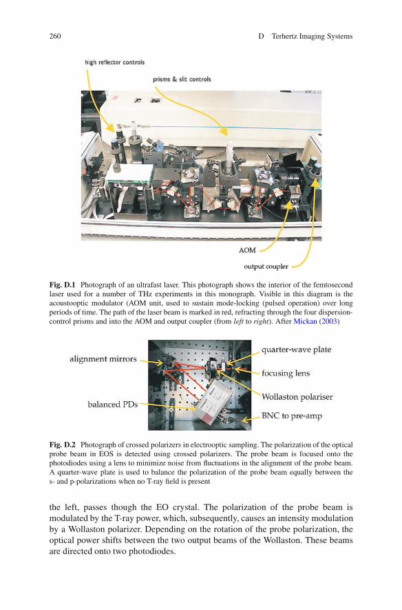
The femtosecond laser illustrated in Fig. D.1 is the key part of the pulsed T-rayspectrometer. The Ti:sapphire laser is used for most of the spectroscopy experimentsin this monograph.
D.1.2 Crossed-Polariser Detection
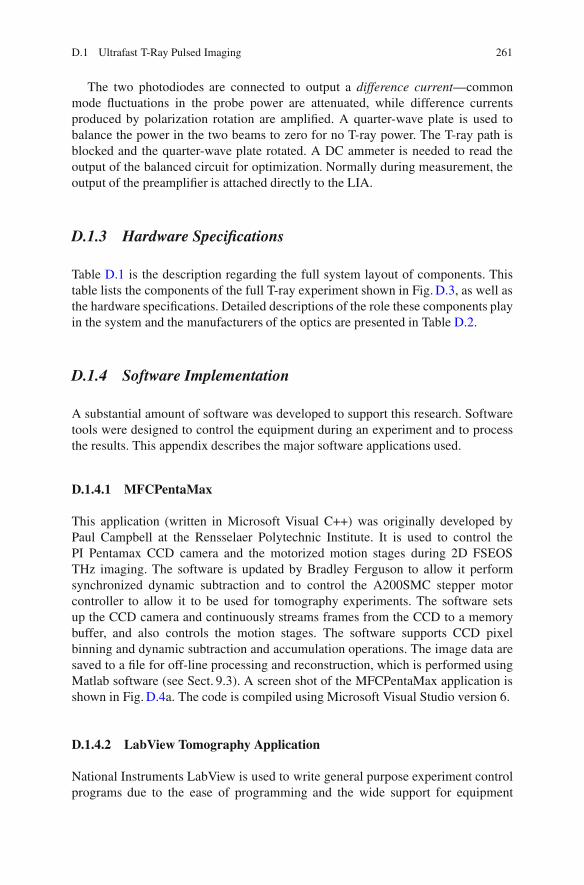
A fundamental part of EOS (see Sect. 2.5.1) is the photodiode detection circuit.A photograph of crossed polarizers in EOS used in a series of THz experiments inthis monograph is shown in Fig. D.2. The probe beam, entering the figure from
X. Yin et al., Terahertz Imaging for Biomedical Applications: Pattern Recognitionand Tomographic Reconstruction, DOI 10.1007/978-1-4614-1821-4,© Springer Science+Business Media, LLC 2012
259
260 D Terhertz Imaging Systems
Fig. D.1 Photograph of an ultrafast laser. This photograph shows the interior of the femtosecondlaser used for a number of THz experiments in this monograph. Visible in this diagram is theacoustooptic modulator (AOM unit, used to sustain mode-locking (pulsed operation) over longperiods of time. The path of the laser beam is marked in red, refracting through the four dispersion-control prisms and into the AOM and output coupler (from left to right). After Mickan (2003)
Fig. D.2 Photograph of crossed polarizers in electrooptic sampling. The polarization of the opticalprobe beam in EOS is detected using crossed polarizers. The probe beam is focused onto thephotodiodes using a lens to minimize noise from fluctuations in the alignment of the probe beam.A quarter-wave plate is used to balance the polarization of the probe beam equally between thes- and p-polarizations when no T-ray field is present
the left, passes though the EO crystal. The polarization of the probe beam ismodulated by the T-ray power, which, subsequently, causes an intensity modulationby a Wollaston polarizer. Depending on the rotation of the probe polarization, theoptical power shifts between the two output beams of the Wollaston. These beamsare directed onto two photodiodes.
D.1 Ultrafast T-Ray Pulsed Imaging 261
The two photodiodes are connected to output a difference current—commonmode fluctuations in the probe power are attenuated, while difference currentsproduced by polarization rotation are amplified. A quarter-wave plate is used tobalance the power in the two beams to zero for no T-ray power. The T-ray path isblocked and the quarter-wave plate rotated. A DC ammeter is needed to read theoutput of the balanced circuit for optimization. Normally during measurement, theoutput of the preamplifier is attached directly to the LIA.
D.1.3 Hardware Specifications
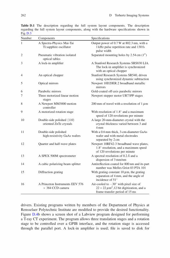
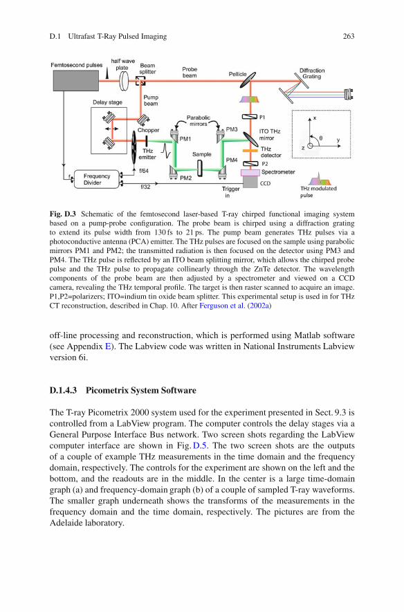
Table D.1 is the description regarding the full system layout of components. Thistable lists the components of the full T-ray experiment shown in Fig. D.3, as well asthe hardware specifications. Detailed descriptions of the role these components playin the system and the manufacturers of the optics are presented in Table D.2.
D.1.4 Software Implementation
A substantial amount of software was developed to support this research. Softwaretools were designed to control the equipment during an experiment and to processthe results. This appendix describes the major software applications used.
D.1.4.1 MFCPentaMax
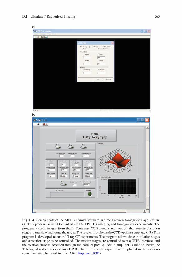
This application (written in Microsoft Visual C++) was originally developed byPaul Campbell at the Rensselaer Polytechnic Institute. It is used to control thePI Pentamax CCD camera and the motorized motion stages during 2D FSEOSTHz imaging. The software is updated by Bradley Ferguson to allow it performsynchronized dynamic subtraction and to control the A200SMC stepper motorcontroller to allow it to be used for tomography experiments. The software setsup the CCD camera and continuously streams frames from the CCD to a memorybuffer, and also controls the motion stages. The software supports CCD pixelbinning and dynamic subtraction and accumulation operations. The image data aresaved to a file for off-line processing and reconstruction, which is performed usingMatlab software (see Sect. 9.3). A screen shot of the MFCPentaMax application isshown in Fig. D.4a. The code is compiled using Microsoft Visual Studio version 6.
D.1.4.2 LabView Tomography Application
National Instruments LabView is used to write general purpose experiment controlprograms due to the ease of programming and the wide support for equipment
262 D Terhertz Imaging Systems
Table D.1 The description regarding the full system layout components. The descriptionregarding the full system layout components, along with the hardware specifications shown inFig. D.3
Number Components Specifications
1 A Spectra-Physics Mai-TaiTi:sapphire oscillator
Output power of 0.7 W at 802.3 nm, with a1 kHz pulse repetition rate and 130 fspulse width
2 Pneumatic vibration isolatedoptical tables
Separated mounting holes by 2.54 cm (1′′)
3 A lock-in amplifier A Stanford Research Systems SRS830 LIA.The lock-in amplifier is synchronizedwith an optical chopper
4 An optical chopper Stanford Research Systems SR540, drivenusing synchronized dynamic subtraction
5 Optical mirrors Newport 10D20ER.2 broadband metallicmirrors
6 Parabolic mirrors Gold-coated off-axis parabolic mirrors7 Three motorized linear motion
stagesNewport stepper motor UR73PP stages
8 A Newport MM3000 motioncontroller
200 mm of travel with a resolution of 1 μm
9 A motorized rotation stage With resolution of 1.8◦ and a maximumspeed of 120 revolutions per minute
10 Double-side polished 〈110〉oriented ZnTe crystals
A large 20-mm-diameter crystal with thecrystal thickness varied between 3 and4 mm
11 Double-side polishedhigh-resistivity GaAs wafers
With a 0.6 mm thick, 3-cm-diameter GaAswafer and with metal electrodesseparated by 2 cm
12 Quarter and half-wave plates Newport 10RF42-3 broadband wave plates,1.8◦ resolution, and a maximum speedof 120 revolutions per minute
13 A SPEX 500M spectrometer A spectral resolution of 0.2 A and adispersion of 3 mm/nm
14 A cubic polarizing beam splitter Antireflection coated for 800 nm and its partnumber was Melles Griot 03 PTA 101
15 Diffraction grating With grating constant 10 μm, the gratingseparation of 4 mm, and the angle ofincidence of 51◦
16 A Princeton Instruments EEV 576× 384 CCD camera
Air-cooled to −30◦ with pixel size of22×22 μm2,12 bit digitization, and aframe-transfer period of 15 ms
drivers. Existing programs written by members of the Department of Physics atRensselaer Polytechnic Institute are modified to provide the desired functionality.Figure D.4b shows a screen shot of a Labview program designed for performinga T-ray CT experiment. The program allows three translation stages and a rotationstage to be controlled over a GPIB interface, and the rotation stage is accessedthrough the parallel port. A lock-in amplifier is used; file is saved to disk for
D.1 Ultrafast T-Ray Pulsed Imaging 263
Fig. D.3 Schematic of the femtosecond laser-based T-ray chirped functional imaging systembased on a pump-probe configuration. The probe beam is chirped using a diffraction gratingto extend its pulse width from 130 fs to 21 ps. The pump beam generates THz pulses via aphotoconductive antenna (PCA) emitter. The THz pulses are focused on the sample using parabolicmirrors PM1 and PM2; the transmitted radiation is then focused on the detector using PM3 andPM4. The THz pulse is reflected by an ITO beam splitting mirror, which allows the chirped probepulse and the THz pulse to propagate collinearly through the ZnTe detector. The wavelengthcomponents of the probe beam are then adjusted by a spectrometer and viewed on a CCDcamera, revealing the THz temporal profile. The target is then raster scanned to acquire an image.P1,P2=polarizers; ITO=indium tin oxide beam splitter. This experimental setup is used in for THzCT reconstruction, described in Chap. 10. After Ferguson et al. (2002a)
off-line processing and reconstruction, which is performed using Matlab software(see Appendix E). The Labview code was written in National Instruments Labviewversion 6i.
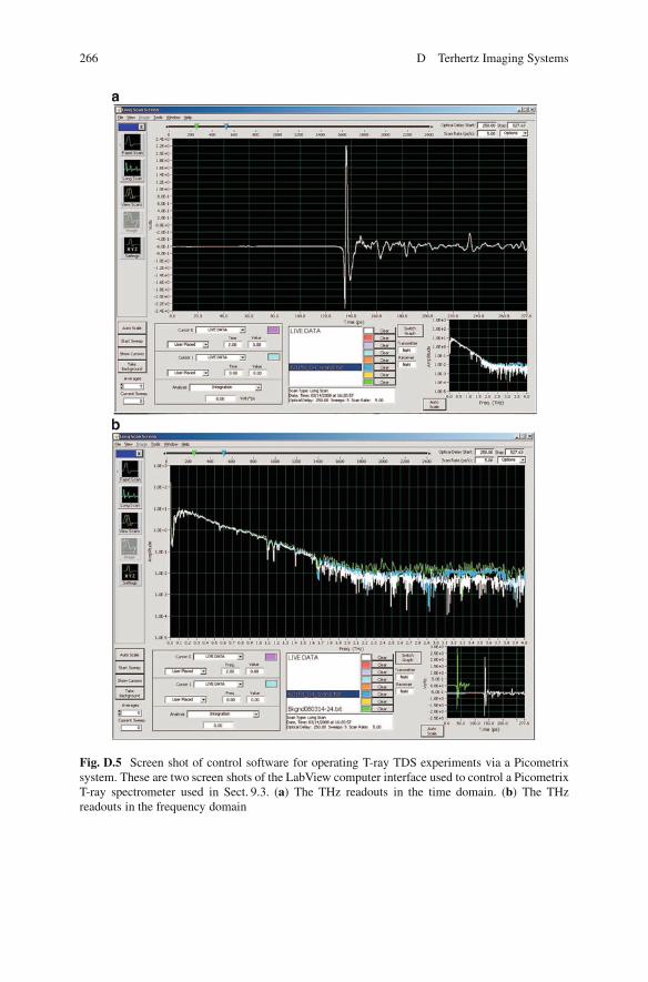
D.1.4.3 Picometrix System Software
The T-ray Picometrix 2000 system used for the experiment presented in Sect. 9.3 iscontrolled from a LabView program. The computer controls the delay stages via aGeneral Purpose Interface Bus network. Two screen shots regarding the LabViewcomputer interface are shown in Fig. D.5. The two screen shots are the outputsof a couple of example THz measurements in the time domain and the frequencydomain, respectively. The controls for the experiment are shown on the left and thebottom, and the readouts are in the middle. In the center is a large time-domaingraph (a) and frequency-domain graph (b) of a couple of sampled T-ray waveforms.The smaller graph underneath shows the transforms of the measurements in thefrequency domain and the time domain, respectively. The pictures are from theAdelaide laboratory.
264 D Terhertz Imaging Systems
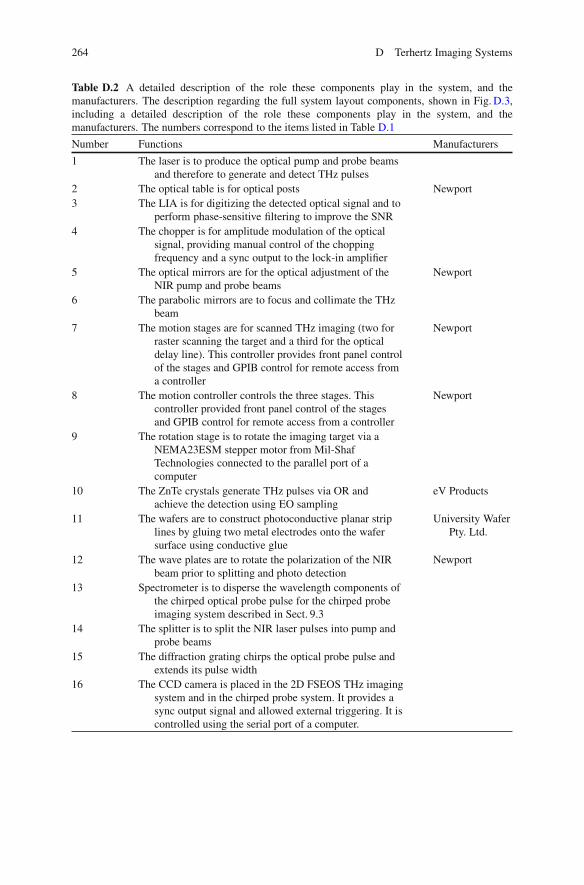
Table D.2 A detailed description of the role these components play in the system, and themanufacturers. The description regarding the full system layout components, shown in Fig. D.3,including a detailed description of the role these components play in the system, and themanufacturers. The numbers correspond to the items listed in Table D.1
Number Functions Manufacturers
1 The laser is to produce the optical pump and probe beamsand therefore to generate and detect THz pulses
2 The optical table is for optical posts Newport3 The LIA is for digitizing the detected optical signal and to
perform phase-sensitive filtering to improve the SNR4 The chopper is for amplitude modulation of the optical
signal, providing manual control of the choppingfrequency and a sync output to the lock-in amplifier
5 The optical mirrors are for the optical adjustment of theNIR pump and probe beams
Newport
6 The parabolic mirrors are to focus and collimate the THzbeam
7 The motion stages are for scanned THz imaging (two forraster scanning the target and a third for the opticaldelay line). This controller provides front panel controlof the stages and GPIB control for remote access froma controller
Newport
8 The motion controller controls the three stages. Thiscontroller provided front panel control of the stagesand GPIB control for remote access from a controller
Newport
9 The rotation stage is to rotate the imaging target via aNEMA23ESM stepper motor from Mil-ShafTechnologies connected to the parallel port of acomputer
10 The ZnTe crystals generate THz pulses via OR andachieve the detection using EO sampling
eV Products
11 The wafers are to construct photoconductive planar striplines by gluing two metal electrodes onto the wafersurface using conductive glue
University WaferPty. Ltd.
12 The wave plates are to rotate the polarization of the NIRbeam prior to splitting and photo detection
Newport
13 Spectrometer is to disperse the wavelength components ofthe chirped optical probe pulse for the chirped probeimaging system described in Sect. 9.3
14 The splitter is to split the NIR laser pulses into pump andprobe beams
15 The diffraction grating chirps the optical probe pulse andextends its pulse width
16 The CCD camera is placed in the 2D FSEOS THz imagingsystem and in the chirped probe system. It provides async output signal and allowed external triggering. It iscontrolled using the serial port of a computer.
D.1 Ultrafast T-Ray Pulsed Imaging 265
Fig. D.4 Screen shots of the MFCPentamax software and the Labview tomography application.(a) This program is used to control 2D FSEOS THz imaging and tomography experiments. Theprogram records images from the PI Pentamax CCD camera and controls the motorized motionstages to translate and rotate the target. The screen shot shows the CCD options setup page. (b) Thisprogram is developed to control T-ray CT experiments. The program allows three translation stagesand a rotation stage to be controlled. The motion stages are controlled over a GPIB interface, andthe rotation stage is accessed through the parallel port. A lock-in amplifier is used to record theTHz signal and is accessed over GPIB. The results of the experiment are plotted in the windowsshown and may be saved to disk. After Ferguson (2004)
266 D Terhertz Imaging Systems
Fig. D.5 Screen shot of control software for operating T-ray TDS experiments via a Picometrixsystem. These are two screen shots of the LabView computer interface used to control a PicometrixT-ray spectrometer used in Sect. 9.3. (a) The THz readouts in the time domain. (b) The THzreadouts in the frequency domain
D.2 Continuous-Wave T-Ray Imaging via THz QCL 267
D.2 Continuous-Wave T-Ray Imaging via THz QCL
D.2.1 THz QCL Imaging
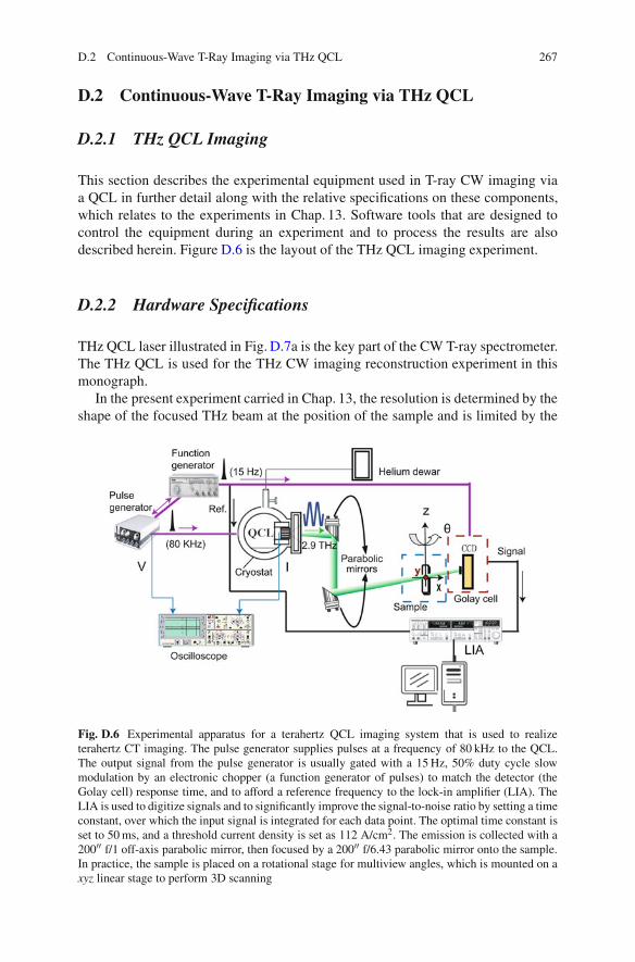
This section describes the experimental equipment used in T-ray CW imaging viaa QCL in further detail along with the relative specifications on these components,which relates to the experiments in Chap. 13. Software tools that are designed tocontrol the equipment during an experiment and to process the results are alsodescribed herein. Figure D.6 is the layout of the THz QCL imaging experiment.
D.2.2 Hardware Specifications
THz QCL laser illustrated in Fig. D.7a is the key part of the CW T-ray spectrometer.The THz QCL is used for the THz CW imaging reconstruction experiment in thismonograph.
In the present experiment carried in Chap. 13, the resolution is determined by theshape of the focused THz beam at the position of the sample and is limited by the
Fig. D.6 Experimental apparatus for a terahertz QCL imaging system that is used to realizeterahertz CT imaging. The pulse generator supplies pulses at a frequency of 80 kHz to the QCL.The output signal from the pulse generator is usually gated with a 15 Hz, 50% duty cycle slowmodulation by an electronic chopper (a function generator of pulses) to match the detector (theGolay cell) response time, and to afford a reference frequency to the lock-in amplifier (LIA). TheLIA is used to digitize signals and to significantly improve the signal-to-noise ratio by setting a timeconstant, over which the input signal is integrated for each data point. The optimal time constant isset to 50 ms, and a threshold current density is set as 112 A/cm2. The emission is collected with a200′′ f/1 off-axis parabolic mirror, then focused by a 200′′ f/6.43 parabolic mirror onto the sample.In practice, the sample is placed on a rotational stage for multiview angles, which is mounted on axyz linear stage to perform 3D scanning
268 D Terhertz Imaging Systems
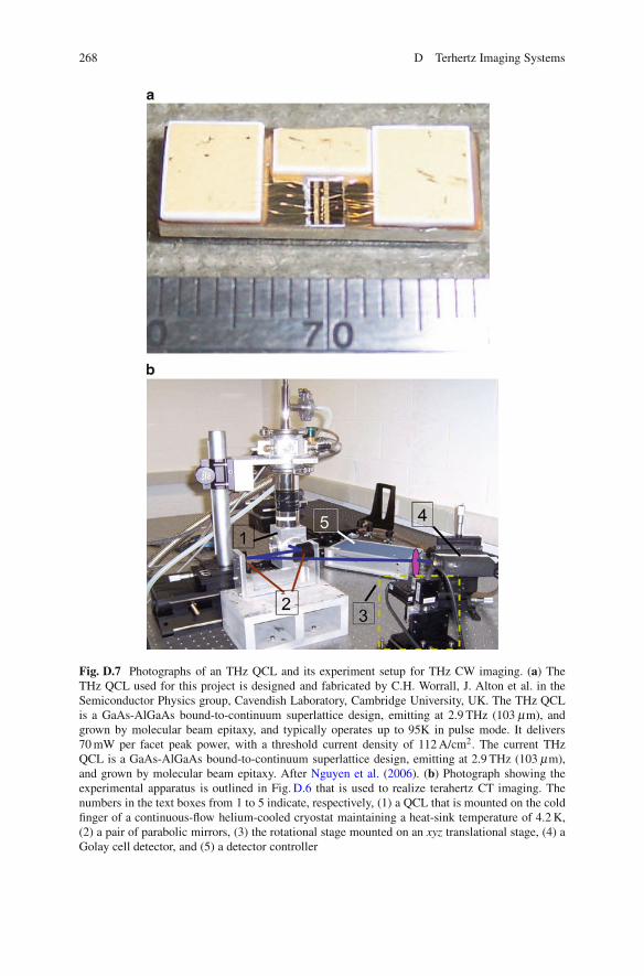
Fig. D.7 Photographs of an THz QCL and its experiment setup for THz CW imaging. (a) TheTHz QCL used for this project is designed and fabricated by C.H. Worrall, J. Alton et al. in theSemiconductor Physics group, Cavendish Laboratory, Cambridge University, UK. The THz QCLis a GaAs-AlGaAs bound-to-continuum superlattice design, emitting at 2.9 THz (103 μm), andgrown by molecular beam epitaxy, and typically operates up to 95K in pulse mode. It delivers70 mW per facet peak power, with a threshold current density of 112 A/cm2. The current THzQCL is a GaAs-AlGaAs bound-to-continuum superlattice design, emitting at 2.9 THz (103 μm),and grown by molecular beam epitaxy. After Nguyen et al. (2006). (b) Photograph showing theexperimental apparatus is outlined in Fig. D.6 that is used to realize terahertz CT imaging. Thenumbers in the text boxes from 1 to 5 indicate, respectively, (1) a QCL that is mounted on the coldfinger of a continuous-flow helium-cooled cryostat maintaining a heat-sink temperature of 4.2 K,(2) a pair of parabolic mirrors, (3) the rotational stage mounted on an xyz translational stage, (4) aGolay cell detector, and (5) a detector controller
D.2 Continuous-Wave T-Ray Imaging via THz QCL 269
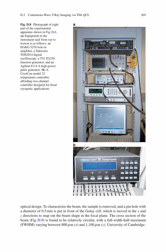
Fig. D.8 Photograph of rightpart of the experimentalapparatus shown in Fig. D.6.(a) Equipment in theinstrument rack from top tobottom is as follows: anEG&G 5210 lock-inamplifier, a TektronixTDS2014 digitaloscilloscope, a TTi TG230function generator, and anAgilent 8114 A high-powerpulse generator. (b) ACryoCon model 32temperature controller,affording two-channelcontroller designed for fixedcryogenic applications
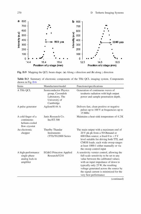
optical design. To characterize the beam, the sample is removed, and a pin hole witha diameter of 0.5 mm is put in front of the Golay cell, which is moved in the x andz directions to map out the beam shape in the focal plane. The cross section of thebeam (Fig. D.9) is found to be relatively circular, with a full-width-half-maximum(FWHM) varying between 800 μm (x) and 1,100 μm (z). University of Cambridge.
270 D Terhertz Imaging Systems
Fig. D.9 Mapping the QCL beam shape. (a) Along x direction and (b) along z direction
Table D.3 Summary of electronic components of the THz QCL imaging system. Componentsshown in Fig. D.6
Items Manufacturer/model Functions/specifications
A THz QCL Semiconductor Physicsgroup, CavendishLaboratory, TheUniversity ofCambridge
Generation of continuous waves ofterahertz radiation with high outputpower and sample penetration depth.
A pulse generator Agilent/8144 A Delivers fast, clean positive or negativepulses up to 100 V at frequencies up to15 MHz
A cold finger of acontinuous-helium-cooledflow cryostat
Janis Research Co.Inc/ST-300
Maintains a heat-sink temperature of 4.2 K
An electronicchopper
Thurlby ThandarInstruments(TTI)/TG2000 Series
The main output with a maximum emf of20 V pk-pk from a 50 Ohmand or600 Ohm source; a fixed 0 to +5 Vlevel suitable for driving both TTL andCMOS loads; each wide sweep rangesat least 1000:1 either manually or viathe sweep control input
A high-performancesingle phaseanalog lock-inamplifier
EG&G Princeton AppliedResearch/5210
A sensitivity vernier control, allowing thefull-scale sensitivity to be set to anyvalue between the calibrated values;with an input impedance of down totypically only 25 W, the resultingvoltage generated across the source bythe signal current is minimized for thevery best performance
(continued)
D.2 Continuous-Wave T-Ray Imaging via THz QCL 271
Table D.3 (continued)
Items Manufacturer/model Functions/specifications
A 100 MHz fourchannel digitalreal-timeoscilloscope
Tektronix/ TDS2014 With up to 200 MHz bandwidth and 2GS/s maximum sample rate, providingaccurate real-time acquisition up totheir full bandwidth, offeringadvanced pulse width triggering andline-selectable video triggering, and11 standard automatic measurements
A temperaturecontroller
Cryocon/model 32 Two-channel controller designed for fixedcryogenic applications. Platinum RTDsensors allow the use of built-in DIN43760 (IEC 750) standard curves for100-W, 1,000-W or 10-KW devices.The standard curve is used fortemperatures from 70 to 1020 K and isextended down to 30 K for cryogenicuse. Operation to about 14 K ispossible with user-supplied curves
Parabolic mirrors Gold-coated off-axisparabolic mirrors
A 200′′ f/1 off-axis parabolic mirror forcollecting the THz radiation, then a200′′ f/6.43 parabolic mirror is used tofocus it onto the sample
Three linear motionstages
Newport/VP-25XA For scanned THz imaging
A motorizedrotation stage
Newport/SR50CC To rotate a sample for multiangle imaging
A Golay cell Cathodean Ltd/IR50 An opto-acoustic detector designed foroperation in the spectral range0.02–20 THz, equipped with a6-mm-diameter polyethylene inputwindow that provides for hightransparency at frequencies up to20 THz. It is mounted on proprietaryvibration-isolating bases
Table D.1 is the description regarding the full system layout of componentsassociated with the terahertz QCL experiments shown in Fig. D.6.
D.2.3 LabVIEWTM Programming Implement for DataAcquisition
The linear and rotational stages on which the sample is mounted are connected to amotion controller (Newport, model: MM4006), which is controlled with a programwritten in LabVIEWTM programming language. LabVIEWTM is an increasinglypopular graphical development environment for signal acquisition, measurement
272 D Terhertz Imaging Systems
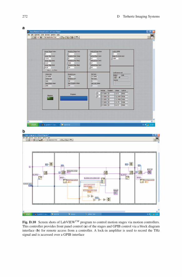
Fig. D.10 Screen shots of LabVIEWT M program to control motion stages via motion controllers.This controller provides front panel control (a) of the stages and GPIB control via a block diagraminterface (b) for remote access from a controller. A lock-in amplifier is used to record the THzsignal and is accessed over a GPIB interface
D.2 Continuous-Wave T-Ray Imaging via THz QCL 273
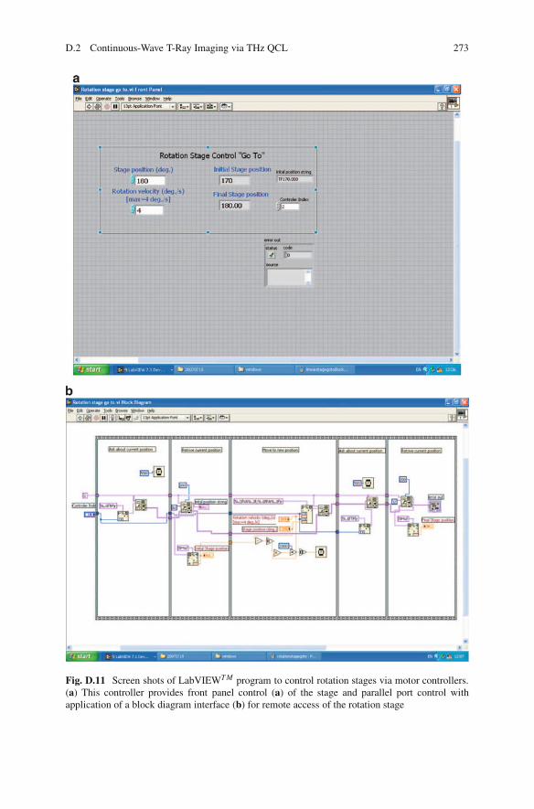
Fig. D.11 Screen shots of LabVIEWT M program to control rotation stages via motor controllers.(a) This controller provides front panel control (a) of the stage and parallel port control withapplication of a block diagram interface (b) for remote access of the rotation stage
274 D Terhertz Imaging Systems
analysis and data presentation (National Instruments, 2004). A LabVIEWTM pro-gram consists of two components. One is a front panel, which serves as userinterface. The front panel is built with controls and indicators, which are interactiveinput and output terminals, respectively. Through the front panels of LabVIEWTM
programs, users enter operating parameters. The other is a block diagram, whichcontains the graphical source code. Objects on the block diagram include terminals,nodes, and functions connected with wires. The block diagram represents theelectronic circuits inside physical instruments. The computer controls the delaystages and acquires data via a General Purpose Interface Bus (GPIB, manufacturer:National Instruments) network. Screen shots of the LabVIEWTM application for thecontrol of linear and rotation stages via front panels and block diagrams are shownin Fig. D.10, and the screen shots regarding front panels and block diagram for thecontrol of a rotation stage are shown in Fig. D.11.
Appendix EMatlab Code
This appendix introduces some of the algorithms used to achieve computer analysisof the raw T-ray waveforms. These algorithms are implemented in MATLAB(manufacturer: The MathWorks, model:7; URL: http://www.mathworks.com) andare available on the attached CD-ROM, as MATLAB files. Also included on theCD-ROM is a directory of raw data files, included as examples of typical T-rayexperimental output, and a pdf file of this monograph.
E.1 Implemented Matlab Toolboxes
Matlab 7 is used to implement all the algorithms described in this monograph. Mat-lab is an interpreted programming language with built-in support for a large numberof mathematical functions and data presentation tools. Mathematical derivationsin this monograph are checked using the symbolic Maple toolbox. The followingMatlab toolboxes are utilized:
1. The wavelet toolbox2. The system identification toolbox3. The SVM and kernel methods matlab toolbox4. The symbolic toolbox5. The signal processing toolbox6. The image processing toolbox
E.2 Code Listings
This section provides a brief description of the Matlab software used to implementmany of the algorithms described in this monograph. Matlab scripts with littlealgorithmic content, including those used to parse input data files and generate plots,
X. Yin et al., Terahertz Imaging for Biomedical Applications: Pattern Recognitionand Tomographic Reconstruction, DOI 10.1007/978-1-4614-1821-4,© Springer Science+Business Media, LLC 2012
275
276 E Matlab Code
have not been included. For more details on the software implementation, pleasecontact the primary author.
The attached CD includes copies of the Matlab files described below:
Powder recognition via frequency-domain features
• WDHeuSURE.m: This function implements the wavelet soft heuristic SUREmethods to achieve preferred denoised performance of THz pulsed responses, aspresented in Chap. 6 (Sect. 6.5). It also demonstrates frequency-domain featuresthat are extracted to be as inputs for classification.
• classify.m: This function is to apply Mahalanobis classifier to classify differenttypes of powders based on extracted frequency-domain features in 3D. Thisalgorithm is described in Chap. 9 (Sects. 9.2 and 9.4).
Cancerous cell recognition functions via autoregressive modeling over waveletsubbands
• WPDencorreAR.m: This function implements the wavelet packet SURE methodsto denoise measured cancerous cell signals ex vivo. AR coefficients are calcu-lated over wavelet subbands to extract important features, aiming to identifycancerous cell signals from normal cell THz responses. It relates to Chap. 9(Sect. 9.4.3.2)
• PronyCorreARMA.m: This function is to apply an improved Prony method toachieve AR moving average (ARMA) modeling over discrete wavelet subbandcoefficients. It is also illustrated that the discrete wavelet-based heuristic SUREmethod is applied in Chap. 9 (Sect. 9.4.3.3) to achieve denoised THz signals ofdifferent types of powder samples.
SVM applications for THz feature subset identification programming
• SVMsDualClas.m: The program is realized via applying SVM and kernelmethods matlab toolbox, abbreviated as SKMT. Dual classification of THz RNAdata is explored. Frequency orientation components are extracted as features tobe input to SVMs. It relates to Chap. 9 (Sect. 9.5.4.1).
• SVMsMulClas.m: This program is for multiclass classification, realized viaapplying One-Against-One algorithms for pairwise classifier designs with useof SKMT. It relates to Chap. 9 (Sect. 9.5.4.2).
Wavelet Scale Correlation-Based Segmentation WaveSegment.m: This functionintroduces wavelet scale correlation-based segmentation for a 3D classificationof the nest structure of a plastic tube inserted in a glass vial. The computedtomography is also illustrated. The contents this function involves are described inChaps. 10 and 11.
Wavelet-Based Local Tomography Reconstruction of THz Pulsed Imaging
• localTomography PulsedTHz.m: This program reconstructs the measured THzpulsed image data along the centered ROI, via applying wavelet and scalingramp filters. This relates to Chap. 12. Time-domain parameters are extracted viaapplying correlation algorithms for the sinogram images.
E.2 Code Listings 277
• FFT scale.m: This function describes the algorithm for wavelet and scaling rampfilters for local reconstruction of THz measurements.
• Sepbackproj.m: This function performs the IRT of wavelet and scaling rampfiltered projections.
Local Reconstruction via THz QCL THz QCL LT.m: This program reconstructsand segments the ROI. Three algorithms are involved: global and local tomographyvia FBP algorithms, and wavelet-based local reconstruction, all of which are appliedon THz CW measurements. This relates to Chap. 13.
References
ABBOTT-D. (2000). Directions in terahertz technology, Proceedings 22nd IEEE GaAs IC Sympo-sium, Seattle, WA, pp. 263–266.
ALIVISATOS-A. P. (1996). Semiconductor clusters, nanocrystals, and quantum dots, Science, 271,pp. 933–937.
ALIVISATOS-A. P. (2004). The use of nanocrystals in biological detection, Nature Biotechnology,22, pp. 47–52.
ALTON-J. (2005). Bound-to-Continuum THz Quantum Cascade Lasers (PhD Thesis), Universityof Cambridge, UK.
ARNONE-D., CIESLA-C., AND PEPPER-M. (2000). Terahertz imaging comes into view, PhysicsWorld, 13(4), pp. 35–40.
PREVITALI-C. M. (2000). Solvent effects on intermolecular electron transfer processes, Pure andApplied Chemistry, 67(1), pp. 127–134.
BEARD-M. C., TURNER-G. M., AND SCHMUTTENMAER-C. A. (2002). Terahertz Spectroscopy,Journal of Physical Chemistry B, 106(29), pp. 7146–7159.
ASHLEY-A., AND PALKA-F. (1973). Transmission cavity and injection stabilization of an X-bandtransferred electron oscillator, IEEE MTT-S International Microwave Symposium Digest, 73(1),pp. 181–182.
ATKESON-C., MOORE-A., AND SCHAAL-S. (1997). Locally weighted learning, Artificial Intelli-gence Review, 11(1-5), pp. 11–73.
AUSTON-D. (1983). Impulse response of photoconductors in transmission lines, IEEE Journal ofQuantum Electronics, 19(4), pp. 639–648.
BARBIERI-S., ALTON-J., BEERE-H. E., FOWLER-J., FORD-J., LINFIELD-E. H., AND
RITCHIE-D. A. (2004). 2.9 THz quantum cascade lasers operating up to 70 K in continuouswave, Applied Physics Letters, 85(10), pp. 1674–1676.
BARTELS-A., CERNA-R., KISTNER-C., THOMA-A., HUDERT-F., JANKE-C., AND DEKORSY-T.(2007). Ultrafast time-domain spectroscopy based on high-speed asynchronous optical sam-pling, Review of Scientific Instruments, 78(3), Art. No. 035107.
BENGIO-Y., AND GRANDVALET-Y. (2004). No unbiased estimator of the variance of k-fold cross-validation, Journal of Machine Learning Research, 5, pp. 1089–1105.
BERENSTEIN-C., AND WALNUT-D. (1994). Local inversion of the Radon transform in evendimensions using wavelets, 75 Years of Radon Transform, pp. 38–58.
BERGER-V., AND SIRTORI-C. (2004). Multispectral classification techniques for terahertz pulsedimaging: an example in histopathology, Semiconductor Science and Technology, 19(8),pp. 964–970.
X. Yin et al., Terahertz Imaging for Biomedical Applications: Pattern Recognitionand Tomographic Reconstruction, DOI 10.1007/978-1-4614-1821-4,© Springer Science+Business Media, LLC 2012
279
280 References
BERRY-E., BOYLE-R., FITZGERALD-A., AND HANDLEY-J. (2005). Time frequency analysis interahertz pulsed imaging, Computer Vision Beyond the Visible Spectrum (Advances in PatternRecognition), Springer Verlag, London, UK, pp. 290–329.
BERRY-E., HANDLEY-J., FITZGERALD-A., MERCHANT-W., BOYLE-R., ZINOV’EV-N., MILES-R.,CHAMBERLAIN-J., AND SMITH-M. (2004). Nonlinear phase matching in THz semiconductorwaveguides, Medical Engineering & Physics, 26(5), pp. 423–430.
BERRYMAN-M. J., AND RAINSFORD-T. (2004). Classification of terahertz data as a tool for thedetection of cancer, Medical Engineering & Physics, 6416(12006), pp. 423–430.
BILGOT-A., PERRIER-V., AND DESBAT-L. (2004). Wavelets, local tomography and interven-tional x-ray imaging, 2004 IEEE on Nuclear Science Symposium Conference Record, 6,pp. 3505–3509.
BOW-S. (2002). Pattern Recognition and Image Preprocessing (Signal Processing and Communi-cations Series), second edition edn, Marcel Dekker, Inc., NY, USA.
BRABEC-T., SPIELMANN-C., CURLEY-P. F., AND KRAUSZ-F. (1992). Kerr lens mode locking,Optics Letters, 17(18), pp. 639–648.
BRINK-J., LIM-J., WANG-G., HEIKEN-J., DEYOE-L., AND VANNIER-M. (1995). Technical opti-mization of spiral CT for depiction of renal artery stenosis: in vitro analysis, Radiology, 194,pp. 157–163.
BROMAGE-J., RADIC-S., AGRAWAL-G. P., STROUD-C. R., FAUCHET-P. M. J., AND
SOBOLEVSKI-R. (1998). Spatiotemporal shaping of half-cycle terahertz pulses by diffractionthrough conductive apertures of finite thickness, Journal of the Optical Society of AmericaB-Optical Physics, 15(7), pp. 1953–1959.
BROWN-E. R., MCINTOSH-K. A., NICHOLS-K. B., AND DENNIS-C. L. (1995). Photomixing up to3.8 THz in low-temperature-grown GaAs, Review of Scientific Instruments, 66(3), pp. 285–287.
BROWN-E. R., SMITH-F. W., AND MCINTOSH-K. A. (1993). Coherent millimeterwave generationby heterodyne conversion in low-temperature-grown GaAs photoconductors, Journal of AppliedPhysics, 73(3), pp. 1480–1484.
BROWN-L. G. (1992). A survey of image registration techniques, Computing-Surveys, 24(4),pp. 325–376.
BROWN-M. S., FIECHTNER-G. J., RUDD-J. V., ZIMDARS-D. A., WARMUTH-M., AND GORD-J. R.(2006). Water-vapor detection using asynchronous THz sampling, Applied Spectroscopy, 60(3),pp. 261–265.
BRUCHERSEIFER-M., NAGEL-M., BOLIVAR-H. P., KURZ-H., BOSSERHOFF-A., AND
BUTTNER-R. (2000). Label-free probing of the binding state of DNA by time-domainterahertz sensing, Applied Physics Letters, 77(24), pp. 4049–4051.
CAI-J., AND LI-Y. (2005). Lecture Notes in Computer Science, Springer, Berlin, Heidelberg.CAI-Y., BRENER-I., LOPATA-J., WYNN-J., PFEIFFER-L., STARK-J. B., WU-Q., ZHANG-X. C.,
AND FEDERICI-J. F. (1998). Design and performance of a THz emission and detection setupbased on a semi-insulating GaAs emitter, Applied Physics Letters, 73(4), pp. 444–446.
CANDES-E. J., DEMANET-L., DONOHO-D., AND YING-L. (2006). Fast discrete curvelet trans-forms, Multiscale Modeling & Simulation, 5, pp. 861–899.
CANTOR-A., CHEO-P., FOSTER-M., AND NEWMAN-L. (1981). Application of submillimeterwave lasers to high voltage cable inspection, IEEE Journal of Quantum Electronics, 17(4),pp. 477–489.
CANU-S., GRANDVALET-Y., GUIGUE-V., AND RAKOTOMAMONJY-A. (2005). SVM and kernelmethods matlab toolbox, Perception Systemes et Information, INSA de Rouen, Rouen, France.
CARIN-L., FELSEN-L. B., KRALJ-D., OH-H. S., LEE-W. C., AND PILLAI-S. U. (1997).Wave-oriented signal processing of dispersive time-domain scattering data, IEEE Transactionson Antennas and Propagation, 45(4), pp. 592–600.
CARRIG-T. J., RODRIGUEZ-G., CLEMENT-T. S., TAYLOR-A. J., AND STEWART-K. R. (1995).Scaling of terahertz radiation via optical rectification in electrooptic crystals, Applied PhysicsLetters, 66(2), pp. 121–123.
CHANG-C.-C., AND LIN-C.-J. (2001). Libsvm: a library for support vector machines, Softwareavailable at http://www.csie.ntu.edu.tw/∼cjlin/libsvm.
References 281
CHAN-W. L., DEIBEL-J., AND MITTLEMAN-D. M. (2007). Imaging with terahertz radiation,Reports on Progress in Physics, 70(8), pp. 1325–1379.
CHEN-Q., AND ZHANG-X C. (2001). Semiconductor dynamic aperture for near-field terahertzwave imaging, IEEE Journal of Selected Topics in Quantum Electronics, 7(4), pp. 608–614.
CHEN-Q., JIANG-Z., XU-G., AND ZHANG-X. C (2000). Near-field terahertz imaging with adynamic aperture, Optics Letters, 25(15), pp. 1122–1124.
CHEVILLE-R. A., AND GRISCHKOWSKY-D. (1995). Time domain terahertz impulse rangingstudies, Applied Physics Letters, 67(14), pp. 1960–1962.
CHEVILLE-R. A., MCGOWAN-R. W., AND GRISCHKOWSKY-D. (1997). Late-time target responsemeasured with terahertz impulse ranging, IEEE Transactions on Antennas and Propogation, 45,pp. 1518-1524.
CHOI-M. K., TAYLOR-K., BETTERMANN-A., AND VAN DERWEIDE-D. (2002). Broadband 10-300GHz stimulus-response sensing for chemical and biological entities, Physics in Medicine andBiology, 47(21), pp. 3777–3789.
CHUANG-K. S., TZENG-H. L., CHEN-S., WU-J., AND CHEN-T. (2006). Fuzzy c-means clusteringwith spatial information for image segmentation, Computerized Medical Imaging and Graphics,30, pp. 9–15.
COGDILL-R. P., FORCHT-R. N., SHEN-Y. C., TADAY-P. F., CREEKMORE-J. R., ANDERSON-C. A.,AND III-J. K. D. (2007). Comparison of terahertz pulse imaging and near-infrared spectroscopyfor rapid, non-destructive analysis of tablet coating thickness and uniformity, Journal ofPharmaceutical Innovation, 2(1-2), pp. 29–36.
COIFMAN-R., AND WICKERHAUSER-M. (1992). Entropy-based algorithms for best basis selec-tion, IEEE Transactions on Information Theory, 38(2), p. 713–718.
COVER-T. M. (1965). Geometrical and statistical properties of systems of linear inequalitieswith applications in pattern recognition, IEEE Transactions on Electronic Computers, 14,pp. 326–334.
CRAWLEY-D., WITHINGTON-S., AND OBRADOVIC-J. (2006). Area-scan camera for terahertzholography, Review of Scientific Instruments, 77(5), Art. No. 053106.
CRISTIANINI-N., AND SHAWE-TAYLOR-J. (2000). An Introduction to Support Vector Machinesand Other Kernel Based Methods, Cambridge University Press, Cambridge, UK.
CUBANSKI-D., AND CYGANSKI-D. (1995). Multivariate classification through adaptive Delaunay-based C spline approximation, IEEE Transactions on Pattern Analysis and Machine Intelligence,17(4), pp. 403–417.
DARMO-J., TAMOSIUNAS-V., FASCHING-G., KROLL-J., UNTERRAINER-K., BECK-M.,GIOVANNINI-M., FAIST-J., KREMSER-C., AND DEBBAGE-P. (2004). Imaging with a terahertzquantum cascade laser, Optics Express, 12(9), pp. 1879–1884.
DAUBECHIES-I. (1988). Orthonormal bases of compactly supported wavelets, Communications onPure & Applied Mathematics, 41(7), pp. 909–996.
DAUBECHIES-I. (1992). Ten lectures on wavelets, Society for Industrial and Applied Mathematics,Philadelphia, PA, USA.
DAVIES-A. G., LINFIELD-E. H., AND JOHNSTON-M. B. (2002). The development of terahertzsources and their applications, Physics in Medicine and Biology, 47(21), pp. 3679–3689.
Defence Research & Development Organization (2008). Web. http://www.drdo.org/pub/techfocus/dec01/multispectral.htm Last Checked: January 14 2008.
DELANEY-A. H., AND BRESLER-Y. (1995). Multiresolution tomographic reconstruction usingwavelets, IEEE Transactions on Image Processing, 4(6), pp. 799–813.
DIVINE-D. V., AND GODTLIEBSEN-F. (2007). Bayesian modeling and significant features explo-ration in wavelet power spectra, Nonlinear Processes in Geophysics, 14, pp. 79–88.
DOBRIN-M., AND SAVIT-C. (1988). Introduction to Geophysical Prospecting, 4th ed., McGraw-Hill, New York.
DOMINGOS-P. (1999). The role of Occam’s Razor in knowledge discovery, Data Mining andKnowledge Discovery, 3, pp. 409–425.
DONOHO-D. L. (1995). De-noising by soft thresholding, IEEE Transactions on InformationTheory, 41(3), pp. 613–627.
282 References
DORNEY-T. D., SYMES-W. W., BARANIUK-R. G., AND MITTLEMAN-D. M. (2002). Terahertzmultistatic reflection imaging, Journal of the Optical Society of America A-Optics ImageScience and Vision, 19(7), pp. 1432–1442.
DUDA-R., AND HART-P. (1973). Pattern Classification and Scene Analysis, 4th edn, John Wileyand Sons Inc, New York, USA.
DUDA-R., HART-P., AND STORK-D. (2001). Pattern Classification, 2nd edn, John Wiley and SonsInc, New York, USA.
DUVILLARET-L., GARET-F., AND COUTAZ-L. (1996). A reliable method for extraction of materialparameters in terahertz time-domain spectroscopy, IEEE Journal of Selected Topics in QuantumElectronics, 2(3), pp. 739–746.
DUVILLARET-L., GARET-F., AND COUTAZ-L. (2000). Influence of noise on the characterizationof materials by terahertz time-domain spectroscopy, Journal of the Optical Society of AmericaB: Optical Physics, 17(3), pp. 452–461.
FAIST-J., BECK-M., AELLEN-T., AND GINI-E. (2001). Quantum-cascade lasers based on a bound-to-continuum transition, Applied Physics Letters, 78(2), pp. 147–149.
FAIST-J., CAPASSO-F., SIVCO-D. L., SIRTORI-C., HUTCHINSON-A. L., AND CHO-A. Y. (1994).Quantum cascade laser, Science, 264(5158), pp. 1023–1025.
FAURE-P. (1976). Stochastic realization algorithms, In System Identification: Advances and CaseStudies, Eds. R. K. Mehra and D. G. Larniotis, Academic Press, New York, USA.
FEDERICI-J. F., GARY-D., BARAT-R., AND MICHALOPOULOU-Z.-H. (2007). T-rays vs. terrorists,IEEE Spectrum, 44, pp. 47–52.
FEDERICI-J. F., MITROFANOV-O., LEE-M., HSU-J. P., BRENER-I., HAREL-R., WYNN-J. D.,PFEIFFER-L. N., AND WEST-K. W. (2002). Terahertz near-field imaging, Physics in Medicineand Biology, 47(21), pp. 3727–3734.
FEDERICI-J. F., SCHULKIN-B., HUANG-F., GARY-D., BARAT-R., OLIVEIRA-F., AND ZIMDARS-D. (2005). THz imaging and sensing for security applications-explosives, weapons and drugs,Semicond. Sci. Technol., 20, pp. S266–S280.
FERGUSON-B. (2004). Three Dimensional T-Ray Inspection Systems (PhD Thesis), University ofAdelaide.
FERGUSON-B., AND ABBOTT-D. (2001a). De-noising techniques for terahertz responses ofbiological samples, Microelectronics Journal (Elsevier), 32(12), pp. 943–953.
FERGUSON-B., AND ABBOTT-D. (2001b). Wavelet de-noising of optical terahertz pulse imagingdata, Fluctuation and Noise Letters, 1(2), pp. L65–L70.
FERGUSON-B., AND ZHANG-X. C. (2002). Materials for terahertz science and technology, NatureMaterials, 1(1), pp. 26–33.
FERGUSON-B., LIU-H., HAY-S., FINLAY-D., ZHANG-X.-C., AND ABBOTT-D. (2004). In vitroosteosacoma biosensing using THz time domain spectroscopy, in J. Neev., and M. Reed. (eds.),Proc. of SPIE BioMEMS and Nanotechnology, Vol. 5275, Bellingham, Australia, pp. 304–316.
FERGUSON-B., WANG-S., GRAY-D., ABBOTT-D., AND ZHANG-X. C. (2002a). T-ray computedtomography, Optics Letters, 27(15), pp. 1312–1314.
FERGUSON-B., WANG-S., GRAY-D., ABBOTT-D., AND ZHANG-X. C. (2002b). Toward functional3D T-ray imaging, Physics in Medicine and Biology (IOP), 47, pp. 3735–3742.
FERGUSON-B., WANG-S., GRAY-D., ABBOTT-D., AND ZHANG-X. C. (2002c). Identification ofbiological tissue using chirped probe THz imaging, Microelectronics Journal (Elsevier), 33(12),pp. 1043–1051.
FERGUSON-B., WANG-S., ZHONG-H., ABBOTT-D., AND ZHANG-X.-C. (2003). Powder retectionwith T-ray imaging, Proceeding of SPIE Terahertz for Military and Security Applications, Vol.5070, pp. 7–16.
FISCHER-B., HOFFMANN-M., H. HELM-G. M., AND JEPSEN-P. U. (2005a). Chemical recognitionin terahertz time-domain spectroscopy and imaging, Semiconductor Science and Technology,20(7), pp. S246–S253.
FISCHER-B., HOFFMANN-M., H. HELM-R. W., RUTZ-F., KLEINE-OSTMANN-T., KOCH-M., AND
JEPSEN-P. U. (2005b). Terahertz time-domain spectroscopy and imaging of artificial RNA,Optics Express, 13(14), pp. 5205–5215.
References 283
FITZGERALD-A. J., BERRY-E., ZINOVEV-N. N., WALKER-G. C., SMITH-M. A., AND
CHAMBERLAIN-J. M. (2002). An introduction to medical imaging with coherent terahertzfrequency radiation, Physics in Medicine and Biology, 47(7), pp. R67–R84.
FITZGERALD-A. J., WALLACE-V. P., JIMENEZ-LINAN-M., BOBROW-L., PYE-R. J.,PURUSHOTHAM-A. D., AND ARNONE-D. D. (2006). Terahertz pulsed imaging of humanbreast tumors, Radiology, 239(2), pp. 533–540.
FITZGERALD-A., PICKWELL-E., WALLACE-V., PURUSHOTHAM-A., PINDER-S., LINAN-M.,PYE-R., AND HA-T. (2005). Medical applications of broadband terahertz pulsed radiation, The18th Annual Meeting of the IEEE Lasers and Electro-Optics Society, pp. 120–121.
FLEMING-J. (1974). High resolution submillimeter-wave Fourier-transform spectrometry of gases,IEEE Transactions on Microwave Theory and Techniques, MT-22(12), pp. 1023–1025.
FLEMING-J. (2001). An adaptive version of the boost by majority algorithm, Machine Learning,43(3), pp. 293–318.
Florida State University (2005). Web. http://micro.magnet.fsu.edu/index.html Last Checked: Au-gust 19 2005.
FROHLICH-H. (1980). The biological effects of microwaves and related questions, Adv. Electronicsand Electron Physics, 53, pp. 85–152.
FUKUNAGA-K., AND HUMMELS-D. M. (1989). Leave-one-out procedures for nonparametric errorestimates, IEEE Transactions on Pattern Analysis and Machine Intelligence, II(4), pp. 421–423.
FUKUNAGA-K., AND KESSELL-D. L. (1973). Nonparametric Bayes error estimation using unclas-sified samples, IEEE Transactions on Information Theory, IT-19(4), pp. 434–440.
GALVAO-R., HADJILOUCAS-S., BECERRA-V. M., AND BOWEN-J. (2005). Subspace systemidentification framework for the analysis of multimoded propagation of THz-transient signals,Measurement Science and Technology, 16(5), pp. 1037–1053.
GALVAO-R., HADJILOUCAS-S., BOWEN-J., AND COELHO-C. (2003). Optimal discrimination andclassification of THz spectra in the wavelet domain, Optics Express, 11(12), pp. 1462–1473.
GALVAO-R., HADJILOUCAS-S., ZAFIROPOULOS-A., WALKER-G. C., BOWEN-J. W., AND
DUDLEY-R. (2007). Optimization of apodization functions in terahertz transient spectrometry,Optics Letters, 32(20), pp. 3008–3010.
GIRAUD-G., AND WYNNE-K. (2003). A comparison of the low-frequency vibrational spectraof liquids obtained through infrared and Raman spectroscopies, Journal of Chemical Physics,119(22), pp. 11753–11764.
GLADKOVA-N., PETROVA-G., NIKULIN-N., LOPOVOK-S., SNOPOVA-L., CHUMAKOV-Y.,NASONOVA-V., GELIKONOV-V., GELIKONOV-G., KURANOV-R., SERGEEV-A., AND
FELDCHTEIN-F. (2000). In vivo optical cohetence tomography imaging of human skin:norm and pathology, Skin Research and Technology, 6(1), pp. 6–16.
GLOBUS-T., WOOLARD-D., AND SAMUELS-A. (2002). Submillimeter-wave fourier transformspectroscopy of biological macromolecules, Journal of Applied Physics, 91(9), pp. 6105–6113.
GMACHL-C., CAPASSO-F., SIVCO-D. L., AND CHO-A. Y. (2001). Recent progress in quantumcascade lasers and applications, Report on Progress in Physics, 64, pp. 1533–1601.
GONZALEZ-R. C., AND WOODS-R. E. (2002). Digital Image Processing, Prentice-Hall, Inc., NewJersey.
GRAVES-E., WEISSLEDER-R., AND NTZIACHRISTOS-V. (2004). Fluorescence molecular imagingof small animal tumour models, Current Molecular Medicine, 4, pp. 419–430.
GREGORY-I., TRIBE-W., COLE-B., BAKER-C., EVANS-M., BRADLEY-I., LINFIELD-E.,DAVIES-A., AND MISSOUS-M. (2004). Phase sensitive continuous-wave THz imagingusing diode lasers, Electronics Letters, 40(2), pp. 143–145.
GROSSE-E. (2002). THz radiation from free electron lasers and its potential for cell and tissuestudies, Physics in Medicine and Biology, 47(21), pp. 3755–3760.
GRUNDLER-W., AND KAISER-F. (1992). Experimental evidence for coherent excitations corre-lated with cell growth nanobiology, Nanobiology, 1, pp. 163–176.
GUEST-M., BUSH-I., VAN DAM-H., SHERWOOD-P., THOMAS-J., VAN LENTHE-J., HAVENITH-R.,AND KENDRICK-J. (2005). The GAMESS-UK electronic structure package: algorithms, devel-opments and applications, Molecular Physics, 103(6-8), pp. 719–747.
284 References
GUO-Y., ZHANG-H., WANG-X., AND CAVALLARO-R. (2001). VLSI implementation of Mallat fastdiscrete wavelet transform algorithm with reduced complexity, IEEE Global Telecommunica-tions Conference, 1, pp. 25–29.
GU-P., CHANG-F., TANI-M., SAKAI-K., AND PAN-C. (1999). Generation of coherent cw-terahertzradiation using a tunable dual-wavelength external cavity laser diode, Japanese Journal ofApplied Physics. Part 2. Letters, 38(11A), pp. L1246–L1248.
GU-P., TANI-M., KONO-S., SAKAI-K., AND ZHANG-X.-C. (2002). Study of terahertz radiationfrom InAs and InSb, Applied Physics Letters, 91(9), pp. 5533–5537.
GUYON-I., WESTON-J., AND BARNHILL-S. (2002). Gene selection for cancer classification usingsupport vector machines, Machine Learning, 46, pp. 389–422.
HADJILOUCAS-S., AND BOWEN-J. (1999). Precision of quasioptical null-balanced bridge tech-niques for transmission and reflection coefficient measurements, Review of Scientific Instru-ments, 70(1), pp. 213–219.
HADJILOUCAS-S., GALVAO-R., AND BOWEN-J. (2002). Analysis of spectroscopic measurementsof leaf water content at THz frequencies using linear transforms, Journal of the Optical Societyof America A, 19(12), pp. 2495–2509.
HADJILOUCAS-S., GALVO-R. K. H., BECERRA-V. M., BOWEN-J. W., MARTINI-R.,BRUCHERSEIFER-M., PELLEMANS-H. P. M., HARING BOLIVAR-P., KURZ-H., AND
CHAMBERLAIN-J. M. (2004). Comparison of state space and ARX models of a waveguide’sTHz transient response after optimal wavelet filtering, IEEE Transactions on MicrowaveTheory and Techniques MTT, 52(10), pp. 2409–2419.
HADJILOUCAS-S., KARATZAS-L. S., AND BOWEN-J. (1999). Measurements of leaf water contentusing terahertz radiation, IEEE Transactions on Microwave Theory and Techniques, 47(2),pp. 142–149.
HANDLEY-J. W., FITZGERALD-A. J., BERRY-E., AND BOYLE-R. D. (2002). Wavelet compressionin medical terahertz pulsed imaging, Physics in Medicine and Biology, 47(21), pp. 3885–3892.
HANGYO-M., NAGASHIMA-T., AND NASHIMA-S. (2002). Spectroscopy by pulsed terahertzradiation, Semiconductor Science and Technology, 13, pp. 1727–1738.
HAN-P., AND ZHANG-X. C. (2001). Free-space coherent broadband terahertz time-domain spec-troscopy, Measurement Science and Technology, 12(11), pp. 1747–1756.
HAN-P., CHO-G., AND ZHANG-X. C. (2000). Time-domain transillumination of biological tissueswith terahertz pulses, Optics Letters, 25(4), pp. 242–244.
HANSON-K. M., AND WECKSUNG-G. W. (1983). Bayesian approach to limited-angle reconstruc-tion in computed tomography, Journal of Optical Society of America, 73(11), pp. 1501–1509.
HARALICK-R., AND SHAPIRO-L. (1985). Image segmentation techniques, Computer Vision,Graphics, and Image Processing, 29, pp. 100–132.
HARAN-G., SUN-W. D., WYNNE-K., AND HOCHSTRASSER-R. M. (1997). Femtosecond far-infrared pump-probe spectroscopy: a new tool for studying low-frequency vibrational dynamicsin molecular condensed phases, Chemical Physics Letters, 274(4), pp. 365–371.
HASTIE-T., TIBSHIRANI-R., AND FRIEDMAN-J. H. (2003). The Elements of Statistical Learning:Data Mining, Inference, and Prediction, Springer, New York.
HAUS-H., AND MECOZZI-A. (1993). Noise of mode-locked lasers, IEEE Journal of QuantumElectronics, 29(3), pp. 983–996.
HEARST-M. A. (1998). Trends controversies: Support vector machines, IEEE Intelligent Systems,13(4), pp. 18–28.
HERMAN-G. T. (1980). Image Reconstruction from Projections: The Fundamentals of Computer-ized Tomography, Academic Press, New York, USA.
HERRMANN-M., TABATA-H., AND KAWAI-T. (2005). Terahertz time-domain spectroscopy andimaging of DNA, International Quantum Electronics Conference, pp. 1240–1241.
HERRMANN-M., TANI-M., AND SAKAI-K. (2000). Display modes in time-resolved terahertzimaging, Japanese Journal of Applied Physics Part 1-Regular Papers Short Notes & ReviewPapers, 39(11), pp. 6254–6258.
HOLSCHNEIDER-M. (1991). Inverse radon transforms through inverse wavelet transforms, InverseProblems, 7(6), pp. 853–861.
References 285
HUANG-D., SWANSON-E. A., LIN-C. P., SCHUMAN-J. S., STINSON-W. G., CHANG-W.,HEE-M. R., FLOTTE-T., GREGORY-K., PULIAFITO-C. A., AND FUJIMOTO-J. G. (1991). Opticalcoherence tomography, Science, 254(5035), pp. 1178–1181.
HUBBARD-B. (1998). The World According to Wavelets, 2nd edn, A.K.Peters, Wellesley, Mas-sachusetts.
IZUMIDA-S., ONO-S., LIU-Z., OHTAKE-H., AND SARUKURA-N. (1999). Spectrum control of THzradiation from InAs in a magnetic field by duration and frequency chirp of the excitation pulses,Applied Physics Letters, 75(4), pp. 451–453.
JAIN-S., AND DESHPANDE-G. (2004). Parametric modeling of brain signals, The Proceeding ofIEEE: Technology for Life: North Carolina Symposium on Biotechnology and Bioinformatics,pp. 85–91.
JAN-J. S. (2006). Medical Image Processing, Reconstruction and Restoration: Concepts andMethods, Marcel Dekker Inc, New York, USA.
JANKE-C., FORST-M., NAGEL-M., KURZ-H., AND BARTELS-A. (2005). Asynchronous opticalsampling for high-speed characterization of integrated resonant terahertz sensors, OpticsLetters, 30(11), pp. 1405–1407.
JENSEN-A., AND LA COUR-HARBO-A. (2001). Ripples in Mathematics: The Discrete WaveletTransform, Springer Verlag, Berlin.
JEON-T. I., AND GRISCHKOWSKY-D. (1998). Characterization of optically dense, doped semi-conductors by reflection THz time domain spectroscopy, Applied Physics Letters, 72(23),pp. 3032–3034.
JIANG-Z., AND ZHANG-X.-C. (1998a). Electro-optic measurement of THz field pulses with achirped optical beam, Applied Physics Letters, 72(16), pp. 1945–1947.
JIANG-Z., AND ZHANG-X.-C. (1998b). Electro-optic measurement of thz field pulses with achirped optical beam, Applied Physics Letters, 72(16), pp. 1945–1947.
JIANG-Z., AND ZHANG-X.-C. (1998c). Single-shot spatiotemporal terahertz field imaging, OpticsLetters, 23(14), pp. 1114–1116.
JOHNSON-J. L., DORNEY-T. D., AND MITTLEMANA-D. M. (2001). Enhanced depth resolution interahertz imaging using phase-shift interferometry, Applied Physics Letters, 78(6), pp. 835–837.
JOHNSTONE-M., AND DONOHO-L. (1995). Adapting to unknown smoothness via wavelet shrink-age, Journal of the Statistical Association, 90(432), pp. 1220–1224.
JOHNSTON-M. B. (2007). Superfocusing of terahertz waves, Nature Photonics, 1(1), pp. 14–15.JOLLIFFE-I. (2002). Principal Component Analysis, 2nd edn, Springer-Verlag, New York, Berlin,
Heidelberg.JONES-I., RAINSFORD-T., FISCHER-B., AND ABBOTT-D. (2006). Far-infrared vibrational spectra
of all-trans, 9-cis and 13-cis retinal measured by THz time-domain spectroscopy, VibrationalSpectroscopy, 41(2), pp. 144–154.
KAFKA-J. D., AND BAER-T. (1987). Prism-pair dispersive delay lines in optical pulse compression,Optics Letters, 12, pp. 401–403.
KAISER-G. (1994). A Friendly Guide to Wavelets, Birkhauser Boston, Cambridge, Mass.KAK-A. C., AND SLANEY-M. (1988). Principles of Computerized Tomographic Imaging, IEEE
Press, New York, USA.KALENDER-W., SEISSLER-W., KLOTZ-E., AND VOCK-P. (1990). Spiral volumetric CT with
single-breath-hold technique, continuous transport, and continuous scanner rotation, Radiology,176, pp. 181–183.
KALOUPTSIDIS-N., AND THEODORIDIS-S. (1993). Adaptive System Identification and SignalProcessing Algorithms, Prentice-Hall, Inc, NJ, USA.
KARPOWICZ-N., ZHONG-H., LIN-K.-I., HWANG-J.-S., AND ZHANG-X.-C. (2005a). Comparisonbetween pulsed terahertz time-domain imaging and continuous wave terahertz imaging, Semi-conductor Science and Technology, 20(7), pp. S293–S299.
KARPOWICZ-N., ZHONG-H., ZHANG-C., LIN-K.-I., HWANG-J.-S., XU-J., AND ZHANG-X.-C.(2005b). Compact continuous-wave subterahertz system for inspection applications, AppliedPhysics Letters, 86(5), Art. No. 054105.
286 References
KATO-R., KONDO-S., IGO-T., OKITA-T., KONISHI-T., SUEMINE-S., OKUDA-S., AND ISOYAMA-G. (2000). Lasing at 150 μm wavelength and measurement of the characteristics of thefree-electron laser at ISIR, Osaka University, Nuclear Instruments and Methods in Physics Re-search Section A: Accelerators, Spectrometers, Detectors and Associated Equipment, 445(1-3),pp. 169–172.
KAWASE-K., OGAWA-Y., WATANABE-Y., AND INOUE-H. (2003). Non-destructive terahertz imag-ing of illicit drugs using spectral fingerprints, Optics Express, 11(20), pp. 2549–2554.
KAWASE-K., SATO-M., TANIUCHI-T., AND ITO-H. (1996). Coherent tunable thz-wave generationfrom LiNbO3 with monolithic grating coupler, Physics Letters, 68(18), pp. 2483–2485.
KEARNS-M. (1997). A bound on the error of cross validation using the approximation andestimation rates, with consequences for the training-test split, Journal of the American StatisticalAssociation, 9(5), pp. 1143–1161.
KERECMAN-A. (1973). The tungsten-P type silicon point contact diode, IEEE MTT-S InternationalMicrowave Symposium Digest, 73(1), pp. 30–34.
KIM-K. I., JUNG-K., PARK-S. H., AND KIM-H. J. (2002). Support vector machines for textureclassification, IEEE Transactions on Pattern Analysis and Machine Intelligence, 24(11),pp. 1542–1550.
KIM-S. M., HATAMI-F., HARRIS-J. S., KURIAN-A. W., FORD-J., KING-D., SCALARI-G.,GIOVANNINI-M., HOYLER-N., AND HARRIS-J. F. G. (2006). Biomedical terahertz imagingwith a quantum cascade laser, Applied Physics Letters, 88(15), Art. No. 153903.
KIM-S. M., HATAMI-F., HARRIS-J. S., KURIAN-A. W., FORD-J., KING-D., SCALARI-G.,GIOVANNINI-M., HOYLER-N., FAIST-J., AND HARRIS-G. (2004). Biomedical terahertz imag-ing with a quantum cascade laser, Applied Physics Letters, 88, Art. No. 153903.
KINDT-J. T., AND SCHMUTTENMAER-C. A. (1999). Theory for determination of the low-frequency time-dependent response function in liquids using time-resolved terahertz pulsespectroscopy, Journal of Chemical Physics, 110(17), pp. 8589–8596.
KISTNER-C., ANDRE-A., FISCHER-T., THOMA-A., JANKE-C., BARTELS-A., GISLER-T.,MARET-G., AND DEKORSY-T. (2007). Hydration dynamics of oriented DNA films investigatedby time-domain terahertz spectroscopy, Applied Physics Letters, 90(23), Art. No. 233902.
KOHLER-R., IOTTI-R. C., TREDICUCCI-A., AND ROSSI-F. (2001). Design and simulation ofterahertz quantum cascade lasers, Applied Physics Letters, 79, pp. 3920–3922.
KOHLER-R., TREDICUCCI-A., BELTRAM-F., BEERE-H. E., LINFIELD-E. H., DAVIES-A. G.,AND RITCHIE-D. A. (2002a). High-intensity interminiband terahertz emission from chirpedsuperlattices, Applied Physics Letters, 80(11), pp. 156–159.
KOHLER-R., TREDICUCCI-A., BELTRAM-F., BEERE-H. E., LINFIELD-E. H., DAVIES-A. G.,RITCHIE-D. A., IOTTI-R. C., AND ROSSI-F. (2002b). Terahertz semiconductor-heterostructurelaser, Nature, 417(6885), pp. 156–159.
KOHLER-R., TREDICUCCI-A., MAURO-C., BELTRAM-F., BEERE-H. E., LINFIELD-E. H.,DAVIES-A. G., AND RITCHIE-D. A. (2004). Terahertz quantum-cascade lasers based on aninterlaced photon-phonon cascade, Applied Physics Letters, 84(8), pp. 1266–1268.
KOLEHMAINEN-V., SILTANEN-S., JARVENPAA-S., J.P.-K., KOISTINEN-P., LASSAS-M.,PIRTTILA-J., AND SOMERSALO-E. (2003). Statistical inversion for medical x-ray tomographywith few radiographs: II. application to dental radiology, Physics in Medicine and Biology, 48,pp. 1465–1490.
KRISHNAGOPAL-S., KUMAR-V., MAITI-S., PRABHU-S. S., AND SARKAR-S. K. (2004). Free-electron lasers, Current Science, 87(8), pp. 1066–1078.
KUBLER-C., HUBER-R., AND LEITENSTORFER-A. (2005). Ultrabroadband terahertz pulses:generation and field-resolved detection, Semiconductor Science and Technology, 20,pp. S128–S133.
LAHTINEN-T., NUUTINEN-J., ALANEN-E., TURUNEN-M., NUORTIO-L., USENIUS-T., AND
HOPEWELL-J. (1999). Quantitative assessment of protein content in irradiated human skin,International Journal of Radiation Oncology Biology Physics, 43(3), pp. 635–638.
References 287
LASCH-P., AND NAUMANN-D. (1998). FT-IR microspectroscopic imaging of human carcinomathin sections based on pattern recognition techniques, Cellular and Molecular Biology, 44(1),pp. 189–202.
DAKOVSKI-L. G., AND KUBERA-B., AND SHAN-J. (2005). Localized terahertz generation viaoptical rectification in ZnTe, Journal of the Optical Society of America B, 22(8), pp. 1667–1670.
LEE-A. W. M., QIN-Q., KUMAR-S., WILLIAMS-B. S., AND HU-Q. (2006). Real-time terahertzimaging over a standoff distance (>25 meters), Applied Physics Letters, 89(24), Art. No.141125.
LEE-H., LEE-J., AND KIM-J. (2001). A micromachined photoconductive near-field probe forpicosecond pulse propagation measurement on coplanar transmission lines, IEEE Journal ofSelected Topics in Quantum Electronics, 7(4), pp. 674–682.
LEITENSTORFER-A., HUNSCHE-S., SHAH-J., NUSS-M. C., AND KNOX-W. H. (1999). Femtosec-ond charge transport in polar semiconductors, Physical Review Letters, 82(25), pp. 5140–5143.
LEVINE-M. D. (1985). Vision in Man and Machines, McGraw-Hill, Inc., New York.LIANG-G., WILKES-D. M., AND CADZOW-J. A. (1993). ARMA model order estimation based
on the eigenvalues of the covariance matrix, IEEE Transactions on Signal Processing, 41(10),pp. 3003–3009.
LJUNG-L. (1999). System Identification: Theory for the User, 2nd edn, Prentice Hall PTR, NewJersey, USA.
LOFFLER-T., BAUER-T., SIEBERT-K., ROSKOS-H., FITZGERALD-A., AND CZASCH-S. (2001).Terahertz dark-field imaging of biomedical tissue, Optics Express, 4434(245), pp. 25–36.
LOFFLER-T., SIEBERT-K., CZASCH-S., AND BAUER, T.AND ROSKOS-H. (2002). Visualizationand classification in biomedical terahertz pulsed imaging, Physics in Medicine and Biology,47(2002), pp. 3847–3852.
LUO-H., LIU-H. C., SONG-C. Y., AND WASILEWSKI-Z. R. (2005). Background-limited terahertzquantum-well photodetector, Applied Physics letters, 86(23), Art. No. 231103.
MACQUEEN-J. B. (1967). Some methods for classification and analysis of multivariate observa-tions, Proceedings of 5-th Berkeley Symposium on Mathematical Statistics and Probability,Vol. 1, University of California Press, Berkeley, pp. 281–297.
MADYCH-W. R. (1999). Tomography, approximate reconstruction, and continuous wavelet trans-forms, Applied and Computational Harmonic Analysis, 7(1), pp. 54–100.
MALLAT-S. (1989). A theory for multiresolution signal decomposition: The wavelet representa-tion, IEEE Transactions on Pattern Analysis and Machine Intelligence, 14(7), pp. 674–693.
MALLAT-S. G. (1999). A Wavelet Tour of Signal Processing, San Diego : Academic Press, CA.MARKELZ-A., ROITBERG-A., AND HEILWEIL-E. (2000). Pulsed terahertz spectroscopy of DNA,
bovine serum albumin and collagen between 0.1 and 2.0 THz, Chemical Physics Letters,320(1-2), pp. 42–48.
MARTEL-P., CALMETTES-P., AND HENNION-B. (1991). Vibrational modes of hemoglobin in redblood cells, Biophysical Journal, 59(2), pp. 363–377.
MA-X. F., AND ZHANG-X. C. (1993). Determination of ratios between nonlinear-opticalcoefficients by using subpicosecond optical rectification, Journal of the Optical Society ofAmerica B: Optical Physics (JOSA B), 10(7), pp. 1175–1179.
MCCLATCHEY-K., REITEN-M. T., AND CHEVILLEA-R. A. (2000). Time resolved syntheticaperture terahertz impulse imaging, Applied physics letters, 79(27), pp. 4485–4487.
MCELROY-R., AND WYNNE-K. (1997). Ultrafast dipole solvation measured in the far infrared,Physical Review Letters, 79(16), pp. 3078–3081.
MCLACHLAN-G. J. (1992). Discriminant Analysis and Statisical Pattern Recognition, Wiley,New York, USA
MCLACHLAN-G. J. (2005). Discriminant Analysis and Statistical Pattern Recognition, Wiley-Interscience, NY, USA.
MCLAUGHLIN-R., CORCHIA-A., JOHNSTON-M. B., CHEN-Q., CIESLA-C. M., ARNONE-D. D.,JONES-G. A. C., LINFIELD-E. H., DAVIES-A. G., AND PEPPER-M. (2000). Enhanced coherentterahertz emission from indium arsenide in the presence of a magnetic field, Applied PhysicsLetters, 76(15), pp. 2038–2040.
288 References
MCMANUS-J. B., NELSON-D. D., SHORTER-J. H., JIMENEZ-R., HERNDON-S., SALESKA-S.,AND ZAHNISER-M. (2005). A high precision pulsed quantum cascade laser spectrometerfor measurements of stable isotopes of carbon dioxide, Journal of Modern Optics, 52(16),pp. 2309–2321.
MENIKH-A., MACCOLL-R., MANNELLA-C. A., AND ZHANG-X.-C. (2002). Terahertz biosensingtechnology: Frontiers and progress, ChemPhysChem, 3(8), pp. 655–658.
MENIKH-A., MICKANA-S. P., LIU-H.-B., MACCOLLB-R., AND ZHANG-X. C. (2004). Label-freeamplified bioaffinity detection using terahertz wave technology, Biosensors and Bioelectronics,20(3), pp. 658–662.
MEYER-BASE-A. (2003). Pattern Recognition for Medical Imaging, Elsevier, Inc, California,USA.
MEYER-Y. (1990). Ondelettes et Operateurs, Tome 1, Herrmann, Paris.MICKAN-S., ABBOTT-D., MUNCHB-J., ZHANG-X. C., AND VAN DOORN-T. (2000). Analysis of
system trade-offs for terahertz imaging, Microelectronics Journal, 31(7), pp. 503–514.MICKAN-S. P. (2003). T-ray Biosensing (PhD Thesis), University of Adelaide.MITCHELL-T. M. (1997). Machine Learning, International Edition, McGraw-Hill Book Co.,
Singapore.MITROFANOV-O., BRENER-I., HAREL-R., WYNN-J. D., PFEIFFER-L. N., WEST-K. W., AND
FEDERICI-J. (1998). THz near-field imaging, Optics Communications, 150(1-6), pp. 22–26.MITROFANOV-O., BRENER-I., HAREL-R., WYNN-J. D., PFEIFFER-L. N., WEST-K. W., AND
FEDERICI-J. (2000). Terahertz near-field microscopy based on a collection mode detector,Applied Physics Letters, 77(22), pp. 3496–3498.
MITROFANOV-O., HAREL-R., LEE-M., PFEIFFER-L. N., WEST-K., WYNN-J. D., AND
FEDERICI-J. (2001a). Study of single-cycle pulse propagation inside a thz near-field probe,Applied Physics Letters, 78(2), pp. 252–254.
MITROFANOV-O., LEE-M., HSU-J. W. P., BRENER-I., HAREL-R., FEDERICI-J., WYNN-J. D.,PFEIFFER-L. N., AND WEST-K. W. (2001b). Collection mode near-field imaging with 0.5 THzpulses, IEEE Journal of Selected Topics in Quantum Electronics, 7(4), pp. 600–607.
MITTLEMAN-D., JACOBSEN-R., AND NUSS-M. (1996). T-ray imaging, IEEE Journal of SelectedTopics in Quantum Electronics, 2(3), pp. 679–692.
MITTLEMAN-D. M., HUNSCHE-S., BOIVIN-L., AND NUSS-M. C. (1997). T-ray tomography,Optics Letters, 22(12), pp. 904–906.
MITTLEMAN-D., NEELAMANI-R., RUDD-R. B. J., AND KOCH-M. (1999). Recent advances interahertz imaging, Applied Physics B - Lasers and Optics, 68(6), pp. 1085–1094.
MUKHERJEE-D., PAL-P., AND DAS-J. (1996). Sodar image segmentation by fuzzy c-means, SignalProcessing, 54, pp. 295–301.
MULLEN-K., AND ENNIS-D. (1987). Mathematical formulation of multivariate Euclidean modelsfor discrimination methods, Psychometrika, 52(2), pp. 235–249.
MULLER-K.-R., MIKA-S., RATSCH-G., TSUDA-K., AND SCHOLKOPF-B. (2001). An introduc-tion to kernel-based learning algorithms, IEEE Transactions on Neural Networks, 12(2),pp. 181–201.
NAGASHIMA-T., AND HANGYO-M. (2001). Measurement of complex optical constants of a highlydoped Si wafer using terahertz ellipsometry, Applied Physics Letters, 79(24), pp. 3917–3919.
NASHIMA-S., TAKATA-K., AND HANGYO-M. (2001). Measurement of optical properties of highlydoped silicon by terahertz time domain reflection spectroscopy, Applied Physics Letters, 79(24),pp. 3923–3925.
NG-B. W.-H. (2003). Wavelet Based Image Texture Segmentation using a Modified K-meansAlgorithm (PhD Thesis), University of Adelaide.
NGUYEN-K. L., MICHAEL-L. J., GLADDEN-L., WORRALL-C. H., ALEXANDER, P. BEERE-H. E., PEPPER-M., RITCHIE-D. A., ALTON-J., BARBIERI-S., AND LINFIELD-E. H. (2006).Three-dimensional imaging with a terahertz quantum cascade laser, Optics Express, 14(6),pp. 2123–2129.
References 289
NISHIZAWA-J., SASAKI-T., SUTO-K., TANABE-T., SAITO-K., YAMADA-T., AND KIMURA-T. (2005). THz transmittance measurements of nucleobases and related molecules in the0.4- to 5.8-THz region using a GaP THz wave generator, Optics Communications, 246(1-3),pp. 229–239.
NTZIACHRISTOS-V., RIPOLL-J., WANG-L. V., AND WEISSLEDER-R. (2005). Looking and lis-tening to light: the evolution of whole-body photonic imaging, Nature Biotechnology, 23(3),pp. 313–320.
OLSON-T., AND DESTEFANO-J. (1994). Wavelet localization of the Radon transform, IEEETransactions on Signal Processing, 42(8), pp. 2055–067.
OZYUZER-L., KOSHELEV-A. E., KURTER-C., GOPALSAMI-N., LI-Q., TACHIKI-M.,KADOWAKI-K., YAMAMOTO-T., MINAMI-H., YAMAGUCHI-H., TACHIKI-T., GRAY-K. E., KWOK-W.-K., AND WELP-U. (2007). Emission of coherent THz radiation fromsuperconductors, Science, 318(5854), pp. 1291–1293.
PAIVA-H., AND GALVAO-R. (2006). Wavelet-packet identification of dynamic systems in fre-quency subbands, Signal Processing, 86(8), pp. 2001–2008.
PEARCE-J., AND MITTLEMAN-D. M. (2003). Using terahertz pulses to study light scattering, Pro-ceedings of the Sixth International Conference on Electrical Transport and Optical Propertiesof Inhomogeneous Media, Vol. 338, pp. 92–96.
PEARCE-J., CHOI-H., MITTLEMAN-D. M., WHITE-J., AND ZIMDARS-D. (2005). Terahertz wideaperture reflection tomography, Optics Letters, 30(13), pp. 1653–1655.
PERCIVAL-D., AND WALDEN-A. (2000). Wavelet Methods for Time Series Analysis, CambridgeUniversity Press, Cambridge, England.
PICKWELL-E., AND WALLACE-V. (2006). Biomedical applications of terahertz technology,Journal of Physics D: Applied Physics, 39(17), pp. R301–R310.
PICKWELL-E., COLE-B. E., FITZGERALD-A. J., WALLACE-V., AND PEPPER-M. (2004). Simu-lation of terahertz pulse propagation in biological systems, Applied Physics Letters, 84(12),pp. 2190–2192.
PITTNER-S., AND KAMARTHI-S. V. (1999). Feature extraction from wavelet coefficients forpattern recognition tasks, IEEE Transactions on Pattern Analysis and Machine Intelligence,21(1), pp. 83–88.
POPPE-A., XU-L., KRAUSZ-F., AND SPIELMANN-C. (1998). Noise characterisation of sub-10-fs Ti:sapphire oscillators, IEEE Journal of Selected Topics in Quantum Electronics, 4(2),pp. 179–184.
PRADARUTTI-B., MATTHAUS-G., RIEHEMANN-S., NOTNI-G., NOLTE-S., AND TUNNERMANN-A. (2007). Advanced analysis concepts for terahertz time domain imaging, Optics Communica-tions - Elsevier, 279(2), pp. 248–254.
PROAKIS-J., AND MANOLAKIS-D. (1996). Digital Signal Processing: Principles, Algorithms, andApplications, Prentice-Hall, Inc., NJ, USA.
QIAN-S. (2002). Time-Frequency and Wavelet Transforms, 1st edn, Prentice Hall, Inc., New Jersey,USA
RANTALA-M., VANSKA-S., JARVENPAA-S., KALKE-M., LASSAS-M., MOBERG-J., AND
SILTANEN-S. (2006). Wavelet-based reconstruction for limited-angle X-Ray tomography, IEEETransactions on Medical Imaging, 25, pp. 210–217.
RASHID-FARROKHI-F., LIU-K., BERENSTEIN-C., AND WALNUT-D. (1997). Wavelet-based mul-tiresolution local tomography, IEEE Transactions on Image Processing, 6(10), pp. 1412–1430.
RAUDYS-S. (2001). Statistical and Neural Classifiers: An Integrated Approach to Design, 1st edn,Springer-Verlag London Ltd, London, U.K.
REID-M., AND FEDOSEJEVS-R. (2005). Quantitative comparison of terahertz emission from<100> InAs surfaces and a GaAs large-aperture photoconductive switch at high fluences,Applied Optics, 44(1), pp. 149–153.
RICE-A., JIN-Y., MA-X. F., AND ZHANG-X.-C. (1994). Terahertz optical rectification from <110>zinc-blende crystals, Applied Physics Letters, 64(11), pp. 1324–1326.
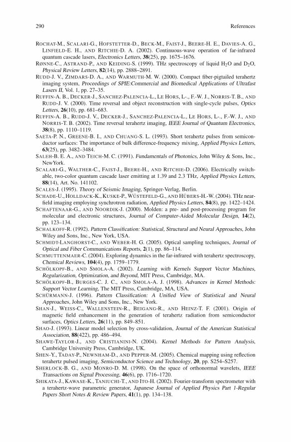
290 References
ROCHAT-M., SCALARI-G., HOFSTETTER-D., BECK-M., FAIST-J., BEERE-H. E., DAVIES-A. G.,LINFIELD-E. H., AND RITCHIE-D. A. (2002). Continuous-wave operation of far-infraredquantum cascade lasers, Electronics Letters, 38(25), pp. 1675–1676.
RØNNE-C., ASTRAND-P., AND KEIDING-S. (1999). THz spectroscopy of liquid H2O and D2O,Physical Review Letters, 82(14), pp. 2888–2891.
RUDD-J. V., ZIMDARS-D. A., AND WARMUTH-M. W. (2000). Compact fiber-pigtailed terahertzimaging system, Proceedings of SPIE:Commercial and Biomedical Applications of UltrafastLasers II, Vol. 1, pp. 27–35.
RUFFIN-A. B., DECKER-J., SANCHEZ-PALENCIA-L., LE HORS, L-., F.-W. J., NORRIS-T. B., AND
RUDD-J. V. (2000). Time reversal and object reconstruction with single-cycle pulses, OpticsLetters, 26(10), pp. 681–683.
RUFFIN-A. B., RUDD-J. V., DECKER-J., SANCHEZ-PALENCIA-L., LE HORS, L-., F.-W. J., AND
NORRIS-T. B. (2002). Time reversal terahertz imaging, IEEE Journal of Quantum Electronics,38(8), pp. 1110–1119.
SAETA-P. N., GREENE-B. I., AND CHUANG-S. L. (1993). Short terahertz pulses from semicon-ductor surfaces: The importance of bulk difference-frequency mixing, Applied Physics Letters,63(25), pp. 3482–3484.
SALEH-B. E. A., AND TEICH-M. C. (1991). Fundamentals of Photonics, John Wiley & Sons, Inc.,NewYork.
SCALARI-G., WALTHER-C., FAIST-J., BEERE-H., AND RITCHIE-D. (2006). Electrically switch-able, two-color quantum cascade laser emitting at 1.39 and 2.3 THz, Applied Physics Letters,88(14), Art. No. 141102.
SCALES-J. (1995). Theory of Seismic Imaging, Springer-Verlag, Berlin.SCHADE-U., HOLLDACK-K., KUSKE-P., WUSTEFELD-G., AND HUBERS-H.-W. (2004). THz near-
field imaging employing synchrotron radiation, Applied Physics Letters, 84(8), pp. 1422–1424.SCHAFTENAAR-G., AND NOORDIK-J. (2000). Molden: a pre- and post-processing program for
molecular and electronic structures, Journal of Computer-Aided Molecular Design, 14(2),pp. 123–134.
SCHALKOFF-R. (1992). Pattern Classification: Statistical, Structural and Neural Approaches, JohnWiley and Sons, Inc., New York, USA.
SCHMIDT-LANGHORST-C., AND WEBER-H. G. (2005). Optical sampling techniques, Journal ofOptical and Fiber Communications Reports, 2(1), pp. 86–114.
SCHMUTTENMAER-C. (2004). Exploring dynamics in the far-infrared with terahertz spectroscopy,Chemical Reviews, 104(4), pp. 1759–1779.
SCHOLKOPF-B., AND SMOLA-A. (2002). Learning with Kernels Support Vector Machines,Regularization, Optimization, and Beyond, MIT Press, Cambridge, MA.
SCHOLKOPF-B., BURGES-C. J. C., AND SMOLA-A. J. (1998). Advances in Kernel Methods:Support Vector Learning, The MIT Press, Cambridge, MA, USA.
SCHURMANN-J. (1996). Pattern Classification: A Unified View of Statistical and NeuralApproaches, John Wiley and Sons, Inc., New York.
SHAN-J., WEISS-C., WALLENSTEIN-R., BEIGANG-R., AND HEINZ-T. F. (2001). Origin ofmagnetic field enhancement in the generation of terahertz radiation from semiconductorsurfaces, Optics Letters, 26(11), pp. 849–851.
SHAO-J. (1993). Linear model selection by cross-validation, Journal of the American StatisticalAssociation, 88(422), pp. 486–494.
SHAWE-TAYLOR-J., AND CRISTIANINI-N. (2004). Kernel Methods for Pattern Analysis,Cambridge University Press, Cambridge, UK.
SHEN-Y., TADAY-P., NEWNHAM-D., AND PEPPER-M. (2005). Chemical mapping using reflectionterahertz pulsed imaging, Semiconductor Science and Technology, 20, pp. S254–S257.
SHERLOCK-B. G., AND MONRO-D. M. (1998). On the space of orthonormal wavelets, IEEETransactions on Signal Processing, 46(6), pp. 1716–1720.
SHIKATA-J., KAWASE-K., TANIUCHI-T., AND ITO-H. (2002). Fourier-transform spectrometer witha terahertz-wave parametric generator, Japanese Journal of Applied Physics Part 1-RegularPapers Short Notes & Review Papers, 41(1), pp. 134–138.
References 291
SIEBE-F., SIEBERT-K., LEONHARDT-R., AND ROSKOS-H. G. (1999). A fully tunable dual-colorCW Ti:AL2O3 laser, IEEE Journal of Quantum Electronics, 35(11), pp. 1731–1736.
SIEBERT-K. J., LOFFLER-T., QUAST-H., THOMSON-M., BAUER-T., LEONHARDT-R., CZASCH-S.,AND ROSKOS-H. G. (2002). All-optoelectronic continuous wave THz imaging for biomedicalapplications, Physics in Medicine and Biology, 47(21), pp. 3743–3748.
SIEGEL-P. H. (2002). Terahertz technology, IEEE Transactions on Microwave Theory andTechniques, 50(3), pp. 910–928.
SIEGEL-P. H. (2004). Terahertz technology in biology and medicine, IEEE Transactions onMicrowave Theory and Techniques, 52(10), pp. 2438–2447.
SIRTORI-C., GMACHL-C., CAPASSO-F., FAIST-J., SIVCO-D. L., HUTCHINSON-A. L., AND
CHO-A. Y. (1998a). Long-wavelength (λ ≈ 8-11.5 μm) semiconductor lasers with waveguidesbased on surface plasmons, Optics Letters, 23(17), pp. 1366–1368.
SIRTORI-C., TREDICUCCI-A., CAPASSO-F., FAIST-J., SIVCO-D. L., HUTCHINSON-A. L., AND
CHO-A. Y. (1998b). Dual-wavelength emission from optically cascaded intersubband transi-tions, Optics Letters, 23(6), pp. 463–465.
SJOBERG-J., ZHANG-Q., LJUNG-L., BENVENISTE-A., DEYLON-B., GLORENNEC-P.-Y.,HJALMARSSON-H., AND JUDITSKY-A. (1995). Nonlinear black-box modeling in systemidentification: a unified overview, Automatica, 31(12), pp. 1691–1724.
SMITH-B. J., AND ADHAMI-R. R. (2000). Enhanced localized computerized tomography em-ploying the discrete wavelet transform, Circuits, Systems, and Signal Processing, 19(5),pp. 399–409.
SMITH-P., AUSTON-D., AND NUSS-M. (1988). Subpicosecond photoconducting dipole antennas,IEEE Journal of Quantum Electronics, 24(2), pp. 255–260.
SMYE-S. W., CHAMBERLAIN-J. M., FITZGERALD-A. J., AND BERRY-E. (2001). The interactionbetween terahertz radiation and biological tissue, Physics in Medicine and Biology, 46(9),pp. R101–R112.
SPARROW-C. (1916). On spectroscopic resolving power, Astrophysics Journal, 44(2), pp. 76–86.STARCK-J.-L., CANDE-E. J., AND DONOHO-D. L. (2002). The curvelet transform for image
denoising, IEEE Transactions on Image Processing, 11, pp. 670–683.STRACHAN-C. J., TADAY-P. F., NEWNHAM-D. A., GORDON-K. C., ZEITLER-J. A., PEPPER-M.,
AND RADES-T. (2004). Using terahertz pulsed spectroscopy to study crystallinity of pharma-ceutical materials, Chemical Physics Letters, 390(1-3), pp. 20–24.
STRACHAN-C. J., TADAY-P. F., NEWNHAM-D. A., GORDON-K. C., ZEITLER-J. A., PEPPER-M.,AND RADES-T. (2005). Using terahertz pulsed spectroscopy to quantify pharmaceutical poly-morphism and crystallinity, Journal of Pharmaceutical Sciences, 94(4), pp. 837–846.
STRANG-G., AND NGUYEN-T. (1996). Wavelets and Filter Banks, 1st edn, Wellesley-CambridgePress, Wellesley, USA.
SUN-B., YAO-J.-Q., ZHANG-B.-G., T.-L.-Z., AND WANG-P. (2007). Theoretical study of phase-matching properties for tunable terahertz-wave generation in isotropic nonlinear crystals,Journal Optoelectronics Letters, 3(2), pp. 152–156.
SUN-F., JI-W., AND ZHANG-X.-C. (2000). Two-photon absorption induced saturation of THzradiation in ZnTe, Conference on Lasers and Electro-Optics’2000, Vol. 39, pp. 479–480.
TADAY-P., BRADLEY-I., ARNONE-D., AND PEPPER-M. (2003). Using terahertz pulse spectroscopyto study the crystalline structure of a drug: A case study of the polymorphs of ranitidinehydrochloride, Journal of Pharmaceutical Sciences, 92(4), pp. 831–838.
TAFLOVE-A., AND HAGNESS-S. (2001). The Finite-Difference Time-Domain Method, ArtechHouse Publishers, Boston, MA.
TEARNEY-G. J., BREZINSKI-M. E., SOUTHERN-J. F., BOUMA-B. E., HEE-M. R., AND
FUJIMOTO-J. G. (1995). Determination of the refractive index of highly scattering human tissueby optical coherence tomography, Optics Letters, 20(21), pp. 2258–2260.
THERRIEN-C., AND OPPENHEIM-A. (1992). Discrete Random Signals and Statistical SignalProcessing, Prentice Hall, New Jersey, USA.
TONOUCHI-M. (2007). Cutting-edge terahertz technology, Nature Photonics, 1(2), pp. 97–105.
292 References
TREACY-E. B. (1969). Optical pulse compression with diffraction gratings, IEEE Journal ofQuantum Electronics, 5(9), pp. 454–458.
TREDICUCCI-A., CAPASSO-F., GMACHL-C., SIVCO-D. L., HUTCHINSON-A. L., AND CHO-A. Y.(1998). High performance interminiband quantum cascade lasers with graded superlattices,Applied Physics Letters, 73(15), pp. 2101–2103.
TREDICUCCI-A., KOHLER-R., MAHLER-L., BEERE-H. E., LINFIELD-E. H., AND RITCHIE-D. A.(2005). Quantum cascade laser, Semiconductor Science and Technology, 20(7), pp. S222–S227.
TRIER-Ø. D., AND JAIN, A. K. TAXT-T. M. (1996). Feature extraction methods for characterrecognition – a survey, Pattern Recognition, 29(4), pp. 641–662.
TUQUN-J., AND VAIDYANATHAN-P. P. (2000). A state-space approach to the design of glob-ally optimal FIR energy compaction filters, IEEE Transaction Signal Processing, 48(10),pp. 2822–2838.
VAIDYANATHAN-P. P. (1993). Multirate Systems and Filter Banks, Prentice Hall, New Jersey,USA.
VAINBERG-E. I., KAZAK-I. A., AND KUROZAEV-V. P. (1981). Reconstruction of the internal three-dimentsional structure of objects based on real-time internal projection, Soviet J. NondestructiveTesting, 17, pp. 415–423.
VAN EXTER-M., AND GRISCHKOWSKY-D. R. (1990). Characterization of an optoelectronicterahertz beam system, IEEE Transactions on Microwave Theory and Techniques, 38(11),pp. 1684–1691.
VAN EXTER-M., FATTINGAR-C. L., AND GRISCHKOWSKY-D. (1989). High-brightness terahertzcharacterised with an ultrafast detector, Applied Physics Letters, 55, pp. 337–339.
VAN OVERSCHEE-P., AND DE MOOR-B. (1994). N4SID: Subspace algorithms for the identifi-cation of combined deterministic-stochastic systems, Automatica, Special Issue on StatisticalSignal Processing and Control, 30(1), pp. 75–93.
VAN OVERSCHEE-P., AND DE MOOR-B. (1995). Choice of state-space basis in combineddeterministic-stochastic subspace identification, Automatica, Special Issue on Trends in SystemIdentification, 31(12), pp. 1877–1883.
VAPNIK-V. N. (1995). The Nature of Statistical Learning Theory, Springer-Verlag, New York.VAPNIK-V. N. (1998). Statistical Learning Theory, John Wiley & Sons, Inc., Wiley, New York.VERGHESE-S., MCINTOSH-K. A., S. CALAWA-W. F. D., DUERR-E. K., AND MOLVAR-K. A.
(1998). Generation and detection of coherent terahertz waves using two photomixers, AppliedPhysics Letters, 73(26), pp. 3743–3748.
VETTERLI-M., AND KOVACEVIC-J. (1995). Wavelets and Subband Coding, Prentice-Hall PTR,New Jersey.
VITIELLO-M. S., SCAMARCIO-G., SPAGNOLO-V., LOSCO-T., GREEN-R. P., TREDICUCCI-A.,BEERE-H. E., AND RITCHIE-D. A. (2006). Electron-lattice coupling in bound-to-continuumTHz quantum-cascade lasers, Physical Review Letters, 88(24), Art. No. 241109.
VOHLAND-M., STOFFELS-J., HAU-C., AND SCHULER-G. (2007). Remote sensing techniques forforest parameter assessment: Multispectral classification and linear spectral mixture analysis,Silva Fennica, 41(3), pp. 441–456.
VOLKOV-V., BUSHUEV-A., KENDJEBULATOV-E., MITYANINA-N., MYAKISHEV-D., PETROV-V.,SEDLYAROV-I., AND TRIBENDIS-A. (2000). VEPP-2000 single mode cavity, Proceedings ofEPAC 2000, Vienna, Austria, pp. 2008–2010.
WALLACE-V., FITZGERALD-A., SHANKAR-S., FLANAGAN-N., PYE-R., CLUFF-J., AND
ARNONE-D. D. (2004a). Terahertz pulsed imaging of basal cell carcinoma ex vivo and in vivo,Journal of Investigative Dermatology, 151(2), pp. 424–432.
WALLACE-V. P., TADAY-P. F., FITZGERALD-A. J., WOODWARD-R. M., CLUFF-J., PYE-R. J.,AND ARNONE-D. D. (2004b). Terahertz pulsed imaging and spectroscopy for biomedical andpharmaceutical applications, Faraday Discussions, 126, pp. 255–263.
WALNUT-D. F. (2001). An Introduction to Wavelet Analysis, 1st edn, Birkhauser, Boston, USA.WALTHER-M. (2003). Modern Spectroscopy on Biological Molecules-Structure and Bonding
investigated by THz time-domain and transient phase-grating spectroscopy (PhD Thesis), theUniversity of Freiburg, Germany.
References 293
WALTHER-M., FISCHER-B., SCHALL-M., HELM-H., AND JEPSEN-P. U. (2000). Far-infraredvibrational spectra of all-trans, 9-cis and 13-cis retinal measured by THz time-domain spec-troscopy, Chemical Physics Letters, 332(3-4), pp. 389–395.
WANG-S., FERGUSON., ABBOTT-D., AND X.-C.-Z. (2003). T-ray imaging and tomography,Journal of Biological Physics, 29(2-3), pp. 247–256.
WANG-S., FERGUSON-B., AND ZHANG-X.-C. (2004a). Pulsed terahertz tomography, Journal ofPhysics D Applied Physics, 37(4), pp. R1–R36.
WANG-S., FERGUSON-B., AND ZHANG-X.-C. (2004b). Pulsed terahertz tomography, Journal ofPhysics D Applied Physics, 37(6), p. 964.
WATANABE-Y., KAWASE-K., IKARI-T., ITO-H., ISHIKAWA-Y., AND MINAMIDE-H. (2003). Spatialpattern separation of chemicals and frequency-independent components using terahertz spectro-scopic imaging, Applied Optics, 42(28), pp. 5744–5748.
WEISS-C., WALLENSTEIN-R., AND BEIGANG-R. (2000). Magnetic-field-enhanced generation ofterahertz radiation in semiconductor surfaces, Applied Physics Letters, 77(25), pp. 4160–4162.
WEISS-S., AND KULIKOWSKI-C. (1991). Computer Systems that Learn, Morgan KaufmannPublishers Inc., San Francisco, CA, USA.
WESTON-J., GRETTON-A., AND ELISSEEFF-A. (2003). SVM practical session (how to get goodresults without cheating), Machine Learning Summer School, Tuebingen, Germany.
WILLIAMS-B. (2007). Terahertz quantum-cascade lasers, Nature Photonics, 1(9), pp. 517–525.WILLIAMS-B., KUMAR-S., HU-Q., AND RENO-J. (2004). Resonant-phonon terahertz quantum-
cascade laser operating at 2.1 THz (λ � 141μm), Electronics Letters, 40(7), pp. 431–433.WILLIAMS-B. S., CALLEBAUT-H., KUMAR-S., HU-Q., AND RENO-J. L. (2003). 3.4-THz quan-
tum cascade laser based on longitudinal-optical-phonon scattering for depopulation, AppliedPhysics Letters, 82(7), pp. 1015–1017.
WITHAYACHUMNANKUL-W., FERGUSON-B., RAINSFORD-T., FINDLAY-D., MICKAN-S. P., AND
ABBOTT-D. (2006). T-ray relevant frequencies for osteosarcoma classification, Proceedings ofSPIE Photonics: Microelectronics, MEMS and Nanotechnology, 6038, Art. No. 60381H.
WITHAYACHUMNANKUL-W., FERGUSON-B., RAINSFORD-T., MICKAN-S., AND ABBOTT-D.(2005). Simple material parameter estimation via terahertz time-domain spectroscopy, IEEElectron Letters, 41(14), pp. 801–802.
WITHAYACHUMNANKUL-W., PNG-G., YIN-X.-X., ATAKARAMIANS-S., JONES-I., LIN-H.-Y.,UNG-S. Y., BALAKRISHNAN-J., NG-B.-H., FERGUSON-B., MICKAN-S., FISCHER-B., AND
ABBOTT-D. (2007). T-ray sensing and imaging, Proceedings of the IEEE, 95(8), pp. 1528–1558.WOODWARD-R. M., COLE-B., WALACE-V. P., ARNONE-D. D., PYE-R., LINFIELD-E. H., PEPPER-
M., AND DAVIES-A. G. (2002). Terahertz pulse imaging in reflection geometry of human skincancer and skin tissue, Physics in Medicine and Biology, 47(21), pp. 3853–3863.
WOODWARD-R. M., WALLACE-V. P., PYE-R. J., COLE-B. E., ARNONE-D. D., LINFIELD-E. H.,AND PEPPER-M. (2003). Terahertz pulse imaging of ex vivo basal cell carcinoma, Journal ofInvestigative Dermatology, 120(1), pp. 72–78.
WOODWARD-R., WALLACE-V., ARNONE-D., LINFIELD-E., AND PEPPER-M. (2004). Terahertzpulsed imaging of skin cancer in the time and frequency domain, Journal of Biological Physics,29(2-3), pp. 257–259.
WOOLARD-D. L., BROWN-E. R., PEPPER-M., AND KEMP-M. (2005). Terahertz frequency sensingand imaging: A time of reckoning future applications?, Proceedings of the IEEE, 93(10),pp. 1722–1743.
WU-Q., HEWITT-T. D., AND ZHANG-X. (1996). Two-dimensional electrooptic imaging of THzbeams, Applied Physics Letters, 69(8), pp. 1026–1028.
XIE-A., VAN DER MEER-A., AND AUSTIN-R. (2002). Excited-state lifetimes of far-infraredcollective modes in proteins, Physical Review Letters, 88(1), Art. No. 018102.
YARIV-A. (1991). Optical Electronics, 4th edn, Oxford University Press, Oxford.YASUI-T., SANEYOSHI-E., AND ARAKI-T. (2005). Asynchronous optical sampling terahertz time-
domain spectroscopy for ultrahigh spectral resolution and rapid data acquisition, Optics Letters,87(6), Art. No. 061101.
294 References
YIN-X.-X., NG-W.-H. B., FERGUSON-B., AND ABBOTT-D. (2007a). Application of auto-regressive models and wavelet sub-bands for classifying terahertz pulse measurements, Journalof Biological Systems, 15, pp. 551–571.
YIN-X.-X., KONG-K. M., LIM-J. W., NG-B. W.-H., FERGUSON-B., MICKAN-S. P., AND
ABBOTT-D. (2007b). Enhanced T-ray signal classification using wavelet preprocessing, Medical& Biological Engineering & Computing (Springer), 45(6), pp. 611–616.
YIN-X.-X., NG-W.-H. B., FERGUSON-B., AND ABBOTT-D. (2007c). Terahertz local tomographyusing wavelets, Joint 32nd International Conference on Infrared and Millimeter Waves and 15thInternational Conference on Terahertz Electronics (IRMMW-THz 2007), Cardiff, UK.
YIN-X.-X., NG-W.-H. B., FERGUSON-B., AND ABBOTT-D. (2007d). Terahertz local tomographyusing wavelets, in W. G. Kropatsch., M. Kampel., and A. Hanbury. (eds.), Computer Analysisof Images and Patterns of Lecture Notes in Computer Science, Vol. 4673, Vienna, Austria,pp. 878–885.
YIN-X.-X., NG-W.-H. B., FERGUSON-B., AND ABBOTT-D. (2008a). Wavelet based lo-cal tomographic image using terahertz techniques, Digital Signal Processing, p. DOI:10.1016/j.dsp.2008.06.009.
YIN-X.-X., NG-W.-H. B., FERGUSON-B., MICKAN-P. S., AND ABBOTT-D. (2007ee). 2-D waveletsegmentation in 3-D T-ray tomography, IEEE Sensors Journal, 7(3), pp. 342–343.
YIN-X.-X., NG-W.-H. B., FERGUSON-B., MICKAN-P. S., AND ABBOTT-D. (2007f). 2D wavelet-based segmentation by fusion in 3D terahertz tomography, IEEE International Symposium onIndustrial Electronics (ISIE), Vigo, Spain, pp. 3409–3414.
YIN-X.-X., NG-W.-H. B., FISCHER-B. M., FERGUSON-B., MICKAN-P. S., AND ABBOTT-D.(2006). Feature extraction from terahertz pulses for classification of RNA data via support vectormachines, in J.-C. Chiao., A. S. Dzurak., C. Jagadish., and D. V. Thiel. (eds.), Proceeding ofSPIE Micro and Nanotechnology: Materials, Processes, Packaging, and Systems III, Vol. 6415,Adelaide, Australia, Art. No. 641516.
YIN-X.-X., NG-B. W.-H., FISCHER-B., FERGUSON-B., AND ABBOTT-D. (2007g). Application ofauto-regressive models and wavelet sub-bands for classifying terahertz pulse measurements,Journal of Biological Systems, 15(4), pp. 551–571.
YIN-X.-X., NG-B. W.-H., FISCHER-B. M., FERGUSON-B., AND ABBOTT-D. (2007h). Supportvector machine applications in terahertz pulsed signals feature sets, IEEE Sensors Journal,7(11-12), pp. 1597–1608.
YIN-X.-X., NG-B. W.-H., ZEITLER-J. A., NGUYEN-K. L., GLADDEN-L., AND ABBOTT-D.(2008b). Local reconstruction for three dimensional terahertz imaging using a CW quantumcascade laser, in H. Arabnia. (ed.), The 2008 International Conference on Image Processing,Computer Vision, and Pattern Recognition (IPCV’08), Vol. I, Las Vegas Nevada, USA,pp. 252–258.
ZEITLER-J. A., SHEN-Y., BAKER-C., TADAY-P. F., PEPPER-M., AND RADES-T. (2007a). Analysisof coating structures and interfaces in solid oral dosage forms by three dimensional terahertzpulsed imaging, Journal of Pharmaceutical Sciences, 96(2), pp. 330–340.
ZEITLER-J. A., TADAY-P. F., NEWNHAM-D. A., PEPPER-M., GORDON-K. C., AND RADES-T.(2007b). Terahertz pulsed spectroscopy and imaging in the pharmaceutical setting - a review,Journal of Pharmacy and Pharmacology, 59(2), pp. 209–223.
ZHANG-X.-C., AND AUSTON-D. H. (1992). Optoelectronic measurement of semiconductor sur-faces and interfaces with femtosecond optics, Journal of Applied Physics, 71(1), pp. 326–338.
ZHANG-X.-C., HU-B. B., DARROW-J. T., AND AUSTON-D. H. (1990). Generation of femtosec-ond electromagnetic pulses from semiconductor surfaces, Physical Review Letters, 56(11),pp. 1011–1013.
ZHANG-X.-C., MA-X. F., JIN-Y., LU-T.-M., BODEN-E. P., PHELPS-P. D., STEWART-K. R., AND
YAKYMYSHYN-C. P. (1992). Terahertz optical rectification from a nonlinear organic crystal,Applied Physics Letters, 61(26), pp. 3080–3082.
ZHANG-Y., WANG-R., AND MONROE-K. (1997). Using wavelet network in nonparametric estima-tion, IEEE Transaction Neural Networks, 8(2), pp. 227–236.
References 295
ZHANG-Y., WANG-R., AND MONROE-K. (2004). Multi-resolution and multi-spectral image fusionfor urban object extraction, Proceedings of XXth ISPRS Congress, Commission III, Istanbul,Turkey, pp. 960–966.
ZHAO-G., SCHOUTEN-N., VAN DER VALK-N., WENCKEBACH-W., AND PLANKEN-P. (2002).Design and performance of a THz emission and detection setup based on a semi-insulatingGaAs emitter, Review of Scientific Instruments, 73(4), pp. 1715–1719.
ZHAO-S. (1999). Wavelet filtering for filtered backprojection in computed tomography, Appliedand Computational Harmonic Analysis, 6(3), pp. 346–373.
Abbreviations
AR Auto regressiveARMA Auto regressive moving averageASOPS Asynchronous optical samplingBCC Basal cell carcinomaBTC Bound-to-continuumCCD Charge-coupled deviceCIL Complex insertion lossCSL Chirped superlatticeCSR Coherent synchrotron radiationCT Computed tomographyCV Cross-validationCW Continuous waveDWPTs Discrete wavelet packet transformsDWT Discrete wavelet transformEM ElectromagneticEO ElectroopticEOS Electrooptic samplingFBP Filtered back projectionFCM Fuzzy c-meansFELs Free-electron lasersFFTs Fast Fourier transformsFIR Far-infraredFIR Finite impulse responseFWT Fast wavelet transformFWHM Full-width-half-maximumGCT Global computed tomographyGCV Generalized cross-validationGPIB General Purpose Interface BusGVD Group velocity dispersionHBO Human bone osteoblasts
X. Yin et al., Terahertz Imaging for Biomedical Applications: Pattern Recognitionand Tomographic Reconstruction, DOI 10.1007/978-1-4614-1821-4,© Springer Science+Business Media, LLC 2012
297
298 Abbreviations
HOS Human osteosarcomaIDWPT Discrete wavelet packet transformIDWT Inverse discrete wavelet transformIRT Inverse Radon transformITO Indium tin oxideIWT Inverse wavelet transformLDA Linear discriminant analysisLCT Local computed tomographyLIA Lock in amplifierLOO Leave-one-outMA Moving averageMQW Multiple-quantum-wellMRI Magnetic resonance imagingMRA Multiresolution analysisMSA Multiscale approximationMSE Mean-square errorNF Near-fieldNHB Normal human boneOAP Off-axis paraboloidOR Optical rectificationPET Positron emission tomographyPC PhotoconductivePC Pockels cellPCA Principal components analysisPCA Photoconductive antennaPE PolyethylenePEV Prediction error variancepoly-A Polyadenylic acidpoly-C Polycytidylic acidQCL Quantum cascade laserQWP Quarter-wave plateRCS Radar cross-sectionRNA Ribonucleic acidROA Region of artifactsROC Receiver operating characteristicROE Region of exposureROI Region of interestRP Resonant-phononSCT Spiral computed tomographySL SuperlatticeS/N Signal-to-noise ratioSNR Signal-to-noise ratioSNIM Scanning near-field imagingSURE Stein’s unbiased risk estimateSVD Singular value decomposition
Abbreviations 299
SVM Support vector machineTDS Time domain spectrometryTE Transverse-electricTFP Thin-film polarizingTHz TerahertzT-ray ThzTM Transverse-magneticTPI Terahertz pulsed imagingTPO THz-wave parametric oscillatorVC Vapnik-ChervonenkisWART Wide aperture reflection tomographyWT Wavelet transformZnTe Zinc Telluride

About the Authors
Xiaoxia (Sunny) Yin was born in Dalian, China. Shereceived the BEng Degree in Industrial Electronics fromDalian University, Liaoning, P. R. China, and completedher PhD degree in 2009 on three dimensional THz-CTunder Derek Abbott and Brian Ng. Her research inter-ests include MRA, segmentation, image reconstruction& classification, and their applications to T-ray imaging.In 2007, she was a visiting scholar at the Universityof Reading, UK, under Dr Silas Hadjiloucas and atthe University of Cambridge, UK, under Prof. Lynn F.Gladden as well as at Rensselaer Polytechnic Institue inTroy, NY, USA, under Prof. X.-C. Zhang.
Dr Yin received a series of awards, including thePhD Scholarship of The University of Adelaide (2005),
WavE Travel Fellowship from Switzerland (2006), The International Associationfor Pattern Recognition (IAPR) travel stipend from Hong Kong (2006), ResearchAbroad Scholarship from The University of Adelaide (2006), the Roger PysdenMemorial Fellowship from Australia Business, ABL State Chamber (2006), astudent scholarship from the 2007 IEEE International Symposium on IndustrialElectronics in Vigo, Spain (2007), a Mutual Community Postgraduate Travel Grantfrom the University of Adelaide (2007), an Overseas Travel Fellowship fromThe Australian Research Council Nanotechnology Network (ARCNN) (2007), aD. R. Stranks Traveling Fellowship from the University of Adelaide (2007), anAustralian Postdoctoral (APD) Fellowship from the Australian Research Council(2008), and the Gertrude Rohan Memorial Prize (2009).
X. Yin et al., Terahertz Imaging for Biomedical Applications: Pattern Recognitionand Tomographic Reconstruction, DOI 10.1007/978-1-4614-1821-4,© Springer Science+Business Media, LLC 2012
301
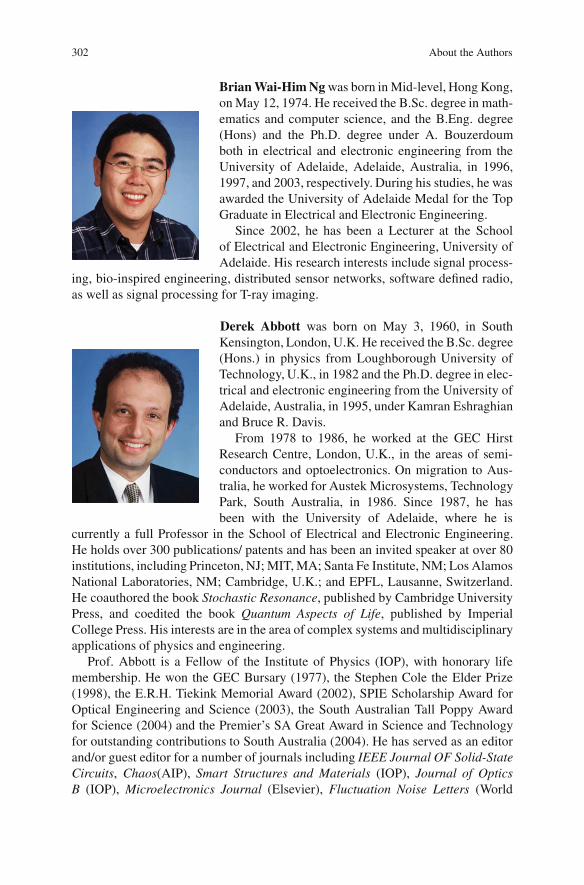
302 About the Authors
Brian Wai-Him Ng was born in Mid-level, Hong Kong,on May 12, 1974. He received the B.Sc. degree in math-ematics and computer science, and the B.Eng. degree(Hons) and the Ph.D. degree under A. Bouzerdoumboth in electrical and electronic engineering from theUniversity of Adelaide, Adelaide, Australia, in 1996,1997, and 2003, respectively. During his studies, he wasawarded the University of Adelaide Medal for the TopGraduate in Electrical and Electronic Engineering.
Since 2002, he has been a Lecturer at the Schoolof Electrical and Electronic Engineering, University ofAdelaide. His research interests include signal process-
ing, bio-inspired engineering, distributed sensor networks, software defined radio,as well as signal processing for T-ray imaging.
Derek Abbott was born on May 3, 1960, in SouthKensington, London, U.K. He received the B.Sc. degree(Hons.) in physics from Loughborough University ofTechnology, U.K., in 1982 and the Ph.D. degree in elec-trical and electronic engineering from the University ofAdelaide, Australia, in 1995, under Kamran Eshraghianand Bruce R. Davis.
From 1978 to 1986, he worked at the GEC HirstResearch Centre, London, U.K., in the areas of semi-conductors and optoelectronics. On migration to Aus-tralia, he worked for Austek Microsystems, TechnologyPark, South Australia, in 1986. Since 1987, he hasbeen with the University of Adelaide, where he is
currently a full Professor in the School of Electrical and Electronic Engineering.He holds over 300 publications/ patents and has been an invited speaker at over 80institutions, including Princeton, NJ; MIT, MA; Santa Fe Institute, NM; Los AlamosNational Laboratories, NM; Cambridge, U.K.; and EPFL, Lausanne, Switzerland.He coauthored the book Stochastic Resonance, published by Cambridge UniversityPress, and coedited the book Quantum Aspects of Life, published by ImperialCollege Press. His interests are in the area of complex systems and multidisciplinaryapplications of physics and engineering.
Prof. Abbott is a Fellow of the Institute of Physics (IOP), with honorary lifemembership. He won the GEC Bursary (1977), the Stephen Cole the Elder Prize(1998), the E.R.H. Tiekink Memorial Award (2002), SPIE Scholarship Award forOptical Engineering and Science (2003), the South Australian Tall Poppy Awardfor Science (2004) and the Premier’s SA Great Award in Science and Technologyfor outstanding contributions to South Australia (2004). He has served as an editorand/or guest editor for a number of journals including IEEE Journal OF Solid-StateCircuits, Chaos(AIP), Smart Structures and Materials (IOP), Journal of OpticsB (IOP), Microelectronics Journal (Elsevier), Fluctuation Noise Letters (World
About the Authors 303
Scientific), and is currently on the Editorial Boards of Proceedings of the IEEE andIEEE Photonics. He has appeared on national and international television and radioand has also received scientific reportage in New Scientist, The Sciences, ScientificAmerican, Nature, The New York Times, and Sciences et Avenir. He has served ona number of IEEE technical program committees, including the IEEE APCCS andthe IEEE GaAs IC Symposium.
Scientific Genealogy
Index
AAbsorption coefficient, 28, 48, 51, 66, 151,
163, 176, 180, 187, 192, 196Approximate sub-images, 193AR models, wavelet subbands
DWPT, 152HBO, 149IDWT, 152Mallat’s algorithm, 151motivation, 151–152NHB and HOS cells classification
block diagram, 154, 155percentage classification accuracy, 155,
156squared error variances, 155, 156
parametric modeling procedure, 149powder sample classification
Durbin’s algorithm, 156–157imaging system adoption, 159Mahalanobis distance classifier, 156scatter plots, 157–158squared error variance, 156, 157
SURE, 152terahertz pulse measurements, 150–151time-domain signals comparison, 149, 152,
153wavelet preprocessing, 152–153
Asynchronous optical sampling (ASOPS)technique, 24–25
Autoregressive (AR) model, 98–99Autoregressive moving average (ARMA)
model, 99–100
BBack-projection algorithms, 253–254Balanced silicon photodiodes, 23
Band-gap semiconductors, 20Basal cell carcinoma (BCC)
internal structure, 59, 60skin cancer, 53terahertz absorption, 48visualized multispectral clustering
techniques, 59Basic Prony method, 99, 156, 157Bayesian models, 247BCC. See Basal cell carcinoma (BCC)Best wavelet packet tree, 113–115Bhattacharyya divergence, 96Binary classifiers. See Support vector machines
(SVMs)Biomedical imaging, 2D and 3D
brain section detection, 55, 56cancer cell detection
breast, 54, 55ductal carcinoma, 55skin, 53–54
cortical cortex, 56gray matter, 56human tooth and pork sample, 53Rayleigh scattering, 52tablet coating detection, 55–56
BiorSplines biorthogonal wavelet,203
BioSplines2.2 biorthogonal wavelet,204
Biorthogonal wavelets, 78, 151, 152Born series, 35Bound-to-continuum (BTC) quantum cascade
laseradvantages, 15design and performance, 15, 16waveguide design, 16
Bound-to-continuum transitions, 11, 222
X. Yin et al., Terahertz Imaging for Biomedical Applications: Pattern Recognitionand Tomographic Reconstruction, DOI 10.1007/978-1-4614-1821-4,© Springer Science+Business Media, LLC 2012
305
306 Index
CCancer cell identification
biomedical imaging, 2D and 3D, 53–55statistical models, 248
Canny edge detector, 197Cauchy–Schwartz inequality, 256Charge coupled device (CCD), 35, 52, 183,
248, 261–265Chirped probe pulse terahertz imaging system,
181, 182Chirped superlattice, 11, 15, 222CIL functions. See Complex insertion loss
(CIL) functionsClassifier design
description, 127, 130evaluation, 127–129learning and adaption, 127, 128overfitting, 129–131
Coherent terahertz radiation detectiontechnique, 22–24
Cold finger of a continuous-helium-cooledflow cryostat, 6, 223, 224, 268, 270
Complex insertion loss (CIL) functionspattern recognition, 177wavelet packet algorithms, 141–144
Complex refractive index, 31, 49, 186Computed tomography (CT) imaging
cross-correlation algorithm, 188description, 179–180diffraction grating pair characteristics
electrooptic detection, 185–186geometry of, 185grating equation, 185GVD, 183simplified pulse compressor, 185
methodologychirped probe pulse terahertz imaging
system, 181, 182FBP algorithm, 180IRT, 180sinogram, 180, 181
reconstruction parametersfrequency-domain sinogram, 186–187time-domain sinogram, 187–188
THz sampling pattern, 183, 184Continuous flow helium-cooled cryostat, 6,
223, 224, 268, 270Continuous wave (CW) THz imaging system
all-optoelectronic, 37, 39drawback, 40logarithmic power transmission, 39–40schematic representation, 37, 39vs. TPI, 3, 4validation, 39–40
Cross-correlation algorithm, 188Cross-validation (CV) methods
K-fold, 128–129LOO, 129SVM, 165–166
Crystalline states, 47, 49CT imaging. See Computed tomography (CT)
imagingCurvelet transforms, 246
DData representation, SVM
powder, 161, 163–164RNA, 161–163TDS functional imaging, 160
Daubechies 4 wavelet, 196, 197Debye relaxation model, 48Depth resolution, 3, 10, 30, 37, 247Deterministic–stochastic models, 108–109Dichotomy, 125Diffraction grating pair
electrooptic detection, 185–186geometry of, 185grating equation, 185GVD, 183simplified pulse compressor, 185
Discrete wavelet packet transforms (DWPTs),152
discrete wavelet packet coefficientcalculation, 87–88
procedure, 86, 88, 89properties of, 86–87time-localization tree, 86wavelet packet bases
cost functions, 90–91evaluation, 88–89linear searching procedure, 91
Discrete wavelet transforms (DWTs)one dimensional, 78–79two dimensional, 83–85, 195–196
DL-mandelic acidclassification results comparison, 141evaluation via discrimination metric, 146identification scheme, 148–149time-domain responses, 142wavelet packet identification, 147
Drude–Lorentz model, 20Durbin’s method, 99, 100, 118, 156, 1572D wavelet-based CT reconstruction
coefficients, 203FBP algorithm, 203ramp filters, 203, 204scaling, 204
Index 307
2D wavelet scale correlation-basedsegmentation
algorithm, 196–197Canny edge detector, 197Otsu’s threshold method, 197
DWPTs. See Discrete wavelet packettransforms (DWPTs)
DWTs. See Discrete wavelet transforms(DWTs)
EEdge detection techniques, 191Eight neighboring pixels, 161, 229, 230Electrooptic rectification. See Optical
rectification mechanismEnhanced T-ray signal classification, wavelet
preprocessingamplitude scatter plot, 138, 139classification, 135, 136experimental results
preprocessed signals, 137–140raw signals, 140
feature extraction, 134–135LOO error estimator, 135–136
Error analysislocal computed tomography
description, 225hole image characteristics, 226–228segmentation strategy, 228–231
local reconstruction, 206–207wavelet-based local reconstruction
Cauchy–Schwartz inequality, 256Radon transform error, 255ramp filters, 257–258relative error, 256ROI, 257
Euclidean discrimination metric, 121–122Euclidean distance classifier, 58, 61, 62
FFast wavelet transform (FWT)
analysis and synthesis stages, 80description, 79inverse transform, 80J-level, 81one-step reconstruction, 81time-frequency boxes, 82
FBP algorithms. See Filtered back-projection(FBP) algorithms
FCM. See Fuzzy c-means (FCM)Feature extraction
considerations, 96–97
enhanced T-ray signal classification,wavelet preprocessing, 134–135
feature matrix calculations, 100–102Fourier transform, 97–98objective, 100parameter estimation
ARMA model, 99–100AR model, 98–99
pattern classification, 131role of, 95–96SVM
kernel selection and parameter tuning,165
via frequency orientation components,164–165
system identificationdescription, 102optimized Mertz apodization functions,
115–117subspace identification algorithm,
103–109wavelet-packet identification, 109–115
Feature selection. See Feature extractionFELs. See Free-electron lasers (FELs)Femtosecond laser-based T-ray functional
imaging system, 133, 134Filtered back-projection (FBP) algorithms
CT imaging, 1802D wavelet-based CT reconstruction, 203
Fourier coefficient spectrummulticlass powder classification via,
174–175RNA classification via, 171–173
Fourier spectrum analysismulticlass classification, 168–171poly-A and ploy-C T-ray pulses, 166–169
Fourier transformfeature extraction, 97–98infrared spectroscopy, 49, 57
Free-electron lasers (FELs), 14Frequency-domain sinogram, 186–187Frequency-domain terahertz spectroscopy
absorption and detectionbiological tissue, 48conformational structure, 49covalent bond, 48liquid dynamics, 48molecular signatures, 48–51solvation, 48water molecule vibrational modes,
47–48electromagnetic (EM) wave interactions,
46, 47Frequency orientation component, 98
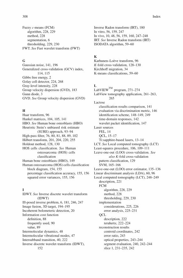
308 Index
Fuzzy c-means (FCM)algorithm, 228, 229method, 228segmentation, 6thresholding, 229, 230
FWT. See Fast wavelet transform (FWT)
GGaussian noise, 141, 196Generalized cross-validation (GCV) index,
114, 115Gibbs free energy, 2Golay cell detector, 224, 268Gray-level intensity, 228Group velocity dispersion (GVD), 183Gunn diode, 3GVD. See Group velocity dispersion (GVD)
HHaar transform, 96Hankel matrices, 104, 105, 141HBO. See Human bone osteoblasts (HBO)Heuristic Stein’s unbiased risk estimate
(SURE) approach, 93–94High-pass filter, 76, 80, 81, 88, 89, 102Hilbert transform, 201, 204, 220, 255Holdout method, 128, 130HOS cells classification. See Human
osteosarcoma (HOS) cellsclassification
Human bone osteoblasts (HBO), 149Human osteosarcoma (HOS) cells classification
block diagram, 154, 155percentage classification accuracy, 155, 156squared error variances, 155, 156
IIDWT. See Inverse discrete wavelet transform
(IDWT)Ill-posed inverse problem, 6, 181, 246, 247Image fusion, 3D target, 194–195Incoherent bolometeric detection, 20Information cost function
definition, 88frequently used, 90value, 89
Intermolecular dynamics, 48Intermolecular vibrational modes, 47Intersubband transition, 40, 222Inverse discrete wavelet transform (IDWT),
152
Inverse Radon transform (IRT), 180In vitro, 56, 159, 247In vivo, 10, 48, 56, 159, 160, 247–248IRT. See Inverse Radon transform (IRT)ISODATA algorithm, 59–60
KKarhunen–Loeve transform, 96K-fold cross-validation, 128–130Kirchhoff migration, 34K-means classifications, 59–60
LLabVIEW
TMprogram, 271–274
LabView tomography application, 261–263,265
Lactoseclassification results comparison, 141evaluation via discrimination metric, 146identification scheme, 148–149, 249time-domain responses, 142wavelet packet identification, 147
Laser sourcesFEL, 14QCL, 15–17Ti:sapphire-based lasers, 13–14
LCT. See Local computed tomography (LCT)Least-squares procedure, 106, 109–111Leave-one-out (LOO) cross-validation. See
also K-fold cross-validationpattern classification, 129SVM, 165–166
Leave-one-out (LOO) error estimator, 135–136Linear discriminant analysis (LDA), 60, 96Local computed tomography (LCT), 248–249
description, 221FCM
algorithm, 228, 229method, 228thresholding, 229, 230
implementationconsiderations, 225, 226error analysis, 225–231
QCLdescription, 222terahertz, 222–224
reconstruction resultscentroid coordinates, 242error ratio, 243optical properties, 243–244segment evaluation, 240, 242–244slice 1, 231–235, 242
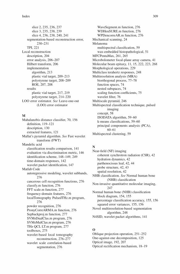
Index 309
slice 2, 235, 236, 237slice 3, 235, 238, 239slice 4, 236, 238, 240, 241
segmentation-based reconstruction error,230–231
TPI, 221Local reconstruction
description, 204error analysis, 206–207Hilbert transform, 206implementation
algorithm, 213plastic vial target, 209–213polystyrene target, 208–209ROE, 207, 208
resultsplastic vial target, 217, 219polystyrene target, 214–220
LOO error estimator. See Leave-one-out(LOO) error estimator
MMahalanobis distance classifier, 70, 156
definition, 119–121description, 130extracted features, 121
Mallat’s pyramid algorithm. See Fast wavelettransform (FWT)
Mandelic acidclassification results comparison, 141evaluation via discrimination metric, 146identification scheme, 148–149, 249time-domain responses, 142wavelet packet identification, 147
Matlab Codeautoregressive modeling, wavelet subbands,
276cancerous cell recognition functions, 276classify.m function, 276FFT scale.m function, 277frequency-domain features, 276localTomography PulsedTHz.m program,
276powder recognition, 276PronyCorreARMA.m function, 276Sepbackproj.m function, 277SVMsDualClas.m program, 276SVMsMulClas.m program, 276THz QCL LT.m program, 277toolboxes, 275wavelet-based local tomography
reconstruction, 276–277wavelet scale correlation-based
segmentation, 276
WaveSegment.m function, 276WDHeuSURE.m function, 276WPDencorreAR.m function, 276
Mechanical scanning, 24Melanoma
multispectral classification, 59wax-embedded histopathological, 51
MFCPentaMax, 261, 265Microbolometer focal-plane array camera, 41Molecular beam epitaxy, 11, 15, 222, 223, 268Morphological operations, 229Multiclass terahertz responses, 248Multiresolution analysis (MRA)
biorthogonal process, 77–78function spaces, 74nested subspaces, 74scaling function coefficients, 75wavelet filter, 76
Multiscale pyramid, 246Multispectral classification technique, pulsed
imagingconcept, 58ISODATA algorithm, 59–60k-means classifications, 59–60principal components analysis (PCA),
60–61Multispectral clustering, 59
NNear-field (NF) imaging
coherent synchrotron radiation (CSR), 42hydration dynamics, 42parthenocissus leaf, 42, 44probe structure, 42, 43spatial resolution, 42
NHB classification. See Normal human bone(NHB) classification
Non-invasive quantitative molecular imaging,247
Normal human bone (NHB) classificationblock diagram, 154, 155percentage classification accuracy, 155, 156squared error variances, 155, 156
Novel multiresolution-based segmentationalgorithm, 248
N4SID, wavelet packet algorithms, 141
OOblique projection operation, 251–252One-against-one decomposition, 125Optical image, 192, 207Optical rectification mechanism, 18–19
310 Index
Optical sampling techniquesASOPS, 24–25coherent terahertz radiation detection,
22–24electro-optic, 22–23photoconductive sampling, 23–24synchronous, 24
Optimized Mertz apodization functions,115–117
Optoelectronic sampling techniques. SeeOptical sampling techniques
Otsu’s threshold, 197, 228Overfitting, 129–131
PParabolic mirrors, 28, 43, 182, 224, 262–264,
268, 271Parameter estimation
ARMA model, 99–100AR model, 98–99
Parametric modeling, 149, 177Parzen estimator, 96Pattern classification
classifier designdescription, 127, 130evaluation, 127–129learning and adaption, 127, 128overfitting, 129–131
Cover’s theorem, 124Euclidean discrimination metric, 121–122feature extraction, 131holdout method, 128kernel function, 124K-fold cross-validation, 128–129LOO cross-validation, 129Mahalanobis distance
definition, 119–121description, 130via extracted features, 121
SVMsbinary classification, 123–125description, 122pairwise classification, 125–126
Pattern recognition/formationAR models, wavelet subbands
DWPT, 152HBO, 149IDWT, 152Mallat’s algorithm, 151motivation, 151–152NHB and HOS cells classification,
154–156parametric modeling procedure, 149
powder samples classification, 156–159SURE, 152terahertz pulse measurements, 150–151time-domain signals comparison, 149,
152, 153wavelet preprocessing, 152–153
biomedical samplesextracted parameters, 58multispectral classification, 58–61parameter combinations, 57SVM, 63wavelet domain classification, 61–62
SVMsadvantages, 160data representation, 161–164feature extraction, 164–165Fourier coefficient spectrum, 171–175Fourier spectrum analysis, 166–171performance assessment, of
classification, 165–166potential of, 160
T-rayschicken and beef sample, 66, 67data acquisition, 68data preprocessing and extraction, 69,
70data recognition system, 68decision classification, 70finite impulse response (FIR), 66, 67goals, 65pattern representation, machine
recognition, 70pattern space, 69preprocessing procedure, 69–70signal processing techniques, 66significance, 65–66SVMs, 159–160THz scanner system, 68
T-ray time-domain techniquesenhanced T-ray signal classification,
wavelet preprocessing, 134–140femtosecond laser-based T-ray
functional imaging system, 133,134
spectroscopy and imaging, 133–134THz-TDS experiment setup, 133, 134
wavelet packet algorithmsCIL functions, 144complex insertion loss, 141–144discrimination metric evaluation,
146–148evaluation, 141–143identification scheme, 148–149N4SID, 141
Index 311
signal processing, noisy background,143–145
sixth-order Butterworth band-pass filter,142
SNRs, 144, 145subspace, 141–144time-domain responses, 142, 143wavelet packet tree, 142, 144
Perfect reconstruction filter banks, 83Perfect reconstruction quadrature mirror filter
banks, 91, 151Per-pixel classification, 228Phase matching condition, 18Photoconductive antenna (PCA)
photoconductive sampling, 24THz response detection, 3
Photoconductive emitter, 30Photoconductive sampling technique, 23–24Photo-Dember effect, 20Photomicrograph, 54, 55Photomixing techniques, 21, 22Picometrix system software, 263, 266Picomolar sensitivity, 2Planar antennas, 9Plastic vial target, local reconstruction
implementation, 209–213results, 217, 219
Pockel effect, 23Polarizing beam splitter, 12, 23, 262Polyadenylic acid (poly-A), 46, 161–163,
166–168, 176Polycytidylic acid (poly-C), 46, 161–163,
166–168, 176Polymorphs, 51, 150Polystyrene target
local reconstruction, 208–209results
centered area images, 214, 216–217off-center area images, 214, 217–220wavelet and scaling ramp filtering, 214,
215Positron emission tomography (PET), 2Powder data representation, SVM, 161,
163–164Prediction error variance (PEV), 99Prism pairs, 13, 14Pump-probe system
delay stage, 24, 25synchronous optical sampling, 24
QQCLs. See Quantum cascade lasers (QCLs)Quadratic programming problem, 117, 124
Quantum cascade lasers (QCLs)description, 222optical analysis, 249terahertz
description, 222–223GaAs-AlGaAs bound-to-continuum
superlattice design, 223imaging system, 223, 224LIA, 223setup, 223, 224
RRadon transformRatioing procedure, 148, 149, 177Reconstruction algorithm, 213Reconstruction filters, 81, 83–85Reflection imaging, 30, 34Refractive index, 13, 18, 28, 31, 42, 46, 49, 51,
57, 69, 176, 180, 186Region-based segmentation, 229Resonant-phonon (RP) scheme, 17Retinal isomers, 50RNA data representation, SVM, 161–163
SSapphire substrate, 42Scaling filter, 75, 76, 204, 209, 213, 255Segmentation-by-fusion, 191Segmentation techniques
description, 191–192extracted object segments, 197, 198segmentation quality, 197–199T-ray CT
imaging, 191signals and spectra, 193, 194slices, 192, 193system, 192
wavelet-based segmentation, by fusiondescription, 1932D wavelet scale correlation-based
segmentation, 196–197DWT, 2D, 195–196image fusion, 3D target, 194–195
Semiconductor sourcesdepletion/accumulation field, 19electron-hole pairs, 19, 20emission bandwidth, 20optical rectification, 18–19photomixing techniques, 21, 22ultrafast charge transport, 19–21
Sigma-Aldrich, 161Signal compression, 73, 92, 98, 152
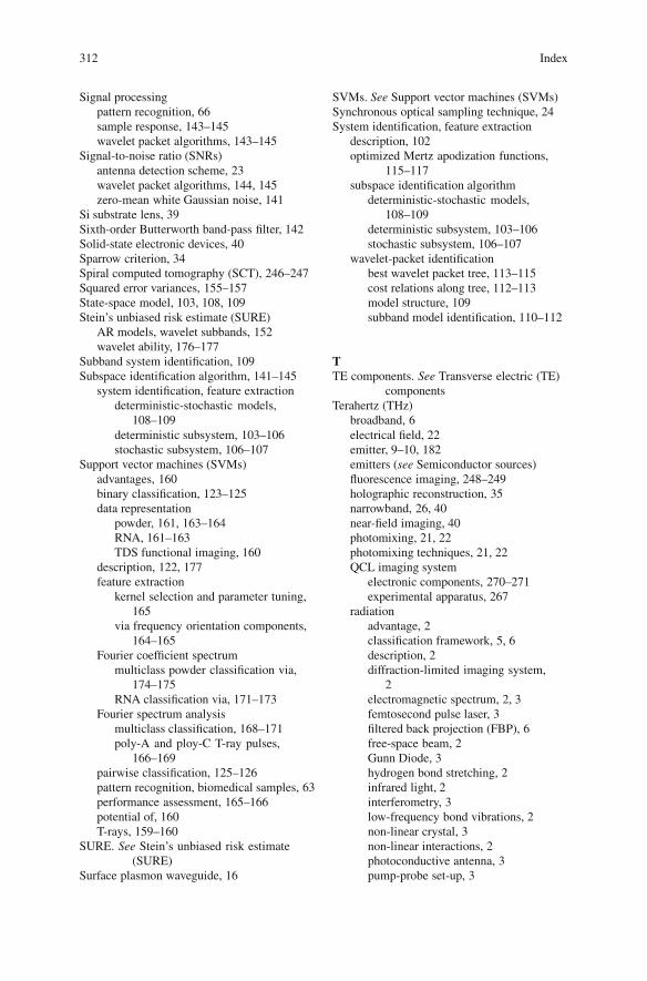
312 Index
Signal processingpattern recognition, 66sample response, 143–145wavelet packet algorithms, 143–145
Signal-to-noise ratio (SNRs)antenna detection scheme, 23wavelet packet algorithms, 144, 145zero-mean white Gaussian noise, 141
Si substrate lens, 39Sixth-order Butterworth band-pass filter, 142Solid-state electronic devices, 40Sparrow criterion, 34Spiral computed tomography (SCT), 246–247Squared error variances, 155–157State-space model, 103, 108, 109Stein’s unbiased risk estimate (SURE)
AR models, wavelet subbands, 152wavelet ability, 176–177
Subband system identification, 109Subspace identification algorithm, 141–145
system identification, feature extractiondeterministic-stochastic models,
108–109deterministic subsystem, 103–106stochastic subsystem, 106–107
Support vector machines (SVMs)advantages, 160binary classification, 123–125data representation
powder, 161, 163–164RNA, 161–163TDS functional imaging, 160
description, 122, 177feature extraction
kernel selection and parameter tuning,165
via frequency orientation components,164–165
Fourier coefficient spectrummulticlass powder classification via,
174–175RNA classification via, 171–173
Fourier spectrum analysismulticlass classification, 168–171poly-A and ploy-C T-ray pulses,
166–169pairwise classification, 125–126pattern recognition, biomedical samples, 63performance assessment, 165–166potential of, 160T-rays, 159–160
SURE. See Stein’s unbiased risk estimate(SURE)
Surface plasmon waveguide, 16
SVMs. See Support vector machines (SVMs)Synchronous optical sampling technique, 24System identification, feature extraction
description, 102optimized Mertz apodization functions,
115–117subspace identification algorithm
deterministic-stochastic models,108–109
deterministic subsystem, 103–106stochastic subsystem, 106–107
wavelet-packet identificationbest wavelet packet tree, 113–115cost relations along tree, 112–113model structure, 109subband model identification, 110–112
TTE components. See Transverse electric (TE)
componentsTerahertz (THz)
broadband, 6electrical field, 22emitter, 9–10, 182emitters (see Semiconductor sources)fluorescence imaging, 248–249holographic reconstruction, 35narrowband, 26, 40near-field imaging, 40photomixing, 21, 22photomixing techniques, 21, 22QCL imaging system
electronic components, 270–271experimental apparatus, 267
radiationadvantage, 2classification framework, 5, 6description, 2diffraction-limited imaging system,
2electromagnetic spectrum, 2, 3femtosecond pulse laser, 3filtered back projection (FBP), 6free-space beam, 2Gunn Diode, 3hydrogen bond stretching, 2infrared light, 2interferometry, 3low-frequency bond vibrations, 2non-linear crystal, 3non-linear interactions, 2photoconductive antenna, 3pump-probe set-up, 3
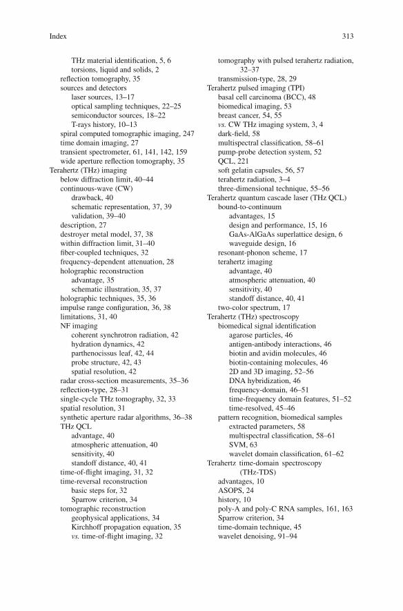
Index 313
THz material identification, 5, 6torsions, liquid and solids, 2
reflection tomography, 35sources and detectors
laser sources, 13–17optical sampling techniques, 22–25semiconductor sources, 18–22T-rays history, 10–13
spiral computed tomographic imaging, 247time domain imaging, 27transient spectrometer, 61, 141, 142, 159wide aperture reflection tomography, 35
Terahertz (THz) imagingbelow diffraction limit, 40–44continuous-wave (CW)
drawback, 40schematic representation, 37, 39validation, 39–40
description, 27destroyer metal model, 37, 38within diffraction limit, 31–40fiber-coupled techniques, 32frequency-dependent attenuation, 28holographic reconstruction
advantage, 35schematic illustration, 35, 37
holographic techniques, 35, 36impulse range configuration, 36, 38limitations, 31, 40NF imaging
coherent synchrotron radiation, 42hydration dynamics, 42parthenocissus leaf, 42, 44probe structure, 42, 43spatial resolution, 42
radar cross-section measurements, 35–36reflection-type, 28–31single-cycle THz tomography, 32, 33spatial resolution, 31synthetic aperture radar algorithms, 36–38THz QCL
advantage, 40atmospheric attenuation, 40sensitivity, 40standoff distance, 40, 41
time-of-flight imaging, 31, 32time-reversal reconstruction
basic steps for, 32Sparrow criterion, 34
tomographic reconstructiongeophysical applications, 34Kirchhoff propagation equation, 35vs. time-of-flight imaging, 32
tomography with pulsed terahertz radiation,32–37
transmission-type, 28, 29Terahertz pulsed imaging (TPI)
basal cell carcinoma (BCC), 48biomedical imaging, 53breast cancer, 54, 55vs. CW THz imaging system, 3, 4dark-field, 58multispectral classification, 58–61pump-probe detection system, 52QCL, 221soft gelatin capsules, 56, 57terahertz radiation, 3–4three-dimensional technique, 55–56
Terahertz quantum cascade laser (THz QCL)bound-to-continuum
advantages, 15design and performance, 15, 16GaAs-AlGaAs superlattice design, 6waveguide design, 16
resonant-phonon scheme, 17terahertz imaging
advantage, 40atmospheric attenuation, 40sensitivity, 40standoff distance, 40, 41
two-color spectrum, 17Terahertz (THz) spectroscopy
biomedical signal identificationagarose particles, 46antigen-antibody interactions, 46biotin and avidin molecules, 46biotin-containing molecules, 462D and 3D imaging, 52–56DNA hybridization, 46frequency-domain, 46–51time-frequency domain features, 51–52time-resolved, 45–46
pattern recognition, biomedical samplesextracted parameters, 58multispectral classification, 58–61SVM, 63wavelet domain classification, 61–62
Terahertz time-domain spectroscopy(THz-TDS)
advantages, 10ASOPS, 24history, 10poly-A and poly-C RNA samples, 161, 163Sparrow criterion, 34time-domain technique, 45wavelet denoising, 91–94
314 Index
Time delay scan, 3Time delay stage, 43Time-domain sinogram, 187–188Time-frequency localisation, 74, 88, 94, 118,
149, 246, 248Time-of-flight imaging, 31, 32Time-resolved terahertz spectroscopy, 45–46Time-reversal reconstruction
basic steps for, 32Sparrow criterion, 34
Ti:sapphire-based lasersprinciple of, 13schematic illustration, 12–14
Toeplitz covariance matrix, 107Tomographic reconstruction
geophysical applications, 34Kirchhoff propagation equation, 35SCT, 246–247statistical reconstruction, 247vs. time-of-flight imaging, 32in vivo THz fluorescence imaging, 247–248
Torsional modes, 49, 50TPI. See Terahertz pulsed imaging (TPI)Traditional back-projection algorithm, 215Transient photocarriers, 24Transient polarization, 19Transverse electric (TE) components, 20Transverse magnetic, 20T-ray computed tomography
imaging, 191signals and spectra, 193, 194slices, 192, 193system, 192
T-ray pattern recognition. See Patternrecognition, T-rays
T-ray time-domain techniquesenhanced T-ray signal classification,
wavelet preprocessingamplitude scatter plot, 138, 139classification, 135, 136feature extraction, 134–135LOO error estimator, 135–136preprocessed signals, 137–140raw signals, 140
femtosecond laser-based T-ray functionalimaging system, 133, 134
spectroscopy and imaging, 133–134THz-TDS experiment setup, 133, 134
Two-channel filter banks, 82–83Two-class terahertz responses, 248Two-color THz QCL spectrum, 17Two-dimensional discrete wavelet transforms,
83–85
UUltrafast charge transport
optical rectification, 19–21semiconductor sources, 19–21
Ultrafast T-ray pulsed imagingcontinuous-wave T-ray imaging
LabVIEWTM
program, 271–274terahertz QCL system, 267–271
crossed-polariser detection, 259–261hardware specifications, 261–263software applications
LabView tomography application,261–263, 265
MFCPentaMax, 261, 265Picometrix system software, 263, 266
ultrafast laser, 259, 260Ultrashort pulse technique, 31Upper state wave function, 16
VVector space, 76Volume space, 246
WWaveguide losses, 16Wavelet and scaling ramp filters, 201, 206,
210, 232Wavelet-based local tomography
reconstructionmatlab Code, 276–277
Wavelet-based segmentation, by fusiondescription, 1932D wavelet scale correlation-based
segmentation, 196–197DWT, 2D, 195–196image fusion, 3D target, 194–195
Wavelet-based terahertz coherent localtomography
description, 2012D IDWT, 204, 2052D wavelet-based CT reconstruction
coefficients, 204FBP algorithm, 203ramp filters, 204, 205scaling, 204
goal, 201implementation
algorithm, 213plastic vial target, 209–213polystyrene target, 208–209ROE, 207, 208
Index 315
local reconstructiondescription, 204error analysis, 206–207Hilbert transform, 206
motivation, 201–203reconstruction algorithm, 213reconstruction results
plastic vial target, 217, 219polystyrene target, 214–220
Wavelet denoisingFIR filters, 92heuristic SURE approach, 93–94procedure, 92reconstruction quadrature mirror filter
banks, 91T-ray project
dyadic downsampling, 93filter bank structure, 93soft threshold operation, 93SURE approach, 93–94time-domain measurement, 92
T-ray pulse noise, 91Wavelet filter, 5, 83–85, 89, 213Wavelet packet algorithms
CIL functions, 144complex insertion loss, 141–144discrimination metric evaluation, 146–148evaluation, 141–143identification scheme, 148–149N4SID, 141signal processing, noisy background,
143–145sixth-order Butterworth band-pass filter,
142SNRs, 144, 145subspace, 141–144time-domain responses, 142, 143wavelet packet tree, 142, 144
Wavelet-packet identificationof lactose responses, 142, 144system identification, feature extraction
best wavelet packet tree, 113–115cost relations along tree, 112–113model structure, 109subband model identification, 110–112
Wavelet preprocessingamplitude scatter plot, 138, 139classification, 135, 136feature extraction, 134–135
LOO error estimator, 135–136preprocessed signals, 137–140raw signals, 140
Wavelet scale correlation, 191–193, 195–197,248, 276
Wavelet series expansion, 78–79Wavelet transforms (WT)
description, 73–74DWPTs, 85–91DWT, 195–196feature extraction, 73image enhancement, 73MRA
biorthogonal process, 77–78function spaces, 74nested subspaces, 74scaling function coefficients, 75wavelet filter, 76
noise removal, 73one dimension
DWT, 78–79FWT, 79–82two-channel filter bank, 82–83wavelet series expansion, 78–79
signal compression, 73two dimensional discrete, 83–85wavelet denoising
FIR filters, 92heuristic SURE approach, 93–94procedure, 92reconstruction quadrature mirror filter
banks, 91T-ray project, 92–94T-ray pulse noise, 91
Wave oscillators, 3White Gaussian process, 98Wide aperture reflection tomography (WART),
35Wiener–Hopf equation, 67WT. See Wavelet transforms (WT)
XX-ray imaging, 222X–y translation stage, 150, 151, 163
ZZnTe sensor, 36, 37