ap biology molecular genetics unit chapters 16 & 17
TRANSCRIPT

AP BiologyMolecular Genetics UnitChapters 16 & 17

Chapter 16 Objectives1. Explain how Griffith and Avery’s experiment with pneumococcus bacteria
contributed to the understanding of molecular genetics. 2. Define transformation. Use Griffith & Avery's results to explain how it occurs.3. Using Hershey & Chase's procedure, use given hypotheses to predict results.
Evaluate each hypothesis based upon the actual results of the experiments.4. Identify the component parts of nucleic acids. Describe their organization into
DNA and RNA.5. Explain how Chargaff's rule supports the base pairing rule.6. Explain how the two strands of a DNA molecule are complementary.7. Explain how DNA replication is semiconservative.8. Outline the process of DNA replication. Include the names and roles of any
enzymes involved in the process.9. Explain why one strand of DNA is replicated continuously, while the other
strand is discontinuous.10. Define Telomere. Explain why the mechanism of replication results in the
shortening of telomeres.11. Explain how DNA condenses into chromosomes in eukaryotic cells.

What part of the cell controls heredity?
• Actual traits are not inherited. Traits are developed. Inheritance must come from something that is in the gametes and in the resulting zygote
• Since the gametes and zygote are cells, the control center of heredity must have some cellular origin
• Cells have 3 fundamental regions• Cell membrane• Cytoplasm• Nucleus
• One of these 3 regions must control heredity & determine how traits will develop in the organism

What part of the cell controls heredity?
• It follows logically that hereditary control is centered either in the membrane, the cytoplasm, or the nucleus.
• We will assume two alternative hypotheses:1. The nucleus controls heredity2. The cytoplasm controls heredity
• An experiment to test these must allow for simple manipulation of cell parts and simple observation of developing traits
• Manipulating cell parts would be simplified by using a single celled organism, but it must be large enough to work with and observe easily, and must have some distinct inherited trait.

1943 - Hammerling’s Experiment
• The giant unicellular algae of the genus Acetabularia are an ideal experimental subject. • They are large,
easy to manipulate, and easy to observe

Hammerling’s Experiment• All Acetabularia
species have a base (which contains the nucleus), a stalk, and a cap. • The base and stalk
are similar between species, but the cap varies greatly
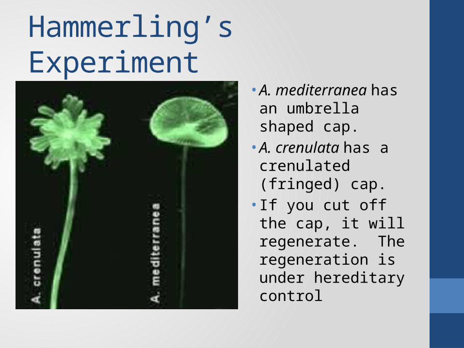
Hammerling’s Experiment• A. mediterranea has
an umbrella shaped cap. • A. crenulata has a
crenulated (fringed) cap.• If you cut off the cap,
it will regenerate. The regeneration is under hereditary control

Experimental Design:
• Cut off the cap of each alga• Remove a section of the stalk (which contains cytoplasm,
but not the nucleus)• Place a stalk from A. crenulata on a base (containing a
nucleus) of A. mediterannea (Experiment 1)• Place a stalk from A. mediterannea on a base of A.
crenulata (Experiment 2)• Observe the regeneration of the cap on each “hybrid”
alga

Exp. #1 – Stalk C on Base M• If the cytoplasm
controls heredity . . .• The cap that
regenerates will follow instructions from the cytoplasm in the stalk. • The cap should
resemble type C
• If the nucleus controls heredity . . . • The cap that
regenerates will follow instructions from the nucleus in the base• The cap should
resemble type M

Exp. #1 – Stalk C on Base M• The algae
regenerated type M caps, suggesting the nucleus in the base of the cell controlled the development of the cap

Exp. #2 – Stalk M on Base C• Similarly,
algae with a nucleus from type C regenerated a crenulated cap.• In both
experiments, the nucleus controlled regeneration

The Nucleus as Control Center• If the nucleus is the
center of hereditary control, there must be something inside of it that actually directs the development of traits.
• The nucleus consists almost entirely of:• Proteins• Nucleic Acids• DNA• RNA

What material in the nucleus controls heredity?• Nucleic Acids?• DNA and RNA• Complex polymer
made of nucleotides• Nitrogenous bases
are variable• 4 types (AGCT)
• Protein?• Complex polymer made
of amino acids• Amino acid side chains
are variable• 20 different types of
essential amino acids commonly found in protein• With more variability,
protein was the favored hypothesis

What material in the nucleus controls heredity?• To test these hypotheses, we need to separate the proteins
from the nucleic acids, treat a test subject with each, and observe the results.
• Viruses are made entirely out of nucleic acids and proteins.• Viruses inject their genetic material into a host cell, take over
the hereditary machinery of the host, and use it to reproduce itself.
• We can use viruses as a test subject if we can find a way to easily identify or manipulate the proteins and the nucleic acids.

Tobacco Mosaic Virus & Holmes Ribgrass Virus
• Tobacco Mosaic Virus (TMV) was the first virus discovered and isolated.
• TMV infects plants, and forms characteristic lesions that are identifiable as tobacco mosaic disease. Holmes ribgrass virus (HRV) also infects plants. The lesions formed in Holmes Ribgrass disease are distinctly different from TMV lesions
• Viruses consist of a protein coat surrounding a core of nucleic acids (RNA in the case of these 2 viruses)
• Viruses inject their host with their genetic material, and take over the host for the purpose of replication

Heinz Fraenkel-Conrat 1955• In the first of a series of experiments, Fraenkel-Conrat
enzymatically digested the protein coat from TMV viruses and isolated the RNA core (and vice versa)
• He then infected tobacco plants with only the protein he derived from the virus. The plants did not develop tobacco mosaic disease
• He also infected tobacco plants with only the RNA from his viruses. These plants developed tobacco mosaic lesions
• These results suggest that nucleic acids control the heredity of the virus and the characteristics associated with the viral disease
• Remember, protein was the favored hypothesis for the hereditary material (due to its greater structural variability)!

Fraenkel-Conrat; Hybrid Viruses
• In this experiment, he created hybrid viruses consisting of the protein coat from HRV and an RNA core from TMV• He infected tobacco
plants with the hybrid viruses

Fraenkel-Conrat; Hybrid Viruses
• If protein was the hereditary material, the plants should exhibit the symptoms of HRV• If the RNA is the
heredity material, the plants should form lesions characteristic of TMV

Results• Not only did the plants show
disease symptoms characteristic of tobacco mosaic disease, but viruses collected from the infected plants were fully formed tobacco mosaic viruses.
• The hereditary material from the TMV core caused the disease symptoms, was replicated in the host cells, and directed the formation of TMV protein coats in the offspring viruses.
• That core material was RNA, not protein as hypothesized
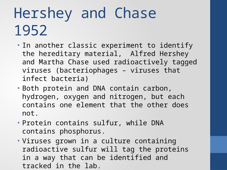
Hershey and Chase 1952• In another classic experiment to identify the hereditary
material, Alfred Hershey and Martha Chase used radioactively tagged viruses (bacteriophages – viruses that infect bacteria)
• Both protein and DNA contain carbon, hydrogen, oxygen and nitrogen, but each contains one element that the other does not.
• Protein contains sulfur, while DNA contains phosphorus.• Viruses grown in a culture containing radioactive sulfur will tag
the proteins in a way that can be identified and tracked in the lab.
• If viruses are grown in a culture containing radioactive phosphorus, their nucleic acids can be tracked

Hershey/Chase Experiment• Produce
radioactively tagged viruses• Allow them to infect
bacteria• Agitate, wash, and
centrifuge the cultures• Test the wash
solution and the bacterial cells for radioactive residue

Predictions:• If protein is the
hereditary material, then the wash solution should contain radioactive phosphorus residue and radioactive sulfur should be detectable in the bacterial cultures
• If DNA is the hereditary material, the wash solution should contain sulfur, while the bacterial culture should contain the tagged phosphorus

Results and conclusions:• The cells that
were infected contained residues of radioactive tagged phosphorus. • DNA was injected
into the host cells• DNA is the
hereditary material
Frederic Griffith 1927
• Pneumonia is a deadly disease. There are various causes, including both bacterial and viral types
• The causes and treatment of bacterial pneumonia has been of enormous importance to medical science for generations
• To determine the pathogen responsible for infectious disease, a researcher will adhere to a series of logical concepts called Koch’s Postulates:• The bacteria must be present in every case of the disease.• The bacteria must be isolated from the host with the disease and
grown in pure culture.• The specific disease must be reproduced when a pure culture of
the bacteria is inoculated into a healthy susceptible host.• The bacteria must be recoverable from the experimentally
infected host.

Smooth vs. Rough Strains
Streptococcus pneumoniae exists in two distinct strains. One which forms rough textured pinpoint colonies and another which forms smooth, spreading colonies

Smooth = Capsulated
We now know that the smooth strains are smooth due to a slime layer, or “capsule” which surrounds the outside of the cell wall

Experiment #1• Inject rough type
pneumococci into healthy mice• Result:• Mice remain healthy• No pneumonia
symptoms are observed• No bacteria are
recovered from blood
• Inject smooth type pneumococci into healthy mice• Result:• Mice exhibit
pneumonia symptoms• Smooth type bacteria
are recovered from the blood of the infected mice

Follow-Up; Experiment #2• Only the smooth type
cause the disease, but is it a result of the action of the cells themselves or is it a result of some poison in the capsule?
• Mice were injected with heat killed smooth type bacteria• Results:• Mice remain healthy• No pneumonia
symptoms are observed• No bacteria are
recovered from blood

OK, so what now?• Clearly it is the cells
themselves, and not the capsule that cause pneumonia, but the capsule must have some significance
• The third experiment in the series involved taking living rough type cultures and growing them in a medium containing the remains of heat killed smooth type bacteria

Experiment #3 - Predictions• Live rough type
hadn’t caused pneumonia in the 1st experiment• Heat killed smooth
didn’t cause pneumonia in the 2nd Experiment
• The only logical assumption would be that since neither caused pneumonia by itself, there would be no reason to expect them to cause pneumonia together

Experiment #3 - Results• Combined live
rough/dead smooth cultures are injected into healthy mice
• Mice develop pneumonia symptoms• Smooth type bacteria
are recovered from the blood of the infected animals

How do we account for that?• So not only did we get
pneumonia from 2 things that were proven not to cause pneumonia, but it sure looks like the smooth type cells came back from the dead like slime covered microscopic pneumonia zombies

Not Likely• OK, so scrap the zombie hypothesis• What appears to have happened is that hereditary material
from the heat killed smooth cells was absorbed by the living rough cells
• Even though the cells were dead, the hereditary material was still operational, and could cause the rough cells to become “transformed”
• The transformed cells expressed the “smooth” genes they picked up from the dead cells. They gained the ability to make a capsule, and because of that they were able to cause pneumonia in the mice
• It turns out that the capsule doesn’t harm the mice, but it does protect the bacteria from the mouse’s immune system, allowing them to stay alive, multiply, and cause the disease

Avery 1943 https://www.youtube.com/watch?v=RWFc8Iqz4Jg

Frederic Griffith and Oswald Avery
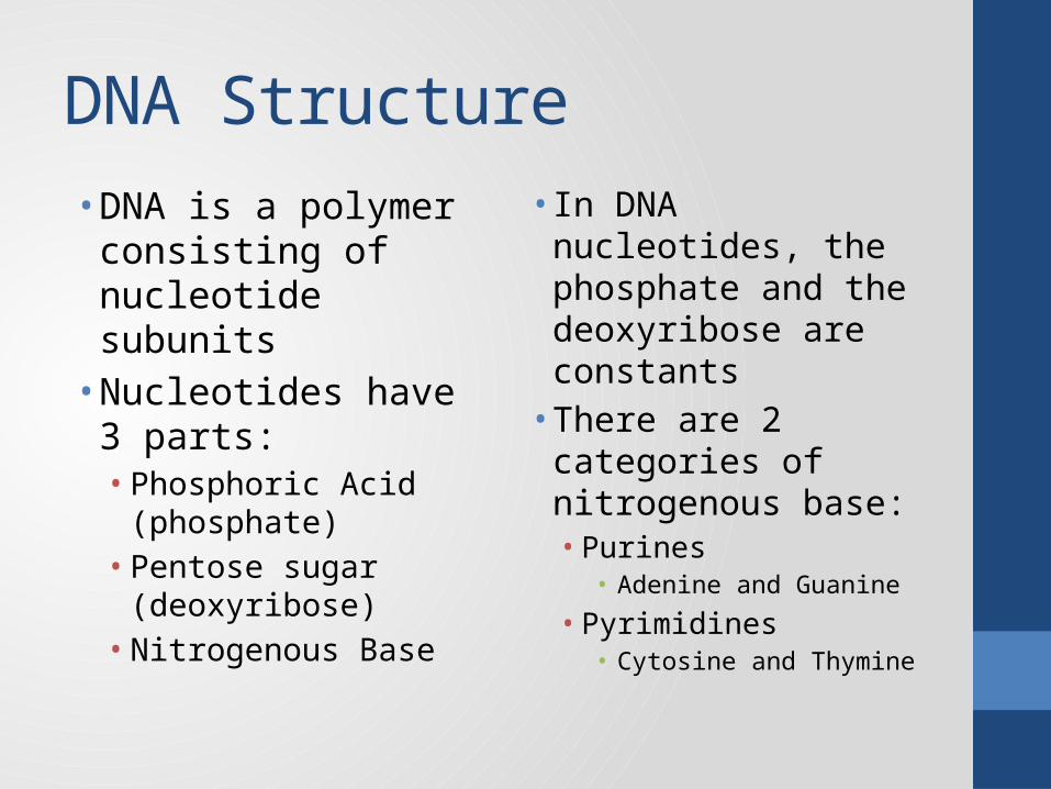
DNA Structure• DNA is a polymer
consisting of nucleotide subunits• Nucleotides have 3
parts:• Phosphoric Acid
(phosphate)• Pentose sugar
(deoxyribose)• Nitrogenous Base
• In DNA nucleotides, the phosphate and the deoxyribose are constants• There are 2
categories of nitrogenous base:• Purines• Adenine and Guanine
• Pyrimidines• Cytosine and Thymine

Pentoses, Purines, and Pyrimidines

DNA Polymer• There are 2
fundamental structural components of a DNA strand, the Sugar/Phosphate backbone and the Nitrogen Base side-chains

Some Vocabulary Clarification
• The deoxyribose is a 5 carbon sugar (pentose)• Structural positions are designated by numbering the carbons• The nitrogen base is at position 1 (1’, read as “1 prime”)• The phosphate is at the 5’ position• The bottom corner (3’) will become the point of attachment
of the next nucleotide in the polymer

Rosalind Franklin - 1951
• Franklin’s x-ray crystallography photographs of DNA demonstrated that DNA consisted of 2 strands twisted in a “double helix”

Erwin Chargaff 1950Chargaff enzymatically digested DNA from a variety of organisms and determined the relative proportion of each base. With great regularity, the proportions of A and T are equal to each other, as are G and C

Base Pairing
• Chargaff’s rule (#A = #T; #G = #C) is explained if the 2 DNA strands align with bases paired across between the strands, A to T and G to C• Note the formation of hydrogen bonds between the
base pairs

The Double Helix Illustrated• The polymer is formed by
the sugar-phosphate backbone• The “phosphodiester” links
are covalent
• The 2 strands are “antiparallel”• The strands are joined by
base pairing• The hydrogen bonds are
weak• They can separate and
rejoin easily

Watson and Crick - 1953• James Watson and
Francis Crick won the Nobel Prize for publishing the structure of DNA • Neither of them is
particularly good looking

Bozeman Videos• DNA and RNA part 1• http://
www.youtube.com/watch?v=qoERVSWKmGk&list=PLFCE4D99C4124A27A&index=34
• DNA and RNA part 2• http://
www.youtube.com/watch?v=W4mYwsr9gGE&list=PLFCE4D99C4124A27A
• Gene Regulation• http://
www.youtube.com/watch?v=3S3ZOmleAj0&list=PLFCE4D99C4124A27A
• Signal Transmission and Gene Expression• http
://www.youtube.com/watch?v=D-usAds_-lU&list=PLFCE4D99C4124A27A

MIT OpenCourseWare Videos• DNA Structure and Classic Experiments, excerpt 1• https://www.youtube.com/watch?v=P-Ry4rRdDbk
• DNA Structure and Classic Experiments, excerpt 2• https://www.youtube.com/watch?v=YCeKtM6Hnmc
• DNA Replication: Fundamentals of Biology• https://www.youtube.com/watch?v=DRBREvFL19g
• Transcription and Translation, excerpt 1• https://www.youtube.com/watch?v=tMr9XH64rtM
• Transcription and Translation, excerpt 2• https://www.youtube.com/watch?v=uBRdfsz_YB4

Replication• “Replica” = exact copy.
Replication is copying of the DNA• DNA is copied in
preparation for cell division • (mitosis/meiosis/binary fission)
• Replication occurs in the S (synthesis) phase of Interphase• In replication, the
entire genome is copied
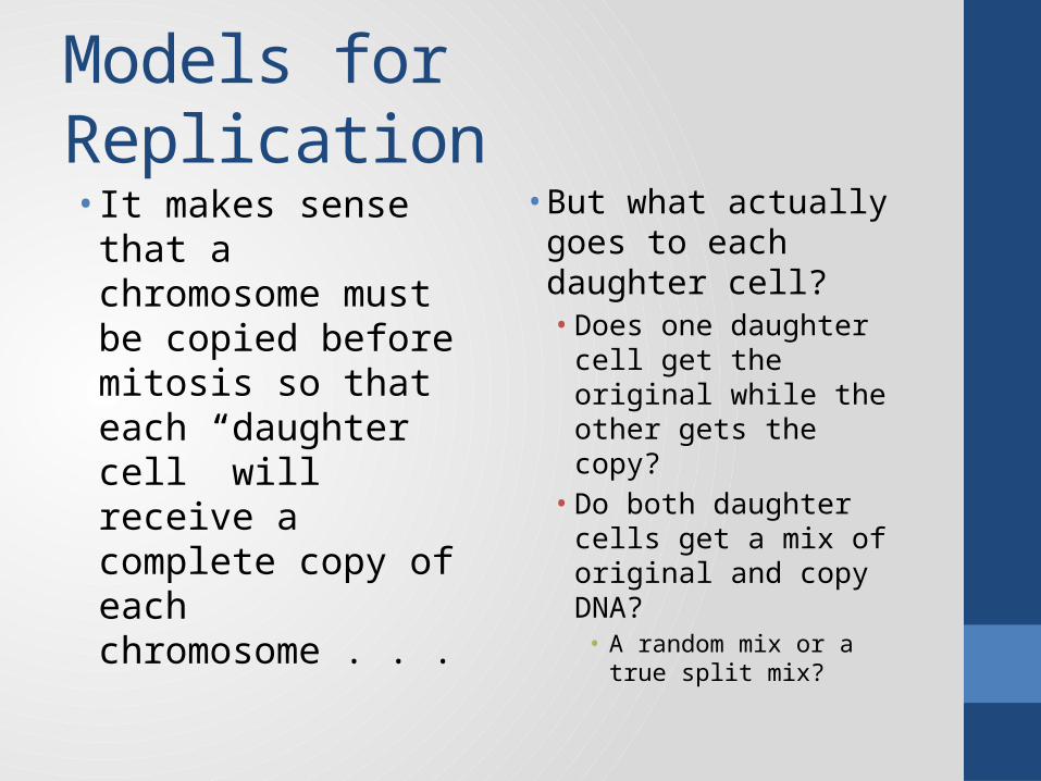
Models for Replication• It makes sense that a
chromosome must be copied before mitosis so that each “daughter cell” will receive a complete copy of each chromosome . . .
• But what actually goes to each daughter cell?• Does one daughter
cell get the original while the other gets the copy?• Do both daughter cells
get a mix of original and copy DNA?• A random mix or a true
split mix?

First, some vocabulary• To “conserve”
something is to allow it to remain intact or unchanged• Remember, the DNA
consists of 2 strands• After replication we’ll
have 2 sets of DNA, each with 2 strands
• If the resulting of replication is 2 original strands staying together, replication is “conservative”• If 1 strand is original
and the other strand is new, replication is “semiconservative”

Replication Models Compared• The diagram shows
the difference between the different models of Replication• Semiconservative• 1 original, 1 new
• Conservative• Originals stay together
• Dispersive• Random mix

Meselson and Stahl – late 50’sMeselson and Stahl did experiments to evaluate the different models of replication by measuring the results of replication using heavy isotopes of nitrogen• read pp.311-
312

Meselson – Stahl Experiment• Bacteria were grown
in cultures containing heavy isotopes of nitrogen (15N)• These bacteria were
transferred to cultures containing normal, light nitrogen (14N)
• After each replication, samples were collected and centrifuged to determine their relative density• The density of the
resulting DNA varied depending upon the proportion of DNA containing the original heavy nitrogen-15

Predicted Results - Molecular

Predicting Results - Observed• The “parent” DNA
had 2 strands containing 15N, so would be consistently very dense• Semiconservative
hybrid DNA would have 1 heavy and 1 light strand, so would be consistently of intermediate density

Predicting Results - Observed• Any subsequent
replications occurring in a 14N environment would produce a small amount of hybrid DNA (from the 2 original conserved strands) and progressively larger amounts of low density DNA

Results and Conclusions
The results of the experiment are precisely what we would predict for Semiconservative replicationhttps://www.youtube.com/watch?v=JcUQ_TZCG0w

Semiconservative Replication• The 2 original DNA
strands are conserved but separated• Each original strand
serves as a template for producing the replica strand
• The process reflects the structure of the DNA molecule. Note:• The sugar phosphate
backbone• Base pairing• “Antiparallel” strand
orientation• https://
www.youtube.com/watch?v=zdDkiRw1PdU&feature=related
• https://www.youtube.com/watch?v=yqESR7E4b_8&feature=related

Replication Process Sequence
STEPS ENZYMES
Unzip
Helix destabilizing protein, DNA helicase, single stranded binding proteins, topoisomerase
form RNA primer RNA primase
base pairing no enzyme required - base pairs align by shape and polarity
attach individual DNA nucleotides to the primer DNA polymerase III
attach individual DNA nucleotides to the growing chain DNA polymerase IIIreplace the primer with DNA nucleotides DNA polymerase I
attach okazaki fragments DNA ligase

Unzipping the strands• The area that unzips forms a “replication fork”• Unzipping is a more complex process than it might seem,
largely because of the helical nature of DNA• The two major players in unzipping are the Helix Destabilizing
Protein and the enzyme DNA Helicase• The Helix Destabilizing Protein does exactly what its name
suggests. It interferes with the twisted shape of the double helix, creating some physical stress between the base pairs
• This creates space for the binding of the DNA Helicase, which will act like a wedge to separate the base pairs from each other• Remember, the sugar-phosphate backbone is permanently linked
together with covalent phosphodiester bonds, but the base pairs are only attracted to each other by hydrogen bonding. Hydrogen bonding is strong as polar attractions go, but weak in comparison to covalent bonds.

Resolving complications • Once unzipping begins, the action of the single strand binding
proteins and topoisomerase become necessary• Single strand binding proteins hold the strands apart so that they
don’t immediately rejoin. Remember, the base pairs are held together by hydrogen bonding. If they come back into contact they will stick back together
• Remember that DNA is a double helix. Disrupting the helix at the replication fork creates stress further down on the molecule. Picture uncoiling an extension cord. Every coil you unwrap compresses a coil you already unwrapped, and pretty soon you have a tangled mess. Add to that the fact that you are turning one double helix into 2 double helices. The enzyme topoisomerase resolves these stresses by cutting the DNA every once in a while to relieve the stress and then bonding it back together afterwards• Don’t try that with your extension cords. They don’t go back together.

Replication

Leading and Lagging Strand• Each of the steps of replication is directed by the action of specific
enzymes• Enzymes and substrates must fit together according to shape and
polarity (“induced fit”)• Nucleotides are not symmetrical, the central deoxyribose is a
pentose. The phosphorylated end is designated as 5’, the flat end (3’) will be the point of attachment of the next nucleotide in the polymer.
• The primary enzyme of replication, DNA polymerase, can only attach single DNA nucleotides to the 3’ end of the chain.
• The strands of DNA are antiparallel, so replication occurs in opposite directions on the two strands
• One strand (the leading strand) will replicate in the same direction as unzipping. The lagging strand replicates opposite the direction of unzipping

Replicating the Leading Strand• The process of replication begins with unzipping. This separates
the base pairs, exposing the genetic code.• Unzipping requires a series of proteins and enzymes
• Once the strands are separated, free triphosphorylated nucleotides will align according to the base pairing rule.
• The first nucleotides to align will be RNA nucleotides, forming an RNA primer. The growing DNA replica will be built off of the 3’ end of the primer• An enzyme called primase controls the formation of the primer
• Base pairing continues with triphosphorylated DNA nucleotides. • Each nucleotide is attached to the 3’ end of the chain by DNA
polymerase III• On the leading strand replication is continuous, the polymerase
follows the helicase without interruption through to completion

Replicating the Lagging Strand• Replication of the lagging strand is largely the same as the
leading strand, but opposite the direction of unzipping.• The lagging strand replicates in shorter segments (okazaki
fragments). Each fragment has an RNA primer.• Each RNA primer must be degraded and replaced with DNA
nucleotides. This is accomplished with a DNA polymerase enzyme (DNA polymerase I)
• Polymerase is not capable of attaching the okazaki fragments together. This is done by an enzyme called DNA ligase
Replication• DNA Replication video• http://
www.youtube.com/watch?v=AGUuX4PGlCc&feature=related• Crash Course: DNA Structure and Replication• http://www.youtube.com/watch?v=8kK2zwjRV0M• Bozeman: DNA Replication• http://www.youtube.com/watch?v=FBmO_rmXxIw

Biocoach Tutorial Links
• DNA Structure and Replication• http://www.phschool.com/science/biology_place/biocoach/dnarep/intro.html
• Transcription• http://www.phschool.com/science/biology_place/biocoach/transcription/intro.html
• Translation• http://www.phschool.com/science/biology_place/biocoach/translation/intro.html
• Lac operon• http://www.phschool.com/science/biology_place/biocoach/lacoperon/intro.html
• Restriction enzyme digest• http://www.phschool.com/science/biology_place/biocoach/red/intro.html

Transcription and Translation• Animation
• http://www.youtube.com/watch?v=41_Ne5mS2ls&feature=related
• Bozeman• http://www.youtube.com/watch?v=h3b9ArupXZg
• MIT• http://www.youtube.com/watch?v=uBRdfsz_YB4

Chapter 17 Objectives1. Describe Beadle and Tatum’s experiment and explain how it led to
the “one gene – one enzyme” hypothesis.2. Extend the “one gene – one enzyme hypothesis into its more
modern version, “one gene – one polypeptide”. Relate this to the “central dogma of molecular genetics”.
3. Explain the process of transcription. Note all molecules needed for transcription as well as the molecules produced.
4. Describe the role of eukaryotic promoter sequences in the initiation of transcription.
5. Define intron and exon. Explain the processing that occurs to convert pre-mRNA into mature mRNA.
6. Explain the process of translation. Discuss the role of mRNA, tRNA, and ribosomes in translation.
7. Define mutation. Discuss several types of mutations and evaluate their effects on the polypeptides produced.
What is a gene?• When we introduce the study of heredity, the emphasis is on
the inheritance of traits. Early experiments and observations, beginning largely with Frederick Griffiths discovery of a “transforming principle”, recognized that heredity has a more fundamental molecular and cellular basis. Traits develop as the result of structural and metabolic changes within cells and tissues.
• We now know that genes code for the production of proteins, and that those proteins are what more directly influence the formation of observable traits.
• Most of those proteins are enzymes which will control steps in metabolic pathways.

Beadle and Tatum 1958

One gene – One enzyme• George Beadle and Edward Tatum performed a series of
experiments with the bread mold neurospora, for which they received the Nobel Prize in 1958. They used X-rays to mutate the mold, and grew pure cultures of the mutated molds. They attempted to grow sample cultures on “minimal media” which did not contain a full nutrient complement (all 20 essential amino acids)
• When they found a strain that could not grow on minimal media, they tested it in media that was supplemented by single amino acids to determine what they needed to survive and grow.
• The pattern of results they observed suggested that the molds had the ability to convert one amino acid into another, and that the mutations disrupted that ability

Lame but easy to visualize:• So you plan an evening in Chicago with friends from different
towns in Northwest Indiana. One from Munster, one from Griffith, one from Merrillville, one from Valpo and one from Michigan City.
• You hear on the radio about an overturned truck that shuts down traffic on Interstate 94, but you miss the part where they say where it happened.
• Only the friends from Munster and Griffith show up• Where was the highway blocked?

Now for real:• Imagine a metabolic pathway, each step controlled by a different
enzyme, which converts an organic precursor molecule into one amino acid, then to another, and so on down the line.
• Suppose that the pathway involved the amino acids citrulline, arginine, and ornithine.
• 3 mutant strains of Neurospora exist which cannot grow on minimal media. All 3 mutant strains can grow on media supplemented with arginine• Strain 1 can grow on media supplemented with any of the three amino
acids, but not on the precursor alone• Strain 2 can grow on media supplemented with arginine or with citrulline,
but not ornithine• Strain 3 can grow with arginine supplements, but not with any of the
others• Hypothesize a sequence of events for the metabolic pathway and
pinpoint the enzyme mutated in each strain

What can we surmise?• The precursor must be the first
step in the pathway because strain 1 can’t grow with it alone but can grow with any of the other 3 supplements• Arginine must be the final product
in the pathway because all three strains could grow on media supplemented with arginine (if they were fed arginine, they didn’t need to make arginine), plus strain 3 could only grow with the arginine supplement
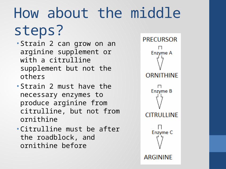
How about the middle steps?• Strain 2 can grow on an
arginine supplement or with a citrulline supplement but not the others• Strain 2 must have the
necessary enzymes to produce arginine from citrulline, but not from ornithine• Citrulline must be after the
roadblock, and ornithine before

What about the mutations?Strain 1 can grow on media supplemented with any of the three amino acids, but not on the precursor aloneThe roadblock must be between the precursor and all of the other enzymes (before ornithine) . • The strain 1 mutation
disrupted the production of enzyme A

What about the mutations?Strain 2 can grow on media supplemented with arginine or with citrulline, but not ornithineThe roadblock must be after ornithine but before citrulline.• The strain 2 mutation
disrupted the production of enzyme B

What about the mutations?Strain 3 can grow with arginine supplements, but not with any of the others The roadblock must be after ornithine and citrulline but before arginine.• The strain 3 mutation
disrupted the production of enzyme C

Conclusion: • The genes that were mutated by the X-Rays disrupted the
ability of the cells to produce particular enzymes• The “one gene – one enzyme” hypothesis is derived from
these experiments.• After Beadle and Tatum we now understand that genes aren’t
so much about traits as they are about enzymes that control metabolism and development.
• Traits result from the actions of these enzymes because the enzymes control the materials that the cell produces
• Modern genetics takes this a step further, recognizing that enzymes are made of protein. So technically, one gene is the instructions for producing one polypeptide

Oh, yeah. One more thing• Genes are made of DNA• DNA is in the nucleus• The machinery used to actually synthesize polypeptides are in
the cytoplasm• The nucleus and cytoplasm are separated by a nuclear
envelope• The nuclear envelope has pores, but those pores are too small
to allow DNA to pass between the nucleus and the cytoplasm• So there must be another molecule, small enough to exit the
nucleus but able to mimic DNA’s genetic code, that carries the genetic information from the nucleus out to the cytoplasm
• Go ahead, say it, you know what it is . . .

The Central Dogma of Molecular Genetics

The Central Dogma of Molecular Genetics
• The copying of a DNA gene into mRNA is called Transcription• The term transcription is appropriate because a transcript is a
copy of the necessary information for a desired task• The process of decoding that gene to form a polypeptide is
called Translation• “Translation” because it is converting from the language of DNA
and RNA (nucleotide sequences) into the language of protein (amino acid sequences)

TranscriptionFundamentally transcription is much like replication, but with several significant differences. • First the DNA strands must unzip • But they must unzip in the proper place to transcribe a particular
gene. Replication copied the entire genome. Transcription copies the instructions for a single protein
• Then RNA nucleotides must align according to the base pairing rule• Remember, RNA has U instead of T, so the base pairing rule is slightly
different• Since the messenger will be RNA, there is no need for an RNA primer
• Then those nucleotides must be joined together to form a polymer• These are RNA nucleotides, so the enzyme that binds them will be
RNA polymerase, not DNA polymerase

Transcription• We can think of transcription as having 3 phases: Initiation,
Elongation, and Termination. Basically it Starts, Happens, and Stops.
• Many of the enzymes used in Replication are not a part of transcription• DNA helicase is unnecessary because the RNA polymerase serves
a dual function. It both unzips the strands and forms the polymer• No RNA primer, so no primase• No topoisomerase because the area unzipping is so small that
there is no stress in the helix to release• Since only one strand is transcribed, there are no Okazaki
fragments and no need for DNA ligase

Initiation• Since transcription only copies a single gene the process must
be initiated at a very specific spot on the chromosome. • Transcribing too much before the actual coding sequence
would be enormously wasteful. Remember, each nucleotide is triphosphorylated. Every nucleotide used but not needed is a waste of energy
• Transcription begins at a particular region “upstream” from the coding sequence called the Promoter region
• The Promoter region will facilitate the binding of the RNA polymerase, either directly or indirectly by binding “transcription factors” which then bind the polymerase
• In eukaryotes, the promoter region generally begins with a series of alternating T and A nucleotides called the TATA box that binds transcription factors
Elongation and Terminaton• Elongation means to add length. RNA polymerase adds RNA
nucleotides to the 3’ end of the mRNA transcript, making the chain grow longer.
• Termination means stop or end. After the coding sequence transcription must end. Every unnecessary nucleotide added is a waste of energy, so a specific stopping point is of great adaptive value
• In eukaryotes, the coding sequence is followed by a “polyadenylation signal” (many A’s) that serve as a stopping point

The Transcription Unit

The Transcription Unit• Where is the actual gene?• Where does the RNA polymerase bind?• Where do the transcription factors bind?• Where does transcription begin? End?• Which strand gets copied?

Prokaryotes vs. Eukaryotes• Prokaryotes (bacteria) do not have a nuclear envelope, so there
is not barrier between the processes of transcription and translation.
• In bacteria, ribosomes will bind to the mRNA even before transcription is completed
• In eukaryotes, however, a series of modifications will take place before the RNA leaves the nucleus:• Addition of a 5’ cap
• The cap consists of a modified G nucleotide. • Prevents degradation and facilitates binding of the ribosome
• Addition of a 3’ poly A tail• 50-250 Adenine nucleotides added to the 3’ end• Prevents degradation
• Splicing • Removal of Intervening Sequences (“introns”)
Why Degradation Matters:• Once the mRNA leaves the nucleus, it almost immediately
starts to fall apart (degrade). Degradation begins at the ends• If the coding sequence began with the first nucleotide,
degradation of even one nucleotide would prevent the protein from being produced correctly. A longer UTR (untranslated region) means that the mRNA will continue to function for a longer period of time before the code begins to be degraded
• What that means is more protein produced from a single transcript, and remember that transcription is very energy expensive
• On the flip side, too much protein from one transcript is wasteful too, not of energy but of amino acids – if you devote amino acids to making protein you don’t need, they aren’t available for building more useful structures or enzymes

Eukaryotic RNA splicing• In eukaryotes, only part of what I represented as the coding
region will actually be translated into an amino acid sequence• The parts of the sequence that will be expressed in the protein
are called Exons. Intervening sequences that will be spliced out and will not be expressed are called Introns.
• Whoever came up with those names should be hung up by their thumbs because they sound like the opposite of what they actually mean, and scientists shouldn’t do stupid stuff like that.
• The Ex in Exon doesn’t mean “out” like it usually would. It means Expressed, so it stays In.
• The In in Intron doesn’t mean it stays in, it means intervening, so it gets cut out.
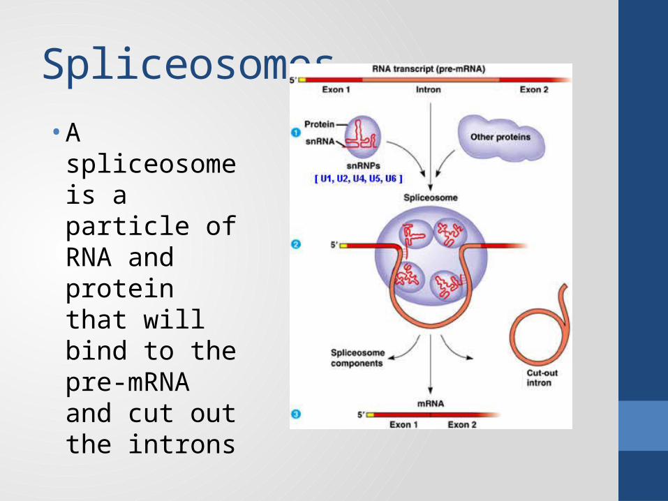
Spliceosomes• A spliceosome is
a particle of RNA and protein that will bind to the pre-mRNA and cut out the introns

Spliceosomes, more detailed

Alternative Splicing• The significance of eukaryotic gene splicing is enormous• A single gene can result in the production of a wide variety of
proteins by transcribing the same gene and processing it differently
• A cell can treat a particular part of the RNA as an Intron and cut it out, or treat it as an Exon, leave it in and express it
• A human cell has about 20,000 genes, but may produce as many as 100,000 different proteins.
• A great deal of genetic variability can be attributed to differential RNA splicing.
• Producing the same enzyme with variations in the allosteric parts of the protein, for example, would serve the same metabolic function but have a great effect on the optimal conditions of the enzymes activity

Translation