antibiotic resistance in lactobacillus reuteri and...
TRANSCRIPT
Antibiotic Resistance in Lactobacillus reuteri and Lactobacillus plantarum
Maria Egervärn Faculty of Natural Resources and Agricultural Sciences
Department of Microbiology
Uppsala
Doctoral Thesis Swedish University of Agricultural Sciences
Uppsala 2009

Acta Universitatis agriculturae Sueciae
2009: 30
ISSN 1652-6880 ISBN 978-91-86195-77-9 © 2009 Maria Egervärn, Uppsala Tryck: SLU Service/Repro, Uppsala 2009
Cover: Antibiotic susceptibility testing by Etest (left; photo: M. Egervärn) and broth microdilution (right; reproduced with the permission of the National Veterinary Institute, Uppsala, Sweden).

3
Antibiotic Resistance in Lactobacillus reuteri and Lactobacillus plantarum
Abstract Lactic acid bacteria (LAB) may act as reservoirs of antibiotic resistance genes that can be transferred via the food chain or within the gastrointestinal tract to pathogenic bacteria. This thesis provides data required for assessing the potential risk of using antibiotic resistant strains of the LAB species Lactobacillus reuteri and Lactobacillus
plantarum as food processing aids or probiotics. Knowledge of the distributions of antibiotic minimum inhibitory concentrations (MICs) for a species is needed when using a phenotypic method to differentiate strains with acquired resistance from susceptible strains or strains with intrinsic resistance. Controlled and standardised conditions are required for antibiotic susceptibility testing of LAB, as demonstrated here during evaluation of the Etest and broth microdilution MIC determination methods used. Inoculum size and incubation time were varied during broth microdilution testing of the susceptibilities of 35 LAB strains to six antibiotics. An increase in either parameter resulted in elevated MICs for all species.
Antibiotic susceptibility profiles were determined for 56 L. reuteri and 121 L. plantarum strains that differed by source and spatial and temporal origin. MIC data obtained with the Etest and the broth microdilution methods corresponded well with each other. All L. plantarum strains were susceptible to ampicillin, gentamicin, erythromycin and clindamycin, and intrinsically resistant to streptomycin. Acquired resistance to tetracycline was associated with plasmid-bound tet(M).
Lactobacillus reuteri strains had acquired resistance to tetracycline (n=28), ampicillin (n=14), erythromycin/clindamycin (n=6) and chloramphenicol (n=1). This resistance was attributed to mutational pbp genes for ampicillin and to added tet(W), erm and cat(TC) genes for the antibiotics inhibiting protein synthesis. Genetic relatedness was observed among L. reuteri strains with high MICs for both ampicillin and tetracycline and among strains with high MICs for both erythromycin and clindamycin. The majority of the antibiotic resistant L. reuteri strains carried the resistance genes on plasmids. Traits of putative transfer machineries adjacent to both plasmid- and chromosome-located resistance genes were demonstrated.
Lactobacillus reuteri as a donor of resistance genes in the human gut was investigated by studying the transferability of the tetracycline resistance gene tet(W) to faecal enterococci, bifidobacteria and lactobacilli. No gene transfer was demonstrated under the conditions tested.
Keywords: Lactobacillus reuteri, Lactobacillus plantarum, antibiotic susceptibility testing, MIC, tetracycline resistance, tet genes, erythromycin resistance, erm genes, gene transferability.
Author’s address: Maria Egervärn, National Food Administration P.O. Box 622, 751 26 Uppsala, Sweden. E-mail: [email protected]

4
5
Contents
List of Publications 7
Abbreviations 9
1 Introduction 11 1.1 Aims 12
2 Antibiotic resistance 13 2.1 Development of antibiotic resistant bacteria 14 2.2 Antibiotic targets 16 2.3 The ribosome as antibiotic target 16
2.3.1 Macrolides 17 2.3.2 Tetracyclines 18
2.4 Resistance mechanisms 19 2.4.1 Macrolide resistance 20 2.4.2 Tetracycline resistance 20
2.5 Transfer of resistance genes 21 2.5.1 Horizontal gene transfer 21
2.6 Intrinsic and acquired resistance 22 2.6.1 Microbiological breakpoints 23 2.6.2 Phenotypic methods 23
3 Lactobacilli 25 3.1 Occurrence and taxonomy 25
3.1.1 Occurrence and taxonomy of L. reuteri and L. plantarum 25 3.2 Industrial applications 26
3.2.1 Industrial applications of L. reuteri and L. plantarum 27 3.3 Safety aspects 27
3.3.1 Safety assessment of lactobacilli 28 3.4 Antibiotic resistance in lactobacilli 29
4 Results and discussion 33 4.1 Antibiotic susceptibility testing (IIIIIIII and III III III III) 33
4.1.1 Evaluation of a broth microdilution method (IIII) 34 4.1.2 Comparison of Etest and broth microdilution MICs (IIIIIIII and IIIIIIIIIIII) 35
4.2 Antibiotic resistance in L. reuteri (IIIIIIII and IVIVIVIV) 36 4.2.1 Tetracycline resistance in L. reuteri 38

6
4.2.2 Erythromycin resistance in L. reuteri 41 4.3 Antibiotic resistance in L. plantarum (IIIIIIIIIIII and IVIVIVIV) 42
4.3.1 Tetracycline resistance in L. plantarum 43 4.3.2 Tentative microbiological breakpoints for L. plantarum 43
4.4 Phenotypic versus genotypic data (IIIIIIII, III III III III, and IV IV IV IV) 44 4.5 In vivo transferability of an L. reuteri tet(W) gene (VVVV) 45
5 Conclusions 49 5.1 Future perspectives 50
References 53
Acknowledgements 67
7
List of Publications
This thesis is based on the work contained in the following papers, which are referred to in the text by their Roman numerals:
I Egervärn, M., Lindmark, H., Roos, S., Huys, G. and Lindgren, S. (2007). Effects of inoculum size and incubation time on broth microdilution susceptibility testing of lactic acid bacteria. Antimicrobial
Agents and Chemotherapy 51(1), 394-396.
II Egervärn, M., Danielsen, M., Roos, S., Lindmark, H. and Lindgren, S. (2007). Antibiotic susceptibility profiles of Lactobacillus reuteri and Lactobacillus fermentum. Journal of Food Protection 70(2), 412-418.
III Flórez, A.B., Egervärn, M., Danielsen, M., Tosi, L., Morelli, L., Lindgren, S. and Mayo, B. (2006). Susceptibility of Lactobacillus plantarum strains to six antibiotics and definition of new susceptibility-resistance cutoff values. Microbial Drug Resistance 12(4), 252-256.
IV Egervärn, M., Roos, S. and Lindmark, H. (2009). Identification and characterisation of antibiotic resistance genes in Lactobacillus reuteri and Lactobacillus plantarum. Accepted for publication in Journal of Applied
Microbiology.
V Egervärn, M., Lindmark, H. and Roos, S. Transferability of a tetracycline resistance gene from probiotic Lactobacillus reuteri to bacteria in the gastrointestinal tract of humans (manuscript).
Papers I-IV are reproduced with the permission of the publishers.

8
My contribution to the papers included in this thesis was as follows:
I Took part in planning the study and analysing the results. Performed all the laboratory work. Main writer of the manuscript.
II Took part in planning the study and analysing the results. Performed the laboratory work concerning L. reuteri. Main writer of the manuscript.
III Took part in planning the study and analysing the results. Performed the laboratory work on 45 of the L. plantarum strains. Minor part in writing the manuscript.
IV Major part in planning the study, analysing the results, performing the laboratory work and writing the manuscript.
V Major part in planning the study and analysing the results. Performed all the laboratory work. Wrote the manuscript.

9
Abbreviations
ACE-ART Assessment and critical evaluation of antibiotic resistance transferability in food chain
CFU Colony forming unit CLSI Clinical Laboratory Standards Institute (previously NCCLS) EFSA European Food Safety Authority EUCAST European Committee for Antimicrobial Susceptibility Testing LAB Lactic acid bacteria LSM LAB susceptibility test medium MIC Minimum inhibitory concentration (µg/mL) MLSB Macrolide, lincosamide, streptogramin B compound PBP Penicillin binding protein PCR Polymerase chain reaction RPP Ribosomal protection protein

10
11
1 Introduction
Lactic acid bacteria (LAB), like all other bacteria, can exchange genes to enhance their survival in antibiotic-containing habitats (Teuber et al., 1999). The close contact between LAB and other bacteria, e.g. in the intestine, on mucosal surfaces or in food, is a precondition for horizontal gene transfer by mobile genetic elements. Ingested and indigenous LAB may therefore contribute to the pool of antibiotic resistance genes that can be transferred via the food chain or within the gastrointestinal tract to other commensal bacteria or to pathogens (Teuber et al., 1999; Salyers et al., 2004).
Antibiotic resistance of LAB used for food, feed and probiotic applications has recently been proposed as a hazard due to the potential risk for transfer to pathogenic bacteria. The European Commission has, as advised by the European Food Safety Authority (EFSA), requested that bacterial strains harbouring transferable antibiotic resistance genes should not be used in animal feeds (European Parliament and Council Regulation EC 429/2008; EC, 2001). No legislation exists so far regarding microorganisms intentionally added to fermented food and probiotics for human use. However, based on the precautionary principle, it is recommended that these products follow similar requirements to feed additives (EFSA, 2007). Measures based on this principle should be considered provisional until more comprehensive data concerning the risk are obtained and analysed.
The present investigation was initially performed as part of the EU project ACE-ART (CT-2003-506214; Morelli, 2008), which aimed to assess and critically evaluate antibiotic resistance transferability in the food chain. The intention was to obtain data related to phenotypic susceptibility testing, identification and characterisation of resistance genes and the potential for gene transferability of different LAB species, and through this prove or discount the current strategy based on the precautionary principle.

12
1.1 Aims
The overall aim of this thesis was to provide data required for assessing the potential risk of using antibiotic resistant strains of the LAB species Lactobacillus reuteri and Lactobacillus plantarum as starter cultures or probiotics. Specific objectives were to:
� Evaluate the effects of inoculum size and incubation time on the
antibiotic susceptibilities of LAB using a broth microdilution method (I), and compare minimum inhibitory concentration (MIC) data obtained by broth microdilution with an Etest method (II and III).
� Determine antibiotic susceptibility profiles of L. reuteri of animal and human origin (II) and L. plantarum of dairy and vegetable origin (III).
� Investigate the genetic relatedness of the L. reuteri strains with atypical antibiotic MICs (II).
� Identify and characterise the resistance genes mediating atypical antibiotic MICs in L. reuteri and L. plantarum (IV).
� Investigate the transferability of an antibiotic resistance gene from a probiotic L. reuteri strain to bacteria in the intestinal tract of humans (V).

13
2 Antibiotic resistance
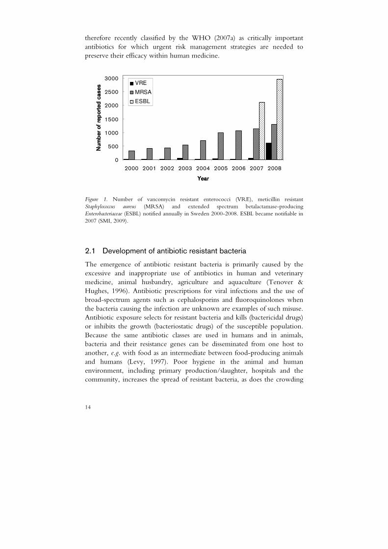
The rapid emergence of antibiotic resistant bacteria is a major threat to public health (ECDC, 2007; WHO, 2007b), the obvious concern being that infections might no longer be treatable with antibiotics. Effective antibiotics are also essential tools for organ transplantation, cancer chemotherapy and orthopaedic surgery (Cars et al., 2008). Surveillance data have shown an increasing incidence of infections caused by antibiotic resistant pathogens in many countries (EARSS, 2007). For example, the prevalence in blood cultures of Klebsiella pneumoniae producing extended-spectrum betalactamases has now approached 50% in some regions of Eastern Europe (EARSS, 2007). In Sweden too, an increase in antibiotic resistant bacteria has occurred during recent years (Fig. 1). However, the situation here is better than in most other countries.
Failure of antibiotic treatment for an infection usually leads to serious consequences for the patient and is associated with increased health care costs for society. In a recent study of more than 1800 Tanzanian children with signs of systemic infection, the mortality rate from Gram-negative bloodstream infections was more than double that of malaria (44% compared with 20%) (Blomberg et al., 2007). Two American studies showed that the mortality rate from bacteraemia and the hospital costs for surgery-related infections were both on average twice as high for patients infected with meticillin resistant Staphylococcus aureus than for patients infected with meticillin susceptible bacteria (Cosgrove et al., 2003; Engemann et al., 2003).
In addition, only two new antibiotic classes (oxazolidinones and lipopeptides) have been developed since the 1960s and few new drugs are currently in the pipeline (Cars et al., 2008). All this has raised concerns about a ‘post-antibiotic era’, a period with no effective antibiotics available. Quinolones, third/fourth-generation cephalosporins and macrolides were
14
therefore recently classified by the WHO (2007a) as critically important antibiotics for which urgent risk management strategies are needed to preserve their efficacy within human medicine.
0
500
1000
1500
2000
2500
3000
2000 2001 2002 2003 2004 2005 2006 2007 2008
YearYearYearYear
Num
ber
of
rep
ort
ed
cases
Num
ber
of
rep
ort
ed
cases
Num
ber
of
rep
ort
ed
cases
Num
ber
of
rep
ort
ed
cases VRE
MRSA
ESBL
Figure 1. Number of vancomycin resistant enterococci (VRE), meticillin resistant Staphylococcus aureus (MRSA) and extended spectrum betalactamase-producing Enterobacteriaceae (ESBL) notified annually in Sweden 2000-2008. ESBL became notifiable in 2007 (SMI, 2009).
2.1 Development of antibiotic resistant bacteria
The emergence of antibiotic resistant bacteria is primarily caused by the excessive and inappropriate use of antibiotics in human and veterinary medicine, animal husbandry, agriculture and aquaculture (Tenover & Hughes, 1996). Antibiotic prescriptions for viral infections and the use of broad-spectrum agents such as cephalosporins and fluoroquinolones when the bacteria causing the infection are unknown are examples of such misuse. Antibiotic exposure selects for resistant bacteria and kills (bactericidal drugs) or inhibits the growth (bacteriostatic drugs) of the susceptible population. Because the same antibiotic classes are used in humans and in animals, bacteria and their resistance genes can be disseminated from one host to another, e.g. with food as an intermediate between food-producing animals and humans (Levy, 1997). Poor hygiene in the animal and human environment, including primary production/slaughter, hospitals and the community, increases the spread of resistant bacteria, as does the crowding

15
of animals on farms and of old and immunocompromised people in hospitals and nursing homes. Increased travel and the worldwide distribution of food are other factors facilitating the rapid global spread (Cars et al., 2008).
To reduce the development of resistant bacteria in food-producing animals, Sweden was the first country to ban the use of antibiotics for growth promotion (Swedish act SFS 1985:295 on feed). In 1998, veterinary
Table 1. Antibiotics, their classes, modes of action and mechanisms of resistance (modified from
Guardabassi & Courvalin, 2006)
Antibiotic Class Mode of action Major resistance mechanism
Ampicillin Betalactams Inhibit transpeptidation step in peptidoglycan synthesis by binding to the PBPs
Betalactamases; mutations in the PBPs
Vancomycin Glycopeptides Bind to D-Ala-D-Ala peptidoglycan precursors, making them inaccessible to the PBPs
Peptidoglycan precursors terminating by D-Ala-D-lactate
Streptomycin, amikacin, gentamicin, kanamycin, neomycin
Aminoglycosides Bind 30S ribosomal subunit
Aminoglycoside modifying enzymes
Linezolid Oxazolidinones Bind 30S ribosomal subunit
Mutation of 23S rRNA
/Oxy/tetracycline Tetracyclines Bind 30S ribosomal subunit
Ribosome protection; efflux
Erythromycin Macrolides Bind 50S ribosomal subunit
Methylation or mutation of 23S rRNA; efflux
Clindamycin Lincosamides Bind 50S ribosomal subunit
Methylation or mutation of 23S rRNA; efflux
Dalfopristin, quinupristin
Streptogramins Bind 50S ribosomal subunit
Methylation or mutation of 23S rRNA; efflux
Chloramphenicol Phenicols Bind 50S ribosomal subunit
Chloramphenicol acetyltransferases
Trimethoprim /sulfonamide
Trimethoprim /sulfonamides
Inhibit tetrahydrofolate pathway
Mutations in the target enzymes

16
medicine accounted for around 50% of global consumption of antibiotics, of which the majority were used for prophylaxis and growth promotion (Wise et al., 1998). Since then, the European Union has banned the use of antibiotics for the latter purpose (European Parliament and Council Regulation EC 1831/2003), but in the US and many developing countries antibiotics are still used as growth promoters.
2.2 Antibiotic targets
Antibiotics are antibacterial compounds that interfere with some structure or process that is essential to bacterial growth or survival, although in most cases harmless to the eukaryotic host harbouring the infecting bacteria. Antibiotics have three main bacterial targets: (i) cell-wall synthesis (e.g. betalactams, glycopeptides); (ii) protein synthesis (e.g. macrolides, tetracyclines and aminoglycosides); and (iii) DNA replication and repair (e.g. fluoroquinolones) (Walsh, 2000). Because lactobacilli are mainly associated with resistance to macrolides and tetracyclines, the mode of action and resistance mechanisms of these drugs are described in detail below. Antibiotics relevant for susceptibility testing of LAB are summarised in Table 1.
2.3 The ribosome as antibiotic target
Many chemically diverse antibiotics act by binding to the bacterial ribosome and thereby interfering with the synthesis of new proteins. High resolution atomic structures of the ribosome obtained by X-ray crystallography have recently revealed how some of these antibiotics target the ribosome (Fig. 2A), as reviewed by Poehlsgaard & Douthwaite (2005). Most of the antibiotics bind reversibly to the ribosome and are bacteriostatic at therapeutic concentrations.
Ribosomes translate the genetic code via messenger RNA (mRNA) to assemble amino acids into proteins. The bacterial ribosome (Fig. 2B) is made up of a small subunit (30S) and a large subunit (50S), both of which consist of a number of ribosomal proteins (r-proteins) and ribosomal RNAs (rRNAs), including 16S rRNA in 30S and 23S rRNA and 5S rRNA in 50S. The 30S subunit contains the A (aminoacyl) site, where the aminoacylated transfer RNA (tRNA) attaches and in which is located the decoding site, where each codon of the mRNA chain interacts with an aminoacylated tRNA anticodon. The 50S subunit contains the catalytic site, i.e. the peptidyl-transferase centre responsible for peptide bond formation,

17
as well as the tunnel through which the newly synthesised peptide chain is channelled before leaving the ribosome (Fig. 2B).
Figure 2. Structure of the small (30S) and large (50S) ribosomal subunits of bacteria with the ribosomal binding sites of some antibiotics inhibiting protein synthesis. A=aminoacyl, P=peptidyl-transferase centre, and E=exit (A). The associated subunits (70S) and the interaction with mRNA and tRNA (B). Reproduced with the permission of the publisher (modified from Poehlsgaard & Douthwaite, 2005).
2.3.1 Macrolides
Macrolides are active against Gram-positive bacteria and many Gram-negative potential pathogens such as Campylobacter, Legionella, Chlamydia, Helicobacter and Mycoplasma. Within Swedish veterinary medicine, macrolides are especially used for therapeutic group treatment of pigs (SVARM, 2007). This family of antibiotics has also been used non-clinically, as growth promoters in food-producing animals.
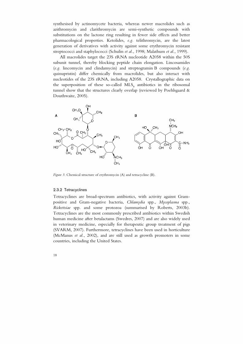
Macrolides consist of a macrocyclic lactone ring of between 14 atoms (e.g. erythromycin, see Fig. 3A) and 16 atoms (e.g. tylosin) with different substituents. Erythromycin and tylosin are naturally occurring antibiotics
18
synthesised by actinomycete bacteria, whereas newer macrolides such as azithromycin and clarithromycin are semi-synthetic compounds with substitutions on the lactone ring resulting in fewer side effects and better pharmacological properties. Ketolides, e.g. telithromycin, are the latest generation of derivatives with activity against some erythromycin resistant streptococci and staphylococci (Schulin et al., 1998; Malathum et al., 1999).
All macrolides target the 23S rRNA nucleotide A2058 within the 50S subunit tunnel, thereby blocking peptide chain elongation. Lincosamides (e.g. lincomycin and clindamycin) and streptogramin B compounds (e.g. quinupristin) differ chemically from macrolides, but also interact with nucleotides of the 23S rRNA, including A2058. Crystallographic data on the superposition of these so-called MLSB antibiotics in the ribosomal tunnel show that the structures clearly overlap (reviewed by Poehlsgaard & Douthwaite, 2005).
Figure 3. Chemical structure of erythromycin (A) and tetracycline (B).
2.3.2 Tetracyclines
Tetracyclines are broad-spectrum antibiotics, with activity against Gram-positive and Gram-negative bacteria, Chlamydia spp., Mycoplasma spp., Rickettsiae spp. and some protozoa (summarised by Roberts, 2003b). Tetracyclines are the most commonly prescribed antibiotics within Swedish human medicine after betalactams (Swedres, 2007) and are also widely used in veterinary medicine, especially for therapeutic group treatment of pigs (SVARM, 2007). Furthermore, tetracyclines have been used in horticulture (McManus et al., 2002), and are still used as growth promoters in some countries, including the United States.

19
Tetracyclines consist of four linear fused cyclic six-membered rings with different substituents attached. Tetracycline (Fig. 3B), chlortetracycline and oxytetracyline are produced by Streptomyces spp., whereas newer derivatives such as doxycycline and minocycline are semi-synthetic compounds. The third and latest generation of tetracyclines is glycylcyclines, e.g. tigecyclin (former GAR-936), which has a bulky side chain designed to overcome resistance in some bacteria (Petersen et al., 1999).
Uptake of tetracyclines into the bacterial cytoplasm favours the chelation of tetracycline with magnesium ions (Chopra & Roberts, 2001). These metal-drug complexes target the A site of the 30S bacterial ribosomal subunit, causing conformational changes and thereby physically preventing the binding of aminoacylated tRNA to the ribosome. Tetracycline primarily interacts with the H34 region of the 16S rRNA at the A-site, which is involved in the binding of aminoacylated tRNA. This was verified by crystallographic structures of the 30S ribosomal subunit, complexed with tetracycline (Brodersen et al., 2000; Pioletti et al., 2001). The functional relevance of the five additional binding sites found is currently not clear. The crystallographic studies in combination with a recent report on simulation of molecular dynamics (Aleksandrov & Simonson, 2008) suggest that the ‘upper’ part of the tetracycline molecule confers receptor binding specificity, whereas the ‘lower’ part controls magnesium binding.
2.4 Resistance mechanisms
Bacteria utilise four major strategies to become resistant to antibiotics (Fig. 4): Decreased intracellular antibiotic concentration by altering cell wall permeability (A) or by efflux (B); enzymatic inactivation of the antibiotic (C); and modification of the antibiotic target (D).
Figure 4. Major mechanisms of resistance to antibiotics in a bacterial cell.
enzymatic
B A
drug
DC
20
2.4.1 Macrolide resistance
The most common macrolide resistance mechanism in both Gram-positive and Gram-negative bacteria is target site modification caused by methylation or mutations. There are currently 33 described erm genes, encoding methyl transferases, which specifically methylate nucleotide A2058 in the 23S rRNA of the 50S ribosomal subunit (Roberts, 2008). Addition of one methyl group by e.g. erm(N) confers high resistance to lincosamides, whereas addition of two methyl groups by e.g. erm(E) confers high resistance to all MLSB antibiotics, including telithromycin (Liu & Douthwaite, 2002). Erm genes are often linked via mobile genetic elements to other resistance genes, especially genes conferring tetracycline resistance (Roberts et al., 1999).
Macrolide and/or MLSB resistance can also be caused by point mutations at A2058 or at adjacent nucleotides in the 23S rRNA and in the ribosomal proteins. Details of various mutations found in Streptococcus pneumoniae, Streptococcus pyogenes and Haemophilus influenzae have been reported by Franceschi et al. (2004). Resistance caused by chromosomal mutations generally presents a low risk of horizontal gene transfer, see below.
Another major resistance mechanism is through efflux pumps, which transport macrolides out of the cell, thereby reducing the intracellular drug concentration. So far, there are 14 described genes encoding these proteins. Efflux pumps confer diverse levels of resistance to the different MLSB antibiotics (reviewed by Roberts et al., 1999; Roberts, 2008). The mef(A) gene, encoding specific macrolide efflux, has been found in a variety of Gram-positive bacteria, including S. pyogenes, where it was first described (Clancy et al., 1996). Roberts and co-workers (1999) standardised the nomenclature for genes conferring macrolide and MLSB resistance.
2.4.2 Tetracycline resistance
Tetracycline resistance is now so widespread among bacteria that the utility of the tetracycline family has been considerably diminished. There are two ribosome-related mechanisms of tetracycline resistance in bacteria, both of which are linked to the primary binding site of the drug on the ribosome. This resistance is caused by ribosomal protection proteins (RPPs; Chopra & Roberts, 2001) or, more rarely, by mutations at nucleotide 1058 of the 16S rRNA (Ross et al., 1998).
The nomenclature and characterisation of 38 tet/otr genes conferring /oxy/tetracycline resistance have previously been reviewed by Roberts et al. (2005). A 42nd tet gene was recently found (Brown et al., 2008), reflecting the ongoing increase in knowledge in the field. Eleven of these genes (e.g.
21
tet(M), tet(O) and tet(W)) encode RPPs, which are cytoplasmic proteins with some sequence homology to elongation factors EF-Tu and EF-G. The RPPs are thought to interact with the H34 protein of the 16S rRNA at the A site, causing allosteric disruption of the primary binding site and thereby releasing tetracycline from the ribosome (Connell et al., 2003). Most of these genes have been found in both Gram-negative and Gram-positive bacteria.
Resistance to tetracycline is also commonly associated with efflux proteins. There are 26 tet efflux genes described so far, all of which confer resistance to tetracycline and doxycycline. Two of these genes, tet(K) and tet(L), are primarily found in Gram-positive bacteria. Efflux genes and genes encoding RPPs are both commonly associated with mobile genetic elements (Roberts, 2005).
2.5 Transfer of resistance genes
Antibiotic resistance traits are passed to daughter cells during replication of a bacterial strain (clone), so-called vertical or clonal dissemination. Horizontal transmission can provide a bacterial host of a different strain, species or even genus with genetic information that can be transiently needed, such as antibiotic resistance genes (Courvalin, 2006). Antibiotic exposure favours bacteria that have acquired resistance determinants at the expense of the susceptible population. Subsequently, wild-type bacteria stop proliferating or die, enabling the resistant bacteria to increase in abundance.
2.5.1 Horizontal gene transfer
There are three known mechanisms for horizontal transfer of resistance genes: conjugation, transformation and transduction. Conjugation is believed to be the most important mode of transfer within the gastrointestinal microbiota (reviewed by Licht & Wilcks, 2006). By this mechanism mobile genetic elements such as plasmids and transposons are transferred from one live bacterium to another through a protein tunnel that temporarily physically connects the bacteria. In Gram-negative bacteria, cell to cell contact is achieved by the formation of sex pili, whereas in Gram-positive bacteria it is achieved through uncharacterised cell surface structures (Grohmann et al., 2003).
Both plasmid-encoded and chromosome-located resistance genes can be transferred via conjugation. Conjugative plasmids are larger than approximately 15 kb and are self-transmissible due to the presence of the origin of transfer (oriT) gene and transfer (tra) genes. Unlike plasmids,

22
conjugative transposons are chromosomal elements that cannot replicate by themselves, but can move within the genome, e.g. from plasmid to plasmid or from chromosome to plasmid and vice versa (Salyers et al., 1995). Smaller, non-conjugative plasmids that carry mobilisation (mob) genes and the oriT sequence can be mobilised by taking advantage of the transfer machinery provided by conjugative elements (Francia et al., 2004). Studies on the in
vivo conjugative transfer of resistance genes in the gastrointestinal microbiota of various animals have recently been reviewed by Licht and Wilcks (2006).
Horizontal gene transfer can also occur by transformation, where DNA released from one bacterium is taken up by a competent bacterium, and by transduction, where DNA is transferred from one bacterium to another via bacteriophages (Licht & Wilcks, 2006). In both cases, the DNA generally must incorporate into the recipient genome by homologous recombination (Frost et al., 2005; Thomas & Nielsen, 2005). This implies narrow-host-range gene transfer, which is in contrast to the much broader host range of e.g. conjugative transposons (Salyers et al., 1995). However, non-homologous recombination is possible during both transformation and transduction, e.g. if plasmid DNA is acquired (Frost et al., 2005; Thomas & Nielsen, 2005). The frequency of gene transfer depends on the genetic material to be transferred, the transfer mechanism, the concentrations of donor and recipient strains and the contacts between these, and the selection pressure (Donohue et al., 1998).
2.6 Intrinsic and acquired resistance
Bacterial resistance to antibiotics can either be intrinsic or acquired
(Courvalin, 2006). Intrinsic resistance is an inherent trait of bacteria and can be due to reduced permeability of certain antibiotics across the cell wall, absence of the antibiotic target or the presence of low affinity targets. Glycopeptide resistance of heterofermentative lactobacilli is an example of intrinsic resistance and is caused by peptidoglycan dipeptides terminating with D-lactate instead of D-alanine, the cell wall target precursor for glycopeptide activity (Billot-Klein et al., 1994; Klein et al., 2000). In contrast, a bacterial strain can acquire resistance either by mutation in indigenous genes or by the uptake of exogenous resistance genes by horizontal transfer from other bacteria. By their nature, intrinsic resistance and resistance due to chromosomal mutation pose a low or even negligible risk for horizontal spread. Added resistance genes, especially those carried by mobile genetic elements, may be more easily transferred between bacteria.
23
2.6.1 Microbiological breakpoints
Bacteria not responding even to maximum doses of a given antibiotic during an infection are defined as clinically resistant. Clinical breakpoints, based on pharmacokinetic, clinical and microbiological data, are used by clinicians to advise on antibiotic therapy in patients (EUCAST, 2000). Bacteria that have acquired resistance mechanisms can grow at a higher antibiotic concentration than the more susceptible wild-type population and are defined as microbiologically resistant. Consequently, the resistant bacteria exhibit a higher MIC, i.e. the lowest concentration of an antibiotic that inhibits bacterial growth.
Microbiological breakpoints, referred to as epidemiological cut-off values by the European Committee for Antimicrobial Susceptibility Testing (EUCAST), are used for distinguishing between strains with and without acquired resistance genes (White et al., 2001). This categorisation is specific for each species and antibiotic and is based on the distribution of antibiotic MICs for a representative number of bacterial strains; 300-600 according to White et al. (2001). Theoretically, a uniform MIC distribution in the lower antibiotic concentration range indicates that all strains are susceptible; a uniform distribution with a high MIC for all strains may be due to an intrinsic trait; and a bimodal MIC distribution indicates that the strains with a high, atypical MIC may have acquired resistance. However, this differentiation is not always clear-cut in practice.
2.6.2 Phenotypic methods
Several methods have been used for phenotypic assessment of the antibiotic susceptibility of bacteria. Agar dilution and broth microdilution (right figure on front page) are dilution methods in which bacterial strains are tested for their ability to produce visible growth on a series of agar plates or in broth in microtitre wells containing serial two-fold dilutions of different antibiotics. The MIC is read as the first plate or well without growth. Dilution methods are the reference methods for antibiotic susceptibility testing in aerobic (CLSI, 2008) and anaerobic bacteria (CLSI, 2007).
The Etest (left figure on front page) is a commercial product and comprises a preformed, predefined gradient of antibiotic concentrations on a plastic strip. When the strip is applied to an inoculated agar plate, the gradient is transferred to the agar and established along the strip. The MIC is read where the edge of the elliptic inhibition zone formed intersects the Etest strip. Another diffusion method is disc diffusion, which comprises a disc containing a specific antibiotic concentration. Radial diffusion of the antibiotic into the inoculated agar creates an inhibition zone, the diameter

24
of which is subsequently measured. In contrast to the latter method, the Etest and both dilution methods give MICs, i.e. quantitative data on the bacterial strains tested.
25
3 Lactobacilli
3.1 Occurrence and taxonomy
Lactobacilli are fastidious bacteria, found in a variety of nutrient-rich environments such as meat and dairy products, plant material, animal and human mucosal surfaces, and sewage and manure (Hammes & Hertel, 2006).
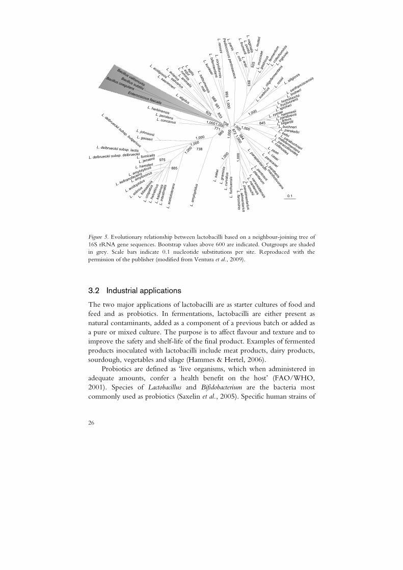
The genus Lactobacillus is a heterogeneous group of LAB, exemplified by the genome G+C content ranging from 32 to 54% of the species included (Schleifer & Ludwig, 1995). Genetic characterisation has also revealed that the former classification of lactobacilli into obligately homofermentative, facultatively heterofermentative and obligately heterofermentative (summarised by Hammes & Vogel, 1995) is only partly connected to phylogeny. Sequencing of the 16S rRNA genes has been used to divide the more than one hundred currently identified Lactobacillus spp. into seven (Hammes & Hertel, 2006) or 12 (Felis & Dellaglio, 2007) phylogenetic groups. However, the grouping of the genus may be subject to future changes as more whole-genome sequences become available and new species are continuously identified (Claesson et al., 2008). The phylogenetic diversity of lactobacilli is displayed in Fig. 5.
3.1.1 Occurrence and taxonomy of L. reuteri and L. plantarum
Lactobacillus reuteri is an obligate heterofermenter belonging to the L. reuteri phylogenetic group, whereas L. plantarum is a facultative heterofermenter belonging to the L. plantarum phylogenetic group (Hammes & Hertel, 2006). These bacteria occur naturally in the gastrointestinal, vaginal and oral tracts of humans and other warm-blooded animals, and L. plantarum is also found naturally in plant material (Hammes & Hertel, 2006).
26
L. mucosae
0.1
L. gastricus
L. coleohominis
L. fermentum
L. ingluviei
L. oligofermentans
L. rossii
L. siligionis
L. suebicus
L. sanfranciscensis
L. lindneri
L. homohio
chii
L. fructivorans
L. spich
eri
L. zymaeL. h
ammesii
L. parabre
vis
L. brevis
L. hilgardii
L. buchneriL. parakefiriL. kefiriL. parabuchneri
L. maleferm
entans
L. paracollinoides
L. collinoides
L. zeaeL. casei
L. paracasei
L. pantheris
L. manihotivorans
L. pentosus
L. paraplantarumL. plantarum
L. versm
oldensis
L. nantensis
L. kim
chii
L. paralimentarius
L. alimentari us
L. f arcimi ni s
L. fuchuensis
L. curvat us
L. graminis
L. sakei
L. amylophilus
L. acetotolerans
L. intestinalis
L. kalixensis
L. helveticus
L. crispatus
L. kitasatonis
L. sobrius
L. acidophilusL. a
mylovorus
L. kefirano
faciensL. a
mylolyticus
L. hamsteri
L. jensenii
L. fornicalis
L. delbrueckii subsp. delbrueck
ii
L. delbrueckii subsp. lactis
L. delbrueckii subsp. bulgaricus
L. johnsoniiL. gasseri
L. concavus
L. perolens
L. harbinensis
L. algidus
L. saerimneri
L. acidipiscis L. salivarius
L. aviariusL. anim
alis
L. ruminis
L. equlL. agilis
L. mali
L. satsumensis
L. kunkeei
L. bifermentans
L. coryniformis
L. rennini
Pediococcus pentosaceus
L. panis
L. orisL. antri
L. vaginalis
L. pontis
L. fru
menti
Bacillus vallismortis
Bacillus subtilis
Bacillus coagulansEnterococcus faecalis
1,000
1,000
1,000 738
885
975 1,000
1,000
1,000
1,000
1,000
872
984
1,000
1,0001,000
771
708
989
681
988
853
925
1,000
993
893
1,000
845
925
L. reuteri
Figure 5. Evolutionary relationship between lactobacilli based on a neighbour-joining tree of 16S rRNA gene sequences. Bootstrap values above 600 are indicated. Outgroups are shaded in grey. Scale bars indicate 0.1 nucleotide substitutions per site. Reproduced with the permission of the publisher (modified from Ventura et al., 2009).
3.2 Industrial applications
The two major applications of lactobacilli are as starter cultures of food and feed and as probiotics. In fermentations, lactobacilli are either present as natural contaminants, added as a component of a previous batch or added as a pure or mixed culture. The purpose is to affect flavour and texture and to improve the safety and shelf-life of the final product. Examples of fermented products inoculated with lactobacilli include meat products, dairy products, sourdough, vegetables and silage (Hammes & Hertel, 2006).
Probiotics are defined as ‘live organisms, which when administered in adequate amounts, confer a health benefit on the host’ (FAO/WHO, 2001). Species of Lactobacillus and Bifidobacterium are the bacteria most commonly used as probiotics (Saxelin et al., 2005). Specific human strains of
27
e.g. L. casei, L. johnsonii, L. rhamnosus, L. plantarum and L. reuteri have been shown to be protective against a variety of gastrointestinal infections and allergic disorders (reviewed by Saxelin et al., 2005; Britton & Versalovic, 2008). However, the mechanisms of action of these bacteria are just beginning to be understood. Putative probiotic mechanisms are related to
production of antimicrobial compounds, interference with pathogens in terms of competition for nutrients or mucosal attachment, enhancement of intestinal barrier function and immunomodulation (Saxelin et al., 2005; Britton & Versalovic, 2008).
3.2.1 Industrial applications of L. reuteri and L. plantarum
Lactobacillus plantarum is commonly used as an inoculant for fermented meat, milk and dairy products as well as for vegetables and silage (Hammes & Hertel, 2006). Both L. reuteri and L. plantarum are associated with lactic acid fermentations of sour dough (Vogel et al., 1994; Hufner et al., 2008) and selected strains are used as probiotics (Casas & Dobrogosz, 2000; Molin, 2001).
Lactobacillus reuteri (strain ATCC 55730) has been shown in several clinical studies to improve outcome in various disorders such as acute rotavirus diarrhoea (Shornikova et al., 1997), colic in babies (Savino et al., 2007), IgE-associated allergy (Abrahamsson et al., 2007), infectious illness in nursery infants (Weizman et al., 2005) and Helicobacter pylori infection (Francavilla et al., 2008). The underlying primary mechanisms conferring the antipathogenic properties of L. reuteri are currently not known (Britton & Versalovic, 2008). Bacteria of the L. reuteri species produce a broad-spectrum antimicrobial compound, reuterin (3-hydroxy-propionaldehyde), when using glycerol as an electron acceptor (Talarico et al., 1988). Reuterin could potentially be applied for decontamination and preservation of meat (El-Ziney et al., 1999), but whether this compound is partly responsible for the probiotic effects of L. reuteri remains to be proven.
3.3 Safety aspects
Lactobacilli have a long history of safe use as food and feed processing aids, and, as previously mentioned, certain Lactobacillus strains confer a health benefit on humans and animals. Despite the ingestion of large numbers of lactobacilli with fermented food and their wide distribution in high numbers in the human microbiota, very few adverse clinical effects have been reported, justifying this safety status (Hammes & Hertel, 2006). However, lactobacilli may function as a reservoir of antibiotic resistance
28
genes (Teuber et al., 1999) (see below), and some strains exhibit certain metabolic activities considered disadvantageous with regard to consumer safety (reviewed by Bernardeau et al., 2006). Metabolites such as D-lactate and biogenic amines can be produced and accumulate in fermented dairy products. Platelet aggregation and bile salt deconjugase activities are other examples of lactobacilli properties of concern (Bernardeau et al., 2006). However, there is currently insufficient evidence available to link some of these metabolic activities to significant safety risks (Connolly et al., 2005; Vankerckhoven et al., 2008).
Lactobacilli have in rare cases caused infections such as bacteraemia and endocarditis (e.g. Salminen et al., 2004; Cannon et al., 2005). The incidence of lactobacilli-induced bacteraemia was less than 1% of the total number of bacteraemia cases reported each year in Sweden between 1998 and 2004 (Sullivan & Nord, 2006). Cannon et al. (2005) reviewed 241 cases of Lactobacillus-associated infection reported worldwide between 1950 and 2003. The majority of these infections occurred in immunocompromised or severely ill patients. Lactobacillus rhamnsous and L. casei were the most frequently isolated species, followed by L. plantarum and L. acidophilus. In four of the cases, the infection was related to heavy dairy consumption and for one patient, with a liver abscess, the probiotic strain L. rhamnosus GG was reported as the causative agent (Rautio et al., 1999). The same strain, originally isolated from the human intestine, caused 11 (12%) cases of Lactobacillus bacteraemia reported between 1990 and 2000 in Finnish patients, all but one having a severe underlying illness (Salminen et al., 2004). However, there was no correlation between the increased probiotic use of L. rhamnosus GG and the incidence of lactobacilli bacteraemia in Finland during 1990-2000 (Salminen et al., 2002). In most cases of Lactobacillus infection, the host's own microbiota is likely to be the source of infection (Vesterlund et al., 2007). One case of bacteraemia caused by an L. reuteri isolate has been reported (Vesterlund et al., 2007).
3.3.1 Safety assessment of lactobacilli
As previously mentioned, legislation exists regarding antibiotic resistant LAB used for feed applications (European Parliament and Council Regulation EC 429/2008), but not for food and probiotic applications. Due to the wide use of lactobacilli as starter cultures and probiotics for humans and to the increasing application of novel strains, especially for probiotic purposes (Temmerman et al., 2003), there is a strong need for valid safety assessments prior to commercial use. At present, there are several unofficial guidelines, which vary in their recommendations, but which taken together
29
suggest assessing properties related to systemic infections, harmful metabolic activity, excessive immune stimulation and transferability of resistance genes (Adams, 1999; Marteau, 2001; FAO/WHO, 2002). However, more recent guidelines consider transferable antibiotic resistance to be the major hazard concerning commercially used lactobacilli (EFSA, 2007; Bernardeau et al., 2008; Vankerckhoven et al., 2008). Currently, these guidelines are not mandatory because they have not been adopted by any authority and hence it is up to the probiotic or starter producer to decide on the safety assessment procedure for a novel strain.
Antibiotic resistance in lactobacilli has been heavily debated during the last decade. Lactobacilli intentionally added to the food chain should not carry transferable antibiotic resistance genes according to EFSA (2007). Thus, such traits are currently a ‘no go area’ in the development of new strains. Alternatively, curative strategies could be applied to health-promoting or starter lactobacilli strains to remove plasmids carrying unwanted antibiotic resistance genes (Huys et al., 2006; Rosander et al., 2008). One such example is the commercial probiotic L. reuteri strain DSM 17938, which was derived from L. reuteri ATCC 55730 by the removal of two resistance plasmids (Fig. 8) without losing any probiotic characteristics (Rosander et al., 2008).
3.4 Antibiotic resistance in lactobacilli
Until 1999, there were only a few systematic studies assessing acquired antibiotic resistance in LAB, including lactobacilli. Teuber et al. (1999) therefore suggested that the potential of commensal bacteria to transfer their antibiotic resistance genes from food to the indigenous human microflora should be investigated. Since the initial publication of the regulation on the use of antibiotic resistant microorganisms as feed additives (EC, 2001) and the report by Danielsen & Wind (2003) opposing the microbiological breakpoints defined by this regulation, antibiotic susceptibility profiles in terms of wild-type MIC distributions have been reported for several individual Lactobacillus spp. (e.g. Klare et al., 2007; Danielsen et al., 2008; Huys et al., 2008; Papers II and III).
Heterofermentative lactobacilli are, as previously mentioned, intrinsically resistant to glycopeptides such as vancomycin, whereas most obligate homofermentative species are susceptible (Danielsen & Wind, 2003). Lactobacilli are generally susceptible to penicillins, and more resistant to cephalosporins such as ceftriaxone, a third-generation cephalosporin (Danielsen & Wind, 2003). The resistance mechanism is not fully

30
elaborated, but cell wall impermeability and non-specific multidrug transporters may be involved (Ammor et al., 2007). Lactobacilli seem to be intrinsically resistant to quinolones, e.g. ciprofloxacin and nalidixic acid, by a currently unknown resistance mechanism (Hummel et al., 2007). Resistance to other inhibitors of nucleic acid synthesis such as trimethoprim and sulphonamides has also been reported as an intrinsic feature (Katla et al., 2001). However, this was probably due to thymidine in the growth medium, which is antagonistic to antibiotic activity (Danielsen et al., 2004).
Lactobacilli are generally susceptible to all protein synthesis inhibitors except aminoglycosides. Intrinsic resistance to the latter group of antibiotics is attributed to the absence of cytochrome-mediated electron transport, enabling antibiotic uptake (Charteris et al., 2001). However, aacA-aphD as well as aadE and aphA3, encoding aminoglycoside modifying enzymes, have previously been found in L. acidophilus and L. salivarius strains isolated from animal faeces (Tenorio et al., 2001) and in L. curvatus from an unpasteurised milk cheese (Danielsen et al., 2005), respectively. Acquired resistance determinants for tetracyclines, chloramphenicol and MLSB antibiotics have been found in lactobacilli isolates from a variety of habitats. Identical genes mediating resistance to these antibiotics have also been found in e.g. streptococci and enterococci, showing that there is no barrier between lactobacilli and pathogenic species (Teuber et al., 1999). Tetracycline resistance genes tet(K, L, M, O, Q, S, W, 36) have been reported in various Lactobacillus species (Chopra & Roberts, 2001; Roberts, 2005; Ammor et al., 2007). The overall most frequently found tet gene, tet(M), has previously been identified in strains of L. plantarum, L. alimentarius, L. curvatus, L. casei, L. acidophilus, L. gasseri, L. crispatus and L. sakei (Gevers et al., 2003a; Klare
et al., 2007), and more recently also in L. reuteri (van Hoek et al., 2008). Ammor et al. (2008) recently identified a plasmid-encoded tet(L) gene and a chromosome-located transposon-associated tet(M) gene in a single L. sakei strain.
MLSB resistance in lactobacilli is frequently associated with the presence of erm(B) and in a few cases erm(C), erm(G) and erm(T) (Roberts, 2003a; Ammor et al., 2007). Besides erm genes, acquired resistance to lincosamides has been caused by lnu(A) in the probiotic strain L. reuteri ATCC 55730 (Kastner et al., 2006) and to streptogramins A by vat(E-1) in an L. fermentum strain from unpasteurised milk (Gfeller et al., 2003), respectively. Macrolide resistance in lactobacilli caused by a point mutation at A2058 in the 23S rRNA has so far only been found in an L. rhamnosus strain of human origin (Florez et al., 2007). Plasmid-encoded genes mediating chloramphenicol resistance (cat) have been identified in L. plantarum from pork (Ahn et al.,

31
1992), L. reuteri from chicken (Lin et al., 1996) and in three L. johnsonii from calf, pig and turkey (van Hoek et al., 2008). The gene of the L. plantarum strain was similar to a streptococcal cat gene and could be transferred by conjugation with a helper plasmid to a Carnobacterium strain (Ahn et al., 1992). Transferable multiresistance in lactobacilli is rare so far, with the exception of erm-vat-tet resistance in an L. fermentum strain (Gfeller et al., 2003) and erm-vat-tet-aad-aph-sat resistance in an L. curvatus strain (Danielsen et al., 2005), both isolated from cheeses made from unpasteurised milk.
Lactobacilli, like other bacteria, have probably acquired most antibiotic resistance genes by conjugation (Ammor et al., 2007), but transfer of resistance genes by transduction has also been reported (Morelli et al., 2004). Natural transformation has not been described in lactobacilli (Ammor et al., 2007), although genes involved in uptake of free DNA have been found in e.g. L. reuteri (Båth et al., 2005; H. Jonsson, pers. comm. 2009). Several mobilisable plasmids have been described, e.g. the tetracycline resistance plasmid pMD5057 of L. plantarum 5057 (Danielsen, 2002) and the erythromycin and streptogramin A resistance plasmid pLME300 of L. fermentum ROT1 (Gfeller et al., 2003). There are to date no reports linking conjugative transposons and antibiotic resistance in lactobacilli (Ammor et al., 2007).
In vitro and in vivo conjugal transfer of the introduced, broad-host-range plasmid pAMβ1 has previously been demonstrated between different lactobacilli and between lactobacilli and enterococci, lactococci or streptococci, respectively (Tannock, 1987; Morelli et al., 1988; McConnell et al., 1991; Vogel et al., 1992). In food, fermented sausages have been the site of observed transfer of pAMβ1 between L. curvatus strains (Vogel et al., 1992). Interspecies gene transfer to E. faecalis of mobilisable erm or tet-containing plasmids from food-related L. plantarum has been demonstrated in vitro (Gevers et al., 2003b; Feld et al., 2008) and in the intestinal tract of gnotobiotic rats (Jacobsen et al., 2007; Feld et al., 2008). Transfer to E. faecalis of an erm gene in an L. reuteri strain from pig has also recently been demonstrated in vitro (Ouoba et al., 2008). However, no erm gene transfer from L. plantarum has been observed in vivo in the presence of a surrounding microbiota, either with or without selection pressure (Feld et al., 2008). Mater et al. (2008) recently showed that probiotic lactobacilli may also acquire antibiotic resistance in vivo. However, the extent to which lactobacilli contribute to dissemination of antibiotic resistance genes in the human gut is not clear and this issue thus needs further attention with regard to consumer safety.

32

33
4 Results and discussion
In the following chapter, the main findings of the papers included in this thesis are summarised and discussed.
4.1 Antibiotic susceptibility testing (IIIIIIII and III III III III)
Distributions of antibiotic MICs for a representative set of strains within a species are needed when using a phenotypic method to assess the presence of acquired resistance genes (White et al., 2001). Antibiotic susceptibility profiles based on wild-type MIC distributions were determined for 56 L. reuteri strains of animal and human origin (Paper II), 56 L. fermentum strains of dairy origin (not discussed further here; Paper II), and for 121 L. plantarum strains of dairy and vegetable origin (Paper III). During the compilation of L. reuteri and L. plantarum strains for this thesis, efforts were made to obtain a wide distribution in terms of source, year of isolation, geographical origin and clonal diversity. The source and spatial and temporal origin of the L. reuteri strains are shown in Fig. 6.
human
vagina; 4
human saliva;
2
pig; 7
bird; 6
rodent; 7
human breast
milk; 8
human
faeces; 10
monkey; 1
dog; 2
cat; 2
unknown; 1
horse; 2
cow; 4
Sweden; 4
Denmark; 1
Finland; 15
USA; 8
Peru; 7
South Africa;
1
Japan; 5
Australia; 1
unknown; 9
Netherlands;
1
Germany; 1
UK; 3
unknown; 62001-; 5
1991-2000;
27
1981-1990;
15
1971-1980; 1
1961-1970; 2
A B C Figure 6. Source (A), geographical origin (B) and year of isolation (C) of the 56 L. reuteri strains.
34
Species confirmation was conducted by sequence analysis of the 16S rRNA gene and subtyping by rep-PCR genomic fingerprinting using the primer (GTG)5, a method that has previously been successfully applied in lactobacilli for this purpose (Gevers et al., 2001). Because L. plantarum, L. pentosus and L. paraplantarum are genotypically closely related and thus have nearly identical 16S rRNA gene sequences (Quere et al., 1997), identification of L. plantarum strains was also confirmed by a species-specific multiplex PCR (Torriani et al., 2001).
The antibiotics tested throughout the studies were ampicillin, tetracycline, erythromycin, clindamycin, streptomycin and gentamicin. The susceptibility of eight additional antibiotics for which EFSA (2005) also defined microbiological breakpoints was tested for L. reuteri (Table 2).
4.1.1 Evaluation of a broth microdilution method (IIII)
Besides testing a variety of strains belonging to the same species, standardised and reliable testing procedures are needed for accurate recognition of strains harbouring acquired resistance genes (White et al., 2001). There is currently no standard method for antibiotic susceptibility testing of Lactobacillus spp. At present, the Clinical and Laboratory Standards Institute (CLSI) recommends broth microdilution for susceptibility testing of clinical Lactobacillus isolates that cause endocarditis and bacteraemia (Jorgensen & Hindler, 2007). However, the suggested testing medium is blood-supplemented Müeller-Hinton, which does not support the growth of all Lactobacillus species (Huys et al., 2002; Klare et al., 2005). In a recent report, no growth was obtained for 3 out of 20 food-related lactobacilli isolates tested as recommended by the CLSI guideline (Ge et al., 2007). Consequently, a variety of methods have been applied for lactobacilli, such as broth microdilution (Flórez et al., 2005; D'Aimmo et al., 2007), agar dilution (Chou et al., 2004; Korhonen et al., 2007), Etest (Danielsen & Wind, 2003; Hummel et al., 2007) and disc diffusion (Temmerman et al., 2003; Kastner et al., 2006). In many of these studies, the testing medium used was MRS, which can exert an antagonistic effect on certain antibiotics (Huys et al., 2002; Klare et al., 2005). LAB susceptibility test medium (LSM; isosensitest 90% (v/v) and MRS 10% (v/v), pH 6.7) was developed by Klare and co-workers (2005) to overcome the disadvantages of previously used media. Other factors that may limit the reproducibility and comparability of MIC data between different laboratories are inoculum size, incubation time, incubation temperature and composition of the atmosphere (White et al., 2001). This led to the study described in Paper I,
35
in which the effects of inoculum size and incubation time on broth microdilution susceptibility testing of some LAB were evaluated.
MICs for 29 LAB reference strains (27 Lactobacillus, 1 Streptococcus
thermophilus and 1 Lactococcus lactis) and six clinical Lactobacillus isolates against six antibiotics were determined using a commercial microdilution panel at inoculum densities ranging from 3 ×××× 104 to 3 ×××× 107 CFU/mL and at 24 and 48 h of incubation. The Lactobacillus reference strains encompassed different phylogenetic groups and sugar fermentation pathways (Table 1 in Paper I). Increased inoculum size and extended incubation time both resulted in elevated antibiotic MICs for all LAB species tested, underlining the importance of controlled and standardised conditions for susceptibility testing of LAB. An inoculum size of 3 ×××× 105
CFU/mL and an incubation time of 48 h were recommended to assess the antibiotic susceptibility of LAB using broth microdilution and LSM.
Standard operating procedures for antibiotic susceptibility testing of lactobacilli using broth microdilution and an Etest method were elaborated within the ACE-ART project as a first step toward standardised methods. These were based on intra- and interlaboratory tests performed within the ACE-ART project (G. Huys, pers. comm. 2009), the use of LSM and the results obtained in Paper I for broth microdilution. The MIC distributions obtained in Papers II and III were subsequently determined according to these protocols. Currently, the broth microdilution protocol is under evaluation at the International Dairy Federation (IDF) for use as an international ISO/IDF standard method (Danielsen & Seifert, 2008).
4.1.2 Comparison of Etest and broth microdilution MICs (IIIIIIII and IIIIIIIIIIII)
Information concerning the comparability of different methods for antibiotic susceptibility testing of lactobacilli is limited. All 56 L. reuteri strains and 72 of the L. plantarum strains were therefore tested for their responses to six antibiotics with both the Etest and the broth microdilution assay. The need for MIC methods in combination with previous experience of these methods in the ACE-ART project were the reasons for using them in this thesis.
For L. reuteri, 86% of the 258 strain-antibiotic combinations resulting in MICs within the test range with both methods were within the accuracy limit of MIC determination tests, i.e. ± one log2 dilution step (CLSI, 2005; Table 2 in Paper II). The MIC agreement was less pronounced for ampicillin and clindamycin. Similar results were obtained for L. plantarum for 329 MICs within the test range (data not shown). The correlation of MICs determined by Etest and broth microdilution on/in LSM has

36
previously been reported for LAB such as L. paraplantarum (Huys et al., 2008). Only 56% of the MIC data were within the accuracy limit, with generally two log2 dilution steps higher MICs obtained by Etest than by broth microdilution for the aminoglycosides tested. However, for Streptococcus thermophilus (Tosi et al., 2007) and members of the L. acidophilus group (Mayrhofer et al., 2008), the percentage of MICs falling within one log2 dilution step was approximately 80%, with the highest discrepancies obtained for clindamycin and tetracycline and, as in Papers II and III, with generally higher MICs obtained by broth microdilution than by Etest. Taken together, MICs obtained by the two methods are comparable and either method could thus be used to assess the presence of acquired antibiotic resistance genes.
In my opinion, broth microdilution provides a simple method to determine MICs for a large number of strains and antibiotics, whereas the Etest could be more suitable for testing single strains. However, resistant and susceptible strains were generally more clearly separated by Etest in the present investigation due to the wider and more precise (MICs between the log2 dilution steps) antibiotic concentration range of the Etest. However, the Etest in particular needs trained eyes to determine correct MICs.
4.2 Antibiotic resistance in L. reuteri (IIIIIIII and IIIIVVVV)
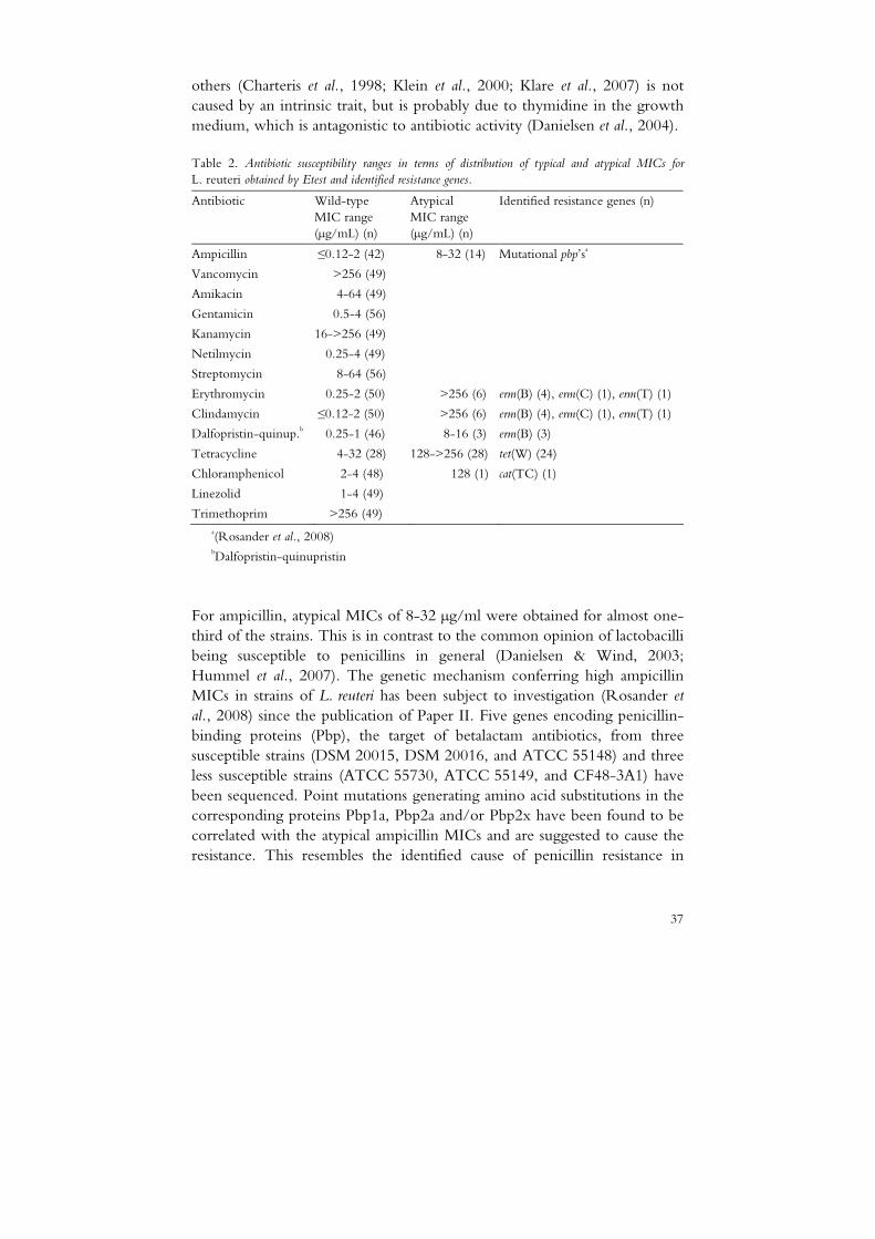
The susceptibility of 56 L. reuteri strains to 14 antibiotics was assessed by Etest and/or broth microdilution (Paper II). Strains exhibiting atypical MICs were subsequently screened by real-time PCR and/or a DNA microarray assay for the presence of known resistance genes (Paper IV). Antibiotic susceptibility ranges and identified resistance genes are summarised in Table 2. The distribution of MICs was uniform for most antibiotics, with MICs in the lower range for linezolid, gentamicin and netilmycin and in the upper range for amikacin and streptomycin. All MICs for kanamycin, vancomycin and trimethoprim were high, with strains exhibiting MICs above the maximum concentration tested. Bimodal distributions of MICs were obtained for ampicillin, chloramphenicol, tetracycline, erythromycin, clindamycin and dalfopristin-quinupristin.
Intrinsic resistance to vancomycin and aminoglycosides such as streptomycin and kanamycin has been reported as a general feature for lactobacilli (Danielsen & Wind, 2003), and stems from the absence of the peptidoglycan D-alanine target precursor and the lack of a cytochrome-mediated transport system required for aminoglycoside uptake, respectively. However, the reduced susceptibility to trimethoprim reported here and by
37
others (Charteris et al., 1998; Klein et al., 2000; Klare et al., 2007) is not caused by an intrinsic trait, but is probably due to thymidine in the growth medium, which is antagonistic to antibiotic activity (Danielsen et al., 2004).
Table 2. Antibiotic susceptibility ranges in terms of distribution of typical and atypical MICs for
L. reuteri obtained by Etest and identified resistance genes.
Antibiotic Wild-type MIC range (µg/mL) (n)
Atypical MIC range (µg/mL) (n)
Identified resistance genes (n)
Ampicillin ≤0.12-2 (42) 8-32 (14) Mutational pbp’sa
Vancomycin >256 (49)
Amikacin 4-64 (49)
Gentamicin 0.5-4 (56)
Kanamycin 16->256 (49)
Netilmycin 0.25-4 (49)
Streptomycin 8-64 (56)
Erythromycin 0.25-2 (50) >256 (6) erm(B) (4), erm(C) (1), erm(T) (1)
Clindamycin ≤0.12-2 (50) >256 (6) erm(B) (4), erm(C) (1), erm(T) (1)
Dalfopristin-quinup.b 0.25-1 (46) 8-16 (3) erm(B) (3)
Tetracycline 4-32 (28) 128->256 (28) tet(W) (24)
Chloramphenicol 2-4 (48) 128 (1) cat(TC) (1)
Linezolid 1-4 (49)
Trimethoprim >256 (49) a(Rosander et al., 2008) bDalfopristin-quinupristin
For ampicillin, atypical MICs of 8-32 µg/ml were obtained for almost one- third of the strains. This is in contrast to the common opinion of lactobacilli being susceptible to penicillins in general (Danielsen & Wind, 2003; Hummel et al., 2007). The genetic mechanism conferring high ampicillin MICs in strains of L. reuteri has been subject to investigation (Rosander et al., 2008) since the publication of Paper II. Five genes encoding penicillin-binding proteins (Pbp), the target of betalactam antibiotics, from three susceptible strains (DSM 20015, DSM 20016, and ATCC 55148) and three less susceptible strains (ATCC 55730, ATCC 55149, and CF48-3A1) have been sequenced. Point mutations generating amino acid substitutions in the corresponding proteins Pbp1a, Pbp2a and/or Pbp2x have been found to be correlated with the atypical ampicillin MICs and are suggested to cause the resistance. This resembles the identified cause of penicillin resistance in
38
streptococci (Hiramatsu et al., 2004). The pbp genes are located on the chromosome and regarded as non-transferable (Rosander et al., 2008).
For chloramphenicol, the wild-type distribution ranging up to 4 µg/mL was in agreement with a previous susceptibility study of L. reuteri (Klare et
al., 2007). Lactobacillus reuteri strain 5010, isolated from dog and exhibiting a 30 times higher chloramphenicol MIC harbours a plasmid-located cat(TC) gene encoding a chloramphenicol acetyltransferase. However, the gene is not identical to the plasmid pTC82 encoded cat(TC) gene of L. reuteri G4 from chicken (Lin et al., 1996), as demonstrated by the negative result using an additional set of primers designed by Cataloluk and co-workers (2004) and covering the whole cat(TC) gene (data not shown). Another L. reuteri strain, ATCC PTA6127, isolated in 1994-95 from a Peruvian dog and displaying a similar rep-PCR fingerprint but with MIC 4 µg/mL for chloramphenicol, was negative in the first PCR screening to the chloramphenicol resistance gene tested. Chloramphenicol is used for certain life-threatening infections such as typhoid fever, but it can cause fatal aplastic anaemia at therapeutic doses in humans, limiting its use within human medicine. However, chloramphenicol is used for several disease conditions in domestic animals (Schwarz et al., 2004), which could be a plausible explanation for the occurrence of the chloramphenicol resistant L. reuteri strain isolated from dog.
4.2.1 Tetracycline resistance in L. reuteri
In total, 28 strains displayed MICs above 64 µg/mL for tetracycline with both Etest and broth microdilution. The wide range of high MICs obtained was in agreement with a previous study assessing antibiotic susceptibility of 43 L. reuteri strains isolated from piglets (Korhonen et al., 2007). Based on the appearance of the tetracycline MIC distribution, it was first believed that the L. reuteri strains harboured different tetracycline resistance genes conferring diverse levels of susceptibility, as reviewed by Chopra & Roberts (2001). However, real-time PCR revealed the presence of tet(W) in 24 of the 28 L. reuteri strains with atypical MIC for tetracycline. None of the other five tetracycline resistance genes tested (tet(K), tet(L), tet(M), tet(O), and tet(S)) were found in any strain including the four tet(W) negative strains Cow 10, ATCC 55148, MF2-3 and MF14-C.
The tet(W) gene is commonly found in human and animal intestinal Gram-positive bacteria, such as various species of Bifidobacterium,
Butyrivibrio, Mitsuokella and Fusobacterium (Kazimierczak et al., 2006; van Hoek et al., 2008). Since the genotypic data of the ACE-ART project became available (van Hoek et al., 2008), it is evident that tet(W) is also
39
found in various Lactobacillus species such as L. amylovorus, L. brevis, L. crispatus, L. gallinarum, L. johnsonii, L. paracasei and L. reuteri. In Papers II and IV, we demonstrated that L. reuteri, displaying 40-42% G+C content (Hammes & Hertel, 2006), is frequently associated with tet(W), whereas the closely related species L. fermentum, displaying 52-54% G+C content (Hammes & Hertel, 2006), is susceptible to tetracycline. Interestingly, this is in contrast to the proposed theory that tet(W), which has a much higher G+C content (53%) than other ribosome-protection-type tet genes, is generally associated with bacterial hosts with a similar G+C-content, such as bifidobacteria and Mitsuokella (Scott et al., 2000).
Comparison of MICs and (GTG)5-PCR genomic fingerprinting data showed that 14 of the 16 L. reuteri strains with high MICs for both ampicillin and tetracycline displayed highly similar rep-PCR fingerprints. Three strains of this so-called group B (Fig. 1 in Paper II) were further characterised. According to size, all three strains seemed to carry the same four plasmids and a tet(W) probe hybridised to the same plasmid of approximately 12 kb. We therefore presumed that all strains of group B, widely distributed in terms of source and geographical origin, contained the same tetracycline resistance plasmid. The other ten strains with atypical MIC for tetracycline were evenly scattered throughout the dendrogram. The apparent genetic heterogeneity of these strains was further demonstrated by the mixed localisation of the tet(W) gene on plasmids or on the chromosome, as determined by Southern blot and/or the ∆∆Ct PCR method described in detail in Paper IV.
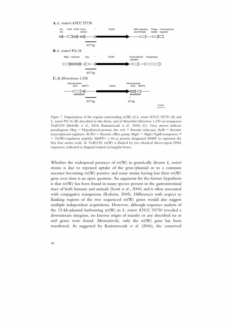
The conservation of the tet(W) gene sequences from different isolates is remarkably high (Scott et al., 2000), as was further confirmed here by the sequence analysis of a chromosome-located tet(W) gene and a plasmid-bound tet(W) gene identified in L. reuteri strains from pig (PA-16) isolated in the 1970s and from human breast milk (ATCC 55730) isolated in 1990, respectively. The whole 1.9 kb gene differs by only two nucleotides in the two L. reuteri strains and by 38 or 40 nucleotides compared with the rumen anaerobe Butyrivibrio fibrisolvens (data not shown), where tet(W) was first identified on a chromosomal transposon (Barbosa et al., 1999). The regions surrounding tet(W) vary in different species of gut bacteria, but contain a conserved core region of 2.6 kb, including the resistance gene, as reported by Kazimierczak et al. (2006). The flanking regions of the two L. reuteri tet(W) genes described in this thesis showed 95-96% similarity to the conserved 657-bp upstream region, but did not contain the conserved 43-bp region downstream of tet(W) (Fig. 7).
40
MgtC Unknown Hyp. * Tet(W) Transcriptional Transposase
regulator
0.5 kb
657 bp
657 bp
657 bp
Nitroreductase Nitroreductase
Nrd1 MAFF1 Tet(W) Nrd2 MAFF2
43 bp
A. L. reuteri ATCC 55730
Ars. ArsR ACR3 Extra- * Tet(W) DNA integrase/ Phage- Transcriptional
red. cellular recombinase related regulator
B. L. reuteri PA-16
C. B. fibrisolvens 1.230
Figure 7. Organisation of the regions surrounding tet(W) of L. reuteri ATCC 55730 (A) and L. reuteri PA-16 (B) described in this thesis, and of Butyrvibrio fibrisolvens 1.230 on transposon TnB1230 (Melville et al., 2004; Kazimierczak et al., 2006) (C). Grey arrows indicate pseudogenes. Hyp. = Hypothetical protein; Ars. red. = Arsenite reductase; ArsR = Arsenite transcriptional regulator; ACR3 = Arsenite efflux pump; MgtC = MgtC/SapB transporter; * = Tet(W)-regulatory peptide; MAFF= a 46-aa protein designated MAFF to represent the first four amino acids. In TnB1230, tet(W) is flanked by two identical direct-repeat DNA sequences, indicated as diagonal striped rectangular boxes.
Whether the widespread presence of tet(W) in genetically diverse L. reuteri strains is due to repeated uptake of the gene/plasmid or to a common ancestor becoming tet(W) positive and some strains having lost their tet(W) gene over time is an open question. An argument for the former hypothesis is that tet(W) has been found in many species present in the gastrointestinal tract of both humans and animals (Scott et al., 2000) and is often associated with conjugative transposons (Roberts, 2005). Differences with respect to flanking regions of the two sequenced tet(W) genes would also suggest multiple independent acquisitions. However, although sequence analysis of the 12-kb-plasmid harbouring tet(W) in L. reuteri ATCC 55730 revealed a downstream integrase, no known origin of transfer or any described tra or mob genes were found. Alternatively, only the tet(W) gene has been transferred. As suggested by Kazimierczak et al. (2006), the conserved

41
surrounding region might function as a mini transfer cassette that has become incorporated into larger mobile elements.
4.2.2 Erythromycin resistance in L. reuteri
Six L. reuteri strains with clearly higher MICs for erythromycin than the majority of strains also had atypical MICs for clindamycin, indicating cross-resistance. Indeed, four of the strains were positive for erm(B) and one strain each was positive for erm(C) and erm(T), as determined with real-time PCR. The resulting dimethylation of the overlapping binding site of the 50S ribosomal subunit confers high resistance to all MLSB antibiotics (Liu & Douthwaite, 2002), thus also explaining the increased MICs to dalfopristin-quinupristin (a mixture of streptogramin A and B). The presence of erm(B) was in agreement with previous studies of three (1048, 1068, 8557:1) of the erythromycin resistant strains (Axelsson et al., 1988; S. Ahrné, pers. comm. 2009).
Comparison of MICs and (GTG)5-PCR genomic fingerprinting data (Fig. 1 in Paper IV) showed that the six strains with atypical MICs for erythromycin and clindamycin were clustered together in the dendrogram, although they did not form a separate group. All erm genes were plasmid-encoded and except for erm(B) in strains 8557:1 and 1068, they were located on plasmids of different sizes, as determined by Southern blot (data not shown), the ∆∆Ct PCR method and/or reported previously (Axelsson
et al., 1988; S. Ahrné, pers. comm. 2009). The unique plasmid profiles of the erm positive strains imply that the erythromycin resistance was not spread clonally, but rather taken up in separate events.
Similarly to the L. reuteri strain LMG 18391, the presence of both erm(B) and tet(W) has recently been reported in an L. paracasei strain (Huys et al., 2008) and two L. crispatus strains (Klare et al., 2007). We found that the two genes were located on the same plasmid, as determined by Southern blot (data not shown for erm(B)). However, it remains to be determined whether the genes are linked to a conjugative transposon, which is often the case with linked erm(B) and tet(M) (Roberts et al., 1999). The other strain (PA-16) with atypical MICs for both tetracycline and erythromycin/clindamycin carried tet(W) and erm(C). Sequence analysis of the erm(C) gene and its flanking regions by direct genome sequencing and a subsequent BLASTP search in Genbank revealed an rRNA methylase with high similarity (95-99% amino acid identity) to erm(C) genes present in various Staphylococcus species. The gene, which is usually located on small plasmids (<5 kb) in staphylococci, was found in the L. reuteri strain on a plasmid of approximately 20 kb. The transposases located downstream of

42
the erm(C) gene and the chromosome-located tet(W) gene of the same strain may be part of transfer machineries, facilitating the spread to other strains.
The macrolide tylosin was the most commonly used antimicrobial agent in pig farming in the European Union until it was banned as an animal growth promoter in 1999. Today it is still used for therapeutic purposes (A. Franklin, pers. comm. 2009). Consequently, bacteria such as enterococci and staphylococci isolated from pigs are frequently resistant to macrolides (Aarestrup & Carstensen, 1998). Here, we found that four of the six L. reuteri strains that tested positive for an erm gene were originally isolated from pigs, although only six of the 56 strains tested in this thesis were from this source. In contrast, Korhonen et al. (2007) previously reported that none of the 43 L. reuteri strains isolated from 30-day-old piglets displayed atypical MICs for erythromycin or clindamycin.
4.3 Antibiotic resistance in L. plantarum (IIIIIIIIIIII and IIIIVVVV)
The susceptibility of up to 121 L. plantarum strains to six antibiotics was assessed by Etest and/or broth microdilution (Paper III and Table E1 of the preceding errata list). Strains with atypical antibiotic MICs were subsequently screened by real-time PCR and/or a DNA microarray assay for the presence of known resistance genes (Paper IV). Antibiotic susceptibility ranges and identified resistance genes are summarised in Table 3. A uniform MIC distribution was obtained for ampicillin, erythromycin and gentamicin, with MICs up to 2 µg/mL, 1-2 µg/mL and 8-16 µg/mL, respectively, depending on the method used. For streptomycin, all strains had MICs in the upper test range or above the maximum concentration tested. Thus, further testing using higher streptomycin concentrations would be needed to define a microbiological breakpoint for L. plantarum to this antibiotic. However, the increased MICs, which are probably due to an intrinsic trait (Danielsen & Wind, 2003), are in accordance with previous results for L. plantarum and other Lactobacillus species, as is the higher observed susceptibility to gentamicin compared with streptomycin (Danielsen & Wind, 2003; Korhonen et al., 2008; Paper II). A distribution with MICs up to 8 µg/mL, but without a clear peak, was obtained for clindamycin. The wide range, covering seven log2 dilution steps, could be due to interlaboratory discrepancies despite the same protocols being used in the four participating laboratories.
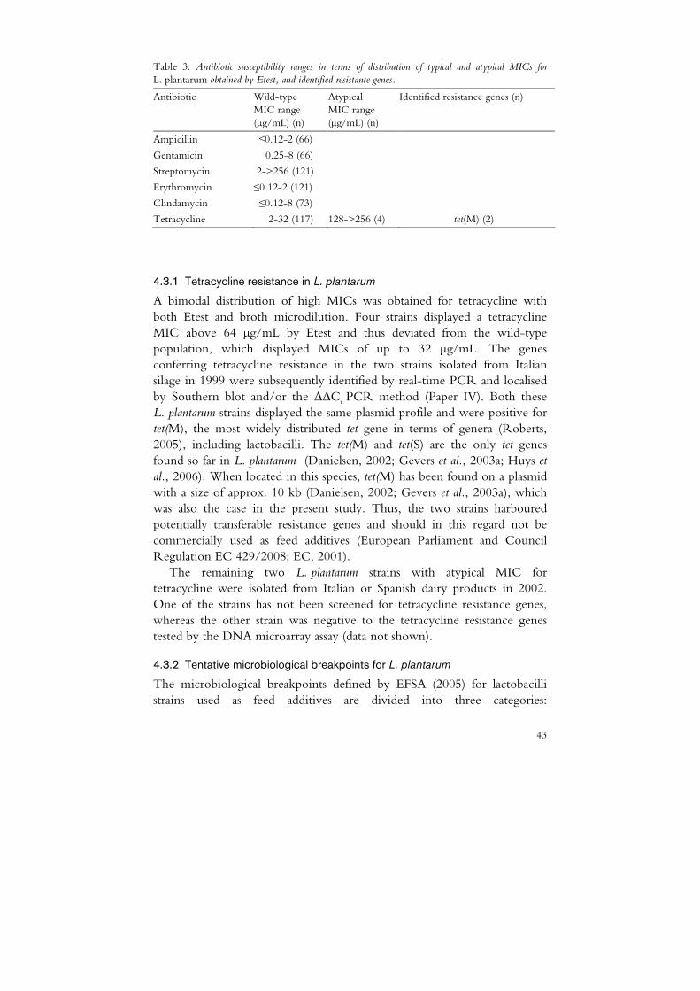
43
Table 3. Antibiotic susceptibility ranges in terms of distribution of typical and atypical MICs for
L. plantarum obtained by Etest, and identified resistance genes.
Antibiotic Wild-type MIC range (µg/mL) (n)
Atypical MIC range (µg/mL) (n)
Identified resistance genes (n)
Ampicillin ≤0.12-2 (66)
Gentamicin 0.25-8 (66)
Streptomycin 2->256 (121)
Erythromycin ≤0.12-2 (121)
Clindamycin ≤0.12-8 (73)
Tetracycline 2-32 (117) 128->256 (4) tet(M) (2)
4.3.1 Tetracycline resistance in L. plantarum
A bimodal distribution of high MICs was obtained for tetracycline with both Etest and broth microdilution. Four strains displayed a tetracycline MIC above 64 µg/mL by Etest and thus deviated from the wild-type population, which displayed MICs of up to 32 µg/mL. The genes conferring tetracycline resistance in the two strains isolated from Italian silage in 1999 were subsequently identified by real-time PCR and localised by Southern blot and/or the ∆∆Ct PCR method (Paper IV). Both these L. plantarum strains displayed the same plasmid profile and were positive for tet(M), the most widely distributed tet gene in terms of genera (Roberts, 2005), including lactobacilli. The tet(M) and tet(S) are the only tet genes found so far in L. plantarum (Danielsen, 2002; Gevers et al., 2003a; Huys et al., 2006). When located in this species, tet(M) has been found on a plasmid with a size of approx. 10 kb (Danielsen, 2002; Gevers et al., 2003a), which was also the case in the present study. Thus, the two strains harboured potentially transferable resistance genes and should in this regard not be commercially used as feed additives (European Parliament and Council Regulation EC 429/2008; EC, 2001).
The remaining two L. plantarum strains with atypical MIC for tetracycline were isolated from Italian or Spanish dairy products in 2002. One of the strains has not been screened for tetracycline resistance genes, whereas the other strain was negative to the tetracycline resistance genes tested by the DNA microarray assay (data not shown).
4.3.2 Tentative microbiological breakpoints for L. plantarum
The microbiological breakpoints defined by EFSA (2005) for lactobacilli strains used as feed additives are divided into three categories:
44
heterofermentative, obligately homofermentive and the species L. plantarum. This has been found to be inadequate in many cases, especially for the large L. delbrueckii group (Korhonen et al., 2008). Tentative microbiological breakpoints, referred to as susceptibility-resistance cut-off values in Paper III, were proposed for L. plantarum to emphasise the need for breakpoints for individual LAB species (Paper III and Table E2 of the preceding errata list). The L. plantarum breakpoints defined in Paper III and by EFSA were conflicting for all antibiotics except tetracycline. Thus, the same four strains with atypical MIC for tetracycline were considered resistant using either breakpoint, whereas e.g. eight strains that were susceptible to clindamycin were considered resistant using the EFSA breakpoints. However, the EFSA breakpoints have recently been updated and are now in agreement with the cut-offs defined in Paper III using broth microdilution for all antibiotics except clindamycin (EFSA, 2008).
4.4 Phenotypic versus genotypic data (IIIIIIII, III III III III, and IVIVIVIV)
The results of the molecular screening correlated well with MIC data on L. reuteri and L. plantarum, except for one tet(W) positive L. reuteri strain with a tetracycline MIC of 16 µg/mL and four presumably tet negative L. reuteri strains with atypical tetracycline MIC (≥256 µg/mL). A weaker hybridisation signal for the tet(W) oligonucleotide was observed on the microarray for the former strain compared with control strains, indicating the presence of a partial or mutated tet(W) gene, or a gene that is similar, rather than identical to tet(W). Sequence analysis of the tet(W) gene in this phenotypically susceptible strain could further elucidate why the gene is non-functional.
Two of the four strains with atypical tetracycline MIC were also tested by microarray analysis, but were negative to the 33 tet genes included in the array. For these strains, there might be other underlying resistance mechanisms such as the presence of multidrug efflux pumps removing tetracycline from its target, as has previously been demonstrated in bifidobacteria for other antibiotics (Margolles et al., 2006). In either case, this shows that phenotypic and molecular tools are both needed to guarantee the presence or absence of acquired resistance genes in strains intended for use in food, feed and probiotic applications, as has also been pointed out by others (Hummel et al., 2007; van Hoek et al., 2008).
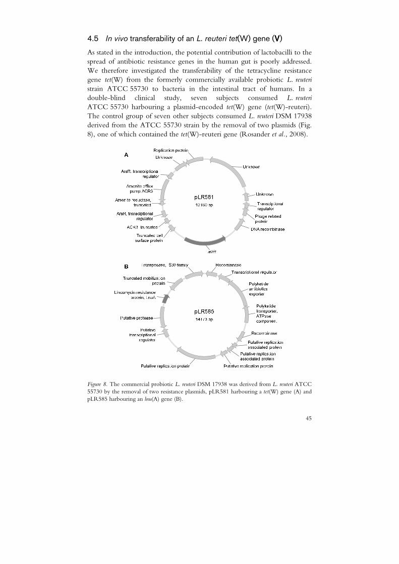
45
4.5 In vivo transferability of an L. reuteri tet(W) gene (VVVV)
As stated in the introduction, the potential contribution of lactobacilli to the spread of antibiotic resistance genes in the human gut is poorly addressed. We therefore investigated the transferability of the tetracycline resistance gene tet(W) from the formerly commercially available probiotic L. reuteri strain ATCC 55730 to bacteria in the intestinal tract of humans. In a double-blind clinical study, seven subjects consumed L. reuteri
ATCC 55730 harbouring a plasmid-encoded tet(W) gene (tet(W)-reuteri). The control group of seven other subjects consumed L. reuteri DSM 17938 derived from the ATCC 55730 strain by the removal of two plasmids (Fig. 8), one of which contained the tet(W)-reuteri gene (Rosander et al., 2008).
Figure 8. The commercial probiotic L. reuteri DSM 17938 was derived from L. reuteri ATCC 55730 by the removal of two resistance plasmids, pLR581 harbouring a tet(W) gene (A) and pLR585 harbouring an lnu(A) gene (B).
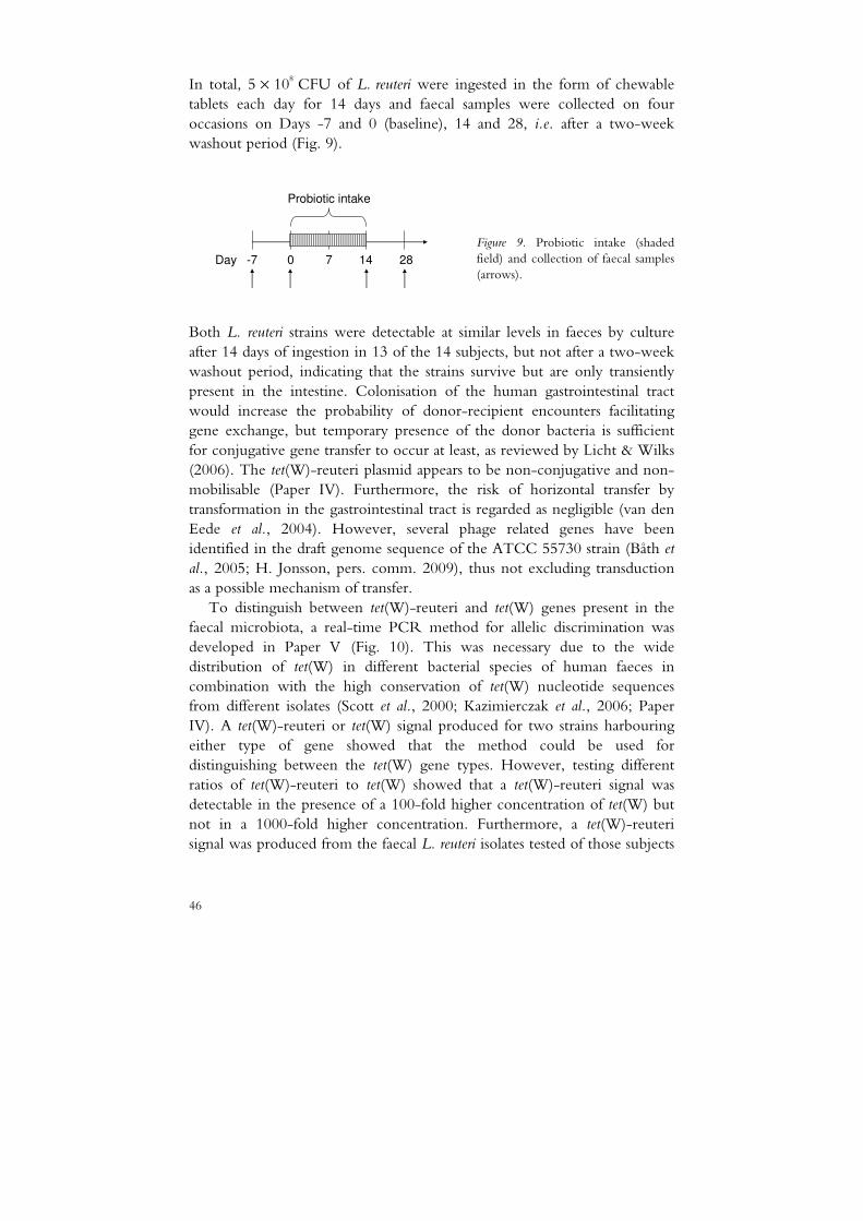
46
In total, 5 × 10⁸ CFU of L. reuteri were ingested in the form of chewable tablets each day for 14 days and faecal samples were collected on four occasions on Days -7 and 0 (baseline), 14 and 28, i.e. after a two-week washout period (Fig. 9).
Figure 9. Probiotic intake (shaded field) and collection of faecal samples (arrows).
Both L. reuteri strains were detectable at similar levels in faeces by culture after 14 days of ingestion in 13 of the 14 subjects, but not after a two-week washout period, indicating that the strains survive but are only transiently present in the intestine. Colonisation of the human gastrointestinal tract would increase the probability of donor-recipient encounters facilitating gene exchange, but temporary presence of the donor bacteria is sufficient for conjugative gene transfer to occur at least, as reviewed by Licht & Wilks (2006). The tet(W)-reuteri plasmid appears to be non-conjugative and non-mobilisable (Paper IV). Furthermore, the risk of horizontal transfer by transformation in the gastrointestinal tract is regarded as negligible (van den Eede et al., 2004). However, several phage related genes have been identified in the draft genome sequence of the ATCC 55730 strain (Båth et
al., 2005; H. Jonsson, pers. comm. 2009), thus not excluding transduction as a possible mechanism of transfer.
To distinguish between tet(W)-reuteri and tet(W) genes present in the faecal microbiota, a real-time PCR method for allelic discrimination was developed in Paper V (Fig. 10). This was necessary due to the wide distribution of tet(W) in different bacterial species of human faeces in combination with the high conservation of tet(W) nucleotide sequences from different isolates (Scott et al., 2000; Kazimierczak et al., 2006; Paper IV). A tet(W)-reuteri or tet(W) signal produced for two strains harbouring either type of gene showed that the method could be used for distinguishing between the tet(W) gene types. However, testing different ratios of tet(W)-reuteri to tet(W) showed that a tet(W)-reuteri signal was detectable in the presence of a 100-fold higher concentration of tet(W) but not in a 1000-fold higher concentration. Furthermore, a tet(W)-reuteri signal was produced from the faecal L. reuteri isolates tested of those subjects
Day -7 0 7 14 28
Probiotic intake
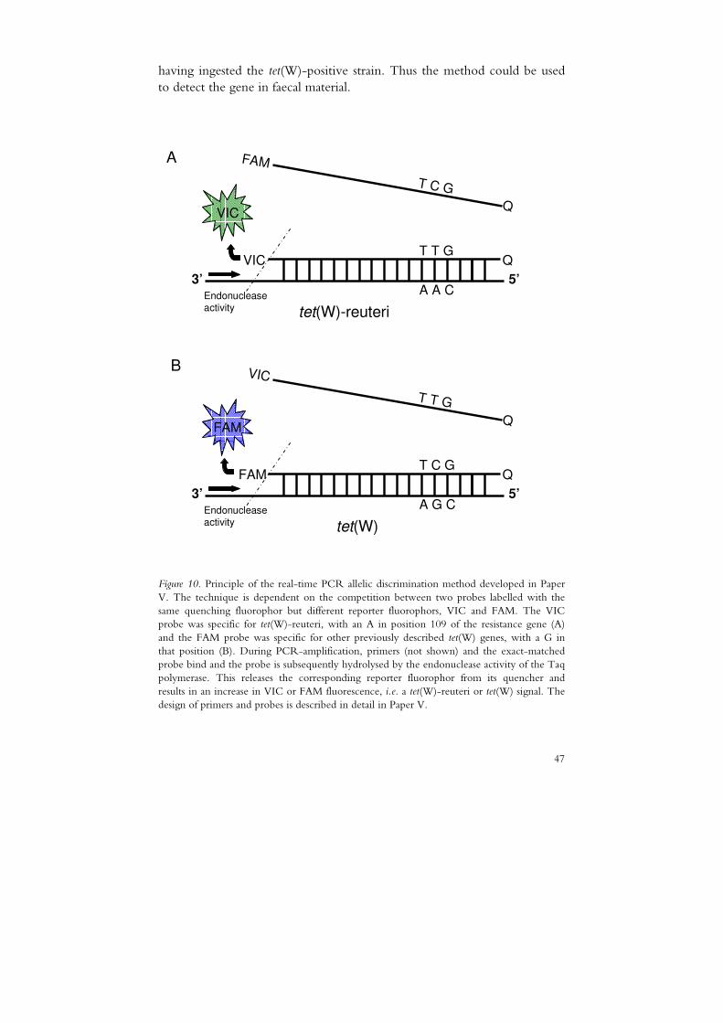
47
having ingested the tet(W)-positive strain. Thus the method could be used to detect the gene in faecal material.
A
B
3’ 5’
FAM
Q
QVICT T G
A A C
T C G
VIC
Endonuclease activity
tet(W)-reuteri
3’ 5’
VIC
Q
QFAMT C G
A G C
T T G
FAM
Endonuclease activity
tet(W)
Figure 10. Principle of the real-time PCR allelic discrimination method developed in Paper V. The technique is dependent on the competition between two probes labelled with the same quenching fluorophor but different reporter fluorophors, VIC and FAM. The VIC probe was specific for tet(W)-reuteri, with an A in position 109 of the resistance gene (A) and the FAM probe was specific for other previously described tet(W) genes, with a G in that position (B). During PCR-amplification, primers (not shown) and the exact-matched probe bind and the probe is subsequently hydrolysed by the endonuclease activity of the Taq polymerase. This releases the corresponding reporter fluorophor from its quencher and results in an increase in VIC or FAM fluorescence, i.e. a tet(W)-reuteri or tet(W) signal. The design of primers and probes is described in detail in Paper V.

48
After enrichment and isolation of bacterial colonies in/on genus-specific tetracycline supplemented media, DNA was extracted and the presence of tet(W)-reuteri was screened by the real-time PCR method developed. A lower Ct value compared with the baseline and the control group obtained by the tet(W)-reuteri detector for the Day 14 and/or Day 28 faecal sample DNA from a subject having ingested tet(W) positive L. reuteri would indicate tet(W)-reuteri gene transfer. However, no tet(W)-reuteri signal was produced from any of the DNA samples. Thus no transfer events were demonstrated under the conditions tested, suggesting that transfer of the tet(W)-reuteri gene during intestinal passage of the probiotic L. reuteri did not occur or occurred at low frequencies undetectable by the method used. As indicated by the tet(W) signals produced, this gene was present in the faecal material from all subjects on one or more sampling occasions, thus verifying its common occurrence within the tetracycline resistant faecal populations of the three genera investigated.
An additional objective was to assess the proportion of tetracycline resistant enterococci, bifidobacteria and lactobacilli present in faeces. The baseline level was 5-12% for one of the eight subjects tested, but in most cases it was less than 0.1%. Our results are in agreement with findings from a similar study in Finland by Saarela et al. (2007), but in contrast to previous findings on the antibiotic susceptibility of faecal strains in Spain, France and Denmark, which suggested a high natural prevalence of tetracycline resistant bifidobacteria, lactobacilli and enterococci in human faeces (Aarestrup et al., 2000; Delgado et al., 2005; Moubareck et al., 2005). Variation in antibiotic use could be a plausible explanation for the higher levels observed in France and Spain (Cars et al., 2001).

49
5 Conclusions
The main findings of this thesis were:
� Controlled and standardised conditions are required for antibiotic susceptibility testing of LAB. � Increased inoculum size or extended incubation time resulted in
elevated antibiotic MICs for all LAB strains using broth microdilution.
� An inoculum of 3 ×××× 105 CFU/mL and an incubation time of 48 h
were recommended for assessing the antibiotic susceptibility of LAB by this method.
� The results obtained have contributed to the development of an international ISO standard method for broth microdilution.
� The MIC data obtained with the Etest and the broth microdilution method were in close agreement for both L. reuteri and L. plantarum.
� All L. plantarum strains tested (n=121) were susceptible to ampicillin,
gentamicin, erythromycin and clindamycin, and intrinsically resistant to streptomycin. Acquired resistance to tetracycline was associated with plasmid-bound tet(M).
� The members of L. reuteri tested (n=56) were susceptible to linezolid, trimethoprim and the aminoglycosides gentamicin and netilmycin, and intrinsically resistant to vancomycin and the aminoglycosides amikacin, kanamycin and streptomycin.
� Lactobacillus reuteri strains with atypical MICs for both tetracycline and
ampicillin were linked to the same genotype, and the strains with

50
atypical MICs for erythromycin and clindamycin were clustered together, although not forming a separate group.
� Lactobacillus reuteri strains had acquired resistance to tetracycline (n=28), ampicillin (n=14), erythromycin/clindamycin (n=6), and chloramphenicol (n=1). This resistance was attributed to mutational pbp genes for ampicillin and to added tet(W), erm and cat(TC) genes for the antibiotics inhibiting protein synthesis.
� The majority of the antibiotic resistant L. reuteri strains harboured plasmid-encoded resistance genes. Traits of putative transfer machineries adjacent to both plasmid- and chromosome-located resistance genes were demonstrated.
� A maximum of two acquired resistance genes were found per L. reuteri strain.
� Transfer of the tet(W) gene from the probiotic strain L. reuteri
ATCC 55730 to faecal enterococci, bifidobacteria and lactobacilli was non-detectable under the conditions tested, although transfer at low frequencies cannot be excluded.
5.1 Future perspectives
LAB intentionally added to the food chain should not carry transferable antibiotic resistance genes (EFSA, 2007). In the absence of reliable scientific data on the potential transfer of antibiotic resistant LAB strains, such strains should be avoided as food processing aids and probiotics according to the precautionary principle. This thesis provides data required to assess the possible risk of using antibiotic resistant strains of the LAB species L. reuteri and L. plantarum as starter cultures or probiotics. However, more data would be of value for further assessment. In particular, more transferability studies in the human gut of both conjugative and non-conjugative antibiotic resistance plasmids/transposons are needed to determine the potential for horizontal transfer. In my opinion, such studies should primarily focus on erm positive lactobacilli due to the clinical importance of macrolides. Sequencing of flanking regions of the remaining plasmid-encoded or chromosome-located tet(W) genes found in L. reuteri could reveal more about the acquisition of this apparently common gene within this species.
51
The considerable increase in available antibiotic susceptibility data for lactobacilli through e.g. ACE-ART (Korhonen et al., 2008) and PROSAFE (Klare et al., 2007) resulted in an recent update of the EFSA breakpoints for LAB used as feed additives and a subsequent split-up of the breakpoints for individual LAB species (EFSA, 2008). A regulation for LAB added to fermented food and probiotics for human use will probably be proposed in the near future. Imposed restrictions have to be based on scientific data, such as these, so that the current strategy based on the precautionary principle can be proven or discounted. Phenotypic tests and molecular tools are both needed to scientifically guarantee the presence or absence of acquired resistance genes in potential starter/probiotic candidates, as pointed out in this thesis and by others (Hummel et al., 2007; van Hoek et al., 2008).
The MIC data presented in this thesis in combination with the results of others will hopefully be used for risk management strategies by organisations such as EUCAST to define rational microbiological breakpoints for the species L. reuteri and L. plantarum. In this thesis, the antibiotics included in the susceptibility testing were those recommended at the time by EFSA (2005). Resistance to quinolones and third/fourth- generation cefalosporins has, as previously mentioned, been reported as an intrinsic feature in lactobacilli. In my opinion, these antibiotics should potentially be tested on a number of Lactobacillus species as more pathogenic bacteria become resistant to the clinically important antibiotics.
Finally, antibiotic resistance of LAB could also be regarded as a beneficial property. A resistant probiotic strain that is co-administered with an antibiotic may reduce the gastrointestinal side effects related to antibiotic treatment (Courvalin, 2006). By identifying strains with potentially non-transferable resistance genes, this field of application might gain wider acceptance and thus have a greater impact in the future.

52

53
References
Aarestrup, F.M., Agerso, Y., Gerner-Smidt, P., Madsen, M. & Jensen, L.B. (2000). Comparison of antimicrobial resistance phenotypes and resistance genes in Enterococcus faecalis and Enterococcus faecium from humans in the community, broilers, and pigs in Denmark. Diagnostic Microbiology and Infectious Disease 37(2), 127-137.
Aarestrup, F.M. & Carstensen, B. (1998). Effect of tylosin used as a growth promoter on the occurrence of macrolide-resistant enterococci and staphylococci in pigs. Microbial Drug Resistance 4(4), 307-312.
Abrahamsson, T.R., Jakobsson, T., Bottcher, M.F., Fredrikson, M., Jenmalm, M.C., Bjorksten, B. & Oldaeus, G. (2007). Probiotics in prevention of IgE-associated eczema: a double-blind, randomized, placebo-controlled trial. Journal of Allergy and Clinical Immunology 119(5), 1174-1180.
Adams, M.R. (1999). Safety of industrial lactic acid bacteria. Journal of Biotechnology 68(2-3), 171-178.
Ahn, C., Collins-Thompson, D., Duncan, C. & Stiles, M.E. (1992). Mobilization and location of the genetic determinant of chloramphenicol resistance from Lactobacillus plantarum caTC2R. Plasmid 27(3), 169-176.
Aleksandrov, A. & Simonson, T. (2008). Binding of tetracyclines to elongation factor Tu, the Tet repressor, and the ribosome: a molecular dynamics simulation study. Biochemistry 47(51), 13594-13603.
Ammor, M.S., Florez, A.B. & Mayo, B. (2007). Antibiotic resistance in non-enterococcal lactic acid bacteria and bifidobacteria. Food Microbiology 24(6), 559-570.
Ammor, M.S., Gueimonde, M., Danielsen, M., Zagorec, M., van Hoek, A.H., de Los Reyes-Gavilan, C.G., Mayo, B. & Margolles, A. (2008). Two different tetracycline resistance mechanisms, plasmid-carried tet(L) and chromosomally located transposon-associated tet(M), coexist in Lactobacillus sakei Rits 9. Applied and Environmental Microbiology 74(5), 1394-1401.

54
Axelsson, L.T., Ahrné, S.E., Andersson, M.C. & Stahl, S.R. (1988). Identification and cloning of a plasmid-encoded erythromycin resistance determinant from Lactobacillus reuteri. Plasmid 20(2), 171-174.
Barbosa, T.M., Scott, K.P. & Flint, H.J. (1999). Evidence for recent intergeneric transfer of a new tetracycline resistance gene, tet(W), isolated from Butyrivibrio fibrisolvens, and the occurrence of tet(O) in ruminal bacteria. Environmental Microbiology 1(1), 53-64.
Bernardeau, M., Guguen, M. & Vernoux, J.P. (2006). Beneficial lactobacilli in food and feed: long-term use, biodiversity and proposals for specific and realistic safety assessments. FEMS Microbiology Reviews 30(4), 487-513.
Bernardeau, M., Vernoux, J.P., Henri-Dubernet, S. & Gueguen, M. (2008). Safety assessment of dairy microorganisms: The Lactobacillus genus. International Journal of Food Microbiology 126(3), 278-285.
Billot-Klein, D., Gutmann, L., Sable, S., Guittet, E. & van Heijenoort, J. (1994). Modification of peptidoglycan precursors is a common feature of the low-level vancomycin-resistant VANB-type Enterococcus D366 and of the naturally glycopeptide-resistant species Lactobacillus casei, Pediococcus pentosaceus, Leuconostoc mesenteroides, and Enterococcus gallinarum. Journal of Bacteriology 176(8), 2398-2405.
Blomberg, B., Manji, K.P., Urassa, W.K., Tamim, B.S., Mwakagile, D.S., Jureen, R., Msangi, V., Tellevik, M.G., Holberg-Petersen, M., Harthug, S., Maselle, S.Y. & Langeland, N. (2007). Antimicrobial resistance predicts death in Tanzanian children with bloodstream infections: a prospective cohort study. BMC Infectious Diseases 7, 43.
Britton, R.A. & Versalovic, J. (2008). Probiotics and gastrointestinal infections. Interdisciplinary Perspectives on Infectious Diseases 2008, 290769.
Brodersen, D.E., Clemons, W.M., Jr., Carter, A.P., Morgan-Warren, R.J., Wimberly, B.T. & Ramakrishnan, V. (2000). The structural basis for the action of the antibiotics tetracycline, pactamycin, and hygromycin B on the 30S ribosomal subunit. Cell 103(7), 1143-1154.
Brown, M.G., Mitchell, E.H. & Balkwill, D.L. (2008). Tet 42, a novel tetracycline resistance determinant isolated from deep terrestrial subsurface bacteria. Antimicrobial Agents and Chemotherapy 52(12), 4518-4521.
Båth, K., Roos, S., Wall, T. & Jonsson, H. (2005). The cell surface of Lactobacillus reuteri ATCC 55730 highlighted by identification of 126 extracellular proteins from the genome sequence. FEMS Microbiology Letters 253(1), 75-82.
Cannon, J.P., Lee, T.A., Bolanos, J.T. & Danziger, L.H. (2005). Pathogenic relevance of Lactobacillus: a retrospective review of over
55
200 cases. European Journal of Clinical Microbiology & Infectious Diseases 24(1), 31-40.
Cars, O., Diaz Högberg, L., Murray, M., Nordberg, O., Sivaraman, S., So, A.D. & Tomson, G. (2008). Meeting the challenge of antibiotic resistance. BMJ (British Medical Journal) 337:a1438, 726-728.
Cars, O., Molstad, S. & Melander, A. (2001). Variation in antibiotic use in the European Union. Lancet 357(9271), 1851-1853.
Casas, I.A. & Dobrogosz, W.J. (2000). Validation of the probiotic concept: Lactobacillus reuteri confers broad-spectrum protection against disease in humans and animals. Microbial and Ecological Health and Disease 12, 247-285.
Cataloluk, O. & Gogebakan, B. (2004). Presence of drug resistance in intestinal lactobacilli of dairy and human origin in Turkey. FEMS Microbiology Letters 236(1), 7-12.
Charteris, W.P., Kelly, P.M., Morelli, L. & Collins, J.K. (1998). Antibiotic susceptibility of potentially probiotic Lactobacillus species. Journal of Food Protection 61(12), 1636-1643.
Charteris, W.P., Kelly, P.M., Morelli, L. & Collins, J.K. (2001). Gradient diffusion antibiotic susceptibility testing of potentially probiotic lactobacilli. Journal of Food Protection 64(12), 2007-2014.
Chopra, I. & Roberts, M. (2001). Tetracycline antibiotics: mode of action, applications, molecular biology, and epidemiology of bacterial resistance. Microbiology and Molecular Biology Reviews 65(2), 232-260.
Chou, Y.K., Chen, H.J., Shaio, M.F. & Kuo, Y.M. (2004). In vitro antibiotic susceptibility of lactobacilli isolated from commercial products containing active lactobacilli. Acta paediatrica Taiwanica 45(3), 141-144.
Claesson, M.J., van Sinderen, D. & O'Toole, P.W. (2008). Lactobacillus phylogenomics--towards a reclassification of the genus. International Journal of Systematic and Evolutionary Microbiology 58(Pt 12), 2945-2954.
Clancy, J., Petitpas, J., Dib-Hajj, F., Yuan, W., Cronan, M., Kamath, A.V., Bergeron, J. & Retsema, J.A. (1996). Molecular cloning and functional analysis of a novel macrolide-resistance determinant, mefA, from Streptococcus pyogenes. Molecular Microbiology 22(5), 867-879.
CLSI (2005). Clinical and Laboratory Standards Institute. 2005. Performance standards for antimicrobial susceptibility testing; 15th informational supplement. Publication M100-S15. Clinical and Laboratory Standards Institute, Wayne, Pa.
CLSI (2007). Clinical and Laboratory Standards Institute. 2007. Methods for antimicrobial susceptibility testing of anaerobic bacteria. Approved standard M11-A7. Clinical and Laboratory Standards Institute, Wayne, Pa.
56
CLSI (2008). Clinical and Laboratory Standards Institute. 2008. Methods for dilution antimicrobial susceptibility tests for bacteria that grow aerobically. Approved standard M07-A8. Clinical and Laboratory Standards Institute, Wayne, Pa.
Connell, S.R., Tracz, D.M., Nierhaus, K.H. & Taylor, D.E. (2003). Ribosomal protection proteins and their mechanism of tetracycline resistance. Antimicrobial Agents and Chemotherapy 47(12), 3675-3681.
Connolly, E., Abrahamsson, T. & Bjorksten, B. (2005). Safety of D(-)-lactic acid producing bacteria in the human infant. J Pediatr Gastroenterol Nutr 41(4), 489-492.
Cosgrove, S.E., Sakoulas, G., Perencevich, E.N., Schwaber, M.J., Karchmer, A.W. & Carmeli, Y. (2003). Comparison of mortality associated with methicillin-resistant and methicillin-susceptible Staphylococcus aureus bacteremia: a meta-analysis. Clinical infectious diseases 36(1), 53-59.
Courvalin, P. (2006). Antibiotic resistance: the pros and cons of probiotics. Digestive and Liver Disease 38 Suppl 2, S261-265.
D'Aimmo, M.R., Modesto, M. & Biavati, B. (2007). Antibiotic resistance of lactic acid bacteria and Bifidobacterium spp. isolated from dairy and pharmaceutical products. International Journal of Food Microbiology 115(1), 35-42.
Danielsen, M. (2002). Characterization of the tetracycline resistance plasmid pMD5057 from Lactobacillus plantarum 5057 reveals a composite structure. Plasmid 48(2), 98-103.
Danielsen, M., Andersen, H.S. & Wind, A. (2004). Use of folic acid casei medium reveals trimethoprim susceptibility of Lactobacillus species. Letters in Applied Microbiology 38(3), 206-210.
Danielsen, M., Madsen, H.L., Hammer, K. & Wind, A. (2005). Investigation of Danish and Italian cheeses made from raw milk and pasteurized milk for the presence of antibiotic resistant lactic acid bacteria and the genetic characterisation of these bacteria. Poster Lab8 conference: Symposium on lactic acid bacteria: Genetics, metabolism and applications, Egmond aan Zee, 2005
Danielsen, M., Mayrhofer, S., Domig, K.J., Amtmann, E., Mayer, H.K., Flórez, A.B., Mayo, B., Korhonen, J. & Tosi, L. (2008). Assessment of the antimicrobial wild-type minimum inhibitory concentration distributions of species of the Lactobacillus delbrueckii group. Dairy Science and Technology 88(2), 183-192.
Danielsen, M. & Seifert, J. (2008). The development of an international ISO/IDF standard for susceptibility testing of lactic acid bacteria and bifidobacteria based on contributions from PROSAFE and ACE-ART. International Journal of Probiotics and Prebiotics 3(4), 247-248.
57
Danielsen, M. & Wind, A. (2003). Susceptibility of Lactobacillus spp. to antimicrobial agents. International Journal of Food Microbiology 82(1), 1-11.
Delgado, S., Florez, A.B. & Mayo, B. (2005). Antibiotic susceptibility of Lactobacillus and Bifidobacterium species from the human gastrointestinal tract. Current Microbiology 50(4), 202-207.
Donohue, D.C., Salminen, S. & Marteau, P. (1998). Safety of Probiotic Bacteria. In: Salminen, S., et al. (Eds.) Lactic Acid Bacteria: Microbiology and Functional Aspects. Marcel Dekker, Inc. pp. 369-384.
EARSS (2007). The European Antimicrobial Resistance Surveillance System. Avilable at: http://www.rivm.nl/earss/
EC (2001). European Commission (EC). Opinion of the Scientific Committee on Animal Nutrition on the criteria for assessing the safety of micro-organisms resistant to antibiotics of human clinical and veterinary importance.
ECDC (2007). Annual epidemiological report on communicable diseases in Europe 2005. European Centre for Disease Prevention and Control. Available at: http://ecdc.europa.eu/pdf/ECDC_epi_report_2007.pdf
EFSA (2005). Opinion of the Scientific Panel on Additives and Products or Substances used in Animal Feed on the updating of the criteria used in the assessment of bacteria for resistance to antibiotics of human and veterinary importance. The EFSA Journal 223, 1-12.
EFSA (2007). Opinion of the Scientific Committee on a request from EFSA on the introduction of a Qualified Presumption of Safety (QPS) approach for assessment of selected microorganisms referred to EFSA. The EFSA Journal 187, 1-16.
EFSA (2008). Technical guidance - Update of the criteria used in the assessment of bacterial resistance to antibiotics of human or veterinary importance - Prepared by the Panel on Additives and Products or Substances used in Animal Feed. The EFSA Journal 732, 1-15.
El-Ziney, M.G., van den Tempel, T., Debevere, J. & Jakobsen, M. (1999). Application of reuterin produced by Lactobacillus reuteri 12002 for meat decontamination and preservation. Journal of Food Protection 62(3), 257-261.
Engemann, J.J., Carmeli, Y., Cosgrove, S.E., Fowler, V.G., Bronstein, M.Z., Trivette, S.L., Briggs, J.P., Sexton, D.J. & Kaye, K.S. (2003). Adverse clinical and economic outcomes attributable to methicillin resistance among patients with Staphylococcus aureus surgical site infection. Clinical infectious diseases 36(5), 592-598.
EUCAST (2000). Terminology relating to methods for the determination of the susceptibility of bacteria to antimicrobial agents (EUCAST

58
definitive document E.Def 12). Clinical Microbiology and Infection 6(9), 503-508.
FAO/WHO (2001). Health and Nutritional Properties of Probiotics in Food including Powder Milk with Live Lactic Acid Bacteria. Report of a Joint FAO/WHO Expert Consultation on Evaluation of Health and Nutritional Properties of Probiotics in Food Including Powder Milk with Live Lactic Acid Bacteria.
FAO/WHO (2002). Guidelines for the evaluation of probiotics in food. Report of a Joint FAO/WHO working group on drafting guidelines for the evaluation of probiotics in food. World Health Organization, London, Ontario, Canada.
Feld, L., Schjorring, S., Hammer, K., Licht, T.R., Danielsen, M., Krogfelt, K. & Wilcks, A. (2008). Selective pressure affects transfer and establishment of a Lactobacillus plantarum resistance plasmid in the gastrointestinal environment. Journal of Antimicrobial Chemotherapy 61(4), 845-852.
Felis, G.E. & Dellaglio, F. (2007). Taxonomy of Lactobacilli and Bifidobacteria. Curr Issues Intest Microbiol 8(2), 44-61.
Flórez, A.B., Delgado, S. & Mayo, B. (2005). Antimicrobial susceptibility of lactic acid bacteria isolated from a cheese environment. Canadian Journal of Microbiology 51(1), 51-58.
Florez, A.B., Ladero, V., Alvarez-Martin, P., Ammor, M.S., Alvarez, M.A. & Mayo, B. (2007). Acquired macrolide resistance in the human intestinal strain Lactobacillus rhamnosus E41 associated with a transition mutation in 23S rRNA genes. International Journal of Antimicrobial Agents 30(4), 341-344.
Francavilla, R., Lionetti, E., Castellaneta, S.P., Magista, A.M., Maurogiovanni, G., Bucci, N., De Canio, A., Indrio, F., Cavallo, L., Ierardi, E. & Miniello, V.L. (2008). Inhibition of Helicobacter pylori infection in humans by Lactobacillus reuteri ATCC 55730 and effect on eradication therapy: a pilot study. Helicobacter 13(2), 127-134.
Franceschi, F., Kanyo, Z., Sherer, E.C. & Sutcliffe, J. (2004). Macrolide resistance from the ribosome perspective. Current Drug Targets-Infectious Disorders 4(3), 177-191.
Francia, M.V., Varsaki, A., Garcillan-Barcia, M.P., Latorre, A., Drainas, C. & de la Cruz, F. (2004). A classification scheme for mobilization regions of bacterial plasmids. FEMS Microbiology Reviews 28(1), 79-100.
Frost, L.S., Leplae, R., Summers, A.O. & Toussaint, A. (2005). Mobile genetic elements: the agents of open source evolution. Nature Reviews Microbiology 3(9), 722-732.
Ge, B., Jiang, P., Han, F., Saleh, N.K., Dhiman, N., Fedorko, D.P., Nelson, N.A. & Meng, J. (2007). Identification and antimicrobial

59
susceptibility of lactic acid bacteria from retail fermented foods. Journal of Food Protection 70(11), 2606-2612.
Gevers, D., Danielsen, M., Huys, G. & Swings, J. (2003a). Molecular characterization of tet(M) genes in Lactobacillus isolates from different types of fermented dry sausage. Applied and Environmental Microbiology 69(2), 1270-1275.
Gevers, D., Huys, G. & Swings, J. (2001). Applicability of rep-PCR fingerprinting for identification of Lactobacillus species. FEMS Microbiology Letters 205(1), 31-36.
Gevers, D., Huys, G. & Swings, J. (2003b). In vitro conjugal transfer of tetracycline resistance from Lactobacillus isolates to other Gram-positive bacteria. FEMS Microbiology Letters 225(1), 125-130.
Gfeller, K.Y., Roth, M., Meile, L. & Teuber, M. (2003). Sequence and genetic organization of the 19.3-kb erythromycin- and dalfopristin-resistance plasmid pLME300 from Lactobacillus fermentum ROT1. Plasmid 50(3), 190-201.
Grohmann, E., Muth, G. & Espinosa, M. (2003). Conjugative plasmid transfer in gram-positive bacteria. Microbiology and Molecular Biology Reviews 67(2), 277-301.
Guardabassi, L. & Courvalin, P. (2006). Modes of antimicrobial action and mechansims of bacterial resistance. In: Aarestrup, F.M. (Ed.) Antimicrobial resistance in bacteria of animal origin. Washington, DC, USA: ASM Press. pp. 1-18.
Hammes, W. & Hertel, C. (2006). The genera Lactobacillus and Carnobacterium. In: The Prokaryotes. pp. 320-403. DOI: 10.1007/0-387-30744-3_10.
Hammes, W.P. & Vogel, R.F. (1995). The genus Lactobacillus. In: Wood, B.J.B., et al. (Eds.) The genera of lactic acid bacteria. London: Blackie academic & professional. pp. 19-54.
Hiramatsu, K., Ohama, M., Mijajima, Y., Kishi, K., Mizunoe, S., Tokimatsu, I., Nagai, H., Kadota, J., Saikawa, T. & Nasu, M. (2004). Antimicrobial susceptibilities and analysis of genes related to penicillin or macrolide resistance in Streptococcus pneumoniae. International Journal of Antimicrobial Agents 24(2), 125-129.
Hufner, E., Britton, R.A., Roos, S., Jonsson, H. & Hertel, C. (2008). Global transcriptional response of Lactobacillus reuteri to the sourdough environment. Systemic and Applied Microbiology 31(5), 323-338.
Hummel, A.S., Hertel, C., Holzapfel, W.H. & Franz, C.M. (2007). Antibiotic resistances of starter and probiotic strains of lactic acid bacteria. Applied and Environmental Microbiology 73(3), 730-739.
Huys, G., D'Haene, K., Danielsen, M., Matto, J., Egervärn, M. & Vandamme, P. (2008). Phenotypic and molecular assessment of antimicrobial resistance in Lactobacillus paracasei strains of food origin. Journal of Food Protection 71(2), 339-344.

60
Huys, G., D'Haene, K. & Swings, J. (2002). Influence of the culture medium on antibiotic susceptibility testing of food-associated lactic acid bacteria with the agar overlay disc diffusion method. Letters in Applied Microbiology 34(6), 402-406.
Huys, G., D'Haene, K. & Swings, J. (2006). Genetic basis of tetracycline and minocycline resistance in potentially probiotic Lactobacillus plantarum strain CCUG 43738. Antimicrobial Agents and Chemotherapy 50(4), 1550-1551.
Jacobsen, L., Wilcks, A., Hammer, K., Huys, G., Gevers, D. & Andersen, S.R. (2007). Horizontal transfer of tet(M) and erm(B) resistance plasmids from food strains of Lactobacillus plantarum to Enterococcus faecalis JH2-2 in the gastrointestinal tract of gnotobiotic rats. FEMS Microbiology Ecology 59(1), 158-166.
Jorgensen, J.H. & Hindler, J.F. (2007). New consensus guidelines from the Clinical and Laboratory Standards Institute for antimicrobial susceptibility testing of infrequently isolated or fastidious bacteria. Clinical infectious diseases 44(2), 280-286.
Kastner, S., Perreten, V., Bleuler, H., Hugenschmidt, G., Lacroix, C. & Meile, L. (2006). Antibiotic susceptibility patterns and resistance genes of starter cultures and probiotic bacteria used in food. Systemic and Applied Microbiology 29, 145-155.
Katla, A.K., Kruse, H., Johnsen, G. & Herikstad, H. (2001). Antimicrobial susceptibility of starter culture bacteria used in Norwegian dairy products. International Journal of Food Microbiology 67(1-2), 147-152.
Kazimierczak, K.A., Flint, H.J. & Scott, K.P. (2006). Comparative analysis of sequences flanking tet(W) resistance genes in multiple species of gut bacteria. Antimicrobial Agents and Chemotherapy 50(8), 2632-2639.
Klare, I., Konstabel, C., Müller-Bertling, S., Reissbrodt, R., Huys, G., Vancanneyt, M., Swings, J., Goossens, H. & Witte, W. (2005). Evaluation of new broth media for microdilution antibiotic susceptibility testing of lactobacilli, lactococci, pediococci, and bifidobacteria. Applied and Environmental Microbiology 71(12), 8982-8986.
Klare, I., Konstabel, C., Werner, G., Huys, G., Vankerckhoven, V., Kahlmeter, G., Hildebrandt, B., Muller-Bertling, S., Witte, W. & Goossens, H. (2007). Antimicrobial susceptibilities of Lactobacillus, Pediococcus and Lactococcus human isolates and cultures intended for probiotic or nutritional use. Journal of Antimicrobial Chemotherapy 59(5), 900-912.
Klein, G., Hallmann, C., Casas, I.A., Abad, J., Louwers, J. & Reuter, G. (2000). Exclusion of vanA, vanB and vanC type glycopeptide resistance in strains of Lactobacillus reuteri and Lactobacillus rhamnosus used as probiotics by polymerase chain reaction and hybridization methods. Journal of Applied Microbiology 89(5), 815-824.

61
Korhonen, J., Danielsen, M., Mayo, B., Egervärn, M., Axelsson, L., Huys, G. & A, V.W. (2008). Antimicrobial susceptibility and proposed microbiological cut-off values of lactobacilli by phenotypic determination. International Journal of Probiotics and Prebiotics 3(4), 257-268.
Korhonen, J.M., Sclivagnotis, Y. & von Wright, A. (2007). Characterization of dominant cultivable lactobacilli and their antibiotic resistance profiles from faecal samples of weaning piglets. Journal of Applied Microbiology 103(6), 2496-2503.
Levy, S.B. (1997). Antibiotic resistance: an ecological imbalance. In: Chadwick DJ, Goode J, eds. Antibiotic resistance: origins, evolution, selection and spread. Chichester, John Wiley and Sons.
Licht, T.R. & Wilcks, A. (2006). Conjugative gene transfer in the gastrointestinal environment. Adv Appl Microbiol 58, 77-95.
Lin, C.F., Fung, Z.F., Wu, C.L. & Chung, T.C. (1996). Molecular characterization of a plasmid-borne (pTC82) chloramphenicol resistance determinant (cat-TC) from Lactobacillus reuteri G4. Plasmid 36(2), 116-124.
Liu, M. & Douthwaite, S. (2002). Activity of the ketolide telithromycin is refractory to Erm monomethylation of bacterial rRNA. Antimicrobial Agents and Chemotherapy 46(6), 1629-1633.
Malathum, K., Coque, T.M., Singh, K.V. & Murray, B.E. (1999). In vitro activities of two ketolides, HMR 3647 and HMR 3004, against gram-positive bacteria. Antimicrobial Agents and Chemotherapy 43(4), 930-936.
Margolles, A., Florez, A.B., Moreno, J.A., van Sinderen, D. & de los Reyes-Gavilan, C.G. (2006). Two membrane proteins from Bifidobacterium breve UCC2003 constitute an ABC-type multidrug transporter. Microbiology 152(Pt 12), 3497-3505.
Marteau, P. (2001). Safety aspects of probiotic products. Scandinavian Journal of Nutrition 45, 22-24.
Mater, D.D., Langella, P., Corthier, G. & Flores, M.J. (2008). A probiotic Lactobacillus strain can acquire vancomycin resistance during digestive transit in mice. Journal of Molecular Microbiology and Biotechnology 14(1-3), 123-127.
Mayrhofer, S., Domig, K.J., Mair, C., Zitz, U., Huys, G. & Kneifel, W. (2008). Comparison of broth microdilution, Etest, and agar disk diffusion methods for antimicrobial susceptibility testing of Lactobacillus acidophilus group members. Applied and Environmental Microbiology 74(12), 3745-3748.
McConnell, M.A., Mercer, A.A. & Tannock, G.W. (1991). Transfer of plasmid pAMβ1 between members of the normal microflora inhabiting the murine digestive tract and modification of the plasmid in a Lactobacillus reuteri host. Microbial Ecology in Health and Disease 4, 343-355.
62
McManus, P.S., Stockwell, V.O., Sundin, G.W. & Jones, A.L. (2002). Antibiotic use in plant agriculture. Annual Review of Phytopathology 40, 443-465.
Melville, C.M., Brunel, R., Flint, H.J. & Scott, K.P. (2004). The Butyrivibrio fibrisolvens tet(W) gene is carried on the novel conjugative transposon TnB1230, which contains duplicated nitroreductase coding sequences. Journal of Bacteriology 186(11), 3656-3659.
Molin, G. (2001). Probiotics in foods not containing milk or milk constituents, with special reference to Lactobacillus plantarum 299v. American Journal of Clinical Nutrition 73(2 Suppl), 380S-385S.
Morelli, L. (2008). Introducing the ACE-ART project: From the expression of interest to 42 months of research. International Journal of Probiotics and Prebiotics 3(4), 241-246.
Morelli, L., Sarra, P.G. & Bottazzi, V. (1988). In vivo transfer of pAM beta 1 from Lactobacillus reuteri to Enterococcus faecalis. Journal of Applied Bacteriology 65(5), 371-375.
Morelli, L., Vogensen, F.K. & von Wright, A. (2004). Genetics of Lactic Acid Bacteria In: Salminen, S., et al. (Eds.) Lactic Acid Bacteria- Microbiological and Functional Aspects. Marcel Dekker, Inc. pp. 249-294.
Moubareck, C., Gavini, F., Vaugien, L., Butel, M.J. & Doucet-Populaire, F. (2005). Antimicrobial susceptibility of bifidobacteria. Journal of Antimicrobial Chemotherapy 55(1), 38-44.
Ouoba, L.I., Lei, V. & Jensen, L.B. (2008). Resistance of potential probiotic lactic acid bacteria and bifidobacteria of African and European origin to antimicrobials: Determination and transferability of the resistance genes to other bacteria. International Journal of Food Microbiology 121(2), 217-224.
Petersen, P.J., Jacobus, N.V., Weiss, W.J., Sum, P.E. & Testa, R.T. (1999). In vitro and in vivo antibacterial activities of a novel glycylcycline, the 9-t-butylglycylamido derivative of minocycline (GAR-936). Antimicrobial Agents and Chemotherapy 43(4), 738-744.
Pioletti, M., Schlunzen, F., Harms, J., Zarivach, R., Gluhmann, M., Avila, H., Bashan, A., Bartels, H., Auerbach, T., Jacobi, C., Hartsch, T., Yonath, A. & Franceschi, F. (2001). Crystal structures of complexes of the small ribosomal subunit with tetracycline, edeine and IF3. Embo Journal 20(8), 1829-1839.
Poehlsgaard, J. & Douthwaite, S. (2005). The bacterial ribosome as a target for antibiotics. Nature Reviews. Microbiology 3(11), 870-881.
Quere, F., Deschamps, A. & Urdaci, M.C. (1997). DNA probe and PCR-specific reaction for Lactobacillus plantarum. Journal of Applied Microbiology 82(6), 783-790.
Rautio, M., Jousimies-Somer, H., Kauma, H., Pietarinen, I., Saxelin, M., Tynkkynen, S. & Koskela, M. (1999). Liver abscess due to a
63
Lactobacillus rhamnosus strain indistinguishable from L. rhamnosus strain GG. Clinical infectious diseases 28(5), 1159-1160.
Roberts, M.C. (2003a). Acquired tetracycline and/or macrolide-lincosamides-streptogramin resistance in anaerobes. Anaerobe 9(2), 63-69.
Roberts, M.C. (2003b). Tetracycline therapy: update. Clinical infectious diseases 36(4), 462-467.
Roberts, M.C. (2005). Update on acquired tetracycline resistance genes. FEMS Microbiology Letters 245(2), 195-203.
Roberts, M.C. (2008). Update on macrolide-lincosamide-streptogramin, ketolide, and oxazolidinone resistance genes. FEMS Microbiology Letters 282(2), 147-159.
Roberts, M.C., Sutcliffe, J., Courvalin, P., Jensen, L.B., Rood, J. & Seppala, H. (1999). Nomenclature for macrolide and macrolide-lincosamide-streptogramin B resistance determinants. Antimicrobial Agents and Chemotherapy 43(12), 2823-2830.
Rosander, A., Connolly, E. & Roos, S. (2008). Removal of antibiotic resistance gene-carrying plasmids from Lactobacillus reuteri ATCC 55730 and characterization of the resulting daughter strain, L. reuteri DSM 17938. Applied and Environmental Microbiology 74(19), 6032-6040.
Ross, J.I., Eady, E.A., Cove, J.H. & Cunliffe, W.J. (1998). 16S rRNA mutation associated with tetracycline resistance in a gram-positive bacterium. Antimicrobial Agents and Chemotherapy 42(7), 1702-1705.
Saarela, M., Maukonen, J., von Wright, A., Vilpponen-Salmela, T., Patterson, A.J., Scott, K.P., Hamynen, H. & Matto, J. (2007). Tetracycline susceptibility of the ingested Lactobacillus acidophilus LaCH-5 and Bifidobacterium animalis subsp. lactis Bb-12 strains during antibiotic/probiotic intervention. International Journal of Antimicrobial Agents 29(3), 271-280.
Salminen, M.K., Rautelin, H., Tynkkynen, S., Poussa, T., Saxelin, M., Valtonen, V. & Jarvinen, A. (2004). Lactobacillus bacteremia, clinical significance, and patient outcome, with special focus on probiotic L. rhamnosus GG. Clinical infectious diseases 38(1), 62-69.
Salminen, M.K., Tynkkynen, S., Rautelin, H., Saxelin, M., Vaara, M., Ruutu, P., Sarna, S., Valtonen, V. & Jarvinen, A. (2002). Lactobacillus bacteremia during a rapid increase in probiotic use of Lactobacillus rhamnosus GG in Finland. Clinical infectious diseases 35(10), 1155-1160.
Salyers, A.A., Gupta, A. & Wang, Y. (2004). Human intestinal bacteria as reservoirs for antibiotic resistance genes. Trends in Microbiology 12(9), 412-416.
Salyers, A.A., Shoemaker, N.B., Stevens, A.M. & Li, L.Y. (1995). Conjugative transposons: an unusual and diverse set of integrated gene transfer elements. Microbiological Reviews 59(4), 579-590.
64
Savino, F., Pelle, E., Palumeri, E., Oggero, R. & Miniero, R. (2007). Lactobacillus reuteri (American Type Culture Collection Strain 55730) versus simethicone in the treatment of infantile colic: a prospective randomized study. Pediatrics 119(1), e124-130.
Saxelin, M., Tynkkynen, S., Mattila-Sandholm, T. & de Vos, W.M. (2005). Probiotic and other functional microbes: from markets to mechanisms. Current Opinion in Biotechnology 16(2), 204-211.
Schleifer, K.H. & Ludwig, W. (1995). Phylogeny of the genus Lactobacillus and related genera. Systemic and Applied Microbiology 18, 461-467.
Schulin, T., Wennersten, C.B., Moellering, R.C., Jr. & Eliopoulos, G.M. (1998). In-vitro activity of the new ketolide antibiotic HMR 3647 against gram-positive bacteria. Journal of Antimicrobial Chemotherapy 42(3), 297-301.
Schwarz, S., Kehrenberg, C., Doublet, B. & Cloeckaert, A. (2004). Molecular basis of bacterial resistance to chloramphenicol and florfenicol. FEMS Microbiology Reviews 28(5), 519-542.
Scott, K.P., Melville, C.M., Barbosa, T.M. & Flint, H.J. (2000). Occurrence of the new tetracycline resistance gene tet(W) in bacteria from the human gut. Antimicrobial Agents and Chemotherapy 44(3), 775-777.
Shornikova, A.V., Casas, I.A., Isolauri, E., Mykkanen, H. & Vesikari, T. (1997). Lactobacillus reuteri as a therapeutic agent in acute diarrhea in young children. J Pediatr Gastroenterol Nutr 24(4), 399-404.
SMI (2009). Swedish Institute for Infectious Disease Control. Available at: www.smi.se
Sullivan, A. & Nord, C.E. (2006). Probiotic lactobacilli and bacteraemia in Stockholm. Scandinavian Journal of Infectious Diseases 38(5), 327-331.
SVARM (2007). Swedish Veterinary Antimicrobial Resistance Monitoring. The NVI (SVA), Uppsala, Sweden, 2008. ISSN 1650-6332.
Swedres (2007). A Report on Swedish Antimicrobial Utilisation and Resistance in Human Medicine. Swedish Institute for Infectious Disease Control, Sweden, 2008. ISSN 1400-3473.
Talarico, T.L., Casas, I.A., Chung, T.C. & Dobrogosz, W.J. (1988). Production and isolation of reuterin, a growth inhibitor produced by Lactobacillus reuteri. Antimicrobial Agents and Chemotherapy 32(12), 1854-1858.
Tannock, G.W. (1987). Conjugal transfer of plasmid pAM beta 1 in Lactobacillus reuteri and between lactobacilli and Enterococcus faecalis. Applied and Environmental Microbiology 53(11), 2693-2695.
Temmerman, R., Pot, B., Huys, G. & Swings, J. (2003). Identification and antibiotic susceptibility of bacterial isolates from probiotic products. International Journal of Food Microbiology 81(1), 1-10.
Tenorio, C., Zarazaga, M., Martinez, C. & Torres, C. (2001). Bifunctional enzyme 6'-N-aminoglycoside acetyltransferase-2"-O-aminoglycoside phosphotransferase in Lactobacillus and Pediococcus

65
isolates of animal origin. Journal of Clinical Microbiology 39(2), 824-825.
Tenover, F.C. & Hughes, J.M. (1996). The challenges of emerging infectious diseases. Development and spread of multiply-resistant bacterial pathogens. Journal of the American Medical Association 275(4), 300-304.
Teuber, M., Meile, L. & Schwarz, F. (1999). Acquired antibiotic resistance in lactic acid bacteria from food. Antonie Van Leeuwenhoek 76(1-4), 115-137.
Thomas, C.M. & Nielsen, K.M. (2005). Mechanisms of, and barriers to, horizontal gene transfer between bacteria. Nature Reviews Microbiology 3(9), 711-721.
Torriani, S., Felis, G.E. & Dellaglio, F. (2001). Differentiation of Lactobacillus plantarum, L. pentosus, and L. paraplantarum by recA gene sequence analysis and multiplex PCR assay with recA gene-derived primers. Applied and Environmental Microbiology 67(8), 3450-3454.
Tosi, L., Berruti, G., Danielsen, M., Wind, A., Huys, G. & Morelli, L. (2007). Susceptibility of Streptococcus thermophilus to antibiotics. Antonie Van Leeuwenhoek 92(1), 21-28.
Walsh, C. (2000). Molecular mechanisms that confer antibacterial drug resistance. Nature 406(6797), 775-781.
van den Eede, G., Aarts, H., Buhk, H.J., Corthier, G., Flint, H.J., Hammes, W., Jacobsen, B., Midtvedt, T., van der Vossen, J., von Wright, A., Wackernagel, W. & Wilcks, A. (2004). The relevance of gene transfer to the safety of food and feed derived from genetically modified (GM) plants. Food and Chemical Toxicology 42(7), 1127-1156.
van Hoek, A.H., Margolles, A., Domig, K.J., Korhonen, J., śycka-Krzesińska, J., Bardowski, J., Danielsen, M., Huys, G., Morelli, L. & Aarts, H. (2008). Molecular assessment of erythromycin and tetracycline resistance genes in lactic acid bacteria and bifidobacteria and their relation to the phenotypic resistance. International Journal of Probiotics and Prebiotics 3(4), 271-280.
Vankerckhoven, V., Huys, G., Vancanneyt, M., Vael, C., Klare, I., Romond, M.-B., Entenza, J.M., Moreillon, P., Wind, R.D., Knol, J., Wiertz, E., Pot, B., Vaughan, E.E., Kahlmeter, G. & Goossens, H. (2008). Biosafety assessment of probiotics used for human consumption: recommendations from the EU-PROSAFE project. Trends in Food Science & Technology 19(2), 102-114.
Weizman, Z., Asli, G. & Alsheikh, A. (2005). Effect of a probiotic infant formula on infections in child care centers: comparison of two probiotic agents. Pediatrics 115(1), 5-9.
Ventura, M., O'Flaherty, S., Claesson, M.J., Turroni, F., Klaenhammer, T.R., van Sinderen, D. & O'Toole, P.W. (2009). Genome-scale

66
analyses of health-promoting bacteria: probiogenomics. Nature Reviews Microbiology 7(1), 61-71.
Vesterlund, S., Vankerckhoven, V., Saxelin, M., Goossens, H., Salminen, S. & Ouwehand, A.C. (2007). Safety assessment of Lactobacillus strains: presence of putative risk factors in faecal, blood and probiotic isolates. International Journal of Food Microbiology 116(3), 325-331.
White, D.G., Acar, J., Anthony, F., Franklin, A., Gupta, R., Nicholls, T., Tamura, Y., Thompson, S., Threlfall, E.J., Vose, D., van Vuuren, M., H.C., W. & M.L., C. (2001). Antimicrobial resistance: standardisation and harmonisation of laboratory methodologies for the detection and quantification of antimicrobial resistance. Revue Scientific et Techniques de l'Office International des Epizooties 20(3), 849-858.
WHO (2007a). Critically Important Antimicrobials for Human Medicine: Categorization for the Development of Risk Management Strategies to contain Antimicrobial Resistance due to Non-Human Antimicrobial Use. Report of the Second WHO Expert Meeting Copenhagen, 29–31 May 2007.
WHO (2007b). The world health report 2007 - A safer future: global public health security in the 21st century. World Health Organisation. Available at: http://www.who.int/whr/2007/en/
Wise, R., Hart, T., Cars, O., Streulens, M., Helmuth, R., Huovinen, P. & Sprenger, M. (1998). Antimicrobial resistance. Is a threat to public healt. BMJ (British Medical Journal) 317, 609-610.
Vogel, R.F., Becke-Schmid, M., Entgens, P., Gaier, W. & Hammes, W.P. (1992). Plasmid transfer and segregation in Lactobacillus curvatus LTH1432 in vitro and during sausage fermentations. Systemic and Applied Microbiology 15, 129-136.
Vogel, R.F., Bocker, G., Stolz, P., Ehrmann, M., Fanta, D., Ludwig, W., Pot, B., Kersters, K., Schleifer, K.H. & Hammes, W.P. (1994). Identification of lactobacilli from sourdough and description of Lactobacillus pontis sp. nov. International Journal of Systemic Bacteriology 44(2), 223-229.
67
Acknowledgements
This work was carried out at the National Food Administration, Uppsala, Sweden, with financial support from the European Commission, Sixth Framework Program (CT-2003-506214, ’ACE-ART’). I would like to express my appreciation to:
My group of supervisors, for excellent guidance and for interesting and educational discussions, including those about the province of Jämtland ☺. I have really enjoyed working with you and I will miss being part of our ’team’.
In particular, my main supervisor Stefan Roos, for your never ending enthusiasm and support, for always taking the time and for sharing your knowledge in the field of lactic acid bacteria and genomics.
My supervisor Hans Lindmark, for sharing your knowledge in molecular biology, for always taking the time, for improving my writing in English and teaching me critical thinking ‘Thought about that?’
My former main supervisor Sven Lindgren, for taking me on as a PhD student, for seeing things from an ecological point of view, for your generosity and support and for bringing pastries to our group meetings.
My supervisor Anders Franklin, for being there when needed, sharing your expertise in antibiotic resistance.
All former ACE-ARTists. It has been fun and educational to be part of this project. In particular, Geert Huys (Paper I), Morten Danielsen (Paper II) and Baltasar Mayo (Paper III), for fruitful cooperations, thorough review of the papers and for always taking the time to answer my many questions. Lasse Axelsson, Angela van Hoek, Jenni Korhonen and Sigrid Mayrhofer, for technical advice and efficient e-mail correspondence.

68
Eamonn Connolly, for cooperation, interesting discussions on the safety of probiotics and excellent review of the thesis.
The staff at Good Food Practice. Johan and Birgitta, for helping me organise the clinical study. Eva and Nilla, for excellent assistance and friendly chats.
My present and former colleagues at the National Food Administration, for taking care of me and always being helpful, for providing the fika room with sweets and interesting discussions on ‘Grey’s anatomy’ and other important stuff. In particular, Catarina, Ann and Kerstin for interest in what I do and valuable friendship. Ingela, for teaching me basic microbiological skills in the lab. Jimmy and Jane, for your ability to provide me with customised growth media at short notice. Marianne B., for help and support. Lisa and Sara, for valuable comments on the thesis.
Past and present members of the Department of Microbiology, SLU, for letting me be a part of your gang despite me not showing up very often. In particular, Hasse J., for your interest in the project and for always caring.
Mina vänner utanför doktorandbubblan. Speciellt till mina tjejer Anna, Malin och Maja med familjer - ni vet varför (New York år 2049?). Mina Uppsalapolare Sara, Louise, Marie, Johanna och Åsa med familjer - för att det är tillåtet att älta och för att ni lärt mig gilla sushi. Jonas gamla vänner som nu också är mina: Andreas, Pär, Robin, Andreas E. och Mattias med familjer - önskar vi sågs lite oftare. Gamla Umeågänget Sara, Anna, Annica, Malin och Magnus med familjer - från overall till detta, på något sätt var det med er allt började. Vännerna hemma i Jämtland: Anna B., Olle och Danne med familjer - för allt kul när vi väl ses. Och inte minst våra nyfunna vänner, grannarna på gatan.
Min kära familj: mamma, pappa, svärmor, svärfar, Pia, Stefan, Ludde, Annica, Jimmy och Ulrica - för er värme och närhet trots att ni bor en bit bort, för att ni pysslar om oss så bra var gång vi ses och för gemenskap även under tyngre stunder. Signes alla storkusiner - för att ni tar hand om henne så bra. Svåger Stefan - för snygga bilder till ramberättelsen.
Slutligen, mina ketchup-lovers därhemma. Jonas - för allt stöd och peppning när det gått trögt, för att du gör mig glad och lycklig och för alla goda middagar à la Fredagskocken i slutet av en slitsam arbetsvecka. Du & jag... och så Signe, min egen lilla gos. Skönt att få koppla av i din värld av teletubbisar och Greta Grisar. Det ger perspektiv på tillvaron ☺
Stort tack till er alla!
Version 1, Maj 2009.