antibacterial activity and phenolic content of propolis ... · antibacterial activity and phenolic...
TRANSCRIPT

Nº 20, Vol. 10 (1), 2018. ISSN 2007 – 0705, pp.: 397 - 412
1Universidad Politécnica de Gómez Palacio, Durango, México 2Universidad Autónoma Agraria Antonio Narro. Unidad Laguna. Coahuila, México
3Universidad Autónoma de San Luis Potosí. San Luis Potosí, México. E-mail: [email protected]
© Universidad De La Salle Bajío (México)
Antibacterial activity and phenolic content of propolis extracts obtained by
different extraction methods
Actividad antibacteriana y contenido fenólico de extractos de propóleos
obtenidos por diferentes métodos de extracción
María del Rosario Moncayo Luján1, Alejandro Moreno Reséndez2, Gretel Sagrario Galván
Barrón1, José Luis Reyes Carrillo2 y María Luisa Carrillo Inungaray3
Palabras clave: flavonoides; examen fitoquímico; polifenoles totales
Keywords: flavonoids; phytochemical screening; total polyphenols
Recepción: 19-02-2018 / Aceptación 28-04-2018
Resumen
Introducción: Los datos disponibles sobre la composición química y los procedimientos para la
extracción del propóleo son actualmente inconclusos. En este estudio se identificaron los grupos
químicos presentes en los extractos de una mezcla de propóleos obtenidos por diferentes
métodos. Además, se determinó su contenido de polifenoles y flavonoides y se estudió su
actividad antioxidante y antibacteriana.
Método: Los extractos se obtuvieron por maceración, por el método ultrasónico y mediante
microondas. Los compuestos fenólicos y flavonoides, así como la medición de la actividad
antioxidante, se cuantificaron por métodos espectrofotométricos. La actividad antibacteriana se
estudió a partir del efecto inhibitorio de cada extracto contra Escherichia coli, Salmonella typhi,
Staphylococcus aureus y Proteus mirabilis, así como por el porcentaje de actividad y el Índice de
Susceptibilidad Bacteriana (IBS).
Resultados: El tamiz fitoquímico evidenció la presencia de abundantes fitoquímicos
biológicamente activos. Se encontró una diferencia significativa (p<0.05) en la actividad
antioxidante y antimicrobiana de los extractos de propóleos obtenidos por diferentes métodos.
Conclusión: La presencia de grupos químicos de compuestos bioactivos y su actividad
antioxidante y antibacteriana justifica el uso de estos extractos en la medicina tradicional.
Abstract
Introduction: The data available regarding the chemical composition and procedures for the
extraction of the propolis are currently inconclusive. In this study the chemical groups present in
the extracts of a propolis mixture obtained by different methods were identified. Additionally,

Antibacterial activity and phenolic content of propolis extracts obtained by different extraction methods
Nº 20, Vol. 10 (1), 2018. ISSN 2007 – 0705, pp.: 397 - 412
- 398 -
their content of polyphenols and flavonoids was determined, and antioxidant and antibacterial
activity was studied.
Method: The extracts were obtained by maceration and for the ultrasonic and microwave
method. Phenolic and flavonoid compounds, as well as the measurement of antioxidant activity,
were quantified by spectrophotometric methods. The antibacterial activity was studied from the
inhibitory effect of each extract against Escherichia coli, Salmonella typhi, Staphylococcus
aureus and Proteus mirabilis, and by the percentage of activity and the Index of Bacterial
Susceptibility (IBS).
Results: The phytochemical screening evidenced the presence of abundant compounds with
important biological activity. It was found a significant difference (p<0.05) in the antioxidant and
antimicrobial activity of the extracts of propolis obtained by different methods.
Conclusion: The presence of chemical groups of bioactive compounds and their antioxidant and
antibacterial activity justifies the use of these extracts in traditional medicine.
Introduction
Propolis is a bee product of complex composition, which is obtained by the bees Apis mellifera
L., by addition of wax and salivary secretions to the resinous, gummy or balsamic material that
they collect from various plants (Ríos et al., 2014). In the hive, bees use propolis for different
purposes, such as closing cracks, minimizing access routes, coating and isolating animal remains
that have been introduced into the hive, consolidating structural components, varnishing the
inside of the hive cells for disinfectant purposes, and avoiding vibrations (Chaillou et al., 2004).
The chemical component and biological activity of propolis depend on the flora area of
bee collection and bee species (Vongsak et al., 2015). It consists of 50–55% of resins and balms,
30–40% of beeswax, 5–10% of essential or volatile oils, 5% of pollen, and 5% of several organic
and mineral materials (Toreti et al., 2103). More than 160 compounds were identified, of which
50% are phenolic compounds to which biological properties are attributed. These compounds are
a heterogeneous set of molecules with antioxidant activity that includes acid phenols and
flavonoids (Drago-Serrano et al., 2006) that contribute to preventing diseases linked to oxidative
stress and to controlling the development of microorganisms (López-Airaghi et al., 2013; Mayta-
Tovalino et al., 2012).

Moncayo Luján, María del Rosario et al.
Nº 20, Vol. 10 (1), 2018. ISSN 2007 – 0705, pp.: 397 - 412
- 399 -
In recent years, the use of propolis in natural medicine has increased, being a valuable raw
material in the cosmetics and food industries, which is why knowledge of the bioactive
compounds present in this product is essential. The chemical composition of propolis is highly
variable and complex due to the biodiversity of the vegetation of each region visited by bees
(Mohammadzadeh et al., 2007). However, even within the same country, the propolis
composition may be qualitatively and quantitatively different depending on the region and time
of collection (Koo, 1999), which makes it difficult for a universal standardization of their
solutions (Popova et al., 2010).
Although most studies on the antibacterial activity of propolis have been made from the
extracts obtained with organic solvents (Omer et al., 2016), the aqueous extracts have also shown
some bactericidal activity (Rodríguez, 2000). Most studies on the antimicrobial activity of plants
have focused more on the effect of solvent used and less on the method used to obtain the
extracts. For example, Ertürk et al. (2016) investigated antibacterial and antioxidant activities of
propolis samples from the Rize province of Turkey in different solvents. They discovered that the
method used to obtain the extracts may affect the content and biological activity of chemical
compounds such as polyphenols. This opens up the possibility of using the resources of the
laboratory in a more efficient way, since it will allow to choose the method with which a better
extraction of active compounds of the substance under study is achieved. In addition, the
chemical composition of propolis is highly variable and complex due to the biodiversity of the
vegetation of each region visited by bees (Mohammadzadeh et al., 2007). For the empirical
treatment of various diseases, propolis is prepared by maceration; So, it is interesting to know if
the method used to obtain the extract affects the content of bioactive compounds and their
biological activity. Therefore, the objective of this work was to identify chemical groups of
bioactive compounds and to determine the content of total phenolic compounds and flavonoids,
as well as to study the antioxidant and antimicrobial capacity of the extracts of the mixture of
propolis of the Comarca Lagunera in the Northern Mexico obtained by different methods.
Material and Methods
Reagents
All the chemical reagents that were used as solvents and standards in the procedures were of the
analytical grade of the Sigma-Aldrich® brand (St. Louis, MO, USA); for the microbiological

Antibacterial activity and phenolic content of propolis extracts obtained by different extraction methods
Nº 20, Vol. 10 (1), 2018. ISSN 2007 – 0705, pp.: 397 - 412
- 400 -
assays, the Mueller-Hinton Agar and Mueller-Hinton Broth culture media from Merck®
(Darmstadt, Germany) were used.
Collection of samples
Four samples of native propolis were collected from beehives using a standard plastic mesh trap
of the apiaries located in the municipalities of Torreón and Matamoros, Coahuila state, and the
municipalities of Gómez Palacio and Lerdo, in the Durango state. These municipalities form part
of the Comarca Lagunera, which is located in northern and central Mexico (25° 05' and 26° 54' N
and 101º 40' and 104° 45' W). It has an average of 235 mm of rain, an elevation of 1,139 m and
an average annual temperature of 18.6 °C (CIGEL, 2018). All samples were kept in the darkness
and stored at 4°C until use. With the samples from the four places, a mixture was formed, since
that is how the population traditionally consumes them. This mixture was dried in a conventional
oven (Linderberg/Blue, USA) at 55°C for 48 h. Moisture content was determined according the
method 925.09 of the AOAC (2014).
Obtaining extracts
The extracts of the propolis mixture were obtained by the classical method of maceration, by
microwave-assisted extraction (Alonso-Castro et al., 2016) and by ultrasound (Muñiz et al.,
2013).
Classical method of maceration. 20 g of the dry mixture of raw propolis was placed in a
flask, and 200 mL of 70% ethanol was added; it was kept at 35°C at 150 rpm for 48 h on a shaker
mixer (IKA KS 4000). Ultrasound-assisted extraction. 20 g of the dry mixture of crude propolis
was placed in a flask, and 200 mL of ethanol (70%) was added; the flask was placed in an
ultrasonic bath (Branson®) at 30°C to 300 W for 30 min. Microwave-assisted extraction. 20 g
of the powdered dried stem of propolis was extracted with 315 mL of 70% ethanol using a closed
system of microwave-assisted extraction (Multiwave 3000 Solv, Anton Paar, Graz, Austria). The
extraction was performed at 70°C and 90 bars during 17 min. The extracts obtained by the three
methods were filtered using filter paper 125 mm, No. 3 (Whatman®), and concentrated under
reduced pressure in a rotary evaporator (BUGI®) to remove the solvent. The extracts were
resuspended in distilled water and sterilized by membrane filtration with a 0.m
pore(Whatman®) and stored at 4°C until use. From the stock solution of each extract, the

Moncayo Luján, María del Rosario et al.
Nº 20, Vol. 10 (1), 2018. ISSN 2007 – 0705, pp.: 397 - 412
- 401 -
chemical groups were identified in each of them, and their antioxidant and antimicrobial
activities were studied.
Phytochemical screening
The identification of chemical groups in the extracts of the propolis mixture was carried out by
means of a phytochemical screening. Chemical groups corresponding to: alkaloids, sterols,
unsaturations, sesquiterpenelactones, flavonoids, saponins, coumarins, and phenolic oxydrils
were identified by colorimetric reactions according to the techniques described by Ramman
(2006) and Buvaneswari et al. (2011). Alkaloids: Mayer Test: To 0.5 mL of the extract, 6 drops
of Mayer's reagent were added by carefully adding one side of the test tube. A white-creamy
precipitate indicated the test as positive. Distilled water was used as a negative control and as
positive control caffeine. Sterols: Liebermann–Burchard reaction: 1.0 mL of extract dissolved
with 1.0 mL of chloroform was mixed with 1.0 mL of acetic anhydride and 0.5 mL of
concentrated H2SO4. A series of color changes showed the presence of phytosterols. A blue-green
ring indicated the presence of terpenoids. Distilled water was used as a negative control and as
positive control almond oil. Unsaturations: A solution of 2% KMnO4 was added dropwise in
water to 0.5 mL of the extract; the test was positive if discoloration or formation of a brown
precipitate was observed resulting from the formation of manganese dioxide. Distilled water was
used as a negative control and as positive control olive oil. Sesquiterpenlactones: Baljet test:
two solutions were used which were mixed in equal volumes before use. Solution A: 0.5 g of
picric acid was placed in 50 mL of ethanol. Solution B: 5.0 g of NaOH in 50 mL of water was
added. For the test, 50 μL of the extract and 4 drops of the reagent were added. The test was
positive if the mixture becomes orange or dark red. Distilled water was used as a negative control
and as positive control celery extract. Flavonoids: H2SO4 test: 50 μL of the extract and 10 μL of
concentrated sulfuric acid were placed. The test was positive if yellow colorings were observed
for flavones and flavonols; orange-icing for flavones; red-blue for chalcones and red-purple for
quinones. Distilled water was used as a negative control and as positive control guava extract.
Saponins: Shinoda test: Magnesium filings were added to 0.5 mL of the extract, and then a few
drops of concentrated HCl were added. The test was considered positive with the appearance of
colors orange, red, pink, and pink-blue to violet. Distilled water was used as a negative control
and as positive control cassava. Coumarins: Sodium hydroxide test: 50.0 μL of the extract was

Antibacterial activity and phenolic content of propolis extracts obtained by different extraction methods
Nº 20, Vol. 10 (1), 2018. ISSN 2007 – 0705, pp.: 397 - 412
- 402 -
placed and 10.0 μL of 10% NaOH solution was added; the test was positive when a yellow color
appeared that disappeared when acidulating with 10.0 μL HCl. Distilled water was used with a
negative control and as positive control cinnamon oil. Phenolic oxydriles: FeCl3 test: 0.5 mL
extract and 5% FeCl3 drops were added together. The appearance of a red, violet-blue or green
precipitate was considered positive. Distilled water was used with negative control and as
positive control vanillin.
Antioxidant properties
Sample preparation. For the determination of the polyphenols and the antioxidant capacity of
each propolis extract, 1 g was weighed and placed in a flask with 4.0 mL of water (ratio 1:5); the
mixture was homogenized by constant stirring for 2 h at 500 rpm, protected against the light. It
was then centrifuged at 10,000 rpm and stored in eppendorf tubes, and the respective analyses
were performed immediately.
Determination of total content phenolic compounds
Total content phenolic compounds were determined using Folin–Ciocalteu reagent following the
method described by Singleton et al. (1999). From each propolis extracts, dilutions in ratio 1:10
were made with deionized water. 1 mL of this dilution was obtained and placed in a test tube, and
5 mL of Folin–Ciocalteu reagent (diluted 1:10 with deionized water) was added. It was allowed
to stand for 8 min, and then 4 mL sodium bicarbonate solution (7.5%, w/v) was added, until a
homogeneous mixture was obtained. The tubes were covered with foil to protect them from light
and were incubated for 2 h at room temperature. The absorbance of each mixture was measured
at 740 nm using a spectrophotometer (Genesys 10uv, USA).
To determine the total phenol content, the absorbance value obtained for each of the
evaluated samples was replaced in the equation of a standard curve of gallic acid (Skerget et al.,
2005). The content of total phenolic compounds was expressed as milligrams of gallic acid
equivalents per liter of extract (mg GAE•L-1).
Estimation of total flavonoids content
Estimation of flavonoids was done as described by Lagouri et al. (2014). The concentration of
total flavonoids in the propolis extracts was measured spectrophotometrically at 415 nm. Half a
Moncayo Luján, María del Rosario et al.
Nº 20, Vol. 10 (1), 2018. ISSN 2007 – 0705, pp.: 397 - 412
- 403 -
milliliter of the sample was transferred to a 10-mL volumetric flask containing 2 mL of AlCl3 (to
10% in ethanol) and 3 mL of CH3COONa (50 g•L-1 in ethanol). The controls contained all the
reagents except for the extract. After 2.5 h at 20°C, the absorbance was measured at 440 nm. The
same procedure was repeated for the standard quercetin solutions (25–250 ppm), and the results
were expressed in milligrams of quercetin per gram of propolis and were presented as means of
triplicates.
Antioxidant activity
Was performed according to the methodology described by Brand et al. (1995), which it is based
on inhibition of the stable radical 2,2-Diphenyl-1-picrylhydrazyl (DPPH). A calibration curve
was done from a methanolic stock solution of DPPH radical, with a concentration of 20 mg•L-1
and the (±)-6-Hydroxy-2,5,7,8-tetramethylchromane-2-carboxylic acid (Trolox reagent). In a
Falcon tube, 990 μL of the DPPH radical and 10 μL of the solution of the ethanolic extract of
propolis added, the mixture was shaken with vortex and allowed to stand for 30 min in the
absence of light, and its absorbance was read in a spectrophotometer at 517 nm.
Antibacterial activity
The antibacterial activity was studied from the inhibitory effect of each extract against
Staphylococcus aureus ATCC 35556, Escherichia coli ATCC 25922, Salmonella typhi ATCC
14028 and Proteus mirabilis ATCC 9150. From the revitalized strains, the inoculum was
prepared as described by Cockerill et al. (2012). The percentage of activity of each extract and
the rate of bacterial susceptibility were also calculated.
Inhibitory effect
The inhibitory effect of each extract was measured from the zones of inhibition observed when
using the disk diffusion technique (Souto et al., 2006). 10 μL (1x10⁶ CFU) of bacteria were
inoculated on the surface of Muller-Hinton agar. After that, filter paper discs (Whatman®) of 6.0
mm diameter were impregnated with 10 μL of each extract. Moxifloxacin (50 mg), a broad-
spectrum antibiotic, was used as a positive control, and destilled water was used as a negative
control. All tests were performed in triplicate.

Antibacterial activity and phenolic content of propolis extracts obtained by different extraction methods
Nº 20, Vol. 10 (1), 2018. ISSN 2007 – 0705, pp.: 397 - 412
- 404 -
Percentage of activity
The percentage of activity (equation 1) indicates the total antimicrobial potency of each extract
(López–Malo et al., 2005).
Activity (%) = 100 x Number of strains susceptible to the extract
Total strains tested (Equation 1)
Index bacterial susceptibility (IBS)
The IBS (Equation 2) shows the number of microorganisms susceptible to the extract, assessing
ranges from 0 (resistance extract all samples) to 100 (susceptible to the whole extract) (López-
Malo et al., 2005; Panghal et al., 2011).
IBS = 100 X Number of effective extracts for each bacterial strain
Total number of bacterial strains (Equation 2)
Statistical analysis
Data were statistically analyzed by one-way analysis of variance (ANOVA) to test differences
between extraction methods, and a (Student–Newmal–Keuls) comparison of means was used to
determine the differences between groups when necessary. Statistical analyses were conducted
using Statistica software. Differences were considered statistically significant at 95% of
confidence level.
Results and discussion
Phytochemical screening
The moisture of the dry propolis mixture from which the extracts was 1.66% ± 0.29. The
phytochemical screening of the hydroalcoholic extract of propolis revealed the presence of
important secondary metabolites. Likewise, there was no difference in the presence of
metabolites analyzed in relation to the extraction method used.
The bioclimate and flora of the Comarca Lagunera, whose vegetation is composed of
Prosopis spp., Abies alba Mill., Acacia farnesiana (L.) Willd, Phoenix spp. and Larrea tridentata
(Sessé and Moc. ex DC.) Coville, varieties (Estrada-Rodríguez, 2004), favor the presence of
bioactive compounds in propolis. The result of phytochemical screening of propolis samples
coincides with that reported by Soltani et al. (2017), who by means of gas chromatography and
Moncayo Luján, María del Rosario et al.
Nº 20, Vol. 10 (1), 2018. ISSN 2007 – 0705, pp.: 397 - 412
- 405 -
mass spectrometry, detected: aromatic acid ester, saturated hydrocarbon alkaloids, saturated
hydrocarbons, and aromatic heterocyclic organic compounds, in ethanolic extracts of Algerian
propolis. Among the numerous groups of substances identified in propolis samples from different
localities, the most common are aromatic acids and esters, chalcones, flavonoids, terpenoids, and
waxy acids. Furthermore, most of the biological activities of propolis have been attributed to
these compounds, especially the flavonoids (Trusheva et al., 2006). A significant number of
reports describe its anti-inflammatory, immunomodulatory, antioxidant, anticancer, and
hepatoprotective properties, as well as many other biological activities of propolis (Borges et al.,
2011).
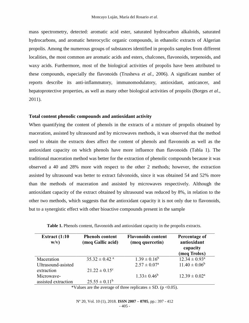
Total content phenolic compounds and antioxidant activity
When quantifying the content of phenols in the extracts of a mixture of propolis obtained by
maceration, assisted by ultrasound and by microwaves methods, it was observed that the method
used to obtain the extracts does affect the content of phenols and flavonoids as well as the
antioxidant capacity on which phenols have more influence than flavonoids (Tabla 1). The
traditional maceration method was better for the extraction of phenolic compounds because it was
observed a 40 and 28% more with respect to the other 2 methods; however, the extraction
assisted by ultrasound was better to extract falvonoids, since it was obtained 54 and 52% more
than the methods of maceration and assisted by microwaves respectively. Although the
antioxidant capacity of the extract obtained by ultrasound was reduced by 8%, in relation to the
other two methods, which suggests that the antioxidant capacity it is not only due to flavonoids,
but to a synergistic effect with other bioactive compounds present in the sample
Table 1. Phenols content, flavonoids and antioxidant capacity in the propolis extracts.
Extract (1:10
w/v)
Phenols content
(meq Gallic acid)
Flavonoids content
(meq quercetin)
Percentage of
antioxidant
capacity
(meq Trolox)
Maceration 35.32 ± 0.42 a 1.39 ± 0.16b 12.34 ± 0.93a
Ultrasound-asisted
extraction
21.22 ± 0.15c
2.57 ± 0.07a 11.40 ± 0.06b
Microwave-
assisted extraction
25.55 ± 0.11b
1.33± 0.46b 12.39 ± 0.02a
*Values are the average of three replicates ± SD. (p <0.05).

Antibacterial activity and phenolic content of propolis extracts obtained by different extraction methods
Nº 20, Vol. 10 (1), 2018. ISSN 2007 – 0705, pp.: 397 - 412
- 406 -
The interest in quantifying phenolic compounds and flavonoids present in propolis is because
they are responsible for the physiological activity of this product (Delgado et al., 2015). Farré et
al. (2004) consider that the phenolic compounds act synergistically with flavonoids, and of these,
quercetin is one of the flavonoids of interest to quantify.
Antibacterial activity
The extracts obtained by the three methods showed 100% of antimicrobial activity and IBS was
of 100%. The inhibitory effect of the extracts (Table 2) varied, not only regarding the extraction
method, but also depending on the bacteria studied. It was observed that the inhibitory effect was
less for E. coli than for the other studied bacteria.
Table 2. Inhibition zones (mm) showing the inhibitory effect of propolis extracts.
Microorganisms
tested
Extract types
Maceration Assisted by
microwave
Assisted by
ultrasound
Moxifloxacin
(50 mg)
Escherichia coli 7.0 ± 0.0a 7.0 ± 0.0a 7.0 ± 0.0a 24.0 ± 0.5
S. typhi 10.3 ± 0.6a 12.3 ± 0.6 a 11.0 ± 1.0 a 29.0 ± 0.0
Staphylococcus aureus 9.0 ± 0.01a 9.0 ± 0.0 5 a 8.0 ± 0.0 a 19.0 ± 0.5
Proteus mirabilis 9.7 ± 0.6 a 10.2 ± 1.2 a 11.2 ± 0.3 a 24.5± 0.6
Values are the average of three replicates ± SD.
The different letters between columns indicate a significant difference (p < 0.05).
There was no significant difference (p <0.5) between the diameters of the zone of inhibition of
extracts obtained by different techniques, which supports the way in which the extract is
traditionally used, that is, by maceration. The least sensitive microorganism was S. typhi, and the
most sensitive was E. coli. Although the three extracts were active against the bacteria studied,
none of them was as effective as the antibiotic used as a positive control. These results are similar
from those reported by Ertürk et al. (2016) and for Koo et al. (2000), who reported that the
ethanolic extract of crude extract of propolis was found the most sensitive microorganism to
propolis was E. coli in the Gram negative group. This activity can be a synergy between
flavonoids, apigenin, chrysin, or other components in raw propolis samples. According to Hegazi
et al. (2014), the propolis samples show a different antimicrobial activity and attribute it to the

Moncayo Luján, María del Rosario et al.
Nº 20, Vol. 10 (1), 2018. ISSN 2007 – 0705, pp.: 397 - 412
- 407 -
complex composition of the resin. In general, propolis is used to treat various diseases in humans.
One of the most important properties of propolis is its antimicrobial activity, which is attributed
mainly to flavonoids, the main biologically active compound in propolis, and it is a substance
that has potential for the treatment of conditions caused by different microorganisms (Tolosa and
Cañizares, 2002). Several trials have demonstrated the antibacterial activity of ethanolic extracts
against Staphylococcus aureus, E. coli and Pseudomonas spp. (Ortega et al., 2011; Díaz-Mena et
al., 2000), and it is currently investigated whether the propolis can be used as a natural food
preservative (Vargas-Sánchez et al., 2013). Dimov et al. (1992) correlated the antibacterial
activity of propolis with the presence of flavonoids; and Farnesi et al. (2009) reported that the
bactericidal effect of these compounds on the metabolic disturbance of ion channels due to paired
phosphorylation / dephosphorylation reactions. The antimicrobial activity is also attributed to
phenolic acids: ferulic, caffeic and cumaric, bioactive compounds found in propolis samples by
Popova et al. (2010). The presence of phenolic compounds in the samples of propolis explain
their antimicrobial activity, which is supported by Marcucci (1995), who reported that caffeic
acid, as well as benzoic acid and cinnamic acid act on the microbial membrane, causing structural
damage to the cells. Until now, no single propolis component has shown to possess more
antibacterial properties than that of the total extract. Some authors cite the highly complex and
variable composition of propolis as a reason for its antimicrobial activity and the data gathered to
date suggest that such activity can be linked to multiple targets, with several constituents acting
in synergy (Scazzocchio, 2006). Propolis affects the cytoplasmic membrane, inhibits bacterial
motility and enzyme activity, exhibits bacteriostatic activity against different bacterial genera and
can act as a bactericide at high concentrations (Mirzoeva, 1997).
Conclusions
The method of obtaining the extract affects the content of phenols and flavonoids, as well as its
antioxidant capacity; however, with the traditional method of maceration, phenols are extracted,
which contributes to their antioxidant capacity. The abundant biologically active phytochemicals,
and their antioxidant and bactericidal activities, seem to validate the classical medicinal uses of
propolis extracts.

Antibacterial activity and phenolic content of propolis extracts obtained by different extraction methods
Nº 20, Vol. 10 (1), 2018. ISSN 2007 – 0705, pp.: 397 - 412
- 408 -
Acknowledgments
Special thanks to the State Council of Science and Technology of the State of Coahuila of
Zaragoza and the Community of Institutions of Higher Education of La Laguna (COECYT-
CIESLAG).
References
Alonso-Castro, A. J., Zapata-Morales, J. R., González-Chávez, M. M., Carranza-Álvarez, C.,
Hernández-Benavides, D. M., y Hernández-Morales, A. (2016). Pharmacological effects
and toxicity of Costus pulverulentus C. Presl (Costaceae). Journal of
Ethnopharmacology, 180, 124–130. doi: 10.1016/j.jep.2016.01.011.
Association of Official Analytical Chemists (AOAC). (2014). Oficial Methods of Analysis of the
Association of Official Analytical Chemists. Gaithersburg. Recuperado de
https://www.aoac.org/aoac_prod_imis/AOAC/Publications/Official_Methods_of_Analysi
s/AOAC_Member/Pubs/OMA/AOAC_Official_Methods_of_Analysis.aspx?hkey=5142c
478-ab50-4856-8939-a7a491756f48 (8 de junio de 2017).
Brand-Williams, W., Cuvelier, M. E. y Berset C. (1995). Use of a free radical method to evaluate
antioxidant activity. Food Science and Technology 28(1), 25– 30.
https://doi.org/10.1016/S0023-6438(95)80008-5
Borges, K. S., Brassesco, M. S., Scrideli, C. A., Soares, A. E. E., y Tone, L. G. (2011).
Antiproliferative Effects of tube-bee propolis in glioblastoma cell lines. Genetics and
Molecular Biology, 34(2), 310-314. doi: 10.1590/S1415-47572011000200024.
Buvaneswari, K., Ramamoorthy, D., y Velanganni, J. (2011). Preliminary Phytochemical and
Antimicrobial Activity Studies on the Leaves of the Indian Plant Thevetia neriifolia Juss.
World Journal of Agricultural Sciences, 7 (6), 659-666.
Centro de Información Georreferenciada de la Región Lagunera (CIGEL). 2018. Mapa Digital de
México de la Región Lagunera. Recuperado de: http://seig-laguna.lag.itesm.mx/CIGEL/
Cockerill, F. R., Wikler, M. A., Alder, J., Dudley, M. A., Eliopoulus. G. M., Ferraro, M. J. y
Zimmer, B. L. (2012). Performance Standards for Antimicrobial Disk Susceptibility
Tests; Approved Standards – Eleventh Edition (Vol. 32). CLSI (Clinical and Labortaory
Standards Institute), Wayne, PA. 58 p.
Moncayo Luján, María del Rosario et al.
Nº 20, Vol. 10 (1), 2018. ISSN 2007 – 0705, pp.: 397 - 412
- 409 -
Chaillou, L. L., HerreraII, H. A., y MaidanaIII, J. F. (2004). Estudio del propoleos de Santiago
del Estero, Argentina. Food Science and Technology 24(1), 11-15.
http://dx.doi.org/10.1590/S0101-20612004000100003
Díaz-Mena, D., Gil-Rodríguez, J. D., y Valdés-González, G. (2000). Determinación de la CMI de
propóleos cubanos a partir de dos técnicas diferentes. Apiciencia 2(2), 1-12.
Delgado-Aceves, M. L., Andrade-Ortega, J. A. y Ramirez-Barragan, C.A. (2015). Physical-
chemical description of propolis collected in La Primavera forest, Zapopan, Jalisco state.
Revista Mexicana de Ciencias Forestales 6(28), 74-87.
Dimov, V., Ivanovska, N., Bankova, V., y Popov, S. (1992). Immunomodulatory action of
propolis: IV. Prophylactic activity against Gram-negative infections and adjuvant effect of
the water soluble derivative. Vaccine, 10(12), 817-823.
Drago-Serrano, M. E., López-López, M., y Saínz-Espuñes, T. D. (2006). Componentes bioactivos
de alimentos funcionales de origen vegetal. Revista Mexicana de Ciencias Farmacéuticas,
37(4), 58-68.
Ertürk, O., Cil, E., Yologlu, N., y Yavuz, C. (2016). An in vitro Study on Antimicrobial and
Antioxidant Activity of Propolis from Rize Province of Turkey. Mellifera, 16(1), 4–18.
Estrada-Rodríguez, J. L. (2004). El cañón de Fernández: anfibios y reptíles. Universidad Juárez
del Estado de Durango, Escuela Superior de Biología, Centro de Estudios Ecológicos,
Instituto de Ecología. Gómez Palacio, Durango, México. 60 p.
Farnesi, A. P., Aquino-Ferreira R., De Jong, D., Bastos, J. K., y Soares. A. E. E. (2009). Effects
of stingless bee and honeybee propolis on four species of bacteria. Genet. Mol. Res., 8(2),
635-640
Farré, R., Frasquet, I., y Sánchez, A. (2004). El propolis y la salud. ARS Pharmaceutica, 45(1),
21-43.
Hegazi, A., Abdou A. M. y Abd Allah, F. (2014). Egyptian Propolis 11: Its antimicrobial activity
with comparison with different localities. Int. J. Curr. Microbiol. App. Sci., 3(9), 530-538.
Koo, H., Gomes, B. P., Rosalen, P. L., Ambrosano, G. M., Park, Y. K. y Cury, J. A. (2000). In
vitro antimicrobial activity of propolis and Arnica montana against oral pathogens.
Archives of Oral Biology, 45(2), 141–148.

Antibacterial activity and phenolic content of propolis extracts obtained by different extraction methods
Nº 20, Vol. 10 (1), 2018. ISSN 2007 – 0705, pp.: 397 - 412
- 410 -
Koo, H., Rosalen, P. L., Cury, J. A., Park, Y. K., Ikegaki, M., y Sattler, A. (1999). Effect of Apis
mellifera propolis from two Brazilian regions on caries development in desalivated rats.
Caries Research 33(5), 393-400.
Lagouri, V., Prasianaki, D., y Crystal, F. (2014). Antioxidant properties and phenolic
composition of greek propolis extracts. International Journal of Food Properties, 17(3),
511–522, doi: 10.1080/10942912.2012.654561.
López-Airaghi, F., Corral, L., Acosta, C., Salguero, A. R., Boggetti, H. J., González, M. y
Tereschuk, M. L. (2013). Evaluación de la actividad antimicrobiana de extractos
convencionales y por fluidos supercríticos de Larrea cuneifolia y propóleos para su uso
en tecnología alimentaria. Investigaciones en Facultades de Ingeniería del NOA,
1853(7871). Recuperado de
https://www.researchgate.net/publication/257409349_Evaluacion_de_la_actividad_antimi
crobiana_de_extractos_convencionales_y_por_fluidos_supercriticos_de_Larrea_cuneifoli
a_y_propoleos_para_su_uso_en_tecnologia_alimentaria
López-Malo, A., Palou, E., Mickey, P., y Michael, D. (2005). Methods for activity assay and
evaluation of results. In: Antimicrobials in Food, Third Edition. CRC Press 659 - 680.
Marcucci, M. C. (1995). Propolis: chemical composition, biological properties and therapeutic
activity. Apidologie, 26(2), 83-99. doi: 10.1051/apido:19950202
Mayta-Tovalino, F., Sacsaquispe-Contreras, S., Ceccarelli-Calle, J., y Alania-Mallqui, J. (2012).
Propóleo peruano: Una nueva alternativa terapéutica antimicrobiana en Estomatología.
Revista Estomatológica Herediana, 22(1), 50-58. doi:
https://doi.org/10.20453/reh.v22i1.159
Mirzoeva, O. K., Grishanin, R. N., y Colder, P. C. (1997). Antimicrobial action of propolis and
some of its components: the effect on growth, membrane potential and motility of
bacteria. Microbiology Research, 152(3), 239-246.
Mohammadzadeh, S., Shariatpanahi, M., Hamedi, M., Ahmadkhaniha, R., Samadi, N., y Ostad,
S. N. (2007). Chemical composition, oral toxicity and antimicrobial activity of Iranian
propolis. Food Chemistry, 103(4), 1097-1103.
https://doi.org/10.1016/j.foodchem.2006.10.006
Muñiz-Márquez, D. B., Martínez-Ávila, G. C., Wong-Paz, J. E., Belmares-Cerda, R. y
Rodríguez-Herrera, R. y Aguilar, C. N. (2013). Ultrasound-assisted extraction of phenolic
Moncayo Luján, María del Rosario et al.
Nº 20, Vol. 10 (1), 2018. ISSN 2007 – 0705, pp.: 397 - 412
- 411 -
compounds from Laurus nobilis L. and their antioxidant activity. Ultrasonics
Sonochemistry, 20(5), 1149-1154. https://doi.org/10.1016/j.ultsonch.2013.02.008
Ertürk, Ö., Çil, E., Yoloğlu, N. y Yavuz, C. (2016). An In vitro Study on Antimicrobial and
Antioxidant Activity of Propolis from Rize Province of Turkey. Mellifera, 16(1), 4–18.
Ortega, N. S., Benitez-Campo, N. y Cabezas-Fajardo, F. A. (2011). Antibacterial activity and
cualitative composition propolis from two climatic regions cauca department. Revista
biológico agropecuaria, 9(1), 8-16.
Panghal, M., Kaushal, V., y Yadav, J. P. (2011). In vitro antimicrobial activity of ten medicinal
plants against clinical isolates of oral cancer cases. Ann Clin Microbiol Antimicrob.,
10(21), 101-111. doi: 10.1186/1476-0711-10-21.
Popova, M., Chen, C. N., Chen, P. Y., Huang, C. Y., y Bankova, V. (2010). A. Validated
spectrophotometric method for quantification of prenylated flavanones in Pacific propolis
from Taiwan, Phytochemical Analyse 21(2), 186-191. doi: 10.1002/pca.1176.
Raaman, N. (2006). Phytochemical Techniques. New Delhi. Publising Agency 19-24.
Ríos, N., Yañez, C., Rojas, L., Mora, F., Usibillaga, A. y Vit. P. (2014). Chemical composition of
essential oil of apis mellifera propolis from Falcon State, Venezuela. Emir. Journal of
Food Agriculture, 26(7), 639-642. doi: 10.9755/ejfa.v26i7.18198.
Rodríguez, H. (2000). La utilidad de las plantas medicinales. EUNA. San José, Costa Rica 61-62
Scazzocchio, F., D'Auriaa, F. D., Alessandrinia, D., y Pantanella, F. (2006). Multifactorial
aspects of antimicrobial activity of propolis. Microbiology Research, 161(4), 327-333.
Singleton, V. L., Orthofer, R.., y Lamuela-Raventos, R. M. (1999). Analysis of total phenols and
other oxidation substrates and antioxidants by means of folin-ciocalteu reagent. Methods
in Enzymology, 299, 152-178.
Skerget, M., Kotonik, P., Hadolin, M., RiznerHras, A., Simonic, M. y Knez, Z. (2005). Phenols,
proanthocyanidins, flavones and flavonols in some plant materials and their antioxidant
activities. Food Chemistry, 89(2), 191-198.
Soltani, E. K., Cerezuela, R., Charef, N., Mezaache-Aichour, S., Esteban, M. A. y Mihoub
Zerroug, M. M. (2017). Algerian propolis extracts: Chemical composition, bactericidal
activity and in vitro effects on gilthead seabream innate immune responses. Fish &
Shellfish Immunology, 62, 57-67. doi: 10.1016/j.fsi.2017.01.009.

Antibacterial activity and phenolic content of propolis extracts obtained by different extraction methods
Nº 20, Vol. 10 (1), 2018. ISSN 2007 – 0705, pp.: 397 - 412
- 412 -
Souto, R., Andrade, A. F. B., Uzeda, M. y Colombo A. P. V. (2006). Prevalence of “non-oral”
Patogenic bacteria in subgingival biofilm of subjects with chronic periodontitis. Brazilian
Journal Microbiology, 37(3), 208-215. http://dx.doi.org/10.1590/S1517-
83822006000300002
Tolosa, L., y Cañizares, E. (2002). Obtención, caracterización y evaluación de la actividad
antimicrobiana de extractos de propóleos de Campeche. ARS Pharmaceutica, 43(1), 187-
204.
Toreti, V. C., Sato, H. H., Pastore, G. M. y Park, Y. K. (2013). Recent Progress of propolis for its
biological and chemical compositions and its botanical origin. Evidence Based
Complementary Alternative Medicine, 2013, 697390. doi: 10.1155/2013/697390.
Trusheva, B., Popova, M., Bankova, V., Simova, S., Marcucci, M. C., Miorin, P. L., da Rocha-
Pasin, F. y Tsvetkova, I. (2006). Bioactive constituents of Brazilian red propolis. Evid
Based Complement Alternat Med., 3(2), 249–254. doi: 10.1093/ecam/nel006.
Vargas-Sánchez, R. D., Torrescano-Urrutia, G. R., y Sanchez-Escalante, A. (2013). El Propóleos:
Conservador Potencial para la Industria Alimentaria. Interciencia, 38(10), 705-711.
Vongsak, B., Kongkiatpaiboon, S., Jaisamut, S.,Machana, S. y Pattarapanich, C. (2015). In vitro
alpha glucosidase inhibition and free-radical scavenging activity of propolis from Thai
stingless bees in mangosteen orchard. Revista Brasileira de Farmacognosia, 25(1), 445–
450.