annals of anatomy - epub.uni-regensburg.de filesuch as singing, clenching and speaking (coskun akar...
TRANSCRIPT

R
Ts
MMa
b
c
a
ARR1A
KLNOS
1
ttmeo
h0
Annals of Anatomy 222 (2019) 79–87
Contents lists available at ScienceDirect
Annals of Anatomy
journa l homepage: www.e lsev ier .com/ locate /aanat
ESEARCH ARTICLE
he morphological variations of the lateral pterygoid muscle: Aystematic review�
atthias Stöckle a,∗, Jochen Fanghänel b, Helge Knüttel c, Christos Alamanos a,ichael Behr a
University Hospital of Regensburg, Department of Prosthodontics, GermanyUniversity Hospital of Regensburg, Department of Orthodontics, GermanyUniversity of Regensburg, Germany
r t i c l e i n f o
rticle history:eceived 19 February 2018eceived in revised form9 September 2018ccepted 10 October 2018
eywords:ateral pterygoid muscleumber of headsrigin/insertionystematic literature review
a b s t r a c t
Background: The lateral pterygoid muscle (LPM) has been described in many anatomical and functionalstudies. The morphology of the LPM is still under debate because of its deep location in the infratemporalfossa and the difficulties to approach this area with different anatomical methods. Although it has beengenerally accepted that this muscle is mainly composed of two separate parts, other forms have beendescribed in the past.Objectives: To conduct a systematic literature review regarding the anatomy and variations of the LPM.Methods: We included studies published in English, German or French employing anatomical and imagingmethods or a combination of the two methods. The cadavers used in the dissections had to be humanand without any pathological alterations. Studies were only included when focusing on the anatomy ofthe LPM or its morphological variations or when taking the frequency of variations into account. Wesearched 26 biomedical databases including MEDLINE, EMBASE, BIOSIS Previews and Science CitationIndex Expanded (part of Web of Science) through October 2014.
The review was followed by the dissection of a hemisected head in two different planes.Results: We identified 4279 records (2200 after deduplication) in the databases searches plus 17 articlesfrom manual searches. 81 studies out of these articles were included in this review. 69 articles usedanatomical methods, 5 imaging methods and 7 studies a combination of the two methods. 11 stud-ies took into account that the LPM may have variations and also considered the relative frequency of
each variation. The frequency of one-headed LPMs ranged between 7.7% and 26.7%, of two-headed LPMsbetween 61.4% and 91.1% and of three-headed LPMs between 4.0% and 35.0%. Our own dissection showeda three-headed version of the LPM.Discussion: In anatomical studies, different preparation techniques seem to be the main reason for diverg-ing results.© 2018 Elsevier GmbH. All rights reserved.
. Introduction
The lateral pterygoid muscle (LPM) plays an important role inhe orofacial system. This muscle is active during protrusion, abduc-ion and mediotrusion and particularly during exact mandibular
ovements such as singing, clenching and speaking (Coskun Akar
t al., 2009; Schumacher, 1997; Tillmann, 2003). The LPM is thenly masticatory muscle with horizontally arranged fibres. Under-� This paper belongs to the special issue Dentistry 2017.∗ Corresponding author.
E-mail address: [email protected] (M. Stöckle).
ttps://doi.org/10.1016/j.aanat.2018.10.006940-9602/© 2018 Elsevier GmbH. All rights reserved.
standing its function or malfunction requires detailed knowledgeof the anatomy and the possible variations of this muscle.
The LPM is located deeply in the infratemporal fossa, andthe inaccessibility of this area and its surrounding tissue makesanatomical dissections very difficult (El Haddioui et al., 2005). Thefact that the LPM consists of two separate heads has been widelyaccepted, but other forms exist. Such variations are termed types,but the terminology differs between authors (Abe et al., 1993;Antonopoulou et al., 2013). In the literature, the anatomy of the LPMhas also been described as one-headed (Naohara, 1989; Abe et al.,
1993; Foucart et al., 1998), two-headed (Choukas and Sicher, 1960;Sümnig et al., 1991; Moritz and Ewers, 1989) and three-headed(Troiano, 1967; Birou et al., 1991; Fujita et al., 2001). The three-
80 M. Stöckle et al. / Annals of Anatomy 222 (2019) 79–87
Table 1Inclusion and exclusion criteria.
Inclusion criteria• Studies with their focus on the topography of the TMJ with its surrounding
tissues and particularly the anatomy of the LPM.• Publications with their own anatomical results, no reviews.• Accepted methods: anatomical dissection, imaging methods (CT and MRT)
and the combination of both.
Exclusion criteria• Animal studies.• Human cadavers with pathological alterations.
ha
l
2
2
atmtFdiaLoE
2
iD1vDi
2
fitadiwat
sy
2
w
Table 2Databases used in the search process.
Code Database name
ZT00 AnimAlt-ZEBETCC00 CCMEDCCTR93 Cochrane Central Register of Controlled TrialsCDSR93 Cochrane Database of Systematic ReviewsDAHTA DAHTA-DatenbankCDAR94 Database of Abstracts of Reviews of EffectsAR96 Deutsches AerzteblattGA03 GmsGM03 Gms MeetingsINAHTA Health Technology Assessment DatabaseMK77 MEDIKATNHSEED NHS Economic Evaluation DatabaseED93 ETHMEDME60 MEDLINECV72 CAB AbstractsCB85 AMEDAZ72 GLOBAL HealthIA70 IPABA70 BIOSIS PreviewsEM47 EMBASEDH64 Derwent Drug BackfileEA08 EMBASE AlertDD83 Derwent Drug FileII78 ISTPB + ISTP/ISSHPIS74 SciSearch (Science Citation Index Expanded)BA26 BIOSIS Previews
Table 3Electronic search strategy in the DIMDI ClassicSearch query language. From left toright: numbers of search statement, numbers of hits and search expressions.
No Hits Search expression
C= 1 141,732,059 ZT00; CC00; CCTR93; CDSR93; DAHTA;CDAR94; AR96; GA03; GM03; INAHTA;MK77; NHSEED; ED93; ME60; CV72;CB85; AZ72; IA70; BA70; EM47; DH64;EA08; DD83; II78; IS74; BA26
S= 2 7817 CT = PTERYGOID MUSCLES3 6989 2 AND BASE = EM474 1961 3 AND (ANATOMY;HISTOLOGY;
INNERVATION;INSERTION;ORIGIN)5 406 4 AND LATERAL?6 929 3 AND LATERAL?7 828 2 NOT 38 1234 7;59 43 MUSCULUS PTERYGOIDEUS LATERALIS10 75 (M;MUSC?) PTERYGOIDEUS LATERALIS11 1904 LATERAL PTERYGOID MUSCLE#12 0 LATERAL? FLUGELMUSK?13 2 LATERAL? FLUEGELMUSK?14 122 PTERYGOID?, # # # LATERALIS. AND
MUSC?15 2402 PTERYGOID?, # # # LATERAL. AND
MUSC?16 379 (M;MUSC?), # # # PTERYGOIDEUS.17 4018 (M;MUSC?), # # # PTERYGOID.18 34 (PTERYGOID;PTERYGOIDEUS), # # #
MYOTOMY.19 40 PTERYGOID, # # # ELECTROMYO?.20 0 PTERYGOIDEUS, # # # ELECTROMYO?.21 3 PTERYGOID, # # # MECHANOMYO?.22 0 PTERYGOIDEUS, # # #
MECHANOMYO?.23 4687 8–2224 524 23 AND BASE = (BA26;BA70) AND CT D
PRIMATES25 3755 23 NOT BASE = (BA26;BA70)26 4279 25;24
• Publication languages other than English, German and French.
eaded variation consists of a superior (LPMS) a medial (LPMM)nd an inferior head (LPMI).
The aim of this study was to provide a systematic review of theiterature on the number and function of the heads of the LPM.
. Material and methods
.1. Eligibility criteria
The LPM can be examined by means of different methods:natomical methods (conventional dissection of cadavers), func-ional methods (electromyographical examination) and imaging
ethods (magnetic resonance tomography (MRT) and computeromography (CT)). The different methods can also be combined.unctional studies were excluded from this review because theyo not provide any new anatomical knowledge. The cadavers used
n the dissections had to be human and without any pathologicallterations. Studies were only when focusing on the anatomy of thePM or its morphological variations or when taking the frequencyf variations into account. We only considered studies published innglish, German or French (Table 1).
.2. Information sources
The search was conducted on October 9, 2014, using all 26 med-cal databases (Table 2) hosted at the German Institute of Medicalocumentation and Information (DIMDI) at that time (a total of41,732,059 records). This included MEDLINE, EMBASE, BIOSIS Pre-iews and Science Citation Index Expanded (part of Web of Science).atabase coverage was from the inception up to the date of search-
ng.
.3. Search
A single search concept “lateral pterygoid muscle” was identi-ed in the research question. We selected appropriate thesaurus
erms for the databases and a broad range of synonyms to cre-te a highly sensitive search strategy that was run on all selectedatabases simultaneously. In the database search we did not
nclude any language restrictions. For records from BIOSIS Previewse employed the controlled term “primates” as a filter to exclude
rticles solely on non-primate species. The full search strategy inhe DIMDI ClassicSearch query language is given in Table 3.
In addition to the electronical search we conducted a manualearch of the bibliographies of all articles selected for full-text anal-sis.
.4. Data processing
Deduplication of records stemming from different databasesas done by an automatic process of the database host DIMDI
27 3691 26 NOT SU = MEDLINE28 2200 check duplicates: unique in s = 27
M. Stöckle et al. / Annals of Anatomy 222 (2019) 79–87 81
Table 4Breakdown of search results by database before deduplication.
Database code Database name Number of hits
CCOO CCMED 1CCTR93 Cochrane Central Register of Controlled Trials 19CDAR94 Database of Abstracts of Reviews of Effects 1GA03 Gms 5GM03 Gms Meetings 6MK77 MEDIKAT 6ME60 MEDLINE 1376CV72 CAB Abstracts 38CB85 AMED 8AZ72 GLOBAL Health 14BA70 BIOSIS Previews 502EM47 EMBASE 1417EA08 EMBASE Alert 3DD83 Derwent Drug File 5II78 ISTPB + ISTP/ISSHP 36IS74 SciSearch 820
(baeEbsts
2
sFmeitveWe
3
TTtaaf
nirs
mt
rhp
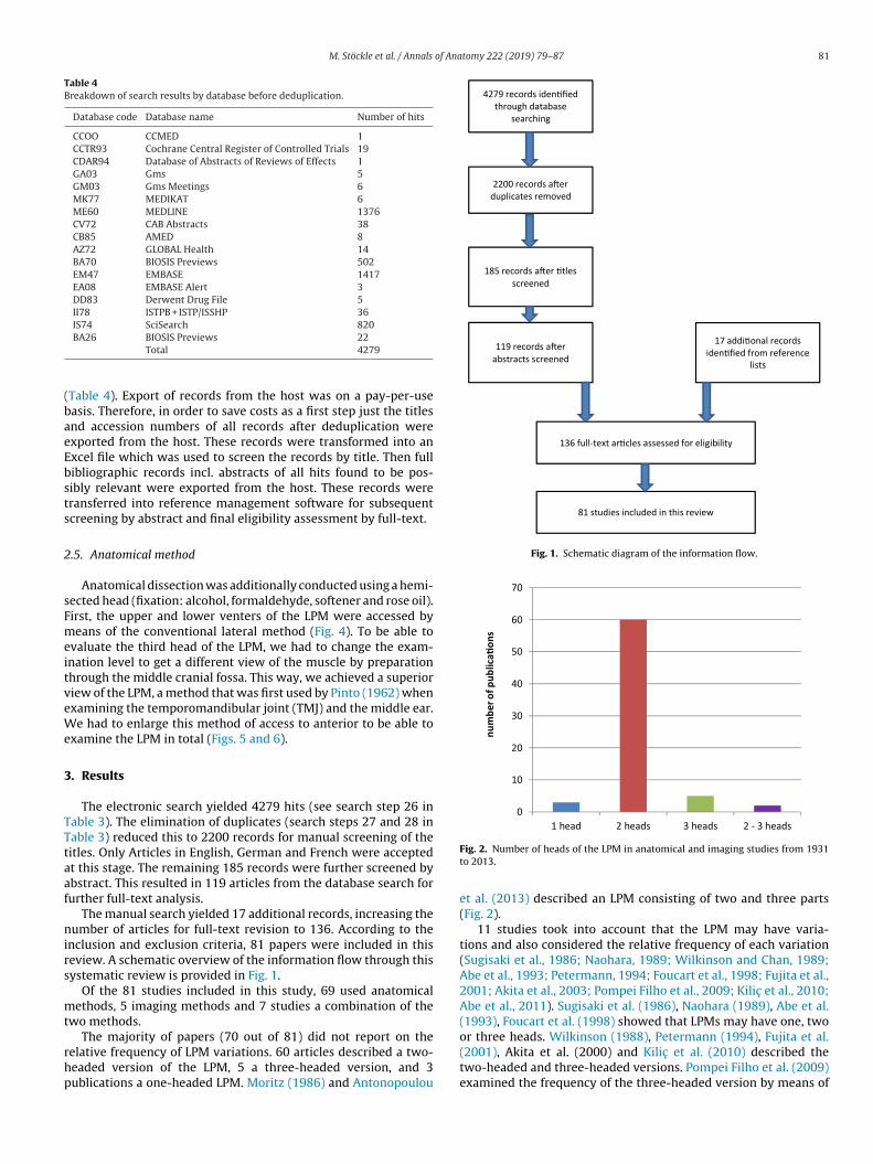
4279 records iden fied through database
searching
2200 records a er duplicates removed
185 records a er tles screened
119 records a er abstracts screened
17 addi onal recordsiden fied from reference
lists
136 full-text ar cles assessed for eligibility
81 studies included in this review
Fig. 1. Schematic diagram of the information flow.
BA26 BIOSIS Previews 22Total 4279
Table 4). Export of records from the host was on a pay-per-useasis. Therefore, in order to save costs as a first step just the titlesnd accession numbers of all records after deduplication werexported from the host. These records were transformed into anxcel file which was used to screen the records by title. Then fullibliographic records incl. abstracts of all hits found to be pos-ibly relevant were exported from the host. These records wereransferred into reference management software for subsequentcreening by abstract and final eligibility assessment by full-text.
.5. Anatomical method
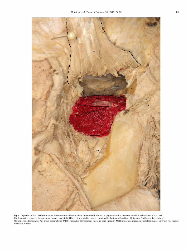
Anatomical dissection was additionally conducted using a hemi-ected head (fixation: alcohol, formaldehyde, softener and rose oil).irst, the upper and lower venters of the LPM were accessed byeans of the conventional lateral method (Fig. 4). To be able to
valuate the third head of the LPM, we had to change the exam-nation level to get a different view of the muscle by preparationhrough the middle cranial fossa. This way, we achieved a superioriew of the LPM, a method that was first used by Pinto (1962) whenxamining the temporomandibular joint (TMJ) and the middle ear.
e had to enlarge this method of access to anterior to be able toxamine the LPM in total (Figs. 5 and 6).
. Results
The electronic search yielded 4279 hits (see search step 26 inable 3). The elimination of duplicates (search steps 27 and 28 inable 3) reduced this to 2200 records for manual screening of theitles. Only Articles in English, German and French were acceptedt this stage. The remaining 185 records were further screened bybstract. This resulted in 119 articles from the database search forurther full-text analysis.
The manual search yielded 17 additional records, increasing theumber of articles for full-text revision to 136. According to the
nclusion and exclusion criteria, 81 papers were included in thiseview. A schematic overview of the information flow through thisystematic review is provided in Fig. 1.
Of the 81 studies included in this study, 69 used anatomicalethods, 5 imaging methods and 7 studies a combination of the
wo methods.
The majority of papers (70 out of 81) did not report on theelative frequency of LPM variations. 60 articles described a two-eaded version of the LPM, 5 a three-headed version, and 3ublications a one-headed LPM. Moritz (1986) and Antonopoulou
Fig. 2. Number of heads of the LPM in anatomical and imaging studies from 1931to 2013.
et al. (2013) described an LPM consisting of two and three parts(Fig. 2).
11 studies took into account that the LPM may have varia-tions and also considered the relative frequency of each variation(Sugisaki et al., 1986; Naohara, 1989; Wilkinson and Chan, 1989;Abe et al., 1993; Petermann, 1994; Foucart et al., 1998; Fujita et al.,2001; Akita et al., 2003; Pompei Filho et al., 2009; Kilic et al., 2010;Abe et al., 2011). Sugisaki et al. (1986), Naohara (1989), Abe et al.(1993), Foucart et al. (1998) showed that LPMs may have one, twoor three heads. Wilkinson (1988), Petermann (1994), Fujita et al.
(2001), Akita et al. (2000) and Kilic et al. (2010) described thetwo-headed and three-headed versions. Pompei Filho et al. (2009)examined the frequency of the three-headed version by means of
82 M. Stöckle et al. / Annals of Anatomy 222 (2019) 79–87
F atomico riatio
ah
sa
t(Aeedd
atK
t
fsOmff
r6
3
sfh
4
hbbn
ig. 3. Relative frequencies of the variations of the LPM in 11 publications with annly the occurrence of the three-headed type with a MRT; thus, data of the other va
n MRT, and Abe et al. (2011) described LPMs with one and twoeads (Fig. 3).
Altogether, the authors of these 11 studies investigated 521ubjects or cadavers. 343 TMJs were investigated by means ofnatomical methods and 178 with imaging methods such as MRT.
In all of these 11 studies, information about the ethnic origin ofhe anatomic samples was missing. Sugisaki et al. (1986), Naohara1989), Wilkinson (1988), Abe et al. (1993), Petermann (1994) andkita et al. (2003) did not note facts about age distribution of theirxamined subjects. Foucart et al. (1998), Fujita et al. (2001), Akitat al. (2003) and Abe et al. (2011) gave information about the gen-er distribution but did not identify a correlation between genderistribution and morphology of the LPM.
Pompei Filho et al. (2009) and Kilic et al. (2010) reported about correlation between gender distribution and the prevalence of ahird head of the LPM. Pompei Filho et al. (2009) used imaging andilic et al. (2010) anatomical methods.
Pompei Filho et al. (2009) examined only the prevalence of ahird head of the LPM.
From 178 investigated healthy individuals, 12 males and 24emales had a three-headed LPM. Kilic et al. (2010) inspected 49pecimens. 32 of them were two-headed and 17 had three heads.f these 17 specimens one was sorted out and was investigatedicroscopically (no information was given if this was a male or a
emale sample) and nine of them were male and seven of thememale (Table 5).
The relative frequency of the one-headed version of the LPManges between 7.7%–26.7%, the two-headed version between1.4%–91.1% and the three-headed version between 4.0%–35.0%.
.1. Anatomical examination
When examining and preparing the hemisected head, we foundimilar results to those detected by Troiano (1967). Removal of theascia surrounding the LPM showed a clear division of the upperead in a medial (LPMM) and a lateral (LPML) part (Figs. 5 and 6).
. Discussion
Publications on anatomical methods and the preparation of
uman cadavers provide detailed information on the structure toe examined. Descriptions of the three-dimensional proportionsetween the LPM and the surrounding structures, such as bone,erves and vessels, are also widely available.al and imaging methods from 1986 to 2011. *Pompei Filho et al. (2009) examinedns were missing.
Preparation of the LPM is very difficult due to its deep locationin the infratemporal fossa and the fact that the muscle is coveredand surrounded by tissues, which makes access to the muscle itselfdifficult.
A stereomicroscope is often used to examine small structuresand to trace the origin or insertion of muscle fibres (Abe et al.,1993; Sümnig et al., 1991). However, important parts of muscles,nerves or vessels may be damaged by inappropriate preparation(Poland, 1890). Abe et al. (1993) argued that the method of dis-section for examining the LPM is negligible, whereas El Haddiouiet al. (2005) reported that conventional preparation techniques arelimited when used to examine the LPM in its deep location.
However, the experience of the author or the anatomical dis-sector is of great importance. Troiano (1967) and Poland (1890)showed that careless dissection may damage the structure tobe examined, which, in the worst case, could lead to misinter-pretations of the anatomy of the LPM. Parts of the LPM or itssurrounding structures may be accidentally removed if the dissec-tor is unaware of the correct anatomy of the infratemporal fossa andits surrounding structures. Therefore, presence of a two-headedinstead of a three-headed LPM may be assumed. A similar prob-lem occurred during our anatomical examination. When only thelateral dissection method is applied, the anatomy of the LPM maybe misinterpreted, because the LPMM is often covered by the morelaterally positioned LPMS. This situation could be another reasonfor incorrect interpretations of the anatomy of the LPM in the past.
Troiano (1967) discovered a third medial head at the same levelof the LPMS. During our own dissection, we detected the sameform of a third head that appears to be covered by the LPMS whenviewed from a lateral or superior position. Therefore, a multi-levelapproach seems to be indicated for the correct examination of theLPM. When only the lateral approach is applied, the possible exis-tence of a third part of the LPM may be overlooked. Furthermore,anatomical and imaging methods can be combined to reduce mis-interpretations of the anatomy of the LPM.
The one-headed LPM was mentioned in 68 publications, but 60out of 68 articles (88.2%) identified an LPM with two heads, 5 (7.4%)an LPM with three heads and 3 (4.4%) an LPM with one head. Theseresults should be considered carefully, because some of the studiesmentioned above examined the region of the TMJ but were not
focussed on the LPM and its variations.In contrast, the results published in the following studies weresignificant (Sugisaki et al., 1986; Naohara, 1989; Wilkinson andChan, 1989; Abe et al., 1993; Petermann, 1994; Foucart et al., 1998;
M. Stöckle et al. / Annals of Anatomy 222 (2019) 79–87 83
Fig. 4. Depiction of the LPM by means of the conventional lateral dissection method. The arcus zygomaticus has been removed for a clear view of the LPM.The Separation between the upper and lower head of the LPM is clearly visible (subject provided by Professor Fanghänel, University Greifswald/Regensburg).MT: musculus temporalis; AZ: arcus zygomaticus; MPLS: musculus pterygoideus lateralis, pars superior; MPLI: musculus pterygoideus lateralis, pars inferior; NA: nervusalveolaris inferior.

84 M. Stöckle et al. / Annals of Anatomy 222 (2019) 79–87
Table 5Results of the main eleven articles in this research concerning methods, number of specimens/patients, age distribution, ethnic origin, gender distribution and correlationsbetween these attributes. a) The detailed age range distribution is available in Fujita et al. (2001, p. 561).
Author Methods Number of speci-mens/patients
Age Ethnic origin Gender distribution Correlations
Sugisaki et al. (1986) Anatomic 14 n.a. n.a. n.a. n.a.Naohara (1989) Anatomic 25 n.a. n.a. n.a. n.a.Wilkinson (1988) Anatomic 5 n.a. n.a. n.a. n.a.Abe et al. (1993) Anatomic 79 n.a. n.a. n.a. n.a.Petermann (1994) Anatomic 42 n.a. n.a. n.a. n.a.Foucart et al. (1998) Anatomic 22 60–101 n.a. 5 female, 6 male n.a.Fujita et al. (2001) Anatomic 20 a) n.a. 15 female, 5 male n.a.Akita et al. (2003) Anatomic 45 n.a. n.a. 11 female, 14 male n.a.Pompei Filho et al. (2009) MRT 178 20–45 n.a. 24 female, 12 male 36/178 with a third head, from that 24 female and 12 maleKilic et al. (2010) Anatomic 49 23–76 n.a. 10 female, 16 male 17/49 with a third head, one was sortet out, 9 male, 7 femaleAbe et al. (2011) Anatomic 30 62–85 n.a. 5 female, 10 male n.a.
Fig. 5. Superior approach to the LPM through the medial cranial fossa according to Pinto (1962). The separated upper head of the LPM is clearly visible (subject provided byProfessor Fanghänel, University Greifswald/Regensburg).Dotted line: approach according to Pinto (1962).D in.CM le; A:
FKeTobToLnNacmw
dTh(o
ashed line: extended anterior approach to trace the fibres of the LPM to their origontinuous line: medial cranial fossa.AE: meatus acusticus externus; MAI: meatus acusticus internus; FO: foramen ova
ujita et al., 2001; Akita et al., 2003; Pompei Filho et al., 2009;ilic et al., 2010; Abe et al., 2011), because some of these authorsxamined their subjects from different planes and perspectives.he authors depicted the frequency of each anatomical variationf the LPM. In these 11 articles, 521 subjects were examined: 343y means of anatomical methods and 178 with imaging methods.he frequency of one-headed LPMs ranged between 7.7% and 26.7%,f two-headed LPMs between 61.4% and 91.1% and of three-headedPMs between 4.0% and 35.0%. These data can be considered sig-ificant, because the studies focused on the LPM and its variations.onetheless it would have been of great impact for this system-tic review, if these authors had given more detailed informationoncerning the origin, age and gender distribution of the speci-ens and patients. This would have helped to intepret in whichay variations of the LPM are influenced.
Imaging methods are particularly suitable for examining andepicting the soft tissues of diseased TMJs (Westesson, 1993;askaya-Yilmaz et al., 2005; Pompei Filho et al., 2009). By now, MRT
as replaced arthroscopy and CT in the diagnosis of TMJ diseasesWestesson, 1993). The big advantage of MRT is the lack of emissionf any harmful X-rays. Sagittal pictures of the TMJ are very usefulanterior; P: posterior.
for interpreting the LPM, although misinterpretations are possible.For instance, the tendon of the LPM may be misinterpreted as a partof the discus (Crowley et al., 1996).
Pompei Filho et al. (2009) discovered a three-headed LPM in20.22% of investigated cases, while several other authors arguethat the LPM has two heads (Katzberg et al., 1985; Adachi et al.,1985; Quémar et al., 1993; Pedullà et al., 2009; Mazza et al., 2009;D’Ippolito et al., 2010).
According to Pompei Filho et al. (2009), the complete amountof muscle fibres of the LPMM inserts into the discus. The courseof these muscle fibres indicates that they could have an importantfunction in stabilising the discus. Furthermore, Pompei Filho et al.(2009) assumed that the third head of the LPM may play a role indisplaced or dysfunctional joint discs.
MRT plays an important role in clinical routine, becauseanatomic proportions, pathological alterations and the surroundingsoft tissues can be very well depicted without any harmful invasiveintervention. In contrast to anatomical methods, the surrounding
tissue of the examined area does not have to be dissected andcan consequently not be damaged by manual preparation. Imag-ing methods in combination with anatomical studies are useful to
M. Stöckle et al. / Annals of Anatomy 222 (2019) 79–87 85
ificat
ri
5
Tabmapmfl
R
A
A
A
A
A
B
Fig. 6. Magn
educe misinterpretation and help to arrange an anatomical exam-nation.
. Conclusions
A multi-level approach to examining the LPM or to dissecting theMJ with its adjacent structures en-bloc seems to be indicated tovoid confusion. One possible multi-level approach could be com-ining the superior and the lateral approach. Combining differentethods such as anatomical methods with imaging methods is
lso helpful. Detailed information about the number of examinedatients/subjects with their ethnic origin, age and gender should beentioned. In future, these improvements may minimise the dif-
erences in results obtained by different authors and could at bestead to a more homogenous description of the LPM.
eferences
be, S., Naito, H., Nakao, T., Yamamoto, M., Won, S.-Y., Kim, H.-J., Ide, Y., 2011. Anal-ysis of the intramuscular innervation of the lateral pterygoid muscle. J. HardTissue Biol. 20, 259–264.
be, S., Takasaki, I., Ichikawa, K., Ide, Y., 1993. Investigations of the run and theattachment of the lateral pterygoid muscle in Japanese. Bull. Tokyo Dent. Coll.34, 135–139.
dachi, S., Jumnongthai, K., Takada, K., Oda, Y., Sakuda, M., 1985. Cephalometricappraisal of the orientation of lateral pterygoid muscles. J. Osaka Univ. Dent.Sch. 25, 127–138.
kita, K., Shimokawa, T., Sato, T., 2003. An anatomic study of the positional relation-ships between the lateral pterygoid muscle and its surrounding nerves. Eur. J.Anat. 7, 5–14.
ntonopoulou, M., Iatrou, I., Paraschos, A., Anagnostopoulou, S., 2013. Variations
of the attachment of the superior head of human lateral pterygoid muscle. J.Craniomaxillofac. Surg. 41, 7.irou, G., Garcier, J.M., Guillot, M., Vanneuville, G., Chazal, J., 1991. A study of thelateral pterygoid muscle: anatomic sections and CT appearances. Surg. Radiol.Anat. 13, 307–311.
ion of Fig. 5.
Choukas, N.C., Sicher, H., 1960. The structure of the temporomandibular joint. OralSurg. Oral Med. Oral Pathol. 13, 1203–1213.
Coskun Akar, G., Govsa, F., Ozgur, Z., 2009. Examination of the heads of the lat-eral pterygoid muscle on the temporomandibular joint. J. Craniofac. Surg. 20,219–223.
Crowley, C., Wilkinson, T., Piehslingher, E., Wilson, D., Czerny, C., 1996. Correlationsbetween anatomic and MRI sections of human cadaver temporomandibularjoints in the coronal and sagittal planes. J. Orofac. Pain 10, 199–216.
D’Ippolito, S.M., Borri Wolosker, A.M., D’Ippolito, G., Herbert de Souza, B., Fenyo-Pereira, M., 2010. Evaluation of the lateral pterygoid muscle using magneticresonance imaging. Dentomaxillofac. Radiol. 39, 494–500.
El Haddioui, A., Laison, F., Zouaoui, A., Bravetti, P., Gaudy, J.F., 2005. Functionalanatomy of the human lateral pterygoid muscle. Surg. Radiol. Anat. 27, 271–286.
Foucart, J.M., Girin, J.P., Carpentier, P., 1998. Innervation of the human lateral ptery-goid muscle. Surg. Radiol. Anat. 20, 185–189.
Fujita, S., Iizuka, T., Dauber, W., 2001. Variation of heads of lateral ptery-goid muscle and morphology of articular disc of human temporomandibularjoint—anatomical and histological analysis. J. Oral Rehabil. 28, 560–571.
Katzberg, R.W., Schenck, J., Roberts, D., Tallents, R.H., Manzione, J.V., Hart, H.R., Fos-ter, T.H., Wayne, W.S., Bessette, R.W., 1985. Magnetic resonance imaging ofthe temporomandibular joint meniscus. Oral Surg. Oral Med. Oral Pathol. 59,332–335.
Kilic , C., Dergın, G., Kurt, B., Kutoglu, T., Ozan, H., Balcioglu, H.A., 2010. Insertionsof the lateral pterygoid muscle to the disc-capsule complex of the temporo-mandibular joint and condyle. Turk. J. Med. Sci. 40.
Mazza, D., Marini, M., Impara, L., Cassetta, M., Scarpato, P., Barchetti, F., Di Paolo, C.,2009. Anatomic examination of the upper head of the lateral pterygoid mus-cle using magnetic resonance imaging and clinical data. J. Craniofac. Surg. 20,1508–1511.
Moritz, T., 1986. Der Ansatz des Musculus pterygoideus lateralis am Kiefergelenkdes Menschen.
Naohara, H., 1989. The macroscopic and microscopic study of the human lateralpterygoid muscle. Tsurumi Shigaku 15, 1–26.
Pedullà, E., Meli, G.A., Garufi, A., Cascone, P., Mandalà, M.L., Deodato, L., Palazzo, G.,2009. Morphometric evaluation of the temporomandibular joint and the masti-catory spaces: the role of high-definition MRI. Minerva Stomatol. 58, 127–143.
Petermann, A.K., 1994. Der Ansatz des Musculus pterygoideus lateralis am Kieferge-
lenk des Menschen in Lupenpraeparation/vorgelegt von Arnd Karl Petermann.Pinto, O.F., 1962. A new structure related to the temporomandibular joint and middleear. J. Prosthet. Dent. 1962, 95–103.
8 of Ana
P
P
Q
S
S
S
T
TT
W
W
W
S
tl
bsd
Sm
l2
Ac2
piA
t
sgS
yJ
at
JtR
1o4
6 M. Stöckle et al. / Annals
oland, J., 1890. Variations of the external pterygoid muscle. J. Anat. Physiol. 24,567–572.
ompei Filho, H., Suazo, G.I.C., Guimares, A.S., 2009. Prevalence of the third head ofthe lateral pterygoid muscle: a magnetic resonance image study. Int. J. Morphol.27 (4), 1043–1046.
uémar, J.C., Ravalec, X., Akoka, S., 1993. Parasagittal magnetic resonance imagingof the lateral pterygoid muscle: a preliminary study. J. Orofac. Pain 7, 169–174.
chumacher, G.-H., 1997. Anatomie für Zahnmediziner. Lehrbuch und Atlas, 3rdedition. Hüthig, Heidelberg.
ugisaki, M., Komori, E., Nakazawa, M., Tanabe, H., Kato, S., 1986. Anatomical studiesof the lateral pterygoid muscle by the superior approach and a review of theliterature. Jpn. J. Oral Maxillofac. Surg. 32, 718–730.
ümnig, W., Bartolain, G., Fanghänel, J., 1991. Histologische Untersuchungen überdie morphologischen Beziehungen des Musculus pterygoideus lateralis zumDiscus articularis am menschlichen Kiefergelenk. Anat. Anz. 173, 279–286.
askaya-Yilmaz, N., Ceylan, G., Incesu, L., Muglali, M., 2005. A possible etiology ofthe internal derangement of the temporomandibular joint based on the MRIobservations of the lateral pterygoid muscle. Surg. Radiol. Anat. 27, 19–24.
illmann, B., 2003. Bewegungsapparat, 3rd edition. Thieme, Stuttgart.roiano, M.F., 1967. New concept of the insertion of the lateral pterygoid muscle. J.
Oral Surg. 25, 337–340.estesson, P.L., 1993. Reliability and validity of imaging diagnosis of temporo-
mandibular joint disorder. Adv. Dent. Res. 7 (2), 137–151.ilkinson, T.M., 1988. The relationship between the disk and the lateral pterygoid
muscle in the human temporomandibular joint. J. Prosthet. Dent. 60, 715–724.ilkinson, T., Chan, E.K., 1989. The anatomic relationship of the insertion of the
superior lateral pterygoid muscle to the articular disc in the temporomandibularjoint of human cadavers. Aust. Dent. J. 34, 315–322.
ystematic research
Abe, S., Abe, Y., Ouchi, Y., Ide, H., 1997. Yonezu Perspectives onhe role of the lateral pterygoid muscle and the sphenomandibularigament in temporomandibular joint function. Cranio 15, 203–207.
Akita, K., Shimokawa, T., Sato, T., 2000, Positional relationshipsetween the masticatory muscles and their innervating nerves withpecial reference to the lateral pterygoid and the midmedial andiscotemporal muscle bundles of temporalis. J. Anat. 197, 291–302.
Alomar, X., Medrano, J., Cabratosa, J., Clavero, J.A., Lorente, M.,erra, I., Monill, J.M., Salvador, A., 2007. Anatomy of the temporo-andibular joint Semin. Ultrasound CT MR 28, 170–183.
Ashworth, G.J., 1990. The attachments of the temporomandibu-ar joint meniscus in the human fetus, Br. J. Oral Maxillofac. Surg.8, 246–250.
Aziz, M.A., Cowie, R.J., Skinner, C.E., Abdi, T.S., Orzame, G., 1998.re the two heads of the human lateral pterygoid separate mus-les? A perspective based on their nerve supply. J. Orofac. Pain 12,26–239.
Aziz, M.A., Cowie, R.J., Skinner, C.E., Mosaddad, H., 2001. Com-lete pterygoidectomy—a novel method for the topographical
nvestigation of the deep masticator space and its contents. Clin.nat. 14, 354-362.
Bade, H., 1999. The function of the disco-muscular apparatus inhe human temporomandibular joint. Ann. Anat. 181, 65–67.
Bauer, W., 1931. Anatomische und mikroskopische Unter-uchungen über das Kiefergelenk – mit besonderer Berücksichti-ung der Veränderungen bei Osteo = Arthritis deformans. Österr. Z.tomat. 30, 1136–1160.
Bertilsson, O., Ström, D., 1995. A literature survey of a hundredears of anatomic and functional lateral pterygoid muscle research.. Orofac. Pain 9, 17–23.
Bittar, G.T., Bibb, C.A., Pullinger, A.G., 1994. Histologic char-cteristics of the lateral pterygoid muscle insertion to theemporomandibular joint. J. Orofac. Pain 8, 243–249.
Bravetti, P., Membre, H., El Haddioui, A., Gerard, H., Fyard,.P., Mahler, P., Gaudy, J.F., 2004. Histological study of the humanemporo-mandibular joint and its surrounding muscles. Surg.adiol. Anat. 26, 371–378.
Carpentier, P., Yung, J.P., Marguelles-Bonnet, R., Meunissier, M.,988. Insertions of the lateral pterygoid muscle: an anatomic studyf the human temporomandibular joint. J. Oral Maxillofac. Surg. 46,77–482.
tomy 222 (2019) 79–87
Christo, J.E., Bennett, S., Wilkinson, T.M., Townsend, G.C., 2005.Discal attachments of the human temporomandibular joint. Aust.Dent. J. 50, 152–160.
Cioffi, I., van Ruijven, L.J., Michelotti, A., Langenbach, G.E.J., 2010.Degree of mineralization at the attachment of lateral pterygoid.Anat. Rec. 293, 1387–1392.
Dauber, W., 1987. Die Nachbarschaftsbeziehungen des Dis-cus articularis des Kiefergelenks und ihre funktionelle DeutungSchweiz. Monatsschr. Zahnmed. 97, 427–437.
Davies, J.C., Charles, M., Cantelmi, D., Liebgott, B., Ravichandiran,M., Ravichandiran, K., Agur, A.M., 2012. Lateral pterygoid muscle:a three-dimensional analysis of neuromuscular partitioning. Clin.Anat. 25, 576–583.
Dusek, T.O., Keily, M.L., 1991. Quantification of the superior lat-eral pterygoid inserting on TMJ components. J. Dent. Res. 70, 421.
Dusek, T.O. Keily, M.L., Gowgiel, J.M., 1989. Insertion of the supe-rior lateral pterygoid muscle to the TMJ in humans. A gross andhistomorphic study. Anat. Rec. 223, 35.
Eriksson, P.O., Eriksson, A., Ringqvist, M., Thornell, L.E., 1981.Special histochemical muscle-fibre characteristics of the humanlateral pterygoid muscle. Arch. Oral Biol. 26, 495–507.
Griffin, C.J., Hawthorn, R., Harris, R., 1975. Anatomy and histol-ogy of the human temporomandibular joint. Monogr. Oral Sci. 4,1–26.
Hartmann, F., Sarrat, P., Gaudy, J.F., Cucchi, G., Susini, G., Tren-tinella, C., 1988. The lateral pterygoid muscle: anatomic dissectionand medical imaging. Perspectives in the treatment of M.P.D.S.Actual Odontostomatol. 545–566.
Heylings, D.J., Nielsen, I.L., McNeill, C., 1995. Lateral pterygoidmuscle and the temporomandibular disc. J. Orofac. Pain 9, 9–16.
Honee, G.L., 1972. The anatomy of the lateral pterygoid muscle.Acta Morphol. Neerl. Scand. 10, 331–340.
Honee, Guillaume Leopold Jacques Marie, 1970. De musculuspterygoideus lateralis. Amsterdam, Universiteit van Amsterdam,(OCoLC)39328030.
Hussain, A., Binahmed, A., Karim, A., Sandor, G.K.B., 2008. Rela-tionship of the maxillary artery and lateral pterygoid muscle in acaucasian sample. Oral Surg. Oral Med. Oral Pathol. Oral Radiol.Endod. 105, 32–36.
Isolan, G.R., Rowe, R., Al-Mefty, O., 2007. Microanatomy andsurgical approaches to the infratemporal fossa: an anaglyphicthree-dimensional stereoscopic printing study. Skull Base 17,285–302.
Katori, Y., Yamamoto, M., Asakawa, S., Maki, H., Rodríguez-Vázquez, J.F., Murakami, G., Abe, S., 2012. Fetal developmentalchange in topographical relationship between the human lateralpterygoid muscle and buccal nerve. J. Anat. 220, 384–395.
Kim, H.J., Kwak, H.H., Hu, K.S., Park, H.D., Kang, H.C. Jung, H.S.,Koh, K.S., 2003. Topographic anatomy of the mandibular nervebranches distributed on the two heads of the lateral pterygoid. Int.J. Oral Maxillofac. Surg. 32, 408–413.
Klineberg, I., 1991. The lateral pterygoid muscle: some anatomi-cal, physiological and clinical considerations. Ann. R. Australas. Coll.Dent. Surg. 11, 96–108.
Kwak, H.H., Ko, S.J., Jung, H.S., Park, H.D., Chung, I.H., Kim, H.J.,2003. Topographic anatomy of the deep temporal nerves, with ref-erences to the superior head of lateral pterygoid. Surg. Radiol. Anat.25, 393–399.
Le Toux, G., Duval, J.M., Darnault, P., 1989. The human temporo-mandibular joint: current anatomic and physiologic status. Surg.Radiol. Anat. 11, 283–288.
Loughner, B.A., Larkin, L.H., Mahan, P.E., 1990. Nerve entrapment
in the lateral pterygoid muscle. Oral Surg. Oral Med. Oral Pathol. 69,299–306.
of Ana
ao
JdS
cj
fim
ls
ha
jA
tM
iS
owO
haD
dhM
c
ga
Wongwatana, S., Kronman, J.H., Clark, R.E., Kabani, S., Mehta, N.,1994. Anatomic basis for disk displacement in temporomandibu-lar joint (TMJ) dysfunction. Am. J. Orthod. Dentofacial Orthop. 105,257–264.
M. Stöckle et al. / Annals
Matsunaga, K., Usui, A., Yamaguchi, K., Akita, K., 2009. Annatomical study of the muscles that attach to the articular discf the temporomandibular joint. Clin. Anat. 22, 932–940.
Merida Velasco, J.R., Rodriguez Vazquez, J.F., Jimenez Collado,., 1993. The relationships between the temporomandibular jointisc and related masticatory muscles in humans. J. Oral Maxillofac.urg. 51, 390.
Meyenberg, K., Kubik, S., Palla, S., 1986. Relationships of the mus-les of mastication to the articular disc of the temporomandibularoint. Schweiz. Monatsschr. Zahnmed. 96, 815–834.
Monemi, M., Thornell, L., Eriksson, P., 1999. Diverse changes inbre type composition of the human lateral pterygoid and digastricuscles during aging. J. Neurol. Sci., 171, 38–48.
Moritz, T., 1987. Ewers Der Ansatz des Musculus pterygoideusateralis am Kiefergelenk des Menschen: Eine histologische Unter-uchung. Dtsch. Zahnarztl. Z. 42, 680–685.
Murray, G.M., Phanachet, I., Uchida, S., Whittle, T., 2004. Theuman lateral pterygoid muscle: a review of some experimentalspects and possible clinical relevance. Aust. Dent. J. 49, 2–8.
Naidoo, L.C., 1993. The development of the temporomandibularoint: a review with regard to the lateral pterygoid muscle. J. Dent.ssoc. S. Afr. 48, 189–194.
Naidoo, L.C., 1996. Lateral pterygoid muscle and its relationshipo the meniscus of the temporomandibular joint. Oral Surg. Oral
ed. Oral Pathol. Oral Radiol. Endod. 82, 4–9.Naidoo, L.C., Juniper, R.P., 1997. Morphometric analysis of the
nsertion of the upper head of the lateral pterygoid muscle. Oralurg. Oral Med. Oral Pathol. Oral Radiol. Endod. 83, 441–446.
Ogutcen-Toller, M., Juniper, R.P., 1993. The embryologic devel-pment of the human lateral pterygoid muscle and its relationshipsith the temporomandibular joint disc and Meckel’s cartilage. J.ral Maxillofac. Surg. 51, 772.
Ogutcen-Toller, M., Juniper, R.P., 1994. The development of theuman lateral pterygoid muscle and the temporomandibular jointnd related structures: a three-dimensional approach. Early Hum.ev. 39, 57–68.
Ogutcen-Toller, M., Keskin, M., 2000. Computerized 3-imensional study of the embryologic development of theuman masticatory muscles and temporomandibular joint. J. Oralaxillofac. Surg. 58, 1381–1386.
Porter, M.R., 1970. The attachment of the lateral pterygoid mus-
le to the meniscus. J. Prosthet. Dent. 24, 555–562.Roche, P.H., Fournier, H.D., Laccourreye, L., Mercier, P., 2001. Sur-ical anatomy of the infratemporal fossa using the transmaxillarypproach. Surg. Radiol. Anat. 23, 209–213.
tomy 222 (2019) 79–87 87
Rosenberg, F.A., Sears, S.B., Landers, M.A., 1983. The gnathostom-atic system: an anatomic approach. Int. J. Periodontics RestorativeDent. 3, 22–35.
Sabit, I., Schaefer, S.D., Couldwell, W.T., 2002. Modified infratem-poral fossa approach via lateral transantral maxillotomy: amicrosurgical model. Surg. Neurol. 58, 21–31.
Schmolke, C., 1994. The relationship between the temporo-mandibular joint capsule, articular disc and jaw muscles. J. Anat.184, 335–345.
Shimokawa, T., Akita, K., Sato, T., Ru, F., Yi, S.-Q., Tanaka, S., 2004.Penetration of muscles by branches of the mandibular nerve: apossible cause of neuropathy. Clin. Anat. 17, 2–5.
Siessere, S., Vitti, M., Semprini, M., Regalo, S.C.H., Iyomasa, M.M.,Dias, F.J., Issa, J.P.M., de Sousa, L.G., 2008. Macroscopic and micro-scopic aspects of the temporomandibular joint related to its clinicalimplication. Micron. 39, 852–858.
Terada, S., Sato, T., 1982. Nerve supply of the medial and lateralpterygoid muscles and its morphological significance. OkajimasFolia Anat. Jpn. 59, 251–264.
Usui, A., Akita, K., Yamaguchi, K., 2008. An anatomic study of thedivisions of the lateral pterygoid muscle based on the findings ofthe origins and insertions. Surg. Radiol. Anat. 30, 327–333.
van Eijden, T.M., Koolstra, J.H., Brugman, P., 1995. Architectureof the human pterygoid muscles. J. Dent. Res. 74, 1489–1495.
van Eijden, T.M., Korfage, J.A., Brugman, P., 1997. Architecture ofthe human jaw-closing and jaw-opening muscles. Anat. Rec. 248,464–474.
Vrionis, F.D., Cano, W.G., Heilman, C.B., 1996. Microsurgicalanatomy of the infratemporal fossa as viewed laterally and superi-orly. Neurosurgery 39, 777–785.
Widmalm, S.E., Lillie, J.H., Ash, M.M., 1987. Anatomical andelectromyographic studies of the lateral pterygoid muscle. J. OralRehabil. 14, 429–446.
Wilkinson, T., Maryniuk, G., 1983. The correlation between sagit-tal anatomic sections and computerized tomography of the TMJ. J.Craniomandibular Pract. 1, 37–45.