anÀlegs de somatostatina marcats amb gal·li-68 … · imatge dels rss amb emissors de positrons...
TRANSCRIPT

ANÀLEGS DE SOMATOSTATINA MARCATS AMB GAL·LI-68
ASPECTES CLÍNICS I INTRODUCCIÓ A LA PRÀCTICA CLÍNICA
Joan Castell S Medicina Nuclear
Hospital Universitari Vall d’Heborn

SITUACIÓ I CONTEXT CLÍNIC
IMATGE DELS RSS AMB EMISSORS DE POSITRONS
PERSPECTIVA CLÍNICA: INDICACIONS I RENDIMENT DIAGNÒSTIC
ESPERAT
OPCIONS EN LA INTRODUCCIÓ A CATALUNYA

Los tumores neuroendocrinos (TNE) constituyen un grupo heterogéneo de
neoplasias. Se originan en las células neuroendocrinas de la cresta neural, glándulas
endocrinas, islotes o sistema endocrino difuso.
Estas células durante el desarrollo embrionario se distribuyen por prácticamente todo
el organismo, por este motivo los TNE pueden localizarse en diversos órganos.
Las células neuroendocrinas se caracterizan por producir una serie de moléculas
(neuropéptidos, neuromodulares o neurotransmisores)
SEOM 2014

•Tumores endocrinos gastroenteropancreáticos (TNEGEP)
Son los más frecuentes. Tipos:
Tumores neuroendocrinos pancreáticos (TNP)
Gastrinoma, insulinoma, glucagonoma,
somatostatinoma, VIPoma, no secretores
Tumores carcinoides (TC)
Tracto digestivo: gástricos, ID, apendicular, colon, recto
Boncopulmonares
•Tumores secretores de catecolaminas (feocromocitoma, paraganglioma,
ganglioneuroma, ganglioneuroblastoma, simpatoblastoma, neuroblastoma)
•Carcinoma medular de tiroides
•Tumores adenohipofisarios, suprarrenales
•Tumor de células de Merkel
•Tumores neuroendocrinos de primario desconocido
TUMORES NEUROENDOCRINOS
SEOM 2014

Los TNE son relativamente poco frecuentes, con una incidencia anual
ajustada por edad de 5.25 casos por 100.000 habitantes.
Se ha observado un aumento en su incidencia en los últimos 30 años debido
a varios motivos, principalmente a la mejoría de las técnicas diagnósticas y a
la mejor identificación de los casos.
Su prevalencia es significativa debido a la historia natural de la mayoría de
estos tumores, de lento crecimiento y de larga supervivencia. Así por
ejemplo, los TNE suponen la segunda neoplasia avanzada más prevalente
del tracto digestivo tras el cáncer colorectal.
SEOM 2014
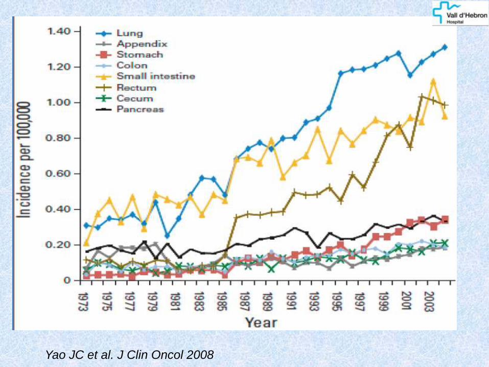
Yao JC et al. J Clin Oncol 2008

CLASIFICACIÓN OMS 2010
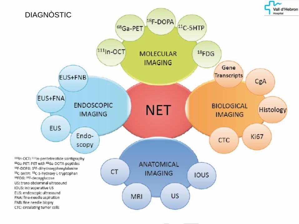
DIAGNÒSTIC

Data are pooled from 52 studies and are mean (95% confidence interval). Data for specificity
and sensitivity are not comparable across studies. S = calculated sensitivity. 11C = carbon-11,
18F = fluorine-18, 68Ga = gallium-68, 64Cu = copper-64. EUS = endoscopic ultrasound, FDG
= fluorodeoxyglucose, IOUS = intraoperative US, SRS = somatostatin receptor scintigraphy
Methods for indentification of primary and metastatic
gastroenteropancreatic NETs. The status of neuroendocrine tumor imaging: from darkness to light?
Bodei L, et al. Neuroendocrinology 2014

TRACTAMENT DELS TNEGEP
MALALTIA LOCALITZADA
Cirurgia
MALALTIA DISSEMINADA ÚNICAMENT AL FETGE I TUMOR PRIMARI RESECABLE
Cirurgia R0 o citoreductora + resecció hepàtica
Alternativament: radiofreqüència, quimio/radioembolització
MALALTIA DISSEMINADA O TUMOR LOCALMENT AVANÇAT IRRESSECABLE
Anàlegs de la somatostatina
Anàlegs de la somatostatina marcats amb beta-emissors (90Y, 177Lu)
131Iode-MIBG (productors de catecolamines)
Quimioterapia: Estreptozocina + Adriamizina / 5Flu
Fàrmacs de diana molecular:
Inhibidors TK i efecte anti-angiogènic: Sunitinib
Inhibidors mTOR: Everolimus
SEOM 2014
Modlin IM. Gastroenteropancreatic neuroendocrine tumours.
Lancet Oncol. 2008

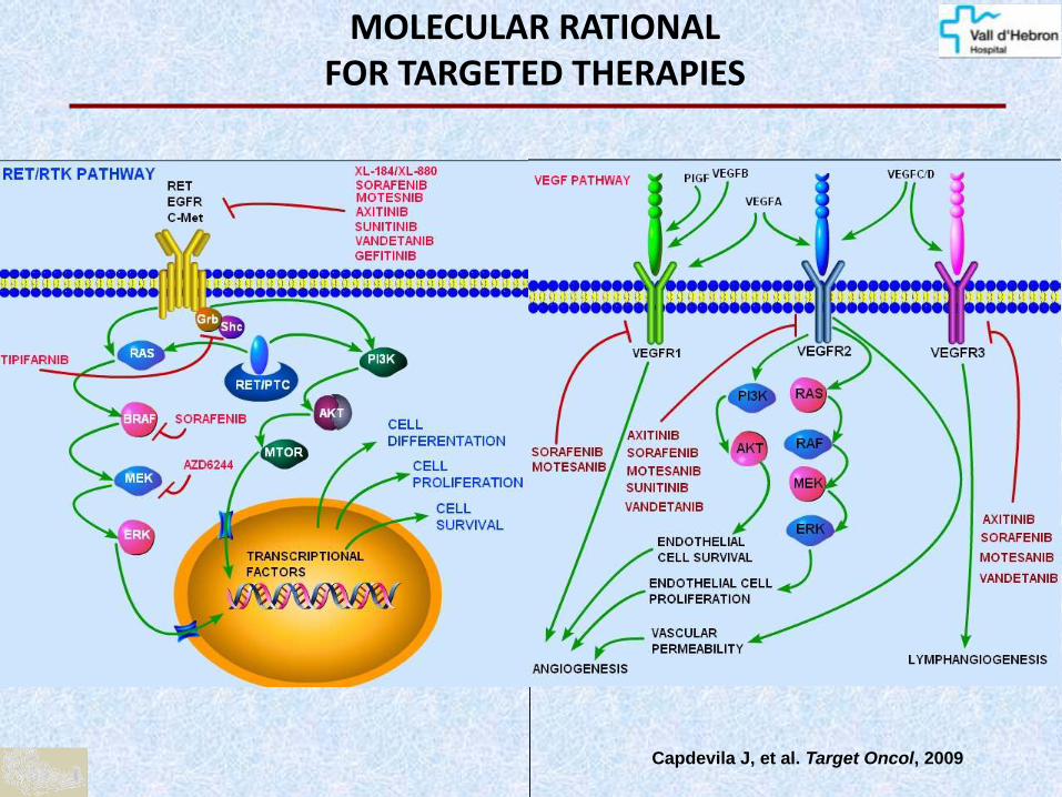
MOLECULAR RATIONAL FOR TARGETED THERAPIES
Capdevila J, et al. Target Oncol, 2009
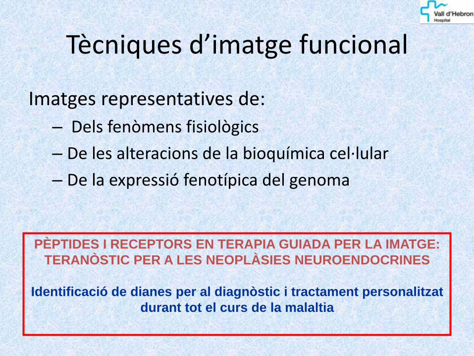
Tècniques d’imatge funcional
Imatges representatives de:
– Dels fenòmens fisiològics
– De les alteracions de la bioquímica cel·lular
– De la expressió fenotípica del genoma
PÈPTIDES I RECEPTORS EN TERAPIA GUIADA PER LA IMATGE:
TERANÒSTIC PER A LES NEOPLÀSIES NEUROENDOCRINES
Identificació de dianes per al diagnòstic i tractament personalitzat
durant tot el curs de la malaltia

VIES D’EXPLORACIÓ D’IMATGE FUNCIONAL DELS TUMORS NEUROENDOCRINS
Secreció de catecolamines (noradrenalina)
SPECT 123/131I-MIBG PET 18F-DOPA
Expressió de receptors de somatostatina
SPECT 111In/ 99mTc -octreòtide
PET 68Ga-DOTA-PÈPTIDS
Augment metabòlic/consum de glucosa/sobre-expresió del GLUT-1
PET 18F-FDG

16
123I-MIBG
99mTc-octreotide
18FDG

MIBG

MIBG


J Nucl Med 2009

Conclusion: This prospective study demonstrated
a sensitivity of 82%-88% and specificity of 82%-
84% for 123I-MIBG imaging used in the diagnostic
assessment of primary or metastatic
pheochromocytoma or paraganglioma

Thirty unrelated patients (16 males, 14 females) with an succinate dehydrogenase gene subunit (SDHB) mutation and a
history of histologically proven abdominal or thoracic PGL were included in this retrospective study of imaging data. All
patients were referred to the National Institutes of Health (NIH) for an outline of an optimal treatment plan for (suspected)
metastatic PGL between November 2000 and May 2006

FDG [18F]fluorodopamine [123I]MIBG
Ferone D, et al. Journal of Molecular Endocrinology (2009)
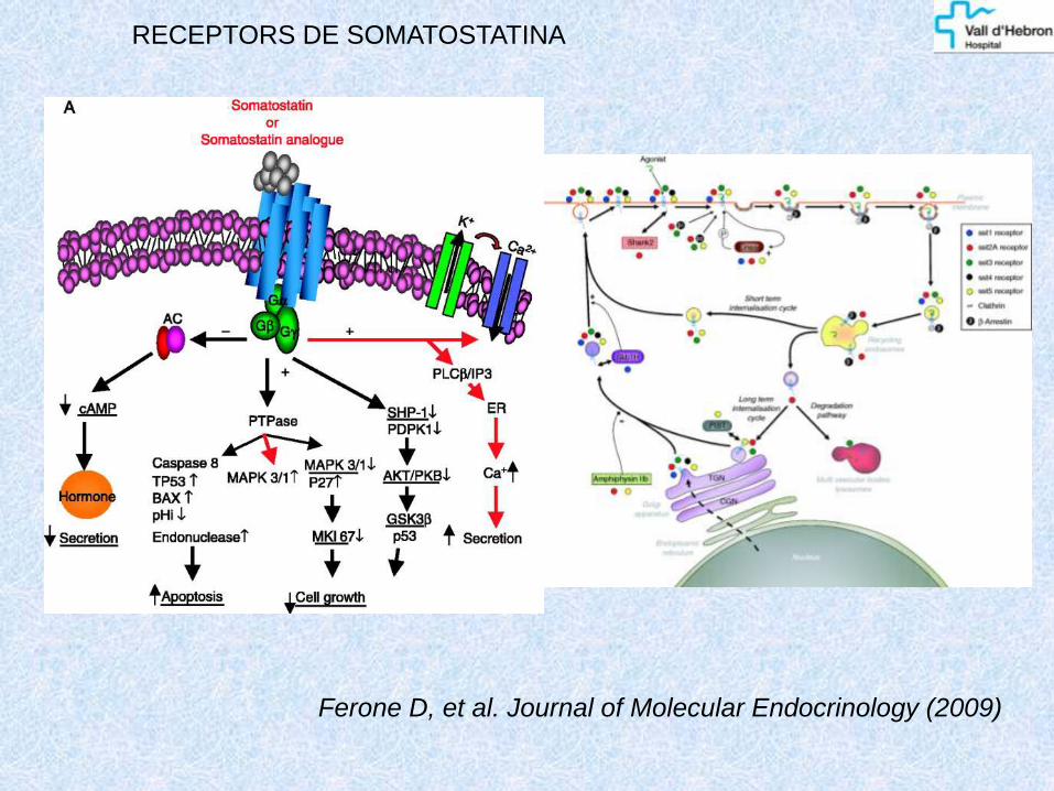
RECEPTORS DE SOMATOSTATINA


99mTc/111In - OCTREÒTIDE: IMATGE DE COS SENCER
HUVH COHORT

99mTc/111In - OCTEÒTRIDE: IMATGE DE FUSIÓ SPECT-TC
HUVH COHORT

99mTc/111In - OCTREÒTIDE
HUVH COHORT

FDG PET-CT
99mTc-TEKTROYD
SPECT-CT
INCIDENTAL PANCREATIC TUMOUR
HUVH COHORT

TUMOR EXPRESION: PROLIFERATIVE INDEX vs RECEPTOR IMAGING
31
FDG
OCTREOTIDE
HUVH COHORT
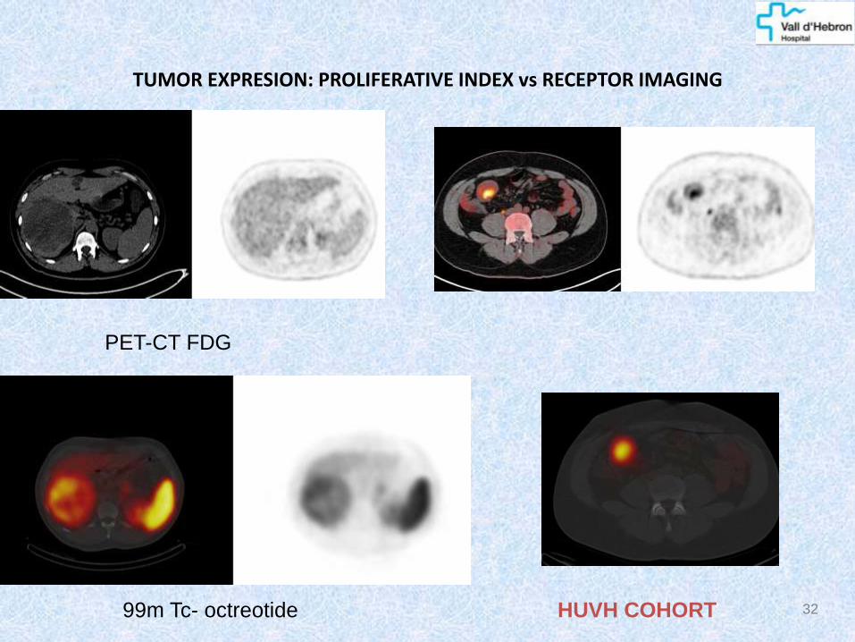
32
PET-CT FDG
99m Tc- octreotide
TUMOR EXPRESION: PROLIFERATIVE INDEX vs RECEPTOR IMAGING
HUVH COHORT

CAL QUE FEM PET AMB ANÀLEGS DE LA SST?
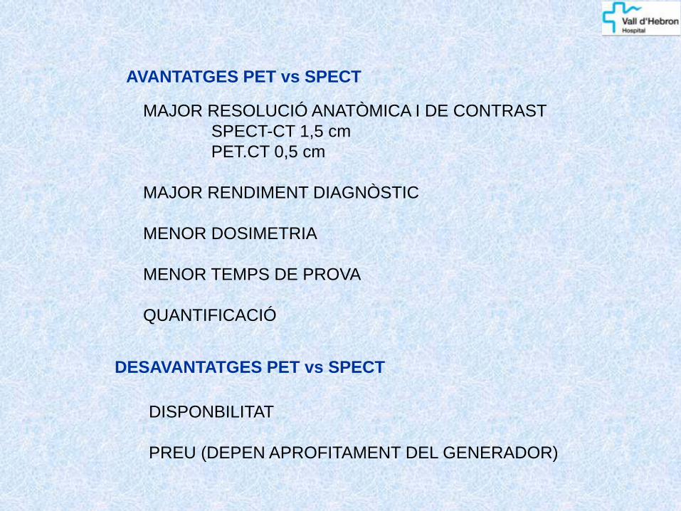
MAJOR RESOLUCIÓ ANATÒMICA I DE CONTRAST
SPECT-CT 1,5 cm
PET.CT 0,5 cm
MAJOR RENDIMENT DIAGNÒSTIC
MENOR DOSIMETRIA
MENOR TEMPS DE PROVA
QUANTIFICACIÓ
AVANTATGES PET vs SPECT
DESAVANTATGES PET vs SPECT
DISPONBILITAT
PREU (DEPEN APROFITAMENT DEL GENERADOR)
11In-DTPA-octreotide
68Ga-DOTATATE
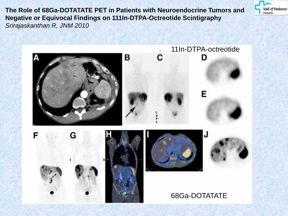
The Role of 68Ga-DOTATATE PET in Patients with Neuroendocrine Tumors and
Negative or Equivocal Findings on 111In-DTPA-Octreotide Scintigraphy
Srirajaskanthan R, JNM 2010

The Role of 68Ga-DOTATATE PET in Patients with Neuroendocrine Tumors and
Negative or Equivocal Findings on 111In-DTPA-Octreotide Scintigraphy
Srirajaskanthan R, JNM 2010

The Role of 68Ga-DOTATATE PET in Patients with Neuroendocrine Tumors and
Negative or Equivocal Findings on 111In-DTPA-Octreotide Scintigraphy
Srirajaskanthan R, JNM 2010

Els TNE expressen habitualment SSTRs predominantment SSTR2 (95%), SSTR (80%) i SSTR 5 (75%)
OCTREOTIDE (111In / 99mTc) Afinitat predominant als SSTR2, i menor als SSTR3 68Ga-DOTA PÈPTIDES DOTATATE: afinitat general SSTR, predominat SSTR2 DOTATOC: afinitat general SSTR, intermèdia a SSTR5 DOTANOC: afinitat general SSTR, intermèdia a SSTR3/5


Tumor receptors imaging
40 M. Schmidt, EJNM 2002
ileal carcinoid 68Ga-DOTATOC
68Ga-DOTATATE

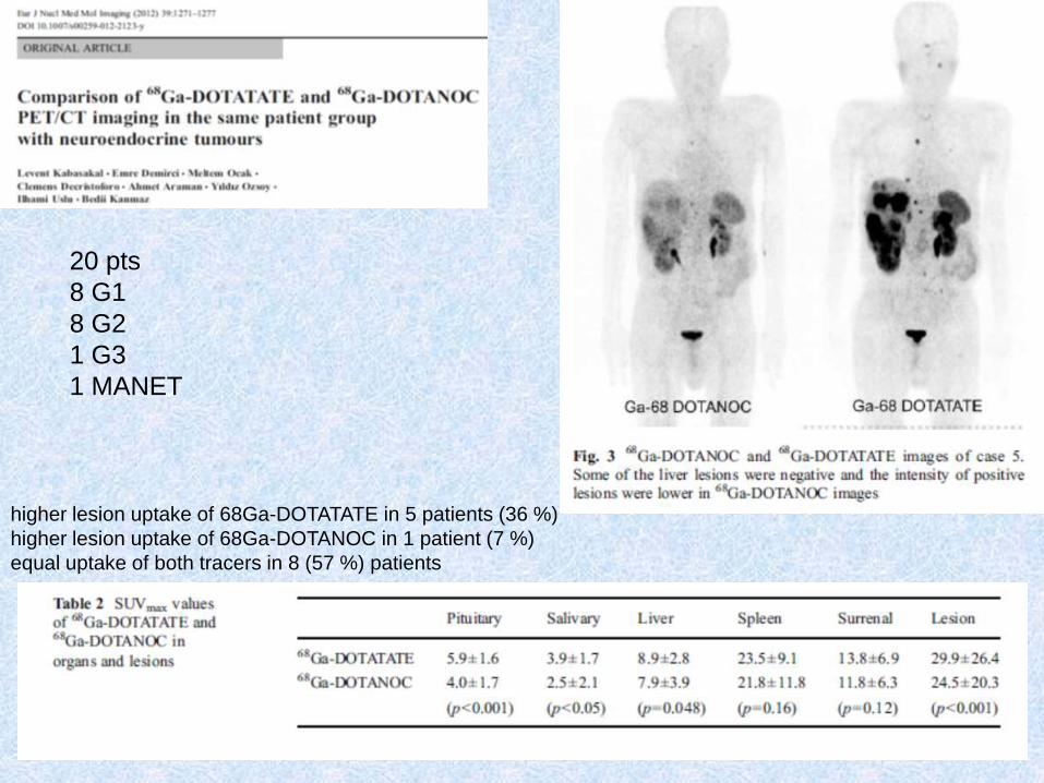
higher lesion uptake of 68Ga-DOTATATE in 5 patients (36 %)
higher lesion uptake of 68Ga-DOTANOC in 1 patient (7 %)
equal uptake of both tracers in 8 (57 %) patients
20 pts
8 G1
8 G2
1 G3
1 MANET

ALTRES TNE SECRETORS

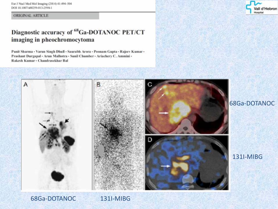
68Ga-DOTANOC
68Ga-DOTANOC
131I-MIBG
131I-MIBG

68Ga-DOTANOC 131I-MIBG
131I-MIBG
68Ga-DOTANOC

T2-weighted 3D imaging with different flip angle evolutions in curve (A), axial (B), and coronal reconstructions (E); 3D volumetric interpolated FATSAT T1-weighted (VIBE) in curve (C), axial (D), and coronal E
Current and Future Trends in the Anatomical and Functional Imaging of Head and Neck Paragangliomas David Taïeb , et al. Seminars in Nuclear Medicine 2013, 43; 462 - 473
carotid body paraganglioma
18F-FDG-PET/CT 18F-FDOPA PET/CT

18F-FDOPA PET-CT 18F-FDG PET-CT
Multifocal and recurrent SDHD-related extraadrenal PGLs
Current and Future Trends in the Anatomical and Functional Imaging of Head and Neck Paragangliomas David Taïeb , et al. Seminars in Nuclear Medicine 2013, 43; 462 - 473
123I-MIBG scintigraphy is highly specific for chromaffin-derived tumors. Its sensitivity ranges from 90% to 100% in sporadic PHEOs, but significantly decreasesin SDHx-related tumors, especially in HNPGLs

CLINICAL INDICATIONS
Localize primary tumours and detect sites of metastatic disease
(staging).
Follow-up patients with known disease to detect residual, recurrent or
progressive disease (restaging).
Determine SST receptor status visually as well by using
semiquantitative parameters like standardized uptake value (patients
with SST receptor-positive tumours are more likely to respond to
octreotide therapy)
Select patients with metastatic disease for SST receptor radionuclide
therapy (with 177Lu or 90Y-DOTA-peptides)

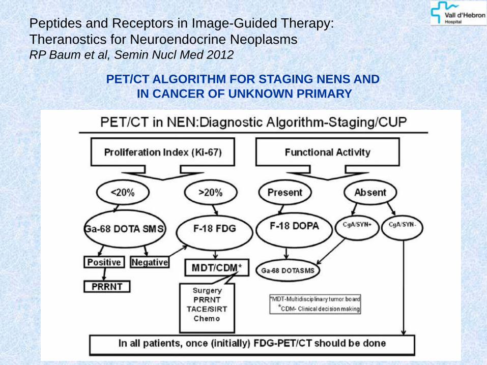
PET/CT ALGORITHM FOR STAGING NENS AND
IN CANCER OF UNKNOWN PRIMARY
Peptides and Receptors in Image-Guided Therapy:
Theranostics for Neuroendocrine Neoplasms RP Baum et al, Semin Nucl Med 2012

68Ga-DOTANOC 18F-fluorodeoxyglucose
Peptides and Receptors in Image-Guided Therapy:
Theranostics for Neuroendocrine Neoplasms RP Baum et al, Semin Nucl Med 2012

Peptides and Receptors in Image-Guided Therapy:
Theranostics for Neuroendocrine Neoplasms RP Baum et al, Semin Nucl Med 2012

ENFOCAMENT ACTUAL DE LA IMATGE MOLECULAR EN ELS TNEs
ESTADIFICACIÓ INICIAL I SELECCIÓ DE TERAPIA
G1 IMATGE Pèptids Anàlegs de la Somatostatina
G2 I G3: IMATGE PAS + IMATGE FDG
Presentacions mosaic / expressió primari vs mets
Rescat per tractament amb β-PAS
SEGUIMENT: ESTADIFICACIÓ DINÀMICA
AVALUACIÓ EFICÀCIA/RESPOSTA TERAPÈUTICA
RF selectiu (*)
RE-AVALUACIÓ DE LA EXPRESSIÓ DE RSS I ÍNDEX PROLIFERATIU
Transformació / desdiferenciació de les lesions “in vivo”
Revisió de les options terapèutiques
(*)Monitoring response to therapy has been proposed but still needs to be
assessed as the change in receptor status does not necessarily indicate therapy
response and dedifferentiation with loss of receptors must be taken into account
EANMMI GUIDELINES

Neuroendocrine Pancreatic Tumor
55
200 mCi 177Lu-octreotate
1ST treatment After the second treatment
After the fourth treatment D. J. Kwekkeboom, EJNM 2003

CONSIDERACIONS SOBRE LA INTRODUCCIÓ CLINICA DEL PET 68Ga-PAS
MODEL DE DISTRIBUCIÓ
Generador 68Ge/68Ga a cada hospital
Distribució des de ciclotró central
APLICACIÓ
Substitució de les GG-SPECT per PET
En tots el casos
disminució del cost per centralització
selecció de pacients – duplicació de proves
Us combinat amb FDG
Selecció de pacients per teràpia amb 177Lu/90Y PAS

Les tècniques d’imatge anatòmica TC i RM es consideren de primera
línia en el diagnòstic i localització dels TNEs però tenen importants
limitacions en la detecció i no permeten la caracterització molecular
inicial ni evolutiva
La determinació de la expressió de les dianes moleculars de cada
tumor permet caracteritzar cada tumor “in vivo” i de forma evolutiva,
més enllà del se perfil inmunohistoquímic inicial.
La imatge molecular PET presenta una millor qualitat (resolució i
sensibilitat) i permet estadificar inicialment la presentació dels tumors
primaris i les metàstasis tant en localització com en la expressió de
receptors i índex proliferatiu
Els R-PET d’activitat metabòlica o PAS s’utilitzaran de forma més
generalitzada i personalitzada en l’imminent futur per la selecció de
teràpies personalitzades i l’avaluació de l’eficàcia terapèutica.
RESUM