animal models for photodynamic therapy (pdt) zenildo ... · animal models for photodynamic therapy...
TRANSCRIPT

Animal models for photodynamic therapy (PDT)
Zenildo Santos Silva Jr.,1,2,3 Sandra Kalil Bussadori3, Kristianne Porta Santos Fernandes3, Ying-Ying Huang1,2, Michael R Hamblin1,2,4 1Massachusetts General Hospital, Wellman Center for Photomedicine, Boston, MA 02114, USA 2Harvard Medical School, Department of Dermatology, Boston, MA 02114, USA 3Postgraduate Program in Biophotonics Applied to Health Sciences, Nove de Julho University, São Paulo, SP, Brazil.
4Harvard-MIT Division of Health Sciences and Technology, Cambridge, MA 02139, USA Author for correspondence +1 617 726 6182; +1 617 726 6643 [email protected]
Abstract
Photodynamic therapy (PDT) employs non-toxic dyes called photosensitizers, which absorb visible light to give the excited singlet state, followed by the long-lived triplet state that can undergo photochemistry. In the presence of ambient oxygen, reactive oxygen species such as singlet oxygen and hydroxyl radicals are formed that are able to kill cancer cells, inactivate microbial pathogens and destroy unwanted tissue. Although there are already several clinically approved photosensitizers for various disease indications, many studies around the world are using animal models to investigate the further utility of PDT. This review will cover the main groups of animal models that have been described in the literature. Cancer comprises the single biggest group of models including syngeneic mouse/rat tumors that can either be subcutaneous or orthotopic, and allow the study of anti-tumor immune response; human tumors that need to be implanted in immunosuppressed hosts; carcinogen-induced tumors; and mice that have been genetically engineered to develop cancer (often by pathways similar to those in patients). Infections are the second biggest class of animal models and the anatomical sites include wounds, burns, oral cavity, ears, eyes, nose etc. Responsible pathogens can include Gram-positive and Gram-negative bacteria, fungi, viruses and parasites. A smaller and diverse group of miscellaneous animal models have been reported that allow PDT to be tested in ophthalmology, atherosclerosis, atrial fibrillation, dermatology, and wound healing. Successful studies using animal models of PDT are blazing the trail for tomorrow’s clinical approvals.
Keywords:
Photodynamic therapy, infection, cancer, subcutaneous tumor, orthotopic tumor, xenograft, autochthonous, genetically engineered mouse model
Acce
pted
Man
uscr
ipt
© 2015 The Author(s) Archiving permitted only in line with the archiving policy of Portland Press Limited.The final version of record will be available under the Creative Commons Attribution Licence 3.0(http://creativecommons.org/licenses/by/3.0/). You are encouraged to use the final version of record.
AC
CE
PT
ED
MA
NU
SC
RIP
T
10.1042/BSR20150188. Please cite using the DOI 10.1042/BSR20150188http://dx.doi.org/up-to-date version is available at
encouraged to use the Version of Record that, when published, will replace this version. The most this is an Accepted Manuscript, not the final Version of Record. You are:Bioscience Reports
). http://www.portlandpresspublishing.com/content/open-access-policy#ArchivingPolicy of Portland Press (which the article is published. Archiving of non-open access articles is permitted in accordance with the Archiving Use of open access articles is permitted based on the terms of the specific Creative Commons Licence under

Summary statement
Photodynamic therapy (PDT) combines visible light and photosensitizing dyes. Different animal models have been used to test PDT for cancer, infectious disease and cardiovascular disease. Mouse models of tumors include subcutaneous, orthotopic, syngeneic, xenograft, autochthonous, and genetically modified.
Introduction to PDT
Photodynamic therapy (PDT) employs non-toxic dyes called photosensitizers (PS), which absorb visible light of the correct wavelength to first give the excited singlet state, followed by a transition to the long-lived excited triplet state that can undergo photochemistry [1]. In the presence of molecular oxygen the photochemical reactions produce a variety of reactive oxygen species (ROS) including singlet oxygen and hydroxyl radicals. These ROS cause oxidative damage to proteins, lipids and nucleic acids and leading to cell death by necrosis and/or apoptosis (Figure 1), PDT was originally developed as a cancer treatment, but has subsequently been investigated as a treatment for choroidal neovascularization secondary to age related macular degeneration, for a range of localized infections and for disorders related to dermatology and imunology.
When PDT is used as a tumor therapy, the PS is usually injected IV, followed by a waiting period known as the “drug light interval” to allow the tumor to accumulate at the site of the tumor. Light can be delivered to the tumor site either by shining a focused spot of light onto the tissue (which can be done endoscopically), or by inserting fiber optics into the tumor tissue in a technique called interstitial light delivery. The antitumor effects of PDT result from the combination of three different in vivo mechanisms namely: direct PDT cytotoxicity to tumor cells, destruction of the tumor microvasculature, and induction of an acute local inflammatory response leading to activation of the host immune system (Figure 2).
Many different types of photoactivable molecules have been synthesized and tested as possible PDT agents. These PS are often based on the tetrapyrrole backbone such as porphyrins, chlorins, bacteriochlorins and phthalocyanines (see Figure 3). The latter three structures possess strong light absorption bands at wavelengths longer than 650 nm and are therefore well suited to the so-called “optical window” required for good tissue penetration of light. Highly effective PS require an absorption maximum between 650 and 800 nm to avoid the absorption by the endogenous tissue chromophores, such as hemoglobin, while still having enough photon energy to carry out photochemistry. The chemical structure of the PS molecule can be tailored to provide high cell uptake, selectivity for cancer cells and endothelial cells, and to provide photostability (i.e. resistance to photobleaching). A recent alternative approach is
Acce
pted
Man
uscr
ipt
© 2015 The Author(s) Archiving permitted only in line with the archiving policy of Portland Press Limited.The final version of record will be available under the Creative Commons Attribution Licence 3.0(http://creativecommons.org/licenses/by/3.0/). You are encouraged to use the final version of record.

to attach the PS covalently or non-covalently to biomolecules that possess a marked targeting ability towards cancer cells such as monoclonal antibodies or specific peptides. A popular alternative to traditional PS, is to use 5-aminolevulinic acid (ALA), a biochemical precursor to the endogenous PS, protoporphyrin IX (PPIX) [2].
Cancer.
Since the first pioneering studies of PDT to cure tumors in the 1970s by Diamond et al [3] and by Dougherty et al. [4], cancer has been the leading indication for PDT. Although much research has been carried our in cell culture studies in vitro, and more recently in three-dimensional tissue culture models [5, 6], more complex systems such as laboratory animals are required to demonstrate that these new PDT approaches could eventually work in clinical situations. The next sections of this review will give an overview of the different animal models that have been employed in studies of PDT for cancer.
Chorioallantoic membrane.
One very simple intermediate model that lies in between in vitro cell culture and laboratory animals is the chorioallantoic membrane (CAM) of fertilized chicken eggs, that have had a window of eggshell removed. This model allows the growth of tumor cells that are applied as a suspension onto the surface of the membrane and turn into “tumors” that go on to develop their own blood supply by the process of angiogenesis (in a similar fashion to real tumors in mice). PS can be injected into these blood vessels, allowed to accumulate in the tumors and then light can easily be delivered and changes in blood flow in the tumor and normal vessels can be observed in real time. PS can also be topically applied to the xenografted tumors on the CAM. The advantages of this model include the ease of in vivo microscopy to study PDT-induced vascular damage, and the lack of regulatory controls on experiments involving eggs [7-11].
Subcutaneous syngeneic mouse and rat tumors.
Using subcutaneous tumors in laboratory mice is very common among investigators aiming to test various PDT regimens (see Table 1). There are a large number of mouse tumor cell lines available, which differ in the type of tissue or organ that the tumor originated from, and also in the particular syngeneic inbred mouse strain that the tumors belong to. Individual laboratory mouse strains (Mus musculus) have particular combinations of major histocompatibility complexes (MHC class I such as H2B, and MHC class II such as Ia) and the tumor cells should have the same combination of MHC molecules as the host mouse to allow them to grow without instant rejection. This syngeneic mouse tumor approach is often used because the mice have intact immune systems, and therefore immunology and anti-tumor immunity effects after PDT can be studied. There are many inbred mouse strains available (over 400), but the most commonly used examples in anticancer PDT studies are BALB/c (albino),
Acce
pted
Man
uscr
ipt
© 2015 The Author(s) Archiving permitted only in line with the archiving policy of Portland Press Limited.The final version of record will be available under the Creative Commons Attribution Licence 3.0(http://creativecommons.org/licenses/by/3.0/). You are encouraged to use the final version of record.

C57BL/6 (black), C3H (brown) and DBA2 (gray). Tumor types include adenocarcinoma lines (colon, lung, breast, pancreas etc), squamous cell carcinoma, fibrosarcoma, lymphoma, melanoma, malignant glioma and many others. Rat tumor cell lines are less common than murine tumor lines, but there are several known belonging to inbred laboratory rat strains (Rattus norwegicus) such as Wistar, Lewis, Sprague-Dawley, Fischer 344 etc. Subcutaneous tumors differ in their rate of growth, degree of vascularization, amount and kind of tumor stroma, and potential to form spontaneous metastasis. In addition to injecting the tumor cells beneath the skin, they can also be injected between the layers of the epidermis and dermis to form intradermal tumors. It has been shown that intradermal tumors are more easily recognized by the immune system compared to subcutaneous tumors [12]. In some cases investigators mix the tumor cells with a preparation of extracellular matrix called Matrigel before injection, and it has been shown that this can make a difference in the response of the tumor to PDT [13]. Many investigators transplant tumors from one animal to another by inserting small pieces obtained from an excised whole tumor under the skin of the next animal.
PDT of subcutaneous mouse tumors is usually carried out by intravenous injection of the PS (often in the tail vein) followed after a certain period of time by light delivery to the tumor including a certain amount of surrounding normal tissue. This time period called the “drug light interval” is an important parameter, that governs the amount of vascular damage that occurs during the treatment and therefore whether the PDT regimen is “vascular’ or “cellular”. Less common is the injection of the PS into the abdominal cavity (intraperitoneal [14], or directly into the tumor (intratumoral [15]). With the recent rise in the study of PDT mediated by a wide range of nanoparticles and multifunctional nanostructures, the use of intratumoral injection has become more common. This is because the phamacokinetics and biodistribution of these engineered nanostructures has as yet not been much investigated.
The response of subcutaneous tumors depends on whether the PDT regimen is predominantly vascular or cellular. For vascular PDT a circumscribed black eschar usually appears about 2 days post PDT (see figure 4). For cellular PDT it is common for the tumor volume to shrink without such a visible black eschar. In many cases a pronounced swelling of the tissue is seen soon after completion of the illumination, due to the acute inflammatory reaction. The duration of the tumor response depends on many factors. In the case of a marked immune response occurring after PDT, tumors do not recur and the animals are regarded as cured after 90 days of being tumor-free. Local tumor recurrence can occur after PDT, and this recurrence often takes the form of an annulus or “doughnut” of tumor surrounding the original tumor. The reason for the morphology of this recurrence is not completely understood, but may involve the presence of cancer stem cells in the periphery of the original tumor at the “invasive front” [16].
Human xenograft tumors.
Acce
pted
Man
uscr
ipt
© 2015 The Author(s) Archiving permitted only in line with the archiving policy of Portland Press Limited.The final version of record will be available under the Creative Commons Attribution Licence 3.0(http://creativecommons.org/licenses/by/3.0/). You are encouraged to use the final version of record.

Much more is known about the molecular biology and tumor biology of human tumors than is known for analogous mouse tumors. A large amount of knowledge about mutations, oncogenes, impaired tumor suppressor pathways, growth factors, epigenetics and so on, is more or less specific for human tumors, and much less is known about the effects of these factors in mouse tumors. The discovery of immunosuppressed mice that can be either athymic nude mice, or the severe combined immunodeficient (SCID) mice that are T- and B-cell-deficient, allows the engraftment of human tumor cells known as “xenografts” (or sometimes xenographs). Due to the increased susceptibility of these mice to infections, the cell lines must be free of mouse pathogens, and the mice must be kept in specific pathogen-free conditions. Even so, innate immunity, particularly natural killer (NK) cells and tumoricidal macrophages, can limit tumor growth and prevent metastasis in nude mice [17]. A large number of human cancer cell lines grown as xenograft tumors grown in a subcutaneous location in either nude mice or SCID mice have been subjected to PDT (see Table 2).
Othotopic syngeneic mouse and rat tumors.
There has been criticism leveled at the widespread use of subcutaneous tumors in PDT research, to the effect that they fail to replicate the normal biology of human tumors by not being situated in their organ of origin (orthotopic), and are therefore not subject to the appropriate environmental cues and signaling from the host tissue. The blood supply that develops to supply the tumor is very different in orthotopic models, and the propensity to spontaneously metastasize is also higher. For instance brain tumors behave very differently when implanted in the brain, than when grown subcutaneously. Investigators have therefore injected the tumor cells into the tissue of the organ of origin in the mouse or rat. This procedure may need a surgical approach that depends on the actual organ to be implanted with the tumor. This has been done with colon carcinomas (into the wall of the colon), renal cell cancers (into the kidney), mammary carcinomas (into the mammary fat pad), bladder carcinomas (into the bladder wall), prostate carcinoma (into the prostate), pancreatic carcinoma (into the pancreas), and lung cancer (into the bronchi). Moreover the actual PDT treatment approach necessitates being able to get the light to the tumor, which is more difficult for orthotopic tumors.
Orthotopic tumors used to test PDT can either be syngeneic in mice or rats or can be xenografts (see Table 3). There is one rabbit tumor that has been quite often used to create orthotopic tumor models in a medium sized animal, the New Zealand white rabbit. The VX2 tumor cell line was originally derived from a virus-induced skin papilloma [18], but has been widely used to produce tumors in the liver [19], the pancreas [20] and the brain [21], all of which have been treated with PDT.
Autochthonous tumors.
Tumors that arise naturally in the host are called “autochthonous”. In laboratory studies these tumors are usually induced by application of chemical carcinogens,
Acce
pted
Man
uscr
ipt
© 2015 The Author(s) Archiving permitted only in line with the archiving policy of Portland Press Limited.The final version of record will be available under the Creative Commons Attribution Licence 3.0(http://creativecommons.org/licenses/by/3.0/). You are encouraged to use the final version of record.

but viruses and physical carcinogenic stimuli (for instance UV radiation) have also been used. These models effectively recapitulate the time-dependent and multi-stage progression of cancer formation in response to relevant environmental carcinogens and tumor-promoting agents [22]. These models utilize the topical, intraperitoneal, or oral administration of a variety of polycyclic aromatic hydrocarbons or reactive organic chemicals either alone or in combination with known tumor promoters such as phorbol esters, to induce specific cancers in a variety of immunocompetent mice, rats and hamsters. The susceptibility to chemical carcinogenesis and the resultant tumor incidence and multiplicity varies with the protocol, the dosage of carcinogens and promoters, and the age and strain of the rodents used. The Syrian golden hamster model of oral dysplasia and cancer caused by repeated application of 7,12-dimethyl-benz(a)anthracene (DMBA) is particularly noteworthy, as hamsters are chosen because of the presence of the cheek pouch as an example of oral mucosa that is relatively easily manipulated [23]. Moreover the lesions undergo a predictable progression through hyperplasia, mild and severe dysplasia before tumors begin to appear after about 12 weeks of application 3X per week. (Table 4) lists some autochthonous tumor models that have been employed in PDT studies.
Genetically-engineered mouse models of cancer
Genetically engineered mouse models (GEMMs) of cancer have been constructed to more closely mimic the development of human disease in a predictable manner [24] . Since the 1980s, several types of GEMMs have been used, including transgenic, knockout, and knockin mouse models [25]. Mutant mice that express oncogenes or dominant-negative tumor-suppressor genes governed by ectopic promoter and enhancer elements can be generated by pronuclear injection of cDNA constructs that contain promoter elements designed to restrict expression to certain tissues. GEMM can be produced by the direct injection of fertilized oocytes or by lentiviral transduction of embryonic stem cells. Targeted transgenesis provides an attractive alternative to random transgenesis by using integration mediated by recombination at specific sites allowing insertion of only a single copy of the transgene [26].
Many GEMM models of cancer have been developed including lung, colon, stomach, esophagus, pancreas, liver, breast, ovary, prostate, bladder, kidney, brain, and skin [25]. In some of these models an additional application of a carcinogen needs to be used to actually initiate tumor development. Examples of these additional insults could be UV irradiation to initiate skin cancer, or injection of dextran sulfate to initiate colon carcinogenesis. Despite the large number of GEMM, as yet only a few of these have been used to test PDT. These are listed in (Table 5).
In vivo imaging
In recent years there has been an explosion of non-invasive imaging techniques applied to laboratory studies of cancer and anti-cancer therapy. Many of these in vivo imaging modalities are based on adapation of those clinical modalities that
Acce
pted
Man
uscr
ipt
© 2015 The Author(s) Archiving permitted only in line with the archiving policy of Portland Press Limited.The final version of record will be available under the Creative Commons Attribution Licence 3.0(http://creativecommons.org/licenses/by/3.0/). You are encouraged to use the final version of record.

are familiar to oncologists to be used in small animals. This classification includes such modalities as micro-positron emitting tomography (microPET) [27], micro-single photon micro-computed tomography (micro-SPECT) [28], micro-computed tomography (µCT) [29], high resolution ultrasound [30], photoacoustic tomography (PAT) [31]. However another large group of small animal in vivo imaging modalities relies upon genetically encoded reporter molecules and falls into the category of molecular imaging. The two most often used modalities in this class are (1) in vivo fluorescence imaging relying on engineered fluorescent protein reporters such as green fluorescent protein (GFP); and (2) in vivo bioluminescent imaging relying on engineered luciferase reporters such as the firefly or marine luciferase enzymes.
Infections.
After cancer, infections represent the next most frequent application of PDT. The use of PDT against infections is motivated by the widespread and growing problem of antibiotic resistance, often called “the single biggest problem facing global health” [32]. A very large number of studies have been published on the use of a wide variety of PS to kill or inactivate numerous classes of microorganisms including Gram-positive bacteria, Gram-negative bacteria, fungi, parasites and viruses. However the number of studies on PDT for actual infections in animal models is much more limited (see Table 6). Traditional methods for quantifying infection involve the sacrifice of animals at various time-points, removal of the infected tissue, homogenization, serial dilution, plating on agar and colony counting. In recent years the experimental study of PDT for infections has been facilitated by the use of in vivo bioluminescence imaging to non-invasively monitor the progression of the infection. Sensitive cameras can record non-invasively, in real time and with longitudinal monitoring, the anatomical location and growth of infectious microorganisms in living hosts [33]. Figure 5 shows an example of the use of this bioluminescence imaging technique to monitor PDT of a Gram-negative E. coli infection mediated by pL-ce6 conjugate and red light, in excisional wounds on the back of BALB/c mice [34].
An interesting alternative animal model to study the pathogenesis and therapy of infections is the use of invertebrate hosts [35]. The invertebrate host that has been most frequently used is the larva of Galleria mellonella, the greater wax moth. The pathogens are injected into the larva, followed by injection of the PS and then by light delivery. This has been reported for Enterococcus faecium [36] and for Candida albicans [37] both of which were treated with methylene blue and red light.
Miscellaneous indications.
There has been a diverse range of diseases other than cancer and infections that have been treated with PDT in animal models. Some of these are listed in (Table 7).
Acce
pted
Man
uscr
ipt
© 2015 The Author(s) Archiving permitted only in line with the archiving policy of Portland Press Limited.The final version of record will be available under the Creative Commons Attribution Licence 3.0(http://creativecommons.org/licenses/by/3.0/). You are encouraged to use the final version of record.

Conclusions.
It is almost a universally accepted axiom in cancer research, that enormous numbers of mice and rats have been cured of cancer by a wide range of experimental therapeutics, but the translation into clinical practice has been disappointing more often than not. Nevertheless the importance of designing and selecting the most appropriate animal models to investigate the ever-increasing range of new experimental therapeutic approaches has never been greater. This consideration applies even more to the rather complex techniques of PDT and aPDI, than it does to the more traditional testing of new pharmaceutical or biologic therapies. The multi-target nature of anti-cancer PDT (tumor cells, tumor vasculature, and immune response) means that thought must be given to understanding the specific tumor biology of the model chosen. Moreover since the light delivery is by definition a localized process, the anatomical location of orthotopic tumors becomes important.
When the testing of PDT in animal models of infectious disease is undertaken, some other points need to be considered. Firstly it is important to realize that human infections generally develop gradually from a relatively small initial infectious inoculum, rather than from a sudden large application of millions or tens of millions of CFU. Secondly a high level of selectivity for microbial cells over host mammalian cells is needed, since the actual fraction by weight of microbial cells in even a serious infection is still very low. Thirdly when the light is switched off, the generation of microbicidal ROS is halted, and the microbial cells may be free to resume their growth unhindered.
The popularity of testing PDT for non-traditional indications involves designing animal models of non-cancer, non-infectious disease. A diverse assortment of conditions in cardiology, atherosclerosis, ophthalmology, neuroscience could in principle be treated with PDT.
Finally for those laboratories that are not easily able to carry out studies on traditional mammalian animal models there are new models described using invertebrate hosts and fertilized eggs. Large animal models have not been much studied in the PDT field, but as more new indications are being studied, studies on large animals can be expected to become more important.
Acce
pted
Man
uscr
ipt
© 2015 The Author(s) Archiving permitted only in line with the archiving policy of Portland Press Limited.The final version of record will be available under the Creative Commons Attribution Licence 3.0(http://creativecommons.org/licenses/by/3.0/). You are encouraged to use the final version of record.

Acknowledgments.
Z.S.S.Jr. was supported by CAPES-Ministry of Education, Brazil, grant 99999.002158/ 2014-00. Research in the Hamblin laboratory is supported by US NIH grant R01AI050875.
Conflict of Interest.
The authors declare that they have no conflict of interest
Acce
pted
Man
uscr
ipt
© 2015 The Author(s) Archiving permitted only in line with the archiving policy of Portland Press Limited.The final version of record will be available under the Creative Commons Attribution Licence 3.0(http://creativecommons.org/licenses/by/3.0/). You are encouraged to use the final version of record.

Table 1 Subcutaneous syngeneic mouse/rat tumors that have been used for PDT
Cell line Cancer type Mouse strain PDT regimen
Authors
EMT6 Mammary sarcoma BALB/c Photofrin, DLI 24h, 630 nm, 110 J/cm2, 130 mW/cm2
[38]
CT26 (c26, colo26) Colon adenocarcinoma BALB/c Pheophorbide HPPH, DLI 24 h, 665 nm, 100 J/cm2, 75 mW/cm2
[39]
DA3Hi
Breast adenocarcinoma BALB/c male mice
Hypericin, inj IP, DLI 6h, 400-700nm, 60 J/cm2, 20 min. [40]
MS-2 Fibrosarcoma BALB/c Phthalocyanine AIS2Pc, DLI 24h, 670 nm, 60 J/cm2. 100 mW/cm2
[41]
4T1 Breast cancer BALB/c female mice
Verteporfin BPD-MA, DLI 24h, 690 nm,120 J/cm2 [42]
B16 Melanoma C57BL/6 Porphyrin TPP, DLI 9h, 630 nm, 90 J/cm2, 100 mW/cm2,
[43] LLC Lewis lung adenocarcinoma C57BL/6 Photofrin, DLI 24h, 630 nm, 150 J/cm2, 90 mW/cm2
[44]
TC1 Lymphoma C57BL/6 Radachlorin. DLI 3h, 662 nm, 300 J/cm2,
[45]
RIF1 Radiation induced fibrosarcoma C3H Photofrin, DLI 24h, 400 nm, 135 J/cm2, [46]
FsaR Fibrosarcoma C3H/HeN
Photofrin, DLI 24h, 630 nm, 150J/cm2, 110 mW/cm2
[47]
SCCVII Squamous cell carcinoma C3H Foscan mTHPC, DLI 24h, 650 nm, 50 J/cm2, 90 mW/cm2
[48]
DLM-8 Osteosarcoma C3H Acridine orange AO, DLI 2h, xenon flash lamp, 15J per pulse, 60 Hz, 10 min.
[49]
P815 Mastocytoma DBA/2 Verteporfin BPD-MA, DLI 15 min, 690 nm, 120 J/cm2, 100 mW/cm2
[50]
Sarcoma 180 Sarcoma
ICR outbred Hematoporphyrin, inj IP, DLI 24h, 635 nm, 30 J/cm2, 26 mW/cm2 [51]
NXS2 Neuroblastoma Female A/J Pheophorbide HPPH, DLI 24h, 665 nm, 48 J/cm2, 7 mW/cm2 [52] Accepted M
anuscrip
t
© 2015 The Author(s) Archiving permitted only in line with the archiving policy of Portland Press Limited.The final version of record will be available under the Creative Commons Attribution Licence 3.0(http://creativecommons.org/licenses/by/3.0/). You are encouraged to use the final version of record.

VMDK Glioma VM
Porphyrin mTHPP, DLI 24h, 648 nm, 20J/cm2, 300mW/cm2.
[53]
Walker 256 Carcinosarcoma Wistar male rats Porphyrin TSPP, DLI 24h, 685-nm, 50 J/cm2, 25 W/cm2.
[54]
MatLyLu (Dunning) Prostate
Copenhagen rats Verteporfin BPD, DLI 1h, 690-nm, 50J/cm2, 50mW/cm2. [55]
Table 2 Orthotopic syngeneic mouse/rat tumors
Cancer type
Cell line Mouse/rat model PDT regimen Authors
Prostate TRAMP-C2 Albino male C57BL/6 mice
5-ALA, DLI 72h, 635 nm, 100 J/cm2, 200mW/cm2.
[56]
Bladder
AY-27 cells
Female Fischer F344 rats
ALA, DLI 2h, 514 nm, 20 J/cm2, 100 mW/cm2.
[57]
Prostate R3327-MatLyLu Dunning
Male Copenhagen rats Verteporfin DLI 15-3h, 690 nm, 50 J/cm2, 50 mW/cm2, 1,000 s.
[58]
Glioma/brain
9L.E29
Female athymic mice
Phthalocyanine, EGFpep-Au NP-Pc 4; DLI 4h, 672 nm, 50 J/cm2 , 0.1 W/cm2.
[59]
Breast cancer 4T1
BALB/c female mice Photofrin, DLI 120h, 635 nm, 100 J/cm2, 416.7 mW/cm2.
[60]
Glioma/brain C6-9 Sprague-Dawley male rats Foscan, m-THPC DLI 48h, 652 nm, 20 J/cm2,100 mW/cm2.
[61]
Glioma/brain BT4C BD-IX rats ALA (ip), 5h DLI, 632 nm, 26 J, 30 mW. [62]
Accepted M
anuscrip
t
© 2015 The Author(s) Archiving permitted only in line with the archiving policy of Portland Press Limited.The final version of record will be available under the Creative Commons Attribution Licence 3.0(http://creativecommons.org/licenses/by/3.0/). You are encouraged to use the final version of record.

Table 3. Xenograft mouse or rat tumors
Cell line Cancer type Animal model PDT regimen
Authors
OVCAR3 ovarian Nude mice Photofrin, DLI 24h, 635 nm, 200 J/cm2.
[63]
OVCAR5 ovarian Nude mice Verteporfin, BPD-MA, DLI 90 min, 690 nm, 40 J, 320s.
[64]
AsPC-1 and Panc-1
Pancreas Male SCID mice Verteporfin, DLI 1h, 690 nm, 40 J/cm2, 74mW/cm2.
[65]
LNCaP Prostate SCID mice liposomal BPD, DLI 1h, 690 nm,100 J/cm2.
[66]
WISH-PC237 Prostate Male CD1 nude mice Pd-bacteriopheophorbide TOOKAD, DLI zero, 650–800 nm, 360 J/cm2, 30 min.
[67]
Eca109 Esophageal SCC Nude mice (sc or orthotopic)
Photofrin, DLI 24h, 630 nm, 135 J/cm2, 75 mW/cm2
[68]
A549 Lung cancer (NSCLC) nude mice factor VII-targeted Sn(IV) chlorin e6 conjugate, 635 nm, 72J/cm2
[69]
H460 Lung cancer (NSCLC) Ncr-nu/nu female mice Photochlor HPPH, DLI 24h, 661 nm, 200J/cm. 150 mW/cm.
[70]
FaDu Head and neck SCC Female nu/nu CByJ.Cg-Foxn1nu/J.
HPPH, DLI 24h, 665 nm, 48Jcm2, 7mW/cm2,
[52]
MDA-MB 231 Mammary carcinoma
Nude mice Phthalocyanine, AlOH-PC, DLI 10 min, 635 nm, 100 J/cm2, 0.97 W,
7 min.
[71]
MCaIV Breast Female SCID Pyropheophorbide MV6401, DLI 15 min, 664 nm 5 J/cm2, 50 mW/cm2.
[72]
Accepted M
anuscrip
t
© 2015 The Author(s) Archiving permitted only in line with the archiving policy of Portland Press Limited.The final version of record will be available under the Creative Commons Attribution Licence 3.0(http://creativecommons.org/licenses/by/3.0/). You are encouraged to use the final version of record.

HT29 Liver Female Swiss nude mice
Monoclonal antibody chlorin(e6) conjugate, 17.1A-pl-ce6-succ, DLI 3h, 666 nm, 80 J, 100mW, 13.3 min
[73]
NPC Nasopharyngeal carcinoma BALB/c nude mice 5-ALA, DLI 3.5h, 630 nm, 100 J/cm2, 100 mW/cm2.
[74]
Route: IV, IP, IT,
Table 4 - Autochthonous tumors
Cancer type Animal species Carcinogen PDT regimen Authors Oral cancer/dysplasia Male Wistar rats 4-Nitroquinoline-1-oxide (4NQO)
Photofrin, DLI 24h, 625 nm, 100 J/cm2, 60 mW/cm2. [75]
Oral cancer/dysplasia Male CBA mice 4-Nitroquinoline-1-oxide (4NQO)
ALA, DLI 5h, 630 nm, 200 J/cm2, 125 mW/cm2 [76]
Oral cancer/dysplasia
Syrian Golden Hamster 7,12-dimethylbenz-(a)anthracene (DMBA)
ALA, DLI 2.5h, LED 638 nm, 275 J/cm2, 200 mW/cm2, [77]
Oral cancer/dysplasia
Syrian Golden Hamster DMBA Topical Photosan, DLI 16 min, 640 nm, 100J/cm2, 320 mW/cm2, 313 s.
(72)
Mammary tumors virgin Sprague–Dawley female rats
DMBA Photogem® hematoporphyrin, DLI 24h, LED 635 nm, 200 J/cm2,180 mW/cm2.
[78]
Skin Tumor
FVB/N mice DMBA/12-ο-tetradecanoylphorbol-13-acetate (TPA)
ALA, DLI 48h, 635 nm, 120 J/cm2, 120 mW/cm2. [79]
Skin Tumor female hairless mice (NMRI – HR-HR)
UV irradiation induced tumor ALA-Me, DLI 4h, 630–636 nm, 40 J/cm2, 20mW/cm2.
[80]
Accepted M
anuscrip
t
© 2015 The Author(s) Archiving permitted only in line with the archiving policy of Portland Press Limited.The final version of record will be available under the Creative Commons Attribution Licence 3.0(http://creativecommons.org/licenses/by/3.0/). You are encouraged to use the final version of record.

Table 5 - GEMM
Tumor GEMM Inducible PDT regimen
Authors
Breast Male FVB/NTgN(WapHRAS)69Lln YSJL
No Foscan-PEG, SC102; DLI 96h, 652 nm, 40 J/cm2 [81]
Breast FVB.Cg- Tg(WapHRAS)69Lln Chr Y SJL/J
FVB/N-Tg(MMTV-PyVT)634Mul/J
C57BL/6J-Tg(WapTAg)1Knw
5-ALA for fluorescence detection of 3 different BrCa. DLI 75 min
[82]
Pancreas ductal adenocarcinoma
LSL-KRasG12D-p53-floxed-Pdx-1-Cre
No Cathepsin-E cleavable ALA, DLI 1h, 652 nm, 10 J/cm2, 50 mW/cm2, 3.5 min
[83]
Basal cell carcinoma B6C3Fe-a ⁄a-Ptchmes ⁄+ UV exposure 20 weeks
MAL, DLI 3h, 550 nm, 650 H, 7J/cm2, 5 mW/cm2. [84]
Melanoma MT-ret transgenic 304/B6 No 5-ALA, DLI 3h, 630 nm, 200 J/cm2, 100mW/cm2. [85]
Colon dysplasia C57BL/6J-Apc(Min) Dextran sulfate Oral ALA for fluorescence detection of colon tumors, DLI 3h.
[86]
Accepted M
anuscrip
t
© 2015 The Author(s) Archiving permitted only in line with the archiving policy of Portland Press Limited.The final version of record will be available under the Creative Commons Attribution Licence 3.0(http://creativecommons.org/licenses/by/3.0/). You are encouraged to use the final version of record.
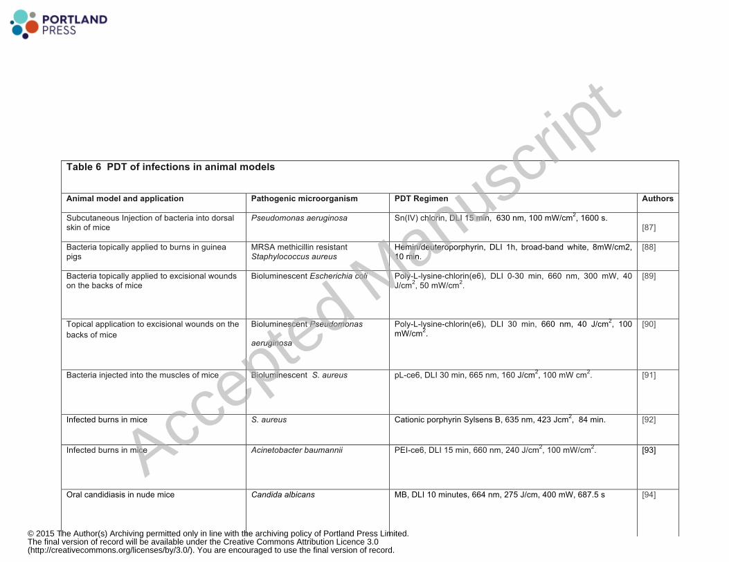
Table 6 PDT of infections in animal models
Animal model and application Pathogenic microorganism PDT Regimen
Authors
Subcutaneous Injection of bacteria into dorsal skin of mice
Pseudomonas aeruginosa
Sn(IV) chlorin, DLI 15 min, 630 nm, 100 mW/cm2, 1600 s.
[87]
Bacteria topically applied to burns in guinea pigs
MRSA methicillin resistant Staphylococcus aureus
Hemin/deuteroporphyrin, DLI 1h, broad-band white, 8mW/cm2, 10 min.
[88]
Bacteria topically applied to excisional wounds on the backs of mice
Bioluminescent Escherichia coli
Poly-L-lysine-chlorin(e6), DLI 0-30 min, 660 nm, 300 mW, 40 J/cm2, 50 mW/cm2.
[89]
Topical application to excisional wounds on the backs of mice
Bioluminescent Pseudomonas
aeruginosa
Poly-L-lysine-chlorin(e6), DLI 30 min, 660 nm, 40 J/cm2, 100 mW/cm2.
[90]
Bacteria injected into the muscles of mice
Bioluminescent S. aureus pL-ce6, DLI 30 min, 665 nm, 160 J/cm2, 100 mW cm2. [91]
Infected burns in mice S. aureus Cationic porphyrin Sylsens B, 635 nm, 423 Jcm2, 84 min. [92]
Infected burns in mice
Acinetobacter baumannii PEI-ce6, DLI 15 min, 660 nm, 240 J/cm2, 100 mW/cm2. [93]
Oral candidiasis in nude mice Candida albicans MB, DLI 10 minutes, 664 nm, 275 J/cm, 400 mW, 687.5 s [94]
Accepted M
anuscrip
t
© 2015 The Author(s) Archiving permitted only in line with the archiving policy of Portland Press Limited.The final version of record will be available under the Creative Commons Attribution Licence 3.0(http://creativecommons.org/licenses/by/3.0/). You are encouraged to use the final version of record.

Leishmaniasis in hamsters Leishmania amazonensis MB, DLI 10 min, LED, 663nm, 12 J/cm2 , 5 mW/cm2, 1h. [95]
Subcutaneous granulomas of tuberculosis in male BALB/c mice
Mycobacterium bovis BCG Verteporfin, BPD, DLI 1h, 690-nm, 60 J/cm2 and 100 J/cm2
[96]
Otitis media with effusion (OME) in Mongolian gerbils
Sttreptococcus pneumoniae or Hemophilus influenzae
Hematoporphyrin, DLI 6h, 632-nm, 90 J, 100 mW, 15 min. [97]
Periodontal disease in Wistar rats.
Natural Gram-negative anaerobic
bacteria
TBO, DLI 1 min, 660nm, 57.14 J/cm2 [98]
Osteomyelitis bacterial infection in Sprague-Dawley rats
Bioluminescent S. aureus IP ALA, DLI 4h, 635nm, 75 J/cm2, 250 mW/cm2 [99]
Murine bacterial arthritis in knee
Bioluminescent methicillin-resistant S. aureus (MRSA)
Intra-articular MB, 660 nm, DLI 5min, 160 J/cm2, 100 mW/cm2. [100]
Oral wound infections in Wistar rats. Streptococcus spp. and Actinomyces viscosus
TBO, 635 nm, DLI 10 min, 48J/cm2, 246 mW, 2min. [101]
Infection in excisional wounds of mice Vibrio vulnificus TBO, DLI 30 min, 560-780 nm, 200J/cm2, 80 mW/cm
2, 20 min. [102]
Accepted M
anuscrip
t
© 2015 The Author(s) Archiving permitted only in line with the archiving policy of Portland Press Limited.The final version of record will be available under the Creative Commons Attribution Licence 3.0(http://creativecommons.org/licenses/by/3.0/). You are encouraged to use the final version of record.

Table 7. PDT for other disease indications
Disease Specialty
Animal model PDT regimen Authors
Choroidal neovasculariztion
Ophthalmology Choriocapillary photo-occusion in rabbits Verteporfin BPD-MA, DLI 3h, 692 nm, 10J/cm2
[103]
Choroidal neovascularization
Ophthalmology Argon-laser induced injuries to choroid in monkeys
Verteporfin, liposomal BPD, DLI 50 min, 692 nm, 150 J/cm2, 600 mW/cm2,
[104]
Atherosclerosis
Cardiology Balloon-injured arteries in cholesterol fed rabbits
Etiopurpurin MV0611, DLI 24h, 542 nm, 18 J/cm2, 90 sec
[105]
Atherosclerosis
Cardiology Balloon-injured arteries in cholesterol fed Yucatan miniswine
Photofrin, DLI 24h, 630 nm, 240 J/cm2, 1.28mW/cm2, 188 s.
[106]
Atrial fibrillation
Cardiology
Intravascular ablation to block cavotricuspid isthmus in dogs
Talaporfin sodium, continuous infusion, 663 nm, 10-W/cm2, 30 s.
[107]
Atrial fibrillation Cardiology Atrioventricular block in rats Talaporfin sodium, DLI 30 min, 670 nm, 10 J/cm2, 150 mW/cm2 [108] Accepted M
anuscrip
t
© 2015 The Author(s) Archiving permitted only in line with the archiving policy of Portland Press Limited.The final version of record will be available under the Creative Commons Attribution Licence 3.0(http://creativecommons.org/licenses/by/3.0/). You are encouraged to use the final version of record.

Vascular wound healing
Vascular surgery Balloon-injured carotid arteries in rats Local MB, 660 nm, 100 J/cm2 , 100mW/cm2
[109]
Wound healing
Dermatology Excisional wounds in rats
ALA IP, DLI 24h, 632 nm, 3 J/cm2, 5 mW,
[110]
Hypertrophic scars Dermatology Excisional wounds in ears of rabbits Topical ALA, DLI 3h, 635nm, 114.6 J/cm2. 20 min [111]
Photoaging
Dermatology UV irradiation of mouse skin 5 days/8 weeks
Topical ALA, DLI 4h, 635 nm, 75 J/cm2
[112]
Accepted M
anuscrip
t
© 2015 The Author(s) Archiving permitted only in line with the archiving policy of Portland Press Limited.The final version of record will be available under the Creative Commons Attribution Licence 3.0(http://creativecommons.org/licenses/by/3.0/). You are encouraged to use the final version of record.

Figure 1
Acce
pted
Man
uscr
ipt
© 2015 The Author(s) Archiving permitted only in line with the archiving policy of Portland Press Limited.The final version of record will be available under the Creative Commons Attribution Licence 3.0(http://creativecommons.org/licenses/by/3.0/). You are encouraged to use the final version of record.

Figure 2
Acce
pted
Man
uscr
ipt
© 2015 The Author(s) Archiving permitted only in line with the archiving policy of Portland Press Limited.The final version of record will be available under the Creative Commons Attribution Licence 3.0(http://creativecommons.org/licenses/by/3.0/). You are encouraged to use the final version of record.

Figure 3
Acce
pted
Man
uscr
ipt
© 2015 The Author(s) Archiving permitted only in line with the archiving policy of Portland Press Limited.The final version of record will be available under the Creative Commons Attribution Licence 3.0(http://creativecommons.org/licenses/by/3.0/). You are encouraged to use the final version of record.

Figure 4
Acce
pted
Man
uscr
ipt
© 2015 The Author(s) Archiving permitted only in line with the archiving policy of Portland Press Limited.The final version of record will be available under the Creative Commons Attribution Licence 3.0(http://creativecommons.org/licenses/by/3.0/). You are encouraged to use the final version of record.

Figure 5
Acce
pted
Man
uscr
ipt
© 2015 The Author(s) Archiving permitted only in line with the archiving policy of Portland Press Limited.The final version of record will be available under the Creative Commons Attribution Licence 3.0(http://creativecommons.org/licenses/by/3.0/). You are encouraged to use the final version of record.

References
1 Agostinis, P., Berg, K., Cengel, K. A., Foster, T. H., Girotti, A. W., Gollnick, S. O., Hahn, S. M., Hamblin, M. R., Juzeniene, A., Kessel, D., Korbelik, M., Moan, J., Mroz, P., Nowis, D., Piette, J., Wilson, B. C. and Golab, J. (2011) Photodynamic therapy of cancer: An update. CA: Cancer J Clin. 61, 250-‐281 2 Krammer, B. and Plaetzer, K. (2008) ALA and its clinical impact, from bench to bedside. Photochemical & photobiological sciences : Official journal of the European Photochemistry Association and the European Society for Photobiology. 7, 283-‐289 3 Diamond, I., Granelli, S. G., McDonagh, A. F., Nielsen, S., Wilson, C. B. and Jaenicke, R. (1972) Photodynamic therapy of malignant tumours. Lancet. 2, 1175-‐1177 4 Dougherty, T. J., Grindey, G. B., Fiel, R., Weishaupt, K. R. and Boyle, D. G. (1975) Photoradiation therapy. II. Cure of animal tumors with hematoporphyrin and light. Journal of the National Cancer Institute. 55, 115-‐121 5 Antoni, D., Burckel, H., Josset, E. and Noel, G. (2015) Three-‐dimensional cell culture: a breakthrough in vivo. Int J Mol Sci. 16, 5517-‐5527 6 Yang, Y., Yang, X., Zou, J., Jia, C., Hu, Y., Du, H. and Wang, H. (2015) Evaluation of photodynamic therapy efficiency using an in vitro three-‐dimensional microfluidic breast cancer tissue model. Lab Chip. 15, 735-‐744 7 Ismail, M. S., Torsten, U., Dressler, C., Diederichs, J. E., Huske, S., Weitzel, H. and Berlien, H. P. (1999) Photodynamic Therapy of Malignant Ovarian Tumours Cultivated on CAM. Lasers in medical science. 14, 91-‐96 8 Hornung, R., Hammer-‐Wilson, M. J., Kimel, S., Liaw, L. H., Tadir, Y. and Berns, M. W. (1999) Systemic application of photosensitizers in the chick chorioallantoic membrane (CAM) model: photodynamic response of CAM vessels and 5-‐aminolevulinic acid uptake kinetics by transplantable tumors. Journal of photochemistry and photobiology. B, Biology. 49, 41-‐49 9 Hoppenheit, C., Huttenberger, D., Foth, H. J., Spitzer, W. J., Reichert, T. E. and Muller-‐Richter, U. D. (2006) Pharmacokinetics of the photosensitizers aminolevulinic acid and aminolevulinic acid hexylester in oro-‐facial tumors embedded in the chorioallantois membrane of a hen's egg. Cancer biotherapy & radiopharmaceuticals. 21, 569-‐578 10 Xiang, L., Xing, D., Gu, H., Yang, D., Yang, S., Zeng, L. and Chen, W. R. (2007) Real-‐time optoacoustic monitoring of vascular damage during photodynamic therapy treatment of tumor. Journal of biomedical optics. 12, 014001 11 Garrier, J., Reshetov, V., Grafe, S., Guillemin, F., Zorin, V. and Bezdetnaya, L. (2013) Factors affecting the selectivity of nanoparticle-‐based photoinduced damage in free and xenografted chorioallantoic membrane model. Journal of drug targeting 12 Bonnotte, B., Gough, M., Phan, V., Ahmed, A., Chong, H., Martin, F. and Vile, R. G. (2003) Intradermal injection, as opposed to subcutaneous injection, enhances immunogenicity and suppresses tumorigenicity of tumor cells. Cancer research. 63, 2145-‐2149
Acce
pted
Man
uscr
ipt
© 2015 The Author(s) Archiving permitted only in line with the archiving policy of Portland Press Limited.The final version of record will be available under the Creative Commons Attribution Licence 3.0(http://creativecommons.org/licenses/by/3.0/). You are encouraged to use the final version of record.

13 Maas, A. L., Carter, S. L., Wileyto, E. P., Miller, J., Yuan, M., Yu, G., Durham, A. C. and Busch, T. M. (2012) Tumor vascular microenvironment determines responsiveness to photodynamic therapy. Cancer research. 72, 2079-‐2088 14 Peng, Q., Moan, J., Kongshaug, M., Evensen, J. F., Anholt, H. and Rimington, C. (1991) Sensitizer for photodynamic therapy of cancer: a comparison of the tissue distribution of Photofrin II and aluminum phthalocyanine tetrasulfonate in nude mice bearing a human malignant tumor. International journal of cancer. Journal international du cancer. 48, 258-‐264 15 Gibson, S. L., van der Meid, K. R., Murant, R. S. and Hilf, R. (1990) Increased efficacy of photodynamic therapy of R3230AC mammary adenocarcinoma by intratumoral injection of Photofrin II. British journal of cancer. 61, 553-‐557 16 Luo, W. R. and Yao, K. T. (2014) Cancer stem cell characteristics, ALDH1 expression in the invasive front of nasopharyngeal carcinoma. Virchows Archiv : an international journal of pathology. 464, 35-‐43 17 Habu, S., Fukui, H., Shimamura, K., Kasai, M., Nagai, Y., Okumura, K. and Tamaoki, N. (1981) In vivo effects of anti-‐asialo GM1. I. Reduction of NK activity and enhancement of transplanted tumor growth in nude mice. Journal of immunology. 127, 34-‐38 18 Shope, R. E. and Hurst, E. W. (1933) Infectious Papillomatosis of Rabbits : With a Note on the Histopathology. The Journal of experimental medicine. 58, 607-‐624 19 Nishiwaki, Y., Nakamura, S. and Sakaguchi, S. (1989) New method of photosensitizer accumulation for photodynamic therapy in an experimental liver tumor. Lasers in surgery and medicine. 9, 254-‐263 20 Elliott, J. T., Samkoe, K. S., Gunn, J. R., Stewart, E. E., Gardner, T. B., Tichauer, K. M., Lee, T. Y., Hoopes, P. J., Pereira, S. P., Hasan, T. and Pogue, B. W. (2015) Perfusion CT estimates photosensitizer uptake and biodistribution in a rabbit orthotopic pancreatic cancer model: a pilot study. Academic radiology. 22, 572-‐579 21 Xiao, H., Liao, Q., Cheng, M., Li, F., Xie, B., Li, M. and Feng, H. (2009) 5-‐Amino-‐4-‐oxopentanoic acid photodynamic diagnosis guided microsurgery and photodynamic therapy on VX2 brain tumour implanted in a rabbit model. Chinese medical journal. 122, 1316-‐1321 22 Ruggeri, B. A., Camp, F. and Miknyoczki, S. (2014) Animal models of disease: pre-‐clinical animal models of cancer and their applications and utility in drug discovery. Biochemical pharmacology. 87, 150-‐161 23 Vairaktaris, E., Spyridonidou, S., Papakosta, V., Vylliotis, A., Lazaris, A., Perrea, D., Yapijakis, C. and Patsouris, E. (2008) The hamster model of sequential oral oncogenesis. Oral oncology. 44, 315-‐324 24 Lee, H. (2014) Genetically engineered mouse models for drug development and preclinical trials. Biomolecules & therapeutics. 22, 267-‐274 25 Frese, K. K. and Tuveson, D. A. (2007) Maximizing mouse cancer models. Nature reviews. Cancer. 7, 645-‐658 26 Beard, C., Hochedlinger, K., Plath, K., Wutz, A. and Jaenisch, R. (2006) Efficient method to generate single-‐copy transgenic mice by site-‐specific integration in embryonic stem cells. Genesis. 44, 23-‐28
Acce
pted
Man
uscr
ipt
© 2015 The Author(s) Archiving permitted only in line with the archiving policy of Portland Press Limited.The final version of record will be available under the Creative Commons Attribution Licence 3.0(http://creativecommons.org/licenses/by/3.0/). You are encouraged to use the final version of record.

27 Koba, W., Jelicks, L. A. and Fine, E. J. (2013) MicroPET/SPECT/CT imaging of small animal models of disease. The American journal of pathology. 182, 319-‐324 28 Jang, B. S. (2013) MicroSPECT and MicroPET Imaging of Small Animals for Drug Development. Toxicological research. 29, 1-‐6 29 Schambach, S. J., Bag, S., Schilling, L., Groden, C. and Brockmann, M. A. (2010) Application of micro-‐CT in small animal imaging. Methods. 50, 2-‐13 30 Renault, G., Bonnin, P., Marchiol-‐Fournigault, C., Gregoire, J. M., Serriere, S., Richard, B. and Fradelizi, D. (2006) [High-‐resolution ultrasound imaging of the mouse]. Journal de radiologie. 87, 1937-‐1945 31 Xia, J. and Wang, L. V. (2014) Small-‐animal whole-‐body photoacoustic tomography: a review. IEEE transactions on bio-‐medical engineering. 61, 1380-‐1389 32 O'Neill, J. (2015) ���������������������������Review on Antimicrobial Resistance: Tackling a Global Health Crisis: Initial Steps. 2015. ed.)^eds.) 33 Demidova, T. N., Gad, F., Zahra, T., Francis, K. P. and Hamblin, M. R. (2005) Monitoring photodynamic therapy of localized infections by bioluminescence imaging of genetically engineered bacteria. Journal of photochemistry and photobiology. B, Biology. 81, 15-‐25 34 Hamblin, M. R., O'Donnell, D. A., Murthy, N., Contag, C. H. and Hasan, T. (2002) Rapid control of wound infections by targeted photodynamic therapy monitored by in vivo bioluminescence imaging. Photochem Photobiol. 75, 51-‐57. 35 Glavis-‐Bloom, J., Muhammed, M. and Mylonakis, E. (2012) Of model hosts and man: using Caenorhabditis elegans, Drosophila melanogaster and Galleria mellonella as model hosts for infectious disease research. Advances in experimental medicine and biology. 710, 11-‐17 36 Chibebe Junior, J., Fuchs, B. B., Sabino, C. P., Junqueira, J. C., Jorge, A. O., Ribeiro, M. S., Gilmore, M. S., Rice, L. B., Tegos, G. P., Hamblin, M. R. and Mylonakis, E. (2013) Photodynamic and Antibiotic Therapy Impair the Pathogenesis of Enterococcus faecium in a Whole Animal Insect Model. PLoS ONE. 8, e55926 37 Chibebe Junior, J., Sabino, C. P., Tan, X., Junqueira, J. C., Wang, Y., Fuchs, B. B., Jorge, A. O., Tegos, G. P., Hamblin, M. R. and Mylonakis, E. (2013) Selective photoinactivation of Candida albicans in the non-‐vertebrate host infection model Galleria mellonella. BMC microbiology. 13, 217 38 Korbelik, M., Krosl, G., Krosl, J. and Dougherty, G. J. (1996) The role of host lymphoid populations in the response of mouse EMT6 tumor to photodynamic therapy. Cancer research. 56, 5647-‐5652 39 Belicha-‐Villanueva, A., Riddell, J., Bangia, N. and Gollnick, S. O. (2012) The effect of photodynamic therapy on tumor cell expression of major histocompatibility complex (MHC) class I and MHC class I-‐related molecules. Lasers in surgery and medicine. 44, 60-‐68 40 Blank, M., Lavie, G., Mandel, M. and Keisari, Y. (2000) Effects of photodynamic therapy with hypericin in mice bearing highly invasive solid tumors. Oncology research. 12, 409-‐418 41 Canti, G., Lattuada, D., Nicolin, A., Taroni, P., Valentini, G. and Cubeddu, R. (1994) Antitumor immunity induced by photodynamic therapy with aluminum disulfonated phthalocyanines and laser light. Anti-‐cancer drugs. 5, 443-‐447
Acce
pted
Man
uscr
ipt
© 2015 The Author(s) Archiving permitted only in line with the archiving policy of Portland Press Limited.The final version of record will be available under the Creative Commons Attribution Licence 3.0(http://creativecommons.org/licenses/by/3.0/). You are encouraged to use the final version of record.

42 Tong, Z. S., Miao, P. T., Liu, T. T., Jia, Y. S. and Liu, X. D. (2012) Enhanced antitumor effects of BPD-‐MA-‐mediated photodynamic therapy combined with adriamycin on breast cancer in mice. Acta pharmacologica Sinica. 33, 1319-‐1324 43 Skidan, I., Dholakia, P. and Torchilin, V. (2008) Photodynamic therapy of experimental B-‐16 melanoma in mice with tumor-‐targeted 5,10,15,20-‐tetraphenylporphin-‐loaded PEG-‐PE micelles. Journal of drug targeting. 16, 486-‐493 44 Merchant, S., Huang, N. and Korbelik, M. (2010) Expression of complement and pentraxin proteins in acute phase response elicited by tumor photodynamic therapy: the engagement of adrenal hormones. International immunopharmacology. 10, 1595-‐1601 45 Kim, Y. W., Bae, S. M., Liu, H. B., Kim, I. W., Chun, H. J. and Ahn, W. S. (2012) Selenium enhances the efficacy of Radachlorin mediated-‐photodynamic therapy in TC-‐1 tumor development. Oncology reports. 28, 576-‐584 46 Adams, K., Rainbow, A. J., Wilson, B. C. and Singh, G. (1999) In vivo resistance to photofrin-‐mediated photodynamic therapy in radiation-‐induced fibrosarcoma cells resistant to in vitro Photofrin-‐mediated photodynamic therapy. Journal of photochemistry and photobiology. B, Biology. 49, 136-‐141 47 Korbelik, M., Sun, J. and Zeng, H. (2003) Ischaemia-‐reperfusion injury in photodynamic therapy-‐treated mouse tumours. British journal of cancer. 88, 760-‐766 48 Separovic, D., Bielawski, J., Pierce, J. S., Merchant, S., Tarca, A. L., Bhatti, G., Ogretmen, B. and Korbelik, M. (2011) Enhanced tumor cures after Foscan photodynamic therapy combined with the ceramide analog LCL29. Evidence from mouse squamous cell carcinomas for sphingolipids as biomarkers of treatment response. International journal of oncology. 38, 521-‐527 49 Satonaka, H., Kusuzaki, K., Matsubara, T., Shintani, K., Nakamura, T., Matsumine, A., Iino, T. and Uchida, A. (2010) In vivo anti-‐tumor activity of photodynamic therapy with intravenous administration of acridine orange, followed by illumination with high-‐power flash wave light in a mouse osteosarcoma model. Oncology letters. 1, 69-‐72 50 Mroz, P., Vatansever, F., Muchowicz, A. and Hamblin, M. R. (2013) Photodynamic therapy of murine mastocytoma induces specific immune responses against the cancer/testis antigen P1A. Cancer research. 73, 6462-‐6470 51 Gamaleia, N. F., Lisnyak, I. A., Shishko, E. D., Mamchur, A. A., Prokopenko, I. V. and Kholin, V. V. (2012) Chronobiological approaches to antiangiogenic photodynamic therapy of tumors: the first experimental evaluation. Experimental oncology. 34, 364-‐366 52 Gil, M., Bieniasz, M., Seshadri, M., Fisher, D., Ciesielski, M. J., Chen, Y., Pandey, R. K. and Kozbor, D. (2011) Photodynamic therapy augments the efficacy of oncolytic vaccinia virus against primary and metastatic tumours in mice. British journal of cancer. 105, 1512-‐1521 53 Lindsay, E. A., Berenbaum, M. C., Bonnett, R. and Thomas, D. G. (1991) Photodynamic therapy of a mouse glioma: intracranial tumours are resistant while subcutaneous tumours are sensitive. British journal of cancer. 63, 242-‐246 54 Nenu, I., Popescu, T., Aldea, M. D., Craciun, L., Olteanu, D., Tatomir, C., Bolfa, P., Ion, R. M., Muresan, A. and Filip, A. G. (2014) Metformin associated with
Acce
pted
Man
uscr
ipt
© 2015 The Author(s) Archiving permitted only in line with the archiving policy of Portland Press Limited.The final version of record will be available under the Creative Commons Attribution Licence 3.0(http://creativecommons.org/licenses/by/3.0/). You are encouraged to use the final version of record.

photodynamic therapy-‐-‐a novel oncological direction. Journal of photochemistry and photobiology. B, Biology. 138, 80-‐91 55 Momma, T., Hamblin, M. R., Wu, H. C. and Hasan, T. (1998) Photodynamic therapy of orthotopic prostate cancer with benzoporphyrin derivative: local control and distant metastasis. Cancer Res. 58, 5425-‐5431 56 Kammerer, R., Buchner, A., Palluch, P., Pongratz, T., Oboukhovskij, K., Beyer, W., Johansson, A., Stepp, H., Baumgartner, R. and Zimmermann, W. (2011) Induction of immune mediators in glioma and prostate cancer cells by non-‐lethal photodynamic therapy. PloS one. 6, e21834 57 Francois, A., Salvadori, A., Bressenot, A., Bezdetnaya, L., Guillemin, F. and D'Hallewin, M. A. (2013) How to avoid local side effects of bladder photodynamic therapy: impact of the fluence rate. The Journal of urology. 190, 731-‐736 58 Chen, B., Pogue, B. W., Zhou, X., O'Hara, J. A., Solban, N., Demidenko, E., Hoopes, P. J. and Hasan, T. (2005) Effect of tumor host microenvironment on photodynamic therapy in a rat prostate tumor model. Clinical cancer research : an official journal of the American Association for Cancer Research. 11, 720-‐727 59 Meyers, J. D., Cheng, Y., Broome, A. M., Agnes, R. S., Schluchter, M. D., Margevicius, S., Wang, X., Kenney, M. E., Burda, C. and Basilion, J. P. (2015) Peptide-‐Targeted Gold Nanoparticles for Photodynamic Therapy of Brain Cancer. Particle & particle systems characterization : measurement and description of particle properties and behavior in powders and other disperse systems. 32, 448-‐457 60 Wang, X., Hu, J., Wang, P., Zhang, S., Liu, Y., Xiong, W. and Liu, Q. (2015) Analysis of the in vivo and in vitro effects of photodynamic therapy on breast cancer by using a sensitizer, sinoporphyrin sodium. Theranostics. 5, 772-‐786 61 Olivier, D., Bourre, L., El-‐Sabbagh, E., Loussouarn, D., Simonneaux, G., Valette, F. and Patrice, T. (2007) Photodynamic effects of SIM01, a new sensitizer, on experimental brain tumors in rats. Surgical neurology. 68, 255-‐263; discussion 263 62 Angell-‐Petersen, E., Spetalen, S., Madsen, S. J., Sun, C. H., Peng, Q., Carper, S. W., Sioud, M. and Hirschberg, H. (2006) Influence of light fluence rate on the effects of photodynamic therapy in an orthotopic rat glioma model. Journal of neurosurgery. 104, 109-‐117 63 Peterson, C. M., Reed, R., Jolles, C. J., Jones, K. P., Straight, R. C. and Poulson, A. M. (1992) Photodynamic therapy of human ovarian epithelial carcinoma, OVCAR-‐3, heterotransplanted in the nude mouse. American journal of obstetrics and gynecology. 167, 1852-‐1855 64 Molpus, K. L., Kato, D., Hamblin, M. R., Lilge, L., Bamberg, M. and Hasan, T. (1996) Intraperitoneal photodynamic therapy of human epithelial ovarian carcinomatosis in a xenograft murine model. Cancer Res. 56, 1075-‐1082 65 Samkoe, K. S., Chen, A., Rizvi, I., O'Hara, J. A., Hoopes, P. J., Pereira, S. P., Hasan, T. and Pogue, B. W. (2010) Imaging tumor variation in response to photodynamic therapy in pancreatic cancer xenograft models. International journal of radiation oncology, biology, physics. 76, 251-‐259 66 Kosharskyy, B., Solban, N., Chang, S. K., Rizvi, I., Chang, Y. and Hasan, T. (2006) A mechanism-‐based combination therapy reduces local tumor growth and metastasis in an orthotopic model of prostate cancer. Cancer research. 66, 10953-‐10958
Acce
pted
Man
uscr
ipt
© 2015 The Author(s) Archiving permitted only in line with the archiving policy of Portland Press Limited.The final version of record will be available under the Creative Commons Attribution Licence 3.0(http://creativecommons.org/licenses/by/3.0/). You are encouraged to use the final version of record.

67 Koudinova, N. V., Pinthus, J. H., Brandis, A., Brenner, O., Bendel, P., Ramon, J., Eshhar, Z., Scherz, A. and Salomon, Y. (2003) Photodynamic therapy with Pd-‐Bacteriopheophorbide (TOOKAD): successful in vivo treatment of human prostatic small cell carcinoma xenografts. International journal of cancer. Journal international du cancer. 104, 782-‐789 68 Wu, D., Liu, Z., Fu, Y., Zhang, Y., Tang, N., Wang, Q. and Tao, L. (2013) Efficacy of 2-‐(1-‐hexyloxyethyl)-‐2-‐devinyl pyropheophorbide-‐a in photodynamic therapy of human esophageal squamous cancer cells. Oncology letters. 6, 1111-‐1119 69 Cheng, J., Xu, J., Duanmu, J., Zhou, H., Booth, C. J. and Hu, Z. (2011) Effective treatment of human lung cancer by targeting tissue factor with a factor VII-‐targeted photodynamic therapy. Current cancer drug targets. 11, 1069-‐1081 70 Grossman, C. E., Pickup, S., Durham, A., Wileyto, E. P., Putt, M. E. and Busch, T. M. (2011) Photodynamic therapy of disseminated non-‐small cell lung carcinoma in a murine model. Lasers in surgery and medicine. 43, 663-‐675 71 Sutoris, K., Vetvicka, D., Horak, L., Benes, J., Nekvasil, M., Jezek, P., Zadinova, M. and Pouckova, P. (2012) Evaluation of topical photodynamic therapy of mammary carcinoma with an experimental gel containing liposomal hydroxyl-‐aluminium phthalocyanine. Anticancer research. 32, 3769-‐3774 72 Dolmans, D. E., Kadambi, A., Hill, J. S., Flores, K. R., Gerber, J. N., Walker, J. P., Borel Rinkes, I. H., Jain, R. K. and Fukumura, D. (2002) Targeting tumor vasculature and cancer cells in orthotopic breast tumor by fractionated photosensitizer dosing photodynamic therapy. Cancer research. 62, 4289-‐4294 73 Del Governatore, M., Hamblin, M. R., Shea, C. R., Rizvi, I., Molpus, K. G., Tanabe, K. K. and Hasan, T. (2000) Experimental photoimmunotherapy of hepatic metastases of colorectal cancer with a 17.1A chlorin(e6) immunoconjugate. Cancer Res. 60, 4200-‐4205 74 Xie, Y., Wei, Z. B., Zhang, Z., Wen, W. and Huang, G. W. (2009) Effect of 5-‐ALA-‐PDT on VEGF and PCNA expression in human NPC-‐bearing nude mice. Oncology reports. 22, 1365-‐1371 75 Nauta, J. M., van Leengoed, H. L., Witjes, M. J., Nikkels, P. G., Star, W. M., Vermey, A. and Roodenburg, J. L. (1997) Photofrin-‐mediated photodynamic therapy of chemically-‐induced premalignant lesions and squamous cell carcinoma of the palatal mucosa in rats. International journal of oral and maxillofacial surgery. 26, 223-‐231 76 Ma, G., Ikeda, H., Inokuchi, T. and Sano, K. (1999) Effect of photodynamic therapy using 5-‐aminolevulinic acid on 4-‐nitroquinoline-‐1-‐oxide-‐induced premalignant and malignant lesions of mouse tongue. Oral oncology. 35, 120-‐124 77 Hsu, Y. C., Yang, D. F., Chiang, C. P., Lee, J. W. and Tseng, M. K. (2012) Successful treatment of 7,12-‐dimethylbenz(a)anthracene-‐induced hamster buccal pouch precancerous lesions by topical 5-‐aminolevulinic acid-‐mediated photodynamic therapy. Photodiagnosis and photodynamic therapy. 9, 310-‐318 78 Ferreira, I., Ferreira, J., Vollet-‐Filho, J. D., Moriyama, L. T., Bagnato, V. S., Salvadori, D. M. and Rocha, N. S. (2013) Photodynamic therapy for the treatment of induced mammary tumor in rats. Lasers in medical science. 28, 571-‐577 79 Kwitniewski, M., Jankowski, D., Jaskiewicz, K., Dziadziuszko, H., Juzeniene, A., Moan, J., Ma, L. W., Peksa, R., Kunikowska, D., Graczyk, A., Kwasny, M., Kaliszewski,
Acce
pted
Man
uscr
ipt
© 2015 The Author(s) Archiving permitted only in line with the archiving policy of Portland Press Limited.The final version of record will be available under the Creative Commons Attribution Licence 3.0(http://creativecommons.org/licenses/by/3.0/). You are encouraged to use the final version of record.
M. and Glosnicka, R. (2009) Photodynamic therapy with 5-‐aminolevulinic acid and diamino acid derivatives of protoporphyrin IX reduces papillomas in mice without eliminating transformation into squamous cell carcinoma of the skin. International journal of cancer. Journal international du cancer. 125, 1721-‐1727 80 Boiy, A., Roelandts, R. and de Witte, P. A. (2011) Photodynamic therapy using topically applied hypericin: comparative effect with methyl-‐aminolevulinic acid on UV induced skin tumours. Journal of photochemistry and photobiology. B, Biology. 102, 123-‐131 81 Walt, H., Nap, M., Dorward, A. M., Leers, M. P., Tennent, B. J., Varga, Z., Stallmach, T., Bjorklund, V. and Beamer, W. G. (2006) Early apoptotic responses in transgenic mouse mammary carcinoma for photodynamic therapy. Photodiagnosis and photodynamic therapy. 3, 227-‐233 82 Dorward, A. M., Fancher, K. S., Duffy, T. M., Beamer, W. G. and Walt, H. (2005) Early neoplastic and metastatic mammary tumours of transgenic mice detected by 5-‐aminolevulinic acid-‐stimulated protoporphyrin IX accumulation. British journal of cancer. 93, 1137-‐1143 83 Abd-‐Elgaliel, W. R., Cruz-‐Monserrate, Z., Wang, H., Logsdon, C. D. and Tung, C. H. (2013) Pancreatic cancer-‐associated Cathepsin E as a drug activator. Journal of controlled release : official journal of the Controlled Release Society. 167, 221-‐227 84 Caty, V., Liu, Y., Viau, G. and Bissonnette, R. (2006) Multiple large surface photodynamic therapy sessions with topical methylaminolaevulinate in PTCH heterozygous mice. The British journal of dermatology. 154, 740-‐742 85 Cordoba, F., Braathen, L. R., Weissenberger, J., Vallan, C., Kato, M., Nakashima, I., Weis, J. and von Felbert, V. (2005) 5-‐aminolaevulinic acid photodynamic therapy in a transgenic mouse model of skin melanoma. Experimental dermatology. 14, 429-‐437 86 Komoike, N., Kato, T., Saijo, H., Arihiro, S., Hashimoto, H., Okabe, M., Ito, M., Koido, S., Homma, S. and Tajiri, H. (2013) Photodynamic diagnosis of colitis-‐associated dysplasia in a mouse model after oral administration of 5-‐aminolevulinic acid. In vivo. 27, 747-‐753 87 Berthiaume, F., Reiken, S. R., Toner, M., Tompkins, R. G. and Yarmush, M. L. (1994) Antibody-‐targeted photolysis of bacteria in vivo. Bio/technology. 12, 703-‐706 88 Orenstein, A., Klein, D., Kopolovic, J., Winkler, E., Malik, Z., Keller, N. and Nitzan, Y. (1997) The use of porphyrins for eradication of Staphylococcus aureus in burn wound infections. FEMS immunology and medical microbiology. 19, 307-‐314 89 Hamblin, M. R., O'Donnell, D. A., Murthy, N., Rajagopalan, K., Michaud, N., Sherwood, M. E. and Hasan, T. (2002) Polycationic photosensitizer conjugates: effects of chain length and Gram classification on the photodynamic inactivation of bacteria. J Antimicrob Chemother. 49, 941-‐951. 90 Hamblin, M. R., Zahra, T., Contag, C. H., McManus, A. T. and Hasan, T. (2003) Optical monitoring and treatment of potentially lethal wound infections in vivo. The Journal of infectious diseases. 187, 1717-‐1725 91 Gad, F., Zahra, T., Francis, K. P., Hasan, T. and Hamblin, M. R. (2004) Targeted photodynamic therapy of established soft-‐tissue infections in mice. Photochem Photobiol Sci. 3, 451-‐458
Acce
pted
Man
uscr
ipt
© 2015 The Author(s) Archiving permitted only in line with the archiving policy of Portland Press Limited.The final version of record will be available under the Creative Commons Attribution Licence 3.0(http://creativecommons.org/licenses/by/3.0/). You are encouraged to use the final version of record.

92 Lambrechts, S. A., Demidova, T. N., Aalders, M. C., Hasan, T. and Hamblin, M. R. (2005) Photodynamic therapy for Staphylococcus aureus infected burn wounds in mice. Photochem Photobiol Sci. 4, 503-‐509 93 Dai, T., Tegos, G. P., Lu, Z., Huang, L., Zhiyentayev, T., Franklin, M. J., Baer, D. G. and Hamblin, M. R. (2009) Photodynamic therapy for Acinetobacter baumannii burn infections in mice. Antimicrob Agents Chemother. 53, 3929-‐3934 94 Teichert, M. C., Jones, J. W., Usacheva, M. N. and Biel, M. A. (2002) Treatment of oral candidiasis with methylene blue-‐mediated photodynamic therapy in an immunodeficient murine model. Oral surgery, oral medicine, oral pathology, oral radiology, and endodontics. 93, 155-‐160 95 Peloi, L. S., Biondo, C. E., Kimura, E., Politi, M. J., Lonardoni, M. V., Aristides, S. M., Dorea, R. C., Hioka, N. and Silveira, T. G. (2011) Photodynamic therapy for American cutaneous leishmaniasis: the efficacy of methylene blue in hamsters experimentally infected with Leishmania (Leishmania) amazonensis. Experimental parasitology. 128, 353-‐356 96 O'Riordan, K., Sharlin, D. S., Gross, J., Chang, S., Errabelli, D., Akilov, O. E., Kosaka, S., Nau, G. J. and Hasan, T. (2006) Photoinactivation of Mycobacteria in vitro and in a new murine model of localized Mycobacterium bovis BCG-‐induced granulomatous infection. Antimicrobial agents and chemotherapy. 50, 1828-‐1834 97 Jung, J. Y., Kwon, P. S., Ahn, J. C., Ge, R., Suh, M. W. and Rhee, C. K. (2009) In vitro and in vivo photodynamic therapy of otitis media in gerbils. The Laryngoscope. 119, 1781-‐1787 98 Fernandes, L. A., de Almeida, J. M., Theodoro, L. H., Bosco, A. F., Nagata, M. J., Martins, T. M., Okamoto, T. and Garcia, V. G. (2009) Treatment of experimental periodontal disease by photodynamic therapy in immunosuppressed rats. Journal of clinical periodontology. 36, 219-‐228 99 Bisland, S. K., Chien, C., Wilson, B. C. and Burch, S. (2006) Pre-‐clinical in vitro and in vivo studies to examine the potential use of photodynamic therapy in the treatment of osteomyelitis. Photochemical & photobiological sciences : Official journal of the European Photochemistry Association and the European Society for Photobiology. 5, 31-‐38 100 Tanaka, M., Mroz, P., Dai, T., Huang, L., Morimoto, Y., Kinoshita, M., Yoshihara, Y., Nemoto, K., Shinomiya, N., Seki, S. and Hamblin, M. R. (2012) Photodynamic Therapy Can Induce a Protective Innate Immune Response against Murine Bacterial Arthritis via Neutrophil Accumulation. PLoS ONE. 7, e39823 101 Lin, J., Bi, L. J., Zhang, Z. G., Fu, Y. M. and Dong, T. T. (2010) Toluidine blue-‐mediated photodynamic therapy of oral wound infections in rats. Lasers in medical science. 25, 233-‐238 102 Wong, T. W., Wang, Y. Y., Sheu, H. M. and Chuang, Y. C. (2005) Bactericidal effects of toluidine blue-‐mediated photodynamic action on Vibrio vulnificus. Antimicrobial agents and chemotherapy. 49, 895-‐902 103 Schmidt-‐Erfurth, U., Hasan, T., Gragoudas, E., Michaud, N., Flotte, T. J. and Birngruber, R. (1994) Vascular targeting in photodynamic occlusion of subretinal vessels. Ophthalmology. 101, 1953-‐1961 104 Kramer, M., Miller, J. W., Michaud, N., Moulton, R. S., Hasan, T., Flotte, T. J. and Gragoudas, E. S. (1996) Liposomal benzoporphyrin derivative verteporfin
Acce
pted
Man
uscr
ipt
© 2015 The Author(s) Archiving permitted only in line with the archiving policy of Portland Press Limited.The final version of record will be available under the Creative Commons Attribution Licence 3.0(http://creativecommons.org/licenses/by/3.0/). You are encouraged to use the final version of record.

photodynamic therapy. Selective treatment of choroidal neovascularization in monkeys. Ophthalmology. 103, 427-‐438 105 Waksman, R., McEwan, P. E., Moore, T. I., Pakala, R., Kolodgie, F. D., Hellinga, D. G., Seabron, R. C., Rychnovsky, S. J., Vasek, J., Scott, R. W. and Virmani, R. (2008) PhotoPoint photodynamic therapy promotes stabilization of atherosclerotic plaques and inhibits plaque progression. Journal of the American College of Cardiology. 52, 1024-‐1032 106 Hsiang, Y. N., Todd, M. E. and Bower, R. D. (1995) Determining light dose for photodynamic therapy of atherosclerotic lesions in the Yucatan miniswine. Journal of endovascular surgery : the official journal of the International Society for Endovascular Surgery. 2, 365-‐371 107 Kimura, T., Takatsuki, S., Miyoshi, S., Fukumoto, K., Takahashi, M., Ogawa, E., Ito, A., Arai, T., Ogawa, S. and Fukuda, K. (2013) Nonthermal cardiac catheter ablation using photodynamic therapy. Circulation. Arrhythmia and electrophysiology. 6, 1025-‐1031 108 Ito, A., Hosokawa, S., Miyoshi, S., Soejima, K. and Arai, T. (2008) Photosensitization reaction-‐induced electrical blockade in myocardial tissue. Conference proceedings : ... Annual International Conference of the IEEE Engineering in Medicine and Biology Society. IEEE Engineering in Medicine and Biology Society. Annual Conference. 2008, 4361-‐4363 109 Heckenkamp, J., Adili, F., Kishimoto, J., Koch, M. and Lamuraglia, G. M. (2000) Local photodynamic action of methylene blue favorably modulates the postinterventional vascular wound healing response. Journal of vascular surgery. 31, 1168-‐1177 110 Jayasree, R. S., Gupta, A. K., Rathinam, K., Mohanan, P. V. and Mohanty, M. (2001) The influence of photodynamic therapy on the wound healing process in rats. Journal of biomaterials applications. 15, 176-‐186 111 Wang, Q., Dong, Y., Geng, S., Su, H., Ge, W. and Zhen, C. (2014) Photodynamic therapy inhibits the formation of hypertrophic scars in rabbit ears by regulating metalloproteinases and tissue inhibitor of metalloproteinase-‐1. Clinical and experimental dermatology. 39, 196-‐201 112 Park, J. Y., Jang, Y. H., Kim, Y. S., Sohn, S. and Kim, Y. C. (2013) Ultrastructural changes in photorejuvenation induced by photodynamic therapy in a photoaged mouse model. European journal of dermatology : EJD. 23, 471-‐477
Acce
pted
Man
uscr
ipt
© 2015 The Author(s) Archiving permitted only in line with the archiving policy of Portland Press Limited.The final version of record will be available under the Creative Commons Attribution Licence 3.0(http://creativecommons.org/licenses/by/3.0/). You are encouraged to use the final version of record.