animal cell culture i
TRANSCRIPT

8/16/2019 Animal Cell Culture I
http://slidepdf.com/reader/full/animal-cell-culture-i 1/55
Animal Cell Culture
W.M.M.S BandaraB.Sc (Hons), MSc (Biotechnology)
MS (USA), M.I.Biol

8/16/2019 Animal Cell Culture I
http://slidepdf.com/reader/full/animal-cell-culture-i 2/55
What is animal Cell Culture?
Removal of cells from an animaland their subsequent growth in a
favorable artificial environment.
Introduction to animal cell culture

8/16/2019 Animal Cell Culture I
http://slidepdf.com/reader/full/animal-cell-culture-i 3/55
•
by enzymatic or mechanical means beforecultivation
• or they may be derived from a cell line or cellstrain that has already been established
Removal of cells from the tissue

8/16/2019 Animal Cell Culture I
http://slidepdf.com/reader/full/animal-cell-culture-i 4/55
8/16/2019 Animal Cell Culture I
http://slidepdf.com/reader/full/animal-cell-culture-i 5/55
History of animal cell culture
• Cell culture was first successfully undertaken
by Ross Harrison in 1907
• Roux in 1885 for the first time maintained
embryonic chick cells in a cell culture
• 1940s: The use of the antibiotics penicillin and
streptomycin in culture medium decreased
the problem of contamination in cell culture.

8/16/2019 Animal Cell Culture I
http://slidepdf.com/reader/full/animal-cell-culture-i 6/55

8/16/2019 Animal Cell Culture I
http://slidepdf.com/reader/full/animal-cell-culture-i 7/55
Historical events in the development of cell culture
• 1878: Claude Bernard proposed that physiologicalsystems of an organism can be maintained in a livingsystem after the death of an organism.
• 1885: Roux maintained embryonic chick cells in a salineculture.
• 1897: Loeb demonstrated the survival of cells isolatedfrom blood and connective tissue in serum and plasma.
• 1903: Jolly observed cell division of salamanderleucocytes in vitro.
•1911: Lewis and Lewis made the first liquid mediaconsisted of sea water, serum, embryo extract, salts andpeptones. They observed limited monolayer growth.
• 1913: Carrel introduced strict aseptic techniques so thatcells could be cultured for long periods.

8/16/2019 Animal Cell Culture I
http://slidepdf.com/reader/full/animal-cell-culture-i 8/55
• 1916: Rous and Jones introduced proteolytic enzymetrypsin for the subculture of adherent cells.
• 1927: Carrel and Rivera produced the first viral vaccine – Vaccinia.
•
1940s: The use of the antibiotics penicillin andstreptomycin in culture medium decreased the problemof contamination in cell culture.
• 1948: Earle isolated mouse fibroblasts which formedclones from single cells. Fischer developed a chemicallydefined medium, CMRL 1066.
• 1952: Gey established a continuous cell line from ahuman cervical carcinoma known as HeLa (Helen Lane)cells. Dulbecco developed plaque assay for animal
viruses using confluent monolayers of cultured cells.
Historical events in the development of cell culture
Contd..
8/16/2019 Animal Cell Culture I
http://slidepdf.com/reader/full/animal-cell-culture-i 9/55
Contd..
•1965: Harris and Watkins were able to fuse human andmouse cells by the use of a virus.
• 1975: Kohler and Milstein produced the first hybridomacapable of secreting a monoclonal antibody.
•
1982: Human insulin became the first recombinantprotein to be licensed as a therapeutic agent.
• 1985: Human growth hormone produced fromrecombinant bacteria was accepted for therapeutic use.
• 1990s: Many recombinant products in clinical trial
• 1980 to date: Tissue culture becomes less of anexperimental research field, and more of a widelyaccepted research tool
Historical events in the development of cell culture

8/16/2019 Animal Cell Culture I
http://slidepdf.com/reader/full/animal-cell-culture-i 10/55
Historical events in the development of cell culture
8/16/2019 Animal Cell Culture I
http://slidepdf.com/reader/full/animal-cell-culture-i 11/55
Safety is very important !
Cell culture laboratory has a number of specific
hazards associated with handling and manipulatinghuman or animal cells and tissues, as well as toxic,corrosive or mutagenic solvents and reagents.
Strict adherence to standard microbiologicalpractices and techniques is essential.
Cell culture laboratory
8/16/2019 Animal Cell Culture I
http://slidepdf.com/reader/full/animal-cell-culture-i 12/55
Cell culture laboratory

8/16/2019 Animal Cell Culture I
http://slidepdf.com/reader/full/animal-cell-culture-i 13/55
Safe Laboratory
• Always wear appropriate personal protective equipment.
Change gloves when contaminated, and dispose of usedgloves.
•
Wash your hands after working with potentiallyhazardous materials.
• Do not eat, drink, smoke, handle contact lenses, apply
cosmetics.
• Be careful when handling of sharps (i.e., needles,
scalpels, pipettes).

8/16/2019 Animal Cell Culture I
http://slidepdf.com/reader/full/animal-cell-culture-i 14/55
•Decontaminate all work surfaces , glasswarebefore and after your experiments, andimmediately after any spill with an appropriatedisinfectant.
• Decontaminate all potentially infectious materialsbefore disposal.
• Report any incidents that may result in exposureto infectious materials to appropriate personnel(e.g., laboratory supervisor, safety officer).
Safe Laboratory
8/16/2019 Animal Cell Culture I
http://slidepdf.com/reader/full/animal-cell-culture-i 15/55
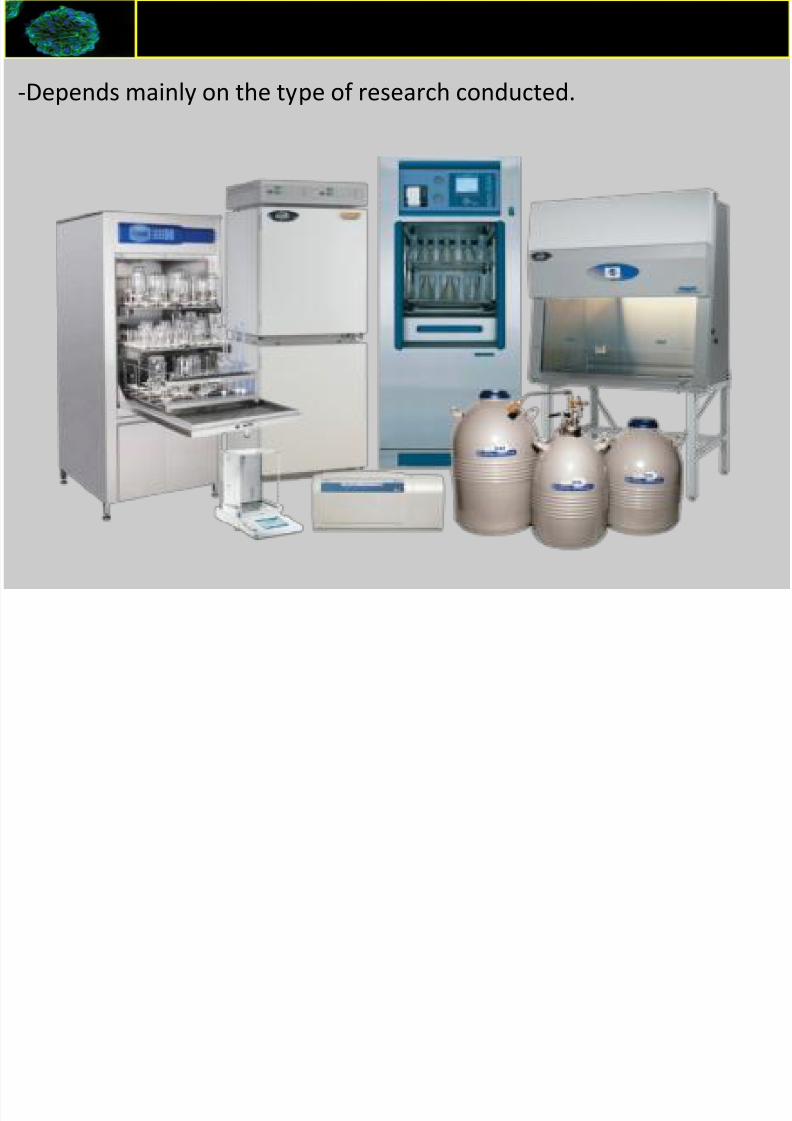
Equipments required for Cell Culture
-Depends mainly on the type of research conducted.

8/16/2019 Animal Cell Culture I
http://slidepdf.com/reader/full/animal-cell-culture-i 16/55
Basic Equipment
• Cell culture hood (i.e., laminar-flow hood or biosafety cabinet)
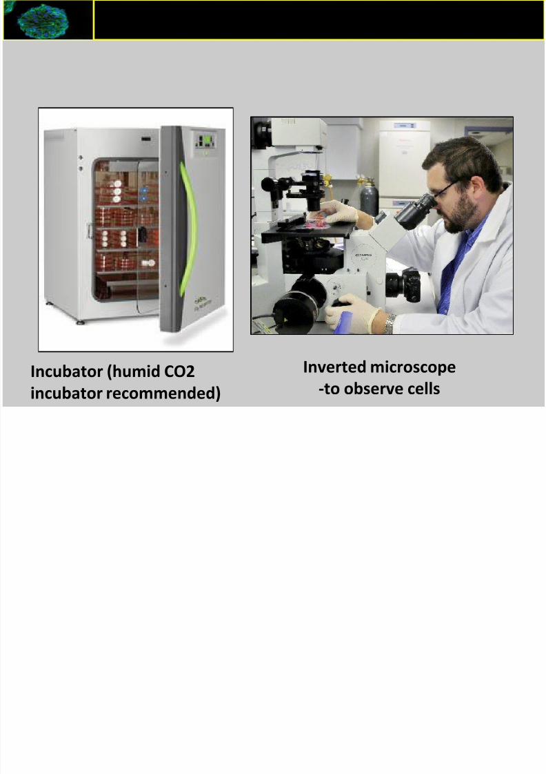
• Incubator (humid CO2 incubator recommended)
• Water bath
• Centrifuge
• Refrigerator and freezer (–20°C)
• Cell counter (e.g., CountessR Automated Cell Counter or
hemacytometer)
• Inverted microscope
• Liquid nitrogen (N2) freezer or cryostorage container• Sterilizer (i.e., autoclave)
Equipments required for Cell Culture
8/16/2019 Animal Cell Culture I
http://slidepdf.com/reader/full/animal-cell-culture-i 17/55
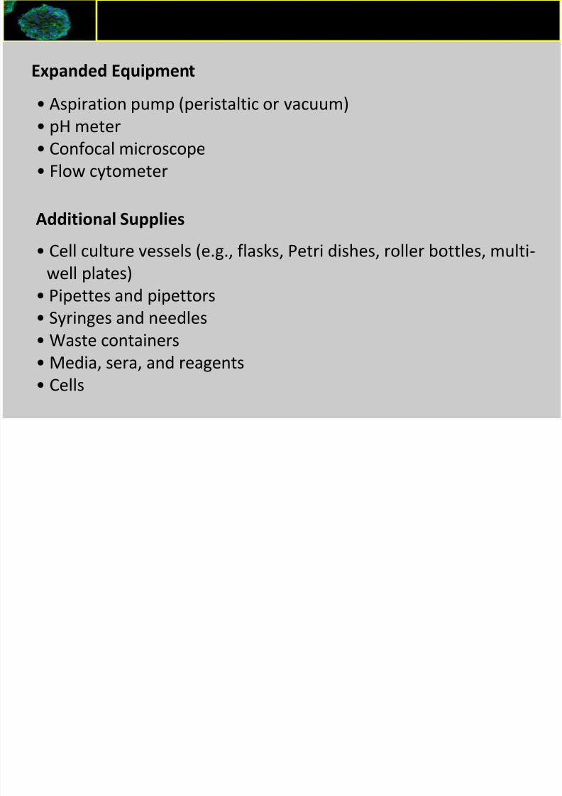
Expanded Equipment
• Aspiration pump (peristaltic or vacuum) • pH meter
• Confocal microscope
• Flow cytometer
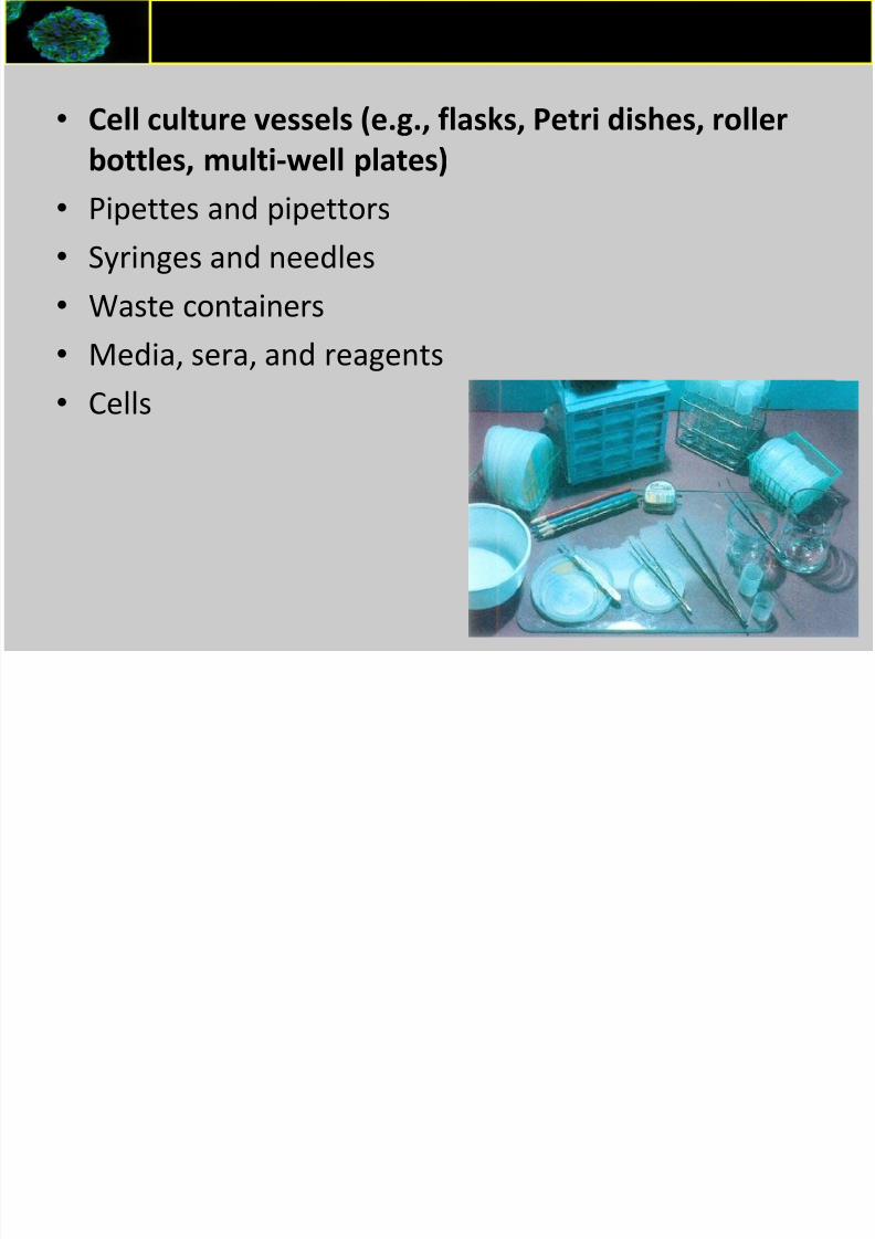
Additional Supplies
• Cell culture vessels (e.g., flasks, Petri dishes, roller bottles, multi-
well plates)
• Pipettes and pipettors• Syringes and needles
• Waste containers
• Media, sera, and reagents
• Cells
Equipments required for Cell Culture

8/16/2019 Animal Cell Culture I
http://slidepdf.com/reader/full/animal-cell-culture-i 18/55
f
8/16/2019 Animal Cell Culture I
http://slidepdf.com/reader/full/animal-cell-culture-i 19/55
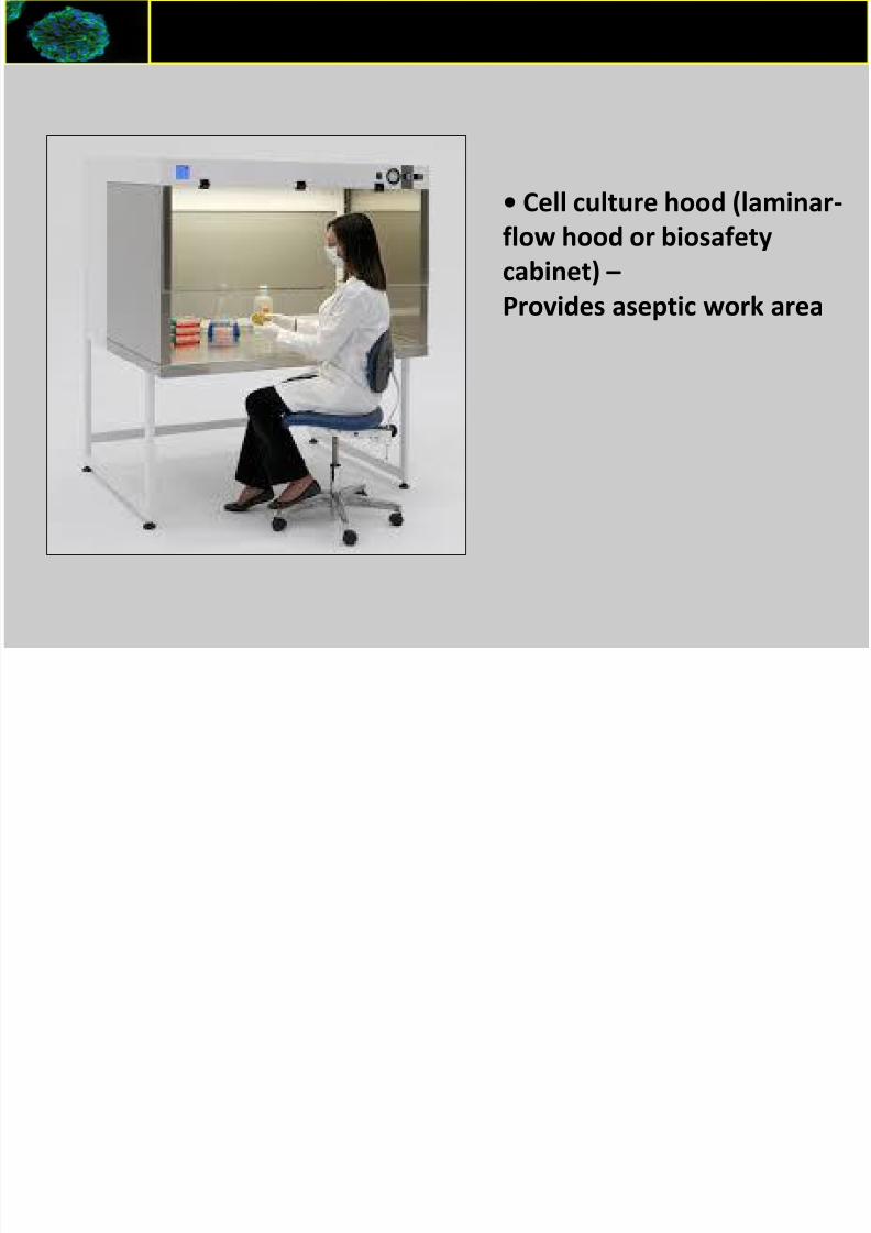
Equipments required for Cell Culture
• Cell culture hood (laminar-
flow hood or biosafety
cabinet) –
Provides aseptic work area

8/16/2019 Animal Cell Culture I
http://slidepdf.com/reader/full/animal-cell-culture-i 20/55
• Horizontal flow -provides protection to the culture (if the air flowing towards the
user) or to the user (if the air is drawn in through the front)
• Vertical flow –provides significant protection to the user and the cell culture.
Basic Layout of a culture hood

8/16/2019 Animal Cell Culture I
http://slidepdf.com/reader/full/animal-cell-culture-i 21/55
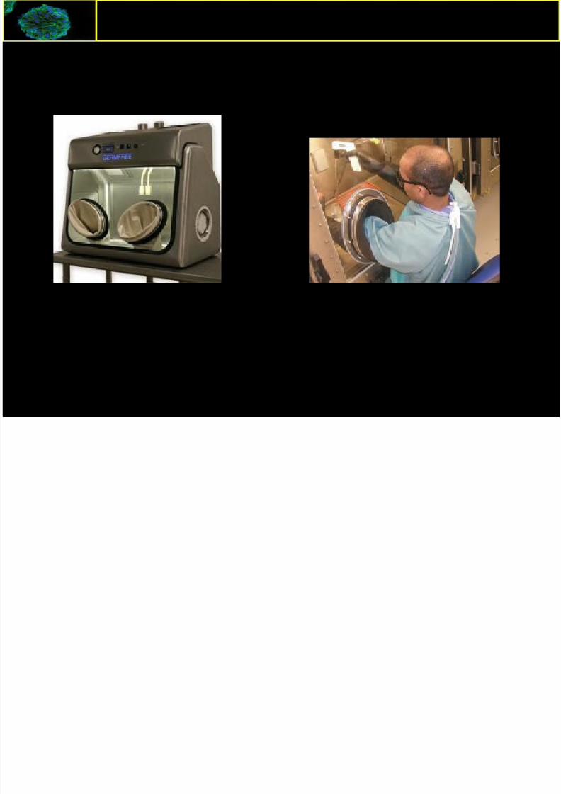
• Class I
an open-front negative pressure cabinet The exhaust air from
the cabinet is filtered by a high-efficiency particulate air(HEPA) filter. The Class I biosafety cabinet will providepersonnel and environmental protection, but not productprotection.
•Class IIan open-front, ventilated cabinet. This cabinet provides aHEPA-filtered, recirculated mass airflow within the workspace. The exhaust air from the cabinet is also filtered byHEPA filters. Thus, the Class II biosafety cabinet will provide
personnel, environment and product protection
• Class III
a totally enclosed ventilated cabinet of gas-tight construction.Operations within the Class III cabinet are conducted through
attached rubber gloves.
Bio safety Cabinets

8/16/2019 Animal Cell Culture I
http://slidepdf.com/reader/full/animal-cell-culture-i 22/55
• Class I
Bio safety Cabinets
f b

8/16/2019 Animal Cell Culture I
http://slidepdf.com/reader/full/animal-cell-culture-i 23/55
• Class II
Bio safety Cabinets
i f bi
8/16/2019 Animal Cell Culture I
http://slidepdf.com/reader/full/animal-cell-culture-i 24/55
• Class III
Bio safety Cabinets
8/16/2019 Animal Cell Culture I
http://slidepdf.com/reader/full/animal-cell-culture-i 25/55
I b
8/16/2019 Animal Cell Culture I
http://slidepdf.com/reader/full/animal-cell-culture-i 26/55
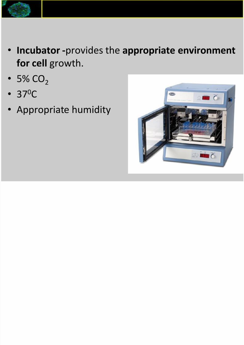
• Incubator -provides the appropriate environment
for cell growth.
• 5% CO2
• 370C
• Appropriate humidity
Incubator
E i t i d f C ll C lt

8/16/2019 Animal Cell Culture I
http://slidepdf.com/reader/full/animal-cell-culture-i 27/55
Other equipments
• Water bath
• Centrifuge
• Refrigerator and freezer
( –20°C)
• Cell counter (Automated
Cell Counter or
hemacytometer
• Liquid nitrogen (N2)
cryostorage container
• Sterilizer (i.e., autoclave)
Equipments required for Cell Culture

8/16/2019 Animal Cell Culture I
http://slidepdf.com/reader/full/animal-cell-culture-i 28/55
E i t i d f C ll C lt
8/16/2019 Animal Cell Culture I
http://slidepdf.com/reader/full/animal-cell-culture-i 29/55
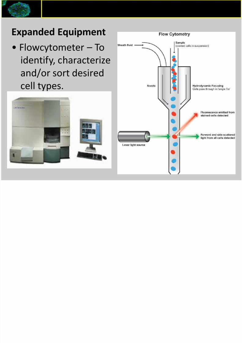
Expanded Equipment
• Flowcytometer – To
identify, characterize
and/or sort desired
cell types.
Equipments required for Cell Culture
8/16/2019 Animal Cell Culture I
http://slidepdf.com/reader/full/animal-cell-culture-i 30/55
Equipments required for Cell Culture
8/16/2019 Animal Cell Culture I
http://slidepdf.com/reader/full/animal-cell-culture-i 31/55
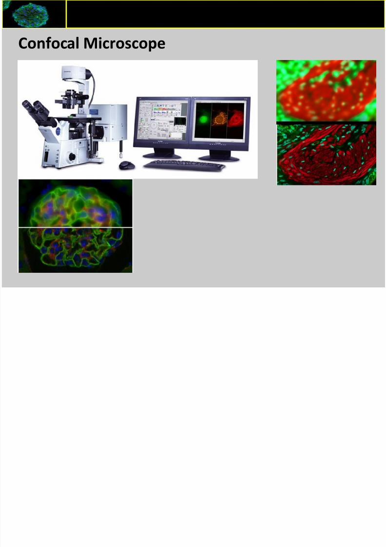
Confocal Microscope
Equipments required for Cell Culture
Additi l S li
8/16/2019 Animal Cell Culture I
http://slidepdf.com/reader/full/animal-cell-culture-i 32/55
Additional Supplies
• Cell culture vessels (e.g., flasks, Petri dishes, roller
bottles, multi-well plates)• Pipettes and pipettors
• Syringes and needles
•Waste containers
• Media, sera, and reagents
• Cells

8/16/2019 Animal Cell Culture I
http://slidepdf.com/reader/full/animal-cell-culture-i 33/55
C ti ll

8/16/2019 Animal Cell Culture I
http://slidepdf.com/reader/full/animal-cell-culture-i 34/55
Counting cells
Square # cells
1 5
2 6
3 5
4 8
Total No of Cells: 24
Total No of cells per square = 6
Total volume = 10 4
Dilution factor = D
Cell concentration = 6 X 10 4 X D / mL
C ti Vi bl ll
8/16/2019 Animal Cell Culture I
http://slidepdf.com/reader/full/animal-cell-culture-i 35/55
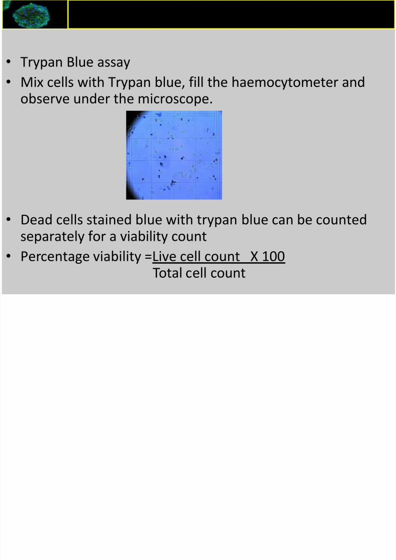
• Trypan Blue assay
• Mix cells with Trypan blue, fill the haemocytometer andobserve under the microscope.
• Dead cells stained blue with trypan blue can be countedseparately for a viability count
• Percentage viability =Live cell count X 100
Total cell count
Counting Viable cells
8/16/2019 Animal Cell Culture I
http://slidepdf.com/reader/full/animal-cell-culture-i 36/55

8/16/2019 Animal Cell Culture I
http://slidepdf.com/reader/full/animal-cell-culture-i 37/55
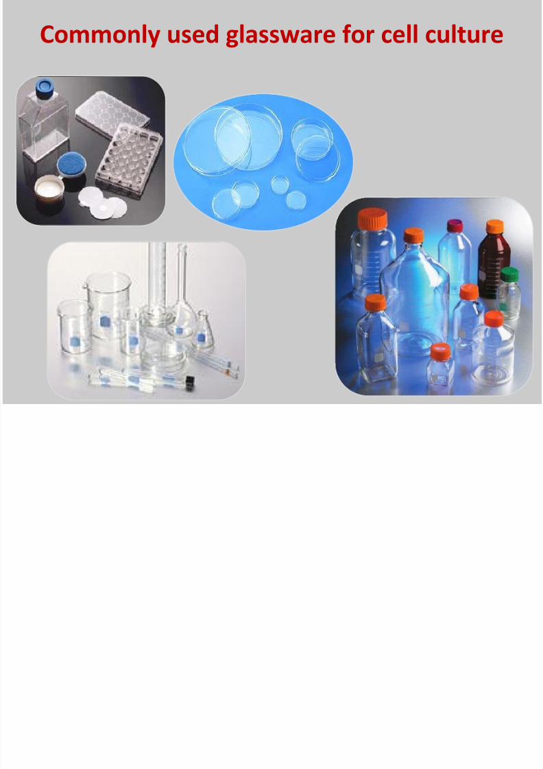
Cell culture vessels
• Flat-bottom and coated, cell
culture vessels are needed for cell
growth.
• Three types of culture vessels are
commonly used : flasks, dishes,and multiwell plates.
• All three types are available in
different sizes with different
surface areas.
• The choice of the vessel depends
on the nature of the procedures
and personal preference.

8/16/2019 Animal Cell Culture I
http://slidepdf.com/reader/full/animal-cell-culture-i 38/55
Culture Flasks
• PYREX glass flasks and Petri dishes
• Disposable plastic culture vessels are
commonly used
• Flasks, dishes and 96 well plates
made up of polystyrene, a longcarbon chain polymer with benzene
rings attached to every other carbon
are available commercially
• Polystyrene was chosen because it
has excellent optical clarity, easy to
mold and can be sterilized by
irradiation.

8/16/2019 Animal Cell Culture I
http://slidepdf.com/reader/full/animal-cell-culture-i 39/55
Tissue Culture Treated Flasks
• Drawback of polystyrene: it is a very
hydrophobic - cells have difficulty attaching.
• Hydrophobic polystyrene surface are modified
to a more hydrophilic surface.
• Corona discharge , extracellular matrix,
collagen, laminin and fibronectin, and heparin
sulfate, hyaluronidate and chondroitin sulfate,poly-D-lysine (PDL), as coatings
d d l l d

8/16/2019 Animal Cell Culture I
http://slidepdf.com/reader/full/animal-cell-culture-i 40/55
Vented and Plug sealed caps
Flasks with vented caps
are preferred, since non-
vented caps need to beloose when the flasks are
in the incubator to allow
for exchange of gases

8/16/2019 Animal Cell Culture I
http://slidepdf.com/reader/full/animal-cell-culture-i 41/55
-Commercially available disposableculture vessels are sterile
-No need to sterilize
-Transfer of media must be done using a
sterile pipettes
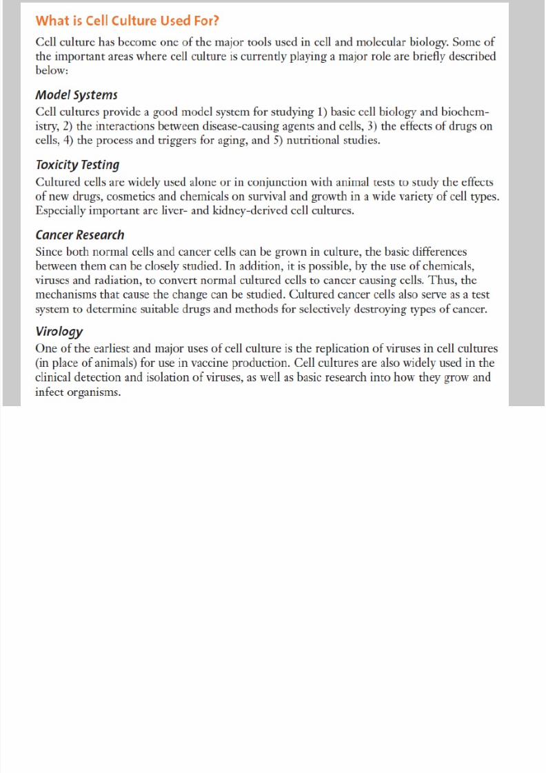
Applications of Cell Culture

8/16/2019 Animal Cell Culture I
http://slidepdf.com/reader/full/animal-cell-culture-i 42/55
Applications of Cell Culture
Study the effects of
drugs and toxiccompounds on the cells
Excellent model systems
for studying the normal
physiology and
biochemistry of cells (e.g.,
metabolic studies, aging)
Drug screening and
development
Large scale manufacturing
of biological compounds
(e.g., vaccines, therapeutic
proteins)
8/16/2019 Animal Cell Culture I
http://slidepdf.com/reader/full/animal-cell-culture-i 43/55

8/16/2019 Animal Cell Culture I
http://slidepdf.com/reader/full/animal-cell-culture-i 44/55

8/16/2019 Animal Cell Culture I
http://slidepdf.com/reader/full/animal-cell-culture-i 45/55
Advantages and disadvantages of cell culture
8/16/2019 Animal Cell Culture I
http://slidepdf.com/reader/full/animal-cell-culture-i 46/55
• The major advantage of using cell culture is the
consistency and reproducibility of results that can be
obtained from using a batch of clonal cells.
• Represent the best experimental models for in vivo
situations.• Can have the same karyotype as the parent tissue
normal or abnormal.
• Can maintain cells in “undedifferentiated” states.
Advantages and disadvantages of cell culture
Advantages and disadvantages of cell culture

8/16/2019 Animal Cell Culture I
http://slidepdf.com/reader/full/animal-cell-culture-i 47/55
Advantages and disadvantages of cell culture
• Control of the environment
• Characterization and homogeneity of the samples
• In vitro modeling of in vivo conditions
• Economy, scale and mechanization of culture
• Avoid animal experiments
• Mimic of in vivo cell behavior (e.g cancer cells)

8/16/2019 Animal Cell Culture I
http://slidepdf.com/reader/full/animal-cell-culture-i 48/55
Cell Culture associated problems
8/16/2019 Animal Cell Culture I
http://slidepdf.com/reader/full/animal-cell-culture-i 49/55
Two main categories
1. Chemical contaminants such as impurities in
media, sera, and water, endotoxins, etc.
2. Biological contaminants such as bacteria, molds,
yeasts, viruses, mycoplasma, as well as cross
contamination by other cell lines.
It is essential to have understanding of the sources of
contaminants and follow good aseptic technique.
Cell Culture associated problems
Cell culture contaminations - Bacteria
8/16/2019 Animal Cell Culture I
http://slidepdf.com/reader/full/animal-cell-culture-i 50/55
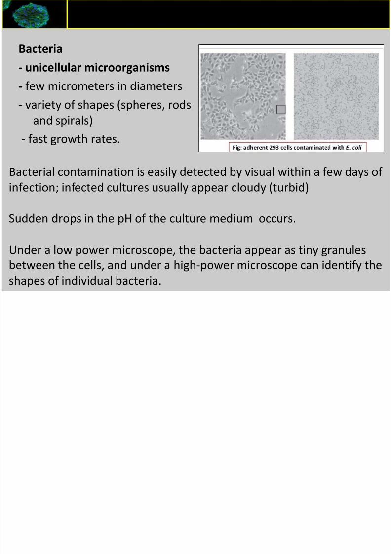
Bacteria
- unicellular microorganisms- few micrometers in diameters
- variety of shapes (spheres, rods
and spirals)
- fast growth rates.
Cell culture contaminations - Bacteria
Bacterial contamination is easily detected by visual within a few days of
infection; infected cultures usually appear cloudy (turbid)
Sudden drops in the pH of the culture medium occurs.
Under a low power microscope, the bacteria appear as tiny granules
between the cells, and under a high-power microscope can identify the
shapes of individual bacteria.
Cell culture contaminations - Yeast

8/16/2019 Animal Cell Culture I
http://slidepdf.com/reader/full/animal-cell-culture-i 51/55
• Yeasts - unicellular eukaryotic microorganisms (Fungi)
• cultures become turbid, especially if the contamination isin an advanced stage.
• little change in the pH of the culture at the initiail stages.
•
Under microscopy, yeast appear as individual ovoid orspherical particles, that may bud off smaller particles.
Cell culture contaminations - Yeast
Cell culture contaminations - Yeast

8/16/2019 Animal Cell Culture I
http://slidepdf.com/reader/full/animal-cell-culture-i 52/55
Cell culture contaminations Yeast
Cell culture contaminations - Molds

8/16/2019 Animal Cell Culture I
http://slidepdf.com/reader/full/animal-cell-culture-i 53/55
• Molds -eukaryotic microorganisms (Fungi)
-grow as multicellular filaments called hyphae
-the pH of the culture remains stable in the initial stages
of contamination
•
Under microscopy, the mycelia usually appear as thin,wisp-like filaments, and sometimes as denser clumps of
spores.
Cell culture contaminations Molds
•Spores of many
mold species can
survive extremely
harsh
environments
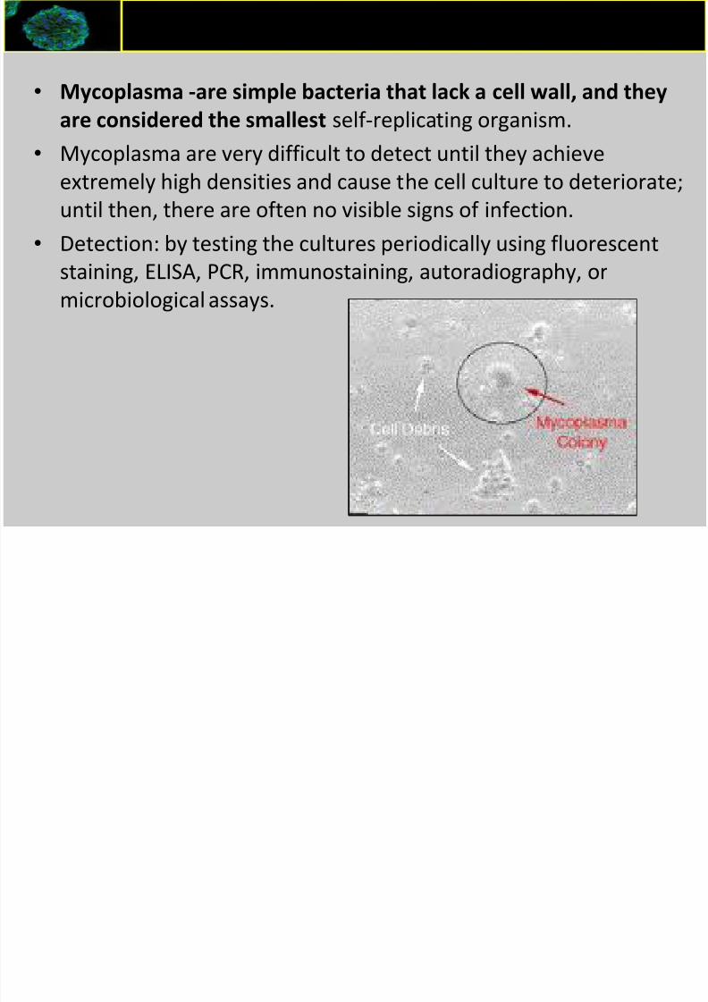
Cell culture contaminations -Mycoplasma
8/16/2019 Animal Cell Culture I
http://slidepdf.com/reader/full/animal-cell-culture-i 54/55
• Mycoplasma -are simple bacteria that lack a cell wall, and they
are considered the smallest self-replicating organism.• Mycoplasma are very difficult to detect until they achieve
extremely high densities and cause the cell culture to deteriorate;
until then, there are often no visible signs of infection.
•
Detection: by testing the cultures periodically using fluorescentstaining, ELISA, PCR, immunostaining, autoradiography, or
microbiological assays.
Cell culture contaminations Mycoplasma
Cell culture contaminations - Viruses

8/16/2019 Animal Cell Culture I
http://slidepdf.com/reader/full/animal-cell-culture-i 55/55
• Viruses -microscopic infectious agents that take over the
host cells machinery to reproduce. T
• heir extremely small size makes them very difficult to detect
in culture, and to remove them from reagents used in cell
culture laboratories. can be a serious health hazard to the
laboratory• Viral infection of cell cultures can be detected by electron
microscopy, immunostaining with a panel of antibodies, ELISA
assays, or PCR with appropriate viral primers.
Cell culture contaminations Viruses