anatomy of the heart blood pressure, blood flow and … pressure, blood flow ... • similar to...
TRANSCRIPT
1
Blood Pressure, Blood Flowand Volume of Blood
Achmad RizalBioSPIN
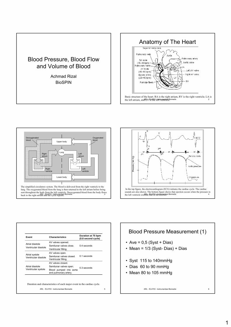
ARL - EL4703 - Isntrumentasi Biomedis 2Basic structure of the heart. RA is the right atrium, RV is the right ventricle; LA is the left atrium, and LV is the left ventricle.
Anatomy of The Heart
ARL - EL4703 - Isntrumentasi Biomedis 3
Lower body
Upper body
Leftatrium
Rightatrium
Leftventicle
Rightventicle
Oxygenatedblood
Deoxygenatedblood
Lung
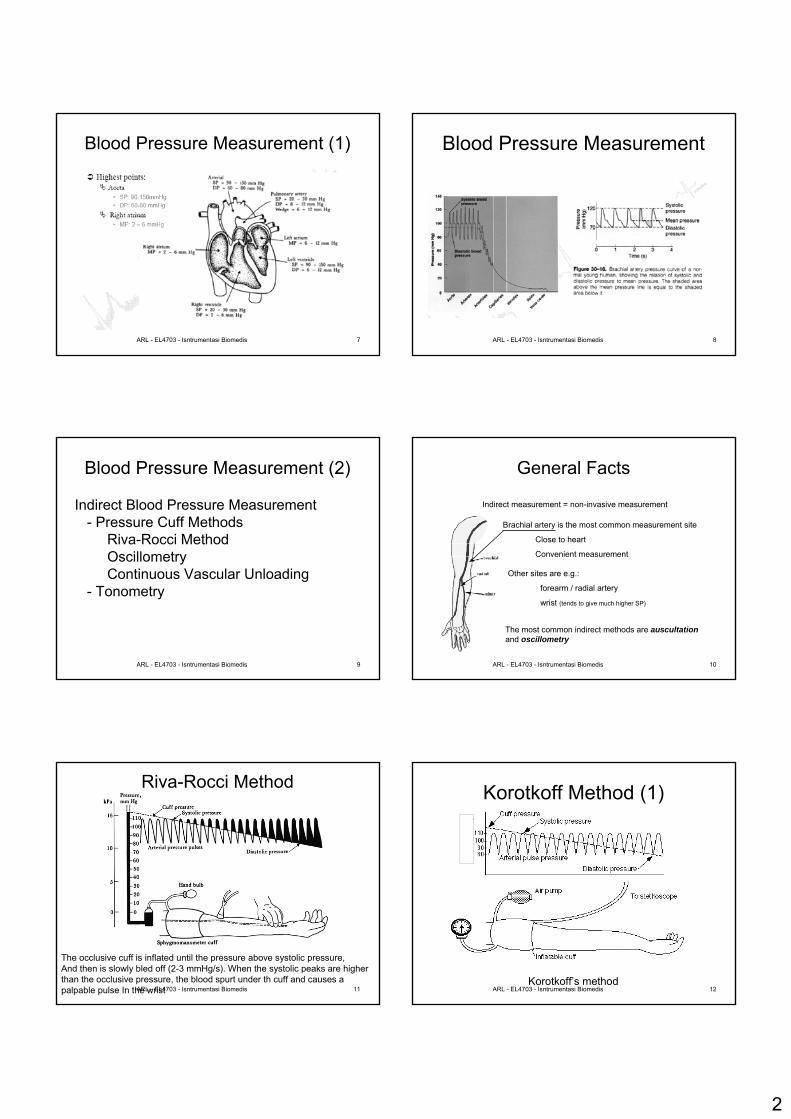
The simplified circulatory system. The blood is delivered from the right ventricle to the lung. The oxygenated blood from the lung is then returned to the left atrium before being sent throughout the body from the left ventricle. Deoxygenated blood from the body flows back to the right atrium and the cycle repeats. ARL - EL4703 - Isntrumentasi Biomedis 4
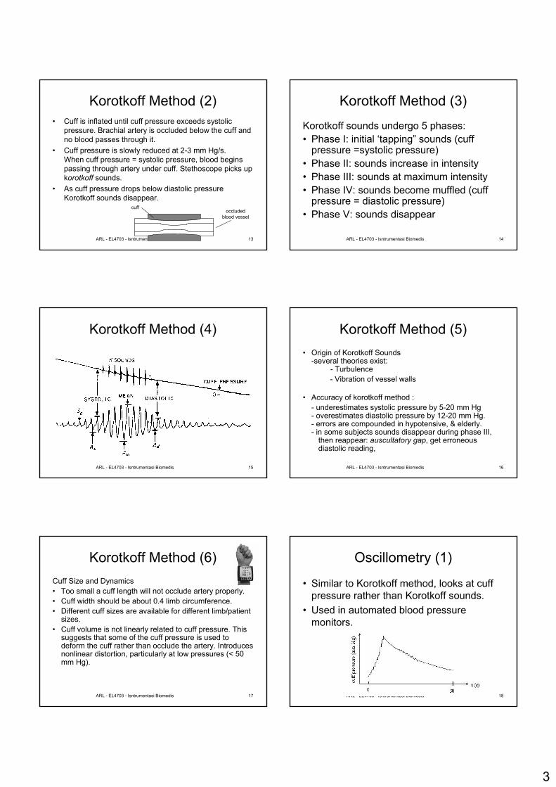
In the top figure, the electrocardiogram (ECG) initiates the cardiac cycle. The cardiac sounds are also shown. The bottom figure shows that ejection occurs when the pressure in the left ventricle exceeds that in the arteries.
ARL - EL4703 - Isntrumentasi Biomedis 5
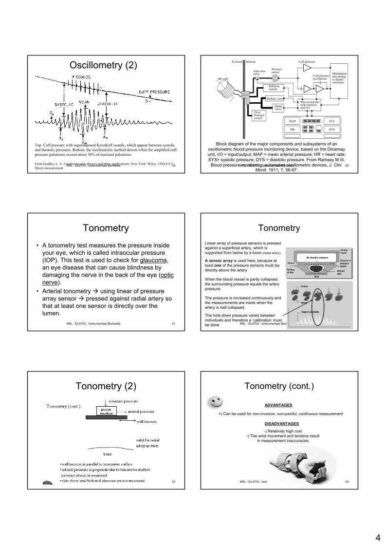
Duration and characteristics of each major event in the cardiac cycle.
AV valves closed.Semilunar valves open.Blood pumped into aorta and pulmonary artery.
AV valves open.Semilunar valves closed. Ventricular filling.
AV valves opened.Semilunar valves close.Ventricular filling.
Characteristics
0.3 seconds Atrial diastoleVentricular systole
0.1 seconds Atrial systoleVentricular diastole
0.4 seconds Atrial diastoleVentricular diastole
Duration at 75 bpm(0.8 second cycle) Event
ARL - EL4703 - Isntrumentasi Biomedis 6
Blood Pressure Measurement (1)
• Ave = 0,5 (Syst + Dias)• Mean = 1/3 (Syst- Dias) + Dias
• Syst 115 to 140mmHg• Dias 60 to 90 mmHg• Mean 80 to 105 mmHg
2
ARL - EL4703 - Isntrumentasi Biomedis 7
Blood Pressure Measurement (1)
ARL - EL4703 - Isntrumentasi Biomedis 8
Blood Pressure Measurement
ARL - EL4703 - Isntrumentasi Biomedis 9
Blood Pressure Measurement (2)
Indirect Blood Pressure Measurement- Pressure Cuff Methods
Riva-Rocci MethodOscillometryContinuous Vascular Unloading
- Tonometry
ARL - EL4703 - Isntrumentasi Biomedis 10
General Facts
Indirect measurement = non-invasive measurement
Brachial artery is the most common measurement site
Close to heart
Convenient measurement
Other sites are e.g.:
forearm / radial artery
wrist (tends to give much higher SP)
The most common indirect methods are auscultationand oscillometry
ARL - EL4703 - Isntrumentasi Biomedis 11
Riva-Rocci Method
The occlusive cuff is inflated until the pressure above systolic pressure,And then is slowly bled off (2-3 mmHg/s). When the systolic peaks are higher than the occlusive pressure, the blood spurt under th cuff and causes a palpable pulse In the wrist ARL - EL4703 - Isntrumentasi Biomedis 12
Korotkoff Method (1)
Korotkoff’s method
3
ARL - EL4703 - Isntrumentasi Biomedis 13
Korotkoff Method (2)• Cuff is inflated until cuff pressure exceeds systolic
pressure. Brachial artery is occluded below the cuff and no blood passes through it.
• Cuff pressure is slowly reduced at 2-3 mm Hg/s.When cuff pressure = systolic pressure, blood beginspassing through artery under cuff. Stethoscope picks upkorotkoff sounds.
• As cuff pressure drops below diastolic pressure Korotkoff sounds disappear.
cuffoccluded
blood vessel
ARL - EL4703 - Isntrumentasi Biomedis 14
Korotkoff Method (3)
Korotkoff sounds undergo 5 phases:• Phase I: initial ‘tapping” sounds (cuff
pressure =systolic pressure)• Phase II: sounds increase in intensity• Phase III: sounds at maximum intensity• Phase IV: sounds become muffled (cuff
pressure = diastolic pressure)• Phase V: sounds disappear
ARL - EL4703 - Isntrumentasi Biomedis 15
Korotkoff Method (4)
ARL - EL4703 - Isntrumentasi Biomedis 16
Korotkoff Method (5)• Origin of Korotkoff Sounds
-several theories exist:- Turbulence- Vibration of vessel walls
• Accuracy of korotkoff method :- underestimates systolic pressure by 5-20 mm Hg- overestimates diastolic pressure by 12-20 mm Hg.- errors are compounded in hypotensive, & elderly.- in some subjects sounds disappear during phase III,
then reappear: auscultatory gap, get erroneous diastolic reading,
ARL - EL4703 - Isntrumentasi Biomedis 17
Korotkoff Method (6)Cuff Size and Dynamics• Too small a cuff length will not occlude artery properly.• Cuff width should be about 0.4 limb circumference.• Different cuff sizes are available for different limb/patient
sizes.• Cuff volume is not linearly related to cuff pressure. This
suggests that some of the cuff pressure is used to deform the cuff rather than occlude the artery. Introduces nonlinear distortion, particularly at low pressures (< 50 mm Hg).
ARL - EL4703 - Isntrumentasi Biomedis 18
Oscillometry (1)
• Similar to Korotkoff method, looks at cuff pressure rather than Korotkoff sounds.
• Used in automated blood pressuremonitors.
4
ARL - EL4703 - Isntrumentasi Biomedis 19
Top: Cuff pressure with superimposed Korotkoff sounds, which appear between systolic and diastolic pressures. Bottom: the oscillometric method detects when the amplified cuff pressure pulsations exceed about 30% of maximal pulsations.
From Geddes, L. A. Cardiovascular devices and their applications. New York: Wiley. 1984.8.9.2 Direct measurement
Oscillometry (2)
ARL - EL4703 - Isntrumentasi Biomedis 20
Block diagram of the major components and subsystems of an oscillometric blood-pressure monitoring device, based on the Dinamapunit, I/O = input/output; MAP = mean arterial pressure; HR = heart rate; SYS= systolic pressure; DYS = diastolic pressure. From Ramsey M III.
Blood pressure monitoring: automated oscillometric devices, J. Clin. Monit. 1911, 7, 56-67.
MAP
HR
SYS
Microcomputerwith memoryand I/O
Multiplexerand analogto digitalconverter
Deflate valve
Dumpvalve
Over-Pressureswitch
Auto-zerovalve
Pressuresensor
Cuff pressure
Cuff pressureoscillations
InternalExternal
BP cuff
Inflationsystem
DYS
ARL - EL4703 - Isntrumentasi Biomedis 21
Tonometry
• A tonometry test measures the pressure inside your eye, which is called intraocular pressure (IOP). This test is used to check for glaucoma, an eye disease that can cause blindness by damaging the nerve in the back of the eye (optic nerve).
• Arterial tonometry using linear of pressure array sensor pressed against radial artery so that at least one sensor is directly over the lumen.
ARL - EL4703 - Isntrumentasi Biomedis 22
Tonometry
A sensor array is used here, because at least one of the pressure sensors must laydirectly above the artery
Linear array of pressure sensors is pressedagainst a superficial artery, which is supported from below by a bone (radial artery).
The pressure is increased continuously and the measurements are made when the artery is half collapsed
When the blood vessel is partly collapsed, the surrounding pressure equals the arterypressure.
The hold-down pressure varies betweenindividuals and therefore a ’calibration’ mustbe done
ARL - EL4703 - Isntrumentasi Biomedis 23
Tonometry (2)
ARL - EL4703 - Isntrumentasi Biomedis 24
Tonometry (cont.)
ADVANTAGES
+) Can be used for non-invasive, non-painful, continuous measurement
DISADVANTAGES
-) Relatively high cost-) The wrist movement and tendons result
in measurement inaccuracies

5
ARL - EL4703 - Isntrumentasi Biomedis 25
Blood Pressure Direct Measurement
• Arterial & venous blood pressure can be measured by inserting a catheter into the blood vessel and maneuvering it until the end is at the site at which the blood pressure is to be measured
• Catheter can be inserted into the artery inside a needle
• The alternative method catheter tip sensor• Very invasive procedure
ARL - EL4703 - Isntrumentasi Biomedis 26
Blood Pressure Direct Measurement
• Sterile saline filled catheter is inserted into blood stream
• The pressure at the tip of catheter is transmitted to an external pressure transducer
• Some blood vessel :Artery: carotid, brachial, femoralVeins : femoral, brachial, subcalian, internal jugulas, CVP central venous pressureRAP right atrium pressure
ARL - EL4703 - Isntrumentasi Biomedis 27
Figure 7.3 Extravascular pressure-sensor system A catheter couples a flush solution (heparinized saline) through a disposable pressure sensor with an integral flush device to the sensing port. The three-way stopcock is used to take blood samples and
zero the pressure sensor.
Sensing port
Roller clamp
Flush solution under pressure
Sample and transducerzero stopcock
Electrical connectorDisposable pressure transducer with an integral flush device
Blood Pressure Direct Measurement
ARL - EL4703 - Isntrumentasi Biomedis 28
Blood Pressure Direct Measurement
Fluid resitance how fluid flows in a catheter, flow f
p = RCf p = p1 – p2p = pressure (N/m2), p1, p2 = gauge pressure at two specific
pointRC = resitance of the tube (Ns/m5)f = flow (Kg/s)
ARL - EL4703 - Isntrumentasi Biomedis 29
Blood Pressure Direct Measurement
• Gauge pressure pressure relative to atmospheric pressure
• If f = 0 , p1 = p2 , in other word, when the flow in a closed tube is zero, pressure at the input equal the pressure at the output
• Tube resistanceRC = 8ηl/(πr4)
η = viscosity, l = tube length, r = radius
ARL - EL4703 - Isntrumentasi Biomedis 30
Blood Flow (2)
Normal blood flow velocity 0,5 m/s – 1 m/s (Systolic, large vessel)
6
ARL - EL4703 - Isntrumentasi Biomedis 31
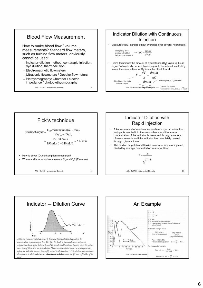
Blood Flow Measurement
How to make blood flow / volumemeasurements? Standard flow meters, such as turbine flow meters, obviously cannot be used!– Indicator-dilution method: cont./rapid injection,
dye dilution, thermodilution– Electromagnetic flowmeters– Ultrasonic flowmeters / Doppler flowmeters– Plethysmography: Chamber / electric
impedance / photoplethysmographyARL - EL4703 - Isntrumentasi Biomedis 32
Indicator Dilution with Continuous Injection
• Measures flow / cardiac output averaged over several heart beats
• Fick’s technique: the amount of a substance (O2) taken up by an organ / whole body per unit time is equal to the arterial level of O2minus the venous level of O2 times the blood flow
va CCdtdm
Cdtdm
dtdVF
−=
Δ==
Blood flow, liters/min(cardiac output)
Consumption of O2 (mL/min)
Arterial and venousconcentration of O2 (mL/L of blood)
dtdVdtdmC =Δ
Change in [] due to continuously added indicator m to volume V
ARL - EL4703 - Isntrumentasi Biomedis 33
Fick’s technique
• How is dm/dt (O2 consumption) measured?• Where and how would we measure Ca and Cv? (Exercise)
minL/5mL/L140L/mL190
minmL/250][O][O
min)(mL/O
22
2
=−
=
−=
va
nconsumptioOutputCardiac
ARL - EL4703 - Isntrumentasi Biomedis 34
Indicator Dilution with Rapid Injection
• A known amount of a substance, such as a dye or radioactive isotope, is injected into the venous blood and the arterial concentration of the indicator is measured through a serious of measurements until the indicator has completely passed through given volume.
• The cardiac output (blood flow) is amount of indicator injected,divided by average concentration in arterial blood.
∫= t
dttC
mF
0)(
ARL - EL4703 - Isntrumentasi Biomedis 35
Indicator – Dilution Curve
After the bolus is injected at time A, there is a transportation delay before the concentration begins rising at time B. After the peak is passed, the curve enters an exponential decay region between C and D, which would continue decaying alone the dotted curve to t1 if there were no recirculation. However, recirculation causes a second peak at E before the indicator becomes thoroughly mixed in the blood at F. The dashed curve indicates the rapid recirculation that occurs when there is a hole between the left and right sides of the heart.
ARL - EL4703 - Isntrumentasi Biomedis 36
An Example
7
ARL - EL4703 - Isntrumentasi Biomedis 37
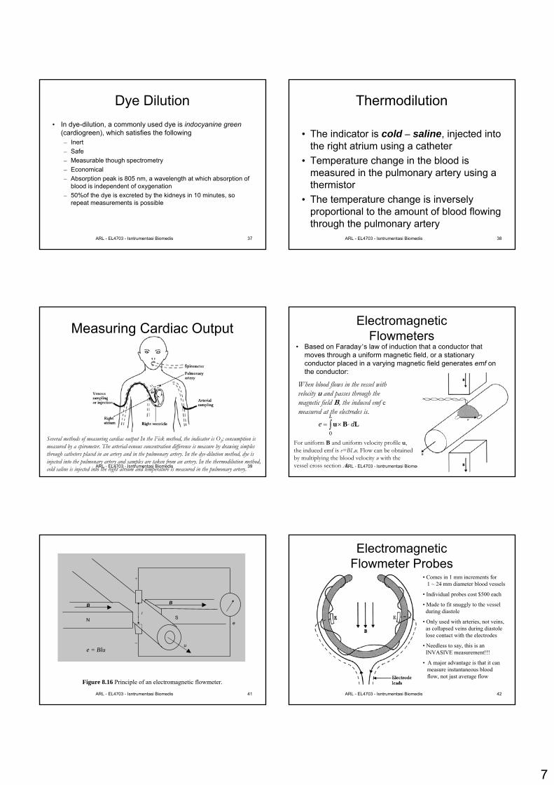
Dye Dilution• In dye-dilution, a commonly used dye is indocyanine green
(cardiogreen), which satisfies the following– Inert– Safe– Measurable though spectrometry– Economical– Absorption peak is 805 nm, a wavelength at which absorption of
blood is independent of oxygenation– 50%of the dye is excreted by the kidneys in 10 minutes, so
repeat measurements is possible
ARL - EL4703 - Isntrumentasi Biomedis 38
Thermodilution
• The indicator is cold – saline, injected into the right atrium using a catheter
• Temperature change in the blood is measured in the pulmonary artery using a thermistor
• The temperature change is inversely proportional to the amount of blood flowing through the pulmonary artery
ARL - EL4703 - Isntrumentasi Biomedis 39
Measuring Cardiac Output
Several methods of measuring cardiac output In the Fick method, the indicator is O2; consumption is measured by a spirometer. The arterial-venous concentration difference is measure by drawing simples through catheters placed in an artery and in the pulmonary artery. In the dye-dilution method, dye is injected into the pulmonary artery and samples are taken from an artery. In the thermodilution method, cold saline is injected into the right atrium and temperature is measured in the pulmonary artery. ARL - EL4703 - Isntrumentasi Biomedis 40
Electromagnetic Flowmeters
• Based on Faraday’s law of induction that a conductor that moves through a uniform magnetic field, or a stationary conductor placed in a varying magnetic field generates emf on the conductor:
When blood flows in the vessel with velocity u and passes through the magnetic field B, the induced emf emeasured at the electrodes is.
∫ ⋅×=L
de0
LBu
For uniform B and uniform velocity profile u, the induced emf is e=BLu. Flow can be obtained by multiplying the blood velocity u with the vessel cross section A.
ARL - EL4703 - Isntrumentasi Biomedis 41
B
N S
B
u −
+
e
l
Figure 8.16 Principle of an electromagnetic flowmeter.
e = Blu
ARL - EL4703 - Isntrumentasi Biomedis 42
ElectromagneticFlowmeter Probes
• Comes in 1 mm increments for 1 ~ 24 mm diameter blood vessels
• Individual probes cost $500 each
• Made to fit snuggly to the vessel during diastole
• Only used with arteries, not veins, as collapsed veins during diastole lose contact with the electrodes
• Needless to say, this is an INVASIVE measurement!!!
• A major advantage is that it can measure instantaneous blood flow, not just average flow

8
ARL - EL4703 - Isntrumentasi Biomedis 43
Ultrasonic Flowmeters• Based on the principle of measuring the time it takes for an
acoustic wave launched from a transducer to bounce off red blood cells and reflect back to the receiver.
• All UT transducers, whether used for flowmeter or other applications, invariably consists of a piezoelectric material, which generates an acoustic (mechanical) wave when excited by an electrical force (the converse is also true)
• UT transducers are typically used with a gel that fills the air gaps between the transducer and the object examined
ARL - EL4703 - Isntrumentasi Biomedis 44
Near / Far Fields• Due to finite diameters, UT transducers produce diffraction
patterns, just like an aperture does in optics.• This creates near and far fields of the UT transducer, in which
the acoustic wave exhibit different properties– The near field extends about dnf=D2/4λ, where D is the
transducer diameter and λ is the wavelength. During this region, the beam is mostly cylindrical (with little spreading), however with nonuniform intensity.
– In the far field, the beam diverges with an angle sinθ=1.2 λ/D, but the intensity uniformly attenuates proportional to the square of the distance from the transducer
Higher frequencies and larger transducers should be used for nearfield operation. Typical operating frequency is 2 ~ 10 MHz.
ARL - EL4703 - Isntrumentasi Biomedis 45
Figure 8.17 Ultrasonic flowmeter. The sensor at the scan head transmits the signal from the oscillator and receives the reflected wave from the blood cells. The RF (radio frequency) amplifier amplifies the received signal and the carrier frequency, then AF (audio frequency) signal is produced by a detector. Adapted from Picot, P. A. and Fenster, A. 1996. Ultrasonic blood volume flow rate meter. US Patent, 5,505,204.
Blood vessel
Doppler angle
Scan head
Skin surface Image plane
RF amplifier
Detector AF
amplifier F/V
converter
Computer
Oscillator
ARL - EL4703 - Isntrumentasi Biomedis 46
UT Flowmeters
Zero-crossingdetector / LPF
Determine direction
High acoustic impedance material
ARL - EL4703 - Isntrumentasi Biomedis 47
Transit time flowmetersEffective velocity of sound in blood: velocity of sound (c) +velocity of flow of blood averaged along the path of the ultrasound (û)
û=1.33ū for laminar flow, û=1.07ū for turbulent flowū: velocity of blood averaged over the cross sectional area, this is differentthan û because the UT path is along a single line not over an averaged of cross sectional area
Transit time in up/down stream direction:
Difference between upstream and downstream directions
θcosˆvelocityconductiondistance
ucDt
±==
2222cosˆ2
)cosˆ(cosˆ2
cuD
ucuDt θ
θθ
≅−
=Δ
ARL - EL4703 - Isntrumentasi Biomedis 48
Transit Time Flowmeters
The quantity ∆T is typically very small and very difficult to measure, particularly in the presence of noise. Therefore phase detection techniques are usually employed rather then measuring actual timing.
9
ARL - EL4703 - Isntrumentasi Biomedis 49
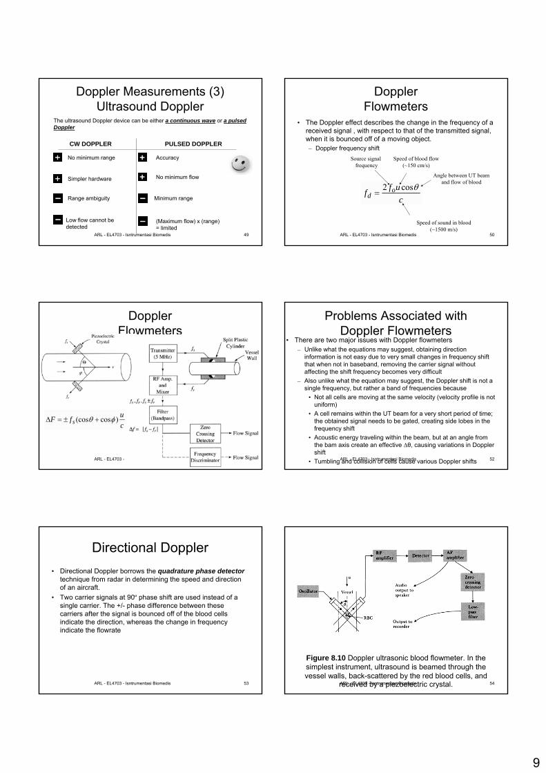
Doppler Measurements (3) Ultrasound Doppler
The ultrasound Doppler device can be either a continuous wave or a pulsedDoppler
CW DOPPLER PULSED DOPPLER
Range ambiguity
Low flow cannot bedetected
No minimum range
Simpler hardware
Minimum range
Accuracy
No minimum flow
(Maximum flow) x (range)= limited
ARL - EL4703 - Isntrumentasi Biomedis 50
Doppler Flowmeters
• The Doppler effect describes the change in the frequency of a received signal , with respect to that of the transmitted signal, when it is bounced off of a moving object.– Doppler frequency shift
cuff o
dθcos2
=
Speed of sound in blood(~1500 m/s)
Angle between UT beamand flow of blood
Speed of blood flow(~150 cm/s)
Source signal frequency
ARL - EL4703 - Isntrumentasi Biomedis 51
Doppler Flowmeters
cufF s )cos(cos φθ +±=Δ
ARL - EL4703 - Isntrumentasi Biomedis 52
Problems Associated withDoppler Flowmeters
• There are two major issues with Doppler flowmeters– Unlike what the equations may suggest, obtaining direction
information is not easy due to very small changes in frequency shift that when not in baseband, removing the carrier signal without affecting the shift frequency becomes very difficult
– Also unlike what the equation may suggest, the Doppler shift is not a single frequency, but rather a band of frequencies because
• Not all cells are moving at the same velocity (velocity profile is not uniform)
• A cell remains within the UT beam for a very short period of time; the obtained signal needs to be gated, creating side lobes in the frequency shift
• Acoustic energy traveling within the beam, but at an angle from the bam axis create an effective ∆θ, causing variations in Doppler shift
• Tumbling and collision of cells cause various Doppler shifts
ARL - EL4703 - Isntrumentasi Biomedis 53
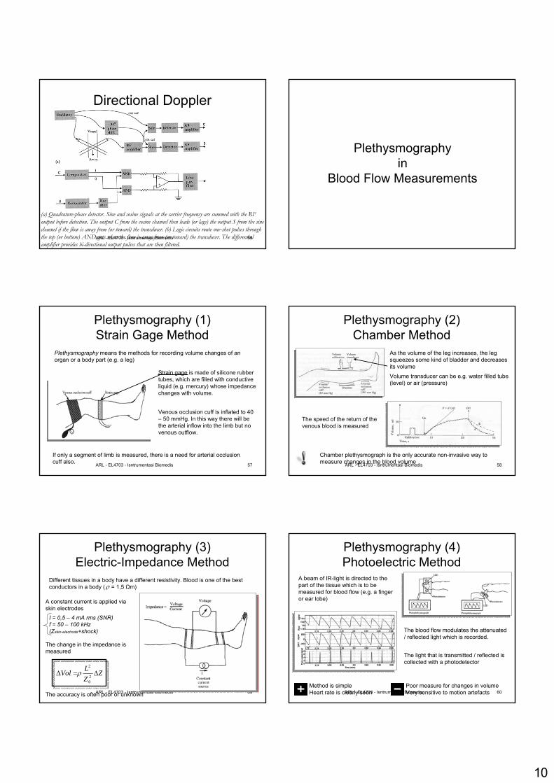
Directional Doppler• Directional Doppler borrows the quadrature phase detector
technique from radar in determining the speed and direction of an aircraft.
• Two carrier signals at 90º phase shift are used instead of a single carrier. The +/- phase difference between these carriers after the signal is bounced off of the blood cells indicate the direction, whereas the change in frequency indicate the flowrate
ARL - EL4703 - Isntrumentasi Biomedis 54
Figure 8.10 Doppler ultrasonic blood flowmeter. In the simplest instrument, ultrasound is beamed through the vessel walls, back-scattered by the red blood cells, and
received by a piezoelectric crystal.
10
ARL - EL4703 - Isntrumentasi Biomedis 55
Directional Doppler
(a) Quadrature-phase detector. Sine and cosine signals at the carrier frequency are summed with the RF output before detection. The output C from the cosine channel then leads (or lags) the output S from the sine channel if the flow is away from (or toward) the transducer. (b) Logic circuits route one-shot pulses through the top (or bottom) AND gate when the flow is away from (or toward) the transducer. The differential amplifier provides bi-directional output pulses that are then filtered.
Plethysmographyin
Blood Flow Measurements
ARL - EL4703 - Isntrumentasi Biomedis 57
Plethysmography (1)Strain Gage Method
Plethysmography means the methods for recording volume changes of an organ or a body part (e.g. a leg)
Strain gage is made of silicone rubbertubes, which are filled with conductiveliquid (e.g. mercury) whose impedancechanges with volume.
Venous occlusion cuff is inflated to 40 – 50 mmHg. In this way there will bethe arterial inflow into the limb but no venous outflow.
If only a segment of limb is measured, there is a need for arterial occlusioncuff also. ARL - EL4703 - Isntrumentasi Biomedis 58
Plethysmography (2)Chamber Method
Chamber plethysmograph is the only accurate non-invasive way to measure changes in the blood volume
As the volume of the leg increases, the legsqueezes some kind of bladder and decreasesits volume
Volume transducer can be e.g. water filled tube(level) or air (pressure)
The speed of the return of the venous blood is measured
ARL - EL4703 - Isntrumentasi Biomedis 59
Plethysmography (3)Electric-Impedance Method
Different tissues in a body have a different resistivity. Blood is one of the bestconductors in a body ( = 1,5 Ωm)ρ
A constant current is applied via skin electrodes
The change in the impedance is measured
The accuracy is often poor or unknown
I = 0,5 – 4 mA rms (SNR)f = 50 – 100 kHz (Zskin-electrode+shock)
ZZLVol Δ=Δ 2
0
2
ρ
ARL - EL4703 - Isntrumentasi Biomedis 60
Plethysmography (4)Photoelectric Method
A beam of IR-light is directed to the part of the tissue which is to bemeasured for blood flow (e.g. a fingeror ear lobe)
The light that is transmitted / reflected is collected with a photodetector
The blood flow modulates the attenuated/ reflected light which is recorded.
Poor measure for changes in volumeVery sensitive to motion artefacts
Method is simpleHeart rate is clearly seen
11
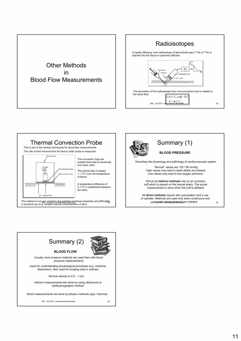
Other Methodsin
Blood Flow Measurements
ARL - EL4703 - Isntrumentasi Biomedis 62
RadioisotopesA rapidly diffusing, inert radioisotope of lipid-soluble gas ( Xe or Kr) is injected into the tissue or passively diffused
133 85
The elimination of the radioisotope from microcirculatory bed is related to the blood flow:
( )ktCtC −= exp)( 0
2/1/2ln tk =
ARL - EL4703 - Isntrumentasi Biomedis 63
Thermal Convection ProbeThe rate of heat removal from the tissue under probe is measured
The concentric rings areisolated thermally & electricallyfrom each other
The central disk is heated1 – 2 C over the temperatureof tissue
A temperature difference of 2- 3 C is established betweenthe disks
o
o
The method is not very common due extreme nonlinear properties and difficultiesin practical use (e.g. variable thermal characteristics of skin)
This is one of the earliest techniques for blood flow measurements
ARL - EL4703 - Isntrumentasi Biomedis 64
Summary (1)BLOOD PRESSURE
Describes the physiology and pathology of cardiocvascular system
”Normal” values are 120 / 80 mmHgHigh values may lead to heart attack and strokes
Low values may lead to low oxygen perfusion
All direct methods require skin punctuation and a useof catheter. Methods are used only when continuous and
accurate measurements are needed.
Almost all indirect methods rely on an occlusivecuff which is placed on the bracial artery. The actual
measurement is done when the cuff is deflated
ARL - EL4703 - Isntrumentasi Biomedis 65
Summary (2)BLOOD FLOW
Used for understanding physiological processes (e.g. medicinedissolution). Also used for locating clots in arteries
Usually more invasive methods are used than with bloodpressure measurements
Normal velocity is 0,5 - 1 m/s
Indirect measurements are done by using ultrasound orplethysmographic method
Direct measurements are done by dilution methods (dye / thermal)