anatomy ii lab practical ii - daytona state … · the academic support center @ daytona state...
TRANSCRIPT
The Academic Support Center @ Daytona State College (Science 56 Page 1 of 63)
S I R E V I E W
ANATOMY II LAB PRACTICAL II

The Academic Support Center @ Daytona State College (Science 56 Page 2 of 63)
By Eddie Hoppe
Includes:
Endocrine System (slides # 2-16)
Blood (# 17-31)
Heart Anatomy and Physiology (# 32-43)
Blood Vessels (44-54)

The Academic Support Center @ Daytona State College (Science 56 Page 3 of 63)
Major Glands
What Hormones they Release
The Class of Chemical
What is the Trigger for Each
What is the Target

The Academic Support Center @ Daytona State College (Science 56 Page 4 of 63)
1 2
3
4
5
6
7
8 9
10

The Academic Support Center @ Daytona State College (Science 56 Page 5 of 63)
1
2
3
4
5
6
7
Al l c lass o f hormones f rom th is g land are pept ide /prote in based hormones
Al l hormones f rom th is g land are top ic (see be low for d iscussion)

The Academic Support Center @ Daytona State College (Science 56 Page 6 of 63)
Target Hormone Ends up
• 1. Ovaries and testes – Follicle-stimulating hormone (FSH) Exocrine
• 2. Ovaries and testes – Luteinizing Hormone (LH) Endocrine
• 3. Thyroid gland -- Thyroid-Stimulating Hormone (TSH) T3/T4
• 4. Adrenal Cortex -- Adrenocorticotropic Hormone (ACTH) Corticosteroids
• 5. Mammary Glands – Prolactin Exocrine
• 6. Liver, bone, muscle, fat – Growth hormone Tissue growth 6
5
4
3
2
1
All hormones released by the Anterior pituitary are peptide/protein based hormones
#6 has both tropic and non-tropic effects

The Academic Support Center @ Daytona State College (Science 56 Page 7 of 63)
Target and effect Hormone
• Kidneys (collecting ducts); conserve water – Antidiuretic Hormone, ADH
• Uterus, mammary glands, brain, -- Oxytocin, OT
1
2
“Make more
Aquaporins!”
These protein based hormones have neuronal triggers (see below)

The Academic Support Center @ Daytona State College (Science 56 Page 8 of 63)
Effect Hormone
• Increases Body Metabolism – T3/T4(thyroid hormones)
• Responds to Hypercalcemia -- Calcitonin
1
2
The thyroid has both hormonal (follicular cells) and humoral (C cells) responses
Follicular cells release monoamines, C cells release peptide based hormones

The Academic Support Center @ Daytona State College (Science 56 Page 9 of 63)
Trigger Hormone
• Responds to Hypocalcemia – Parathyroid Hormone (PTH) 1
This hormone works by promoting the synthesis of calcitriol from the kidneys which promotes calcium
intestinal calcium absorption, inhibition of urinary excretion of calcium, and indirectly simulating
osteoclasts to resorb bone.
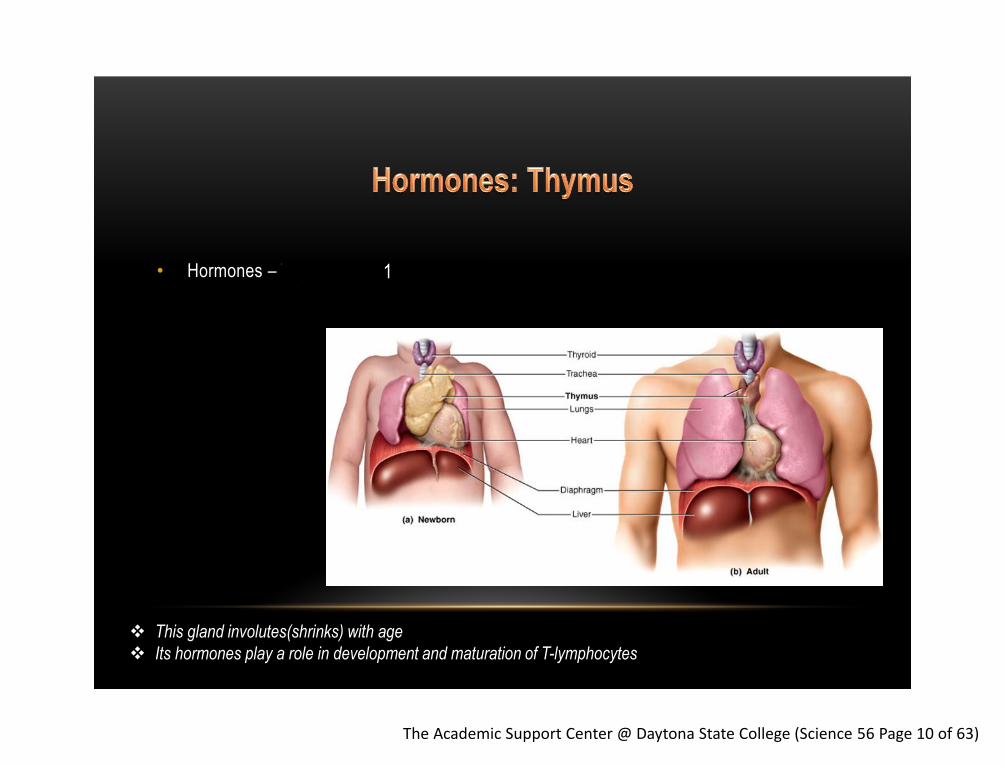
The Academic Support Center @ Daytona State College (Science 56 Page 10 of 63)
• Hormones – Thymosin and Thymopoietin 1
This gland involutes(shrinks) with age
Its hormones play a role in development and maturation of T-lymphocytes

The Academic Support Center @ Daytona State College (Science 56 Page 11 of 63)
• These hormones are released due to a stimulation of the sympathetic nervous system --
Epinephrine/Norepinephrine
• The cells in of this gland are modified postganglionic sympathetic and are called
chromaffin cells
1
1
These monoamine hormones have a neuronal trigger
2
1. What hormones are released?
2. Name the cells which make up the adrenal medulla

The Academic Support Center @ Daytona State College (Science 56 Page 12 of 63)
Category of Hormone Example Hormone and layer Effect
• Mineralocorticoids – Aldosterone from the Zona Glomerulos ↑ Na+ pumps
• Releases Glucocorticoids – Cortisol from the Zona Fasciculata ↓ inflammation
• Releases Gonadocorticoids – Testosterone from the Zona Reticularis
1
2
3
2
1
3
Glucocorticoids and Gonadocorticoids have tropic triggers (ACTH from the anterior pituitary)
Each hormone made and released by this gland are steroids

The Academic Support Center @ Daytona State College (Science 56 Page 13 of 63)
Trigger Hormone - Specific Cell Effect
• Hyperglycemia – Insulin from β cells – Store glucose in the form of glycogen
• Hypoglycemia – Glucagon from α cells – Break down the stored glycogen
1 2
These are antagonistic hormones controlling glucose homeostasis in blood
These protein based hormones have humoral triggers
3 4

The Academic Support Center @ Daytona State College (Science 56 Page 14 of 63)
Hormone
• Interstitial cells produce and release: testosterone
• Responsible for male secondary sex characteristics and libido
1
The testis are dual endocrine/exocrine
Tropic trigger and releases steroids

The Academic Support Center @ Daytona State College (Science 56 Page 15 of 63)
Hormones Released from the Ovaries
• Granulosa cells release estrodiol in the first half of the menstrual cycle
• After ovulation, the corpus luteum produces the above hormone and progesterone
1
The ovaries are dual endocrine/exocrine
Tropic trigger and releases steroids
2

The Academic Support Center @ Daytona State College (Science 56 Page 16 of 63)
• The function of this gland is to regulate circadian rhythm
• Serotonin is release during the day and melatonin is released at night
• Both of these hormones fall under the class of monoamines and have neuronal triggers
1
3 2
4 5
These hormones are monoamines and have a neuronal trigger

The Academic Support Center @ Daytona State College (Science 56 Page 17 of 63)
Thyroid Parathyroid
Adrenal Glands
Gonads: Testis
Pancreatic Islets
Ovary
3. 4.
5. 6.
7. 8.

The Academic Support Center @ Daytona State College (Science 56 Page 18 of 63)
Formed Elements of Blood
Platelets
Differential White Blood Cell Count
Hematocrit

The Academic Support Center @ Daytona State College (Science 56 Page 19 of 63)
Function: 4.8 million/µL in females
• Hemoglobin within the cell transport 5.4 million/µL in males
mostly O2 and some CO2
Characteristics:
• Most numerous of the formed elements
• Biconcave Disk
• Only lives about 100-120 days

The Academic Support Center @ Daytona State College (Science 56 Page 20 of 63)
• Neutrophil: 3-5 lobed nucleus 60–70% of all WBCs
Most numerous (seen first at sight of infection)
Releases a cocktail of chemicals to kill bacteria
Will self destruct when outnumbered by pathogens
Identification Characteristics:
Multi-lobed nucleus
• Eosinophil: Bi-lobed nucleus 2–4% of all WBCs
Seen in high amounts during parasitic infection (they secrete chemicals to destroy parasites/worm)
Phagocytizes antigen-antibody complexes
Seen in higher amounts during worm infestation!
Identification Characteristics:
Bi-lobed Nucleus
Has a rosy pigment

The Academic Support Center @ Daytona State College (Science 56 Page 21 of 63)
• Basophil: cannot see nucleus due to being obscured by many granules 0.5–1% of all WBCs
Appear in many kinds of inflammatory reactions
Secretes chemicals to affect localized blood flow:
histamine (vasodilator)
heparin (anticoagulant: prevents blood clotting)

The Academic Support Center @ Daytona State College (Science 56 Page 22 of 63)
• Lymphocyte: the specialists of the immune system 20–25% of all WBCs
Targets “bad cells” like cancer and virally infected cells
B-lymphocyte = plasma cells secrete antibodies,
T-lymphocytes = Cytotoxic, Helper, and Memory T cells
Natural Killer Cells = checks a certain antigen on the cell
surface called a “self-molecule”
• Monocyte: the cleanup crew 3–8% of all WBCs
Grows up to be a macrophage
Eats anything that isn’t supposed to be there: debris, invaders,
pathogens (after becoming macrophage)

The Academic Support Center @ Daytona State College (Science 56 Page 23 of 63)
2. Monocyte

The Academic Support Center @ Daytona State College (Science 56 Page 24 of 63)
2. Lymphocyte
1. Neutrophil

The Academic Support Center @ Daytona State College (Science 56 Page 25 of 63)
1. Monocyte
2. Lymphocyte

The Academic Support Center @ Daytona State College (Science 56 Page 26 of 63)
1. Neutrophil
3. Lymphocyte
4. Neutrophil
2. Monocyte

The Academic Support Center @ Daytona State College (Science 56 Page 27 of 63)
2. Eosinophil
Lymphocyte 5. Basophil
4.

The Academic Support Center @ Daytona State College (Science 56 Page 28 of 63)
• Also known as thrombocytes 150,000–400,000/µL
• Fragments of a megakaryocyte in the red bone
marrow
• Primary function is to help stop blood loss
by forming a platelet plug
Megakaryocyte: a stem cell producing platelets

The Academic Support Center @ Daytona State College (Science 56 Page 29 of 63)
3. Positive feedback is in action until coagulation is completed
Intrinsic and extrinsic pathways merge on
fibrinogen(soluble protein) to form fibrin(insoluble protein),
which completes the clot.
1. Constriction caused by:
• Neuronal reflex pain receptors
• Local properties of smooth muscle
• Local paracrine signaling
- platelets releasing 5-HT(serotonin)
2. Positive Feedback mechanism:
• Platelets stick to exposed collagen
• Platelets degranulate and release
ADP to attracts other platelets
and thomboxane A2
• More serotonin released

The Academic Support Center @ Daytona State College (Science 56 Page 30 of 63)
• FUNCTION: Differential white blood cell count gives the relative percentage of each type
of white blood cell and also helps reveal abnormal white blood cell populations (note the
normal percentage is listed in blue in the above slides)
• Abnormal amounts will help determine the type of infection present
Example:
• Count 50 leukocytes, categorize them, then multiply 2. You will get X number of
leukocytes out of a 100. 60% neutrophils is a normal count. 80% would indicate a
bacterial infection!
Chances are
They will give
You the numbers!

The Academic Support Center @ Daytona State College (Science 56 Page 31 of 63)
• FUNCTION: to measure the percentage of Red Blood Cells in the body
• Denoting any abnormal conditions
The Academic Support Center @ Daytona State College (Science 56 Page 32 of 63)
Layers of the heart wall and pericardium
Inner Heart Anatomy
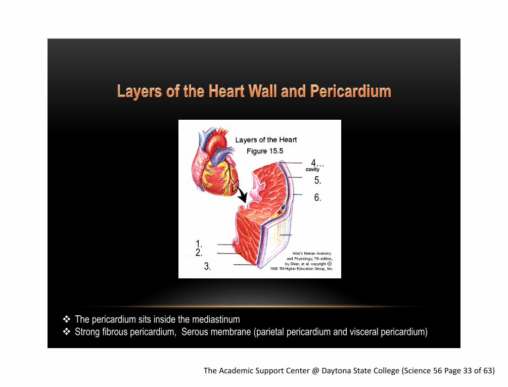
The Academic Support Center @ Daytona State College (Science 56 Page 33 of 63)
1. 2.
3.
4…
5.
6.
The pericardium sits inside the mediastinum
Strong fibrous pericardium, Serous membrane (parietal pericardium and visceral pericardium)

The Academic Support Center @ Daytona State College (Science 56 Page 34 of 63)
21. 20. 19.
18.
17. 16.
15.
14.
13. 12. 11.
10. 1.
2. 3. 4. 5.
6.
7. 8.
9.

The Academic Support Center @ Daytona State College (Science 56 Page 35 of 63)

The Academic Support Center @ Daytona State College (Science 56 Page 36 of 63)
General Terminology
Systolic and diastolic pressures
Blood pressure
Heart Rate
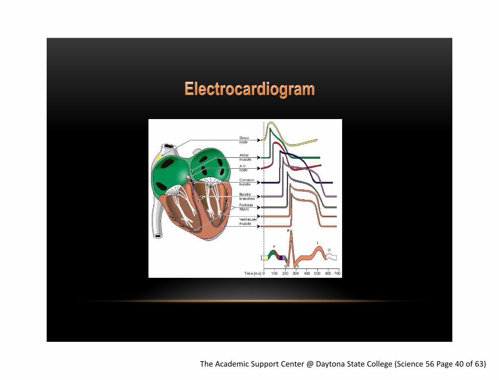
Electrocardiogram

The Academic Support Center @ Daytona State College (Science 56 Page 37 of 63)
• Systolic pressure: the highest arterial blood pressure exerted on the arteries (90-120 in adults)
• Diastolic pressure: the lowest pressure in the arteries (60 – 80 in adults)
• Pulse Pressure: Systolic over diastolic pressures. It represents the force that your heart generates
during each cardiac cycle.
• Sphygmomanometer: a device used to measure blood pressure
• Electrocardiogram: a transthoracic interpretation of the electrical activity of the heart over a period
of time, as detected by electrodes attached to the surface of the skin
• Auscultation: the term for listening to the internal sounds of the body, usually using a stethoscope
• Isovolumetric: unchanging volume
• Blood Pressure: the force which the blood exerts on the walls of the blood vessels
The Academic Support Center @ Daytona State College (Science 56 Page 38 of 63)
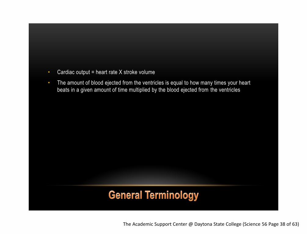
• Cardiac output = heart rate X stroke volume
• The amount of blood ejected from the ventricles is equal to how many times your heart
beats in a given amount of time multiplied by the blood ejected from the ventricles

The Academic Support Center @ Daytona State College (Science 56 Page 39 of 63)
1.
2.
3.
6.
5.
4.
Normal ECG
The Academic Support Center @ Daytona State College (Science 56 Page 40 of 63)

The Academic Support Center @ Daytona State College (Science 56 Page 41 of 63)
• FIRST: understand what is happening during the electrocardiogram
• SECOND: from the ECG, know what the chambers are doing: systole/diastole
• THIRD: from knowing the contraction of the chambers, figure out the pressure that would be in each valve as it contracts and relaxes
• FOUR: from knowing the pressure, determine the position of the heart valves and flow of blood
Example:
1. During the P wave, a depolarization occurs in the atria which is then recorded on the ECG
2. This change in voltage across the atria indicates that slow voltage gated calcium was triggered and muscle contraction was initiated, the atria are in systole
3. Due to contraction of the atria, the pressure goes up
4. This pressure pushes just a little bit of blood into the ventricles, causing the AV valves to close because the pressure in the ventricles are now higher than the pressure in the atria

The Academic Support Center @ Daytona State College (Science 56 Page 42 of 63)
P Wave – records atrial depolarization and therefore atrial systole
QRS Wave – records atrial repolarization and therefore atrial diastole
ventricular depolarization, therefore ventricular systole
T Wave – records ventricular repolarization and therefore ventricular diastole
Quiescent Period – records no electrical change, which indicated the ventricles being relaxed
1. 2.
3. 4.
5. 6.
P
Q
R
S
T
Quiescent Period

The Academic Support Center @ Daytona State College (Science 56 Page 43 of 63)
1.
2.
3.
7.
6.
5.
1. Which phase of contraction?
2. Which valve is doing what?
3. Which valve is doing what?
4. Which phase of relaxation?
5. Which valve is doing what?
6. Which valve is doing what?
7. This is measuring aortic…
4.

The Academic Support Center @ Daytona State College (Science 56 Page 44 of 63)
The Academic Support Center @ Daytona State College (Science 56 Page 45 of 63)
• Does heart rate increase or decrease when initially holding your breath?
It decreases due to mammalian diving reflex
• As a result does time 1 or time 2 change? Does it become longer or shorter?
Time 2 changes, which is the time between ventricular relaxation and atrial contraction
• After strenuous activity, does time 1 or 2 change? Does it become longer or shorter?
Time 2 changes

The Academic Support Center @ Daytona State College (Science 56 Page 46 of 63)
Arteries
Veins
Hepatic Portal System

The Academic Support Center @ Daytona State College (Science 56 Page 47 of 63)

The Academic Support Center @ Daytona State College (Science 56 Page 48 of 63)

The Academic Support Center @ Daytona State College (Science 56 Page 49 of 63)
1.
2.
3.
4.
5.
6.
7.
8.
9.
16.
15. 12.
11.
10.
… Aorta
… Aorta
… Aorta
… Aorta

The Academic Support Center @ Daytona State College (Science 56 Page 50 of 63)
1.
2.
3.
4.
5.
6.
7.

The Academic Support Center @ Daytona State College (Science 56 Page 51 of 63)
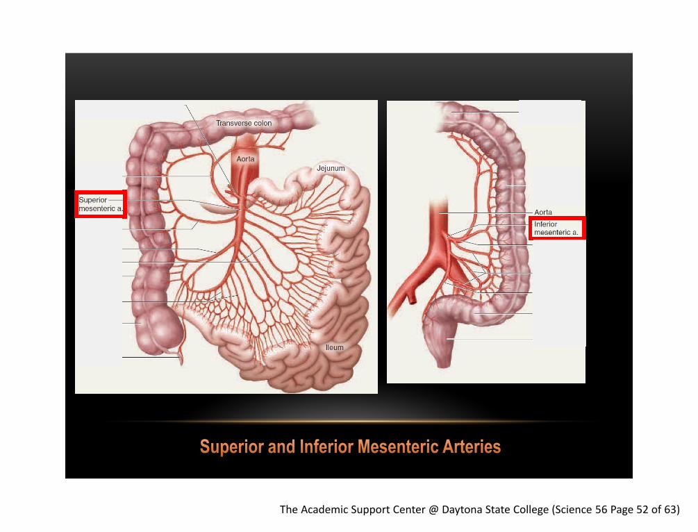
2. 3.
1.
5.
4.
The Academic Support Center @ Daytona State College (Science 56 Page 52 of 63)

The Academic Support Center @ Daytona State College (Science 56 Page 53 of 63)
1.
2.
3.
4.
5.
6.
1. Right common Iliac
2. Right internal Iliac
3. Right external iliac
4. Femoral
5. Popliteal
6. Dorsalis Pedis

The Academic Support Center @ Daytona State College (Science 56 Page 54 of 63)
1. 2.
3.
4. 5.
6.
7.
8.
9.
10.
11. 12.
14. 15. 16.

The Academic Support Center @ Daytona State College (Science 56 Page 55 of 63)
1. 2.
3. 4. 5. 6.
7.

The Academic Support Center @ Daytona State College (Science 56 Page 56 of 63)
Portal System: two capillary beds
2.
3.
4. 5.
6. 1.

The Academic Support Center @ Daytona State College (Science 56 Page 57 of 63)
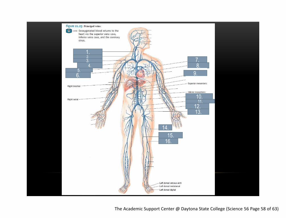
The Academic Support Center @ Daytona State College (Science 56 Page 58 of 63)
1. 2. 3. 4.
5.
6.
7. 8.
9.
14.
15. 16.
13. 12.
10. 11.

The Academic Support Center @ Daytona State College (Science 56 Page 59 of 63)

The Academic Support Center @ Daytona State College (Science 56 Page 60 of 63)
4 nm intercellular cleft Allows solutes like glucose
to pass through
20-100nm
filtration pours
Molecules such as small
Proteins but still retains
RBC and platelets
Big enough to let RBC,
and albumin pass
Examples Skeletal
muscle
Kidneys,
Endocrine
glands,
small
intestines
Liver,
spleen,
Red Bone
Marrow
30-40 µm
Filtration pours
40-70 layers of
elastic sheets
Examples Aorta, subclavian, common carotid,
pulmonary trunk, common iliac
Brachial, femoral, renal, splenic
To various organs
40 layers of
smooth muscle
25 layers of
smooth muscle

The Academic Support Center @ Daytona State College (Science 56 Page 61 of 63)
In general, you will see the following properties throughout arteries and veins. The specific qualities of the tunica media w ill be based on the location of the blood vessel and the pressure of the blood.
• Elastic Fibers
- Seen in areas of high blood pressure (aorta, pulmonary trunk, common carotid, subclavian) and used to absorb/lessen blood pressure for the downstream arteries
• Collagen
- Specifically in places of high blood pressure
• Smooth muscle
- Seen in areas where you need to decrease blood pressure: distributing arteries which control blood flow to large areas of your body (femoral/brachial) and specific organs (splenic and renal)
- Also seen in resistance arteries which control blood to specific organs (resistance arteries)

The Academic Support Center @ Daytona State College (Science 56 Page 62 of 63)
The Academic Support Center @ Daytona State College (Science 56 Page 63 of 63)
Prepared by E. Hoppe – SI Leader Edited by D. Leonard – Learning Specialist & K. Martin – Peer Tutor The Academic Support Center @ Daytona State College http://www.daytonastate.edu/asc/ascsciencehandouts.html
Questions