analysis of kinase effects on viral replication of the
TRANSCRIPT

UNF Digital Commons
UNF Graduate Theses and Dissertations Student Scholarship
2006
Analysis of Kinase Effects on Viral Replication ofthe PapillomavirusJoshua R. RaynesUniversity of North Florida
This Master's Thesis is brought to you for free and open access by theStudent Scholarship at UNF Digital Commons. It has been accepted forinclusion in UNF Graduate Theses and Dissertations by an authorizedadministrator of UNF Digital Commons. For more information, pleasecontact Digital Projects.© 2006 All Rights Reserved
Suggested CitationRaynes, Joshua R., "Analysis of Kinase Effects on Viral Replication of the Papillomavirus" (2006). UNF Graduate Theses andDissertations. 241.https://digitalcommons.unf.edu/etd/241
A~AL YSIS OF E.:INASE EFFECTS ON VIRAL
REPLICATION OF THE P APILLO~/[A VIRlJS
by
Joshua R. Raynes
A thesis submitted to the Department of Biology in
partial fulfillment of the requirements for the degree of
Master of Science in Biology
University of North Florida
College of Arts and Sciences
June 2006
Unpublished work © Joshua R. Raynes

CERTIFICATE OF APPROVAL
The thesis of Joshua R. Raynes is approved by: Date
Com~~ Chairperson
Committee Member
ACCEPTED FOR THE DEPARTMENT:
ACCEPTED FOR THE COLLEGE:
ACCEPTED FOR THE UNIVERSITY:
Dean of the Graduate School
Signature Deleted
Signature Deleted
Signature Deleted
Signature Deleted
Signature Deleted
Signature Deleted

ACI(N0,VLEDGEJ\1E~TS
Jules Hemi Poincare once said "Science is facts; just as houses are made of stone,
so is science made of facts; but a pile of stones is not a house, and a collection of facts is
not necessarily science, " and never has this concept been more real to me than during my
research and the completion of this work. My efforts in the laboratory could have easily
constructed a metaphorical stack of stones, but I could have never forged this thesis
without the help of so many outstanding individuals.
My interest and abilities in the biological sciences were not the product of a life
changing occunence, profound experience, or single mentor. It is simply the culmination
of years of scientific marvel and the· direction and assistance of the great faculty members
in the University ofNorth Florida's department ofbiology. My research experience
served as a great source for my thirst for scientific information and I know that I am
greatly indebted for the initial opportunity, the space to work, and guidance in the
research itself, to Dr. Michael Lentz. In addition to being an outstanding professor and a
great committee leader, he was always there to listen and steer me in the right direction. I
must also thank Dr. Bowers for helping me over a huge expelimental hurdle and for
perpetually reminding me how deep the rabbit hole of knowledge goes. Know that your
instruction and your companionship mattered greatly to me, you are appreciated. To Dr.
Venkat, I offer my thanks for being my non-biology biased opinion and a great friend
during all my years at UNF. Dr. Joe Butler, Dr. Tony Rossi, and Ms. Tammy Hom, you
have my utmost gratitude for helping me every step of the way towards the completion of
lll
this work and my Master's degree. For the aid in image enhancement I thank David
Wilson from CIRT. I must also thank Stan Stevens at the University of Florida
Proteomics Core, Interdisciplinary Center for Biotechnology Research, for all his work
with the mass spectrometric analysis. To my fellow lab-rats, Tess Shideler and Mira
Lambert-Ferland, thanks so much for the bench-work assistance and the company. There
is a light at the end of the tum1el; all those gels and blots do get you somewhere.
Most of all, I would like to thank my wife, Rachel Raynes. I've been fortunate
enough to have many great opportunities and experiences in my life, but by far the best
occurrence my 23 years on this earth has afforded me was meeting her. To a brilliant and
talented woman, who never ceases to inspire me, who has always encouraged me to
attempt to achieve even my highest goals, and is the best support system anyone could
ask for, thank you so much.
This work was supported by NIH AREA Grant Rl5 CA087051 to Dr. Michael
Lentz and summer grants from the UNF Office of Academic Affairs.
lV

TABLE OF CO~TE~TS
Title page I
Certificate of Approval II
Acknowledgements 111
Table of Contents v
List of Table and Figures Vl
Abstract Vlll
Introduction 9
Materials and Methods 16
Results 22
Discussion 42
References 50
Vita 54
v

LIST OF TABLES A~D FIG~URES
Figure 1: A phosphorylation site map of the bovine papillomavirus El protein
including known functional domains and phospho-amino acid locations.
Figure 2: Assay of PKC effects upon viral replication including autoradiograph film
and a table of PKC target sites, surrounding sequence, and method used to predict the
residues.
Figure 3: Assay of CDK effects upon viral replication including autoradiograph film
and a table of CDK target sites, smTounding sequence, and method used to predict the
residues.
Figure 4: Assay of CK2 effects upon viral replication including autoradiograph film
and a table of CK2 target sites, surrounding sequence, and method used to predict the
residues.
Figure 5: Assay of DNAPK effects upon viral replication including autoradiograph
film and a table of DNAPK target sites, surrounding sequence, and method used to
predict the residues.
vi

Figure 6: Effects of chemical inhibitors upon cellular proliferation and viability
including images of control and inhibited cells, percent viabilities post chemical
exposure, and average total cell number of each sample.
Table 1: Summary of all kinases assayed in this study and the specific chemicals
used to inhibit their activity.
Table 2: Composite of all identified phosphorylation sites on the bovine
papillomavirus El protein with locations of each specific phospho-residue in the
sequence and the predicted kinase.
Vll
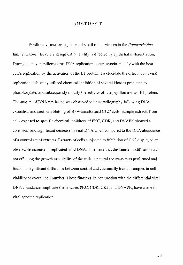
ABSTRACT
Papillomaviruses are a genera of small tumor viruses in the Papovaviridae
family, whose lifecycle and replication ability is directed by epithelial differentiation.
During latency, papillomavirus DNA replication occurs synchronously with the host
cell's replication by the activation of the El protein. To elucidate the effects upon viral
replication, this study utilized chemical inhibition of several kinases predicted to
phosphorylate, and subsequently modify the activity of, the papilloma virus' E 1 protein.
The amount of DNA replicated was observed via autoradiography following DNA
extraction and southern blotting ofBPV-transformed C127 cells. Sample extracts from
cells exposed to specific chemical inhibitors of PKC, CDK, and DNAPK showed a
consistent and significant decrease in viral DNA when compared to the DNA abundance
of a control set of extracts. Extracts of cells subjected to inhibition of CK2 displayed an
observable increase in replicated viral DNA. To ensure that the kinase modification was
not effecting the growth or viability of the cells, a neutral red assay was performed and
found no significant difference between control and chemically treated samples in cell
viability or overall cell number. These findings, in conjunction with the differential viral
DNA abundance, implicate that kinases PKC, CDK, CK2, and DNAPK, have a role in
viral genome replication.
viii

I~TRODl_JCTION
Papillomavimses are small (capsid, 55-nm; genome, 8 kbp) obligate
intracellular parasites that infect epithelial cells of the skin and mucous membranes in a
range ofvertebrate hosts [1-5, 6, 10]. These double-stranded DNA tumor viruses are one
of the two genera within the Papovaviridae family and the non-enveloped virions exhibit
T=7 icosahedral symmehy [28, 30]. Entry ofvims particles occurs via abrasive
mechanism, which exposes the target basal cell layer. This exposure is followed by
clathrin-dependent endocytosis and nucleocapsid L2 protein-mediated egress from late
endosomes into cell cytoplasm [31 ]. The papillomavims has a complex lifecycle which is
closely linked to the differentiation status of the epithelial cells. During latency
papillomavims DNA replication can only occur during the host cellS-phase of the cell
cycle [7, 10, 17, 24]. Following viral attachment, entry, and nucleocapsid disassembly,
initial infection and viral latency are characterized by low viral genome copy number
(50-200 per cell) and minimal gene expression, where only a select few genes are
transcribed in low concentration. The viral genome exists as an extra-chromosomal
episome and remains perpetually present in the stratum basale layer, allowing for the
vims's persistent ability to relapse [32]. Papillomavimses are non-lytic, and mature
virions are shed only as the stratum corneum desquamates.
These vimses primarily infect the basal layer, or stratum basale, of stratified
squamous (both keratinizing and non-keratinizing) epithelial tissue. The stratum basale
cells, found just superficial to the dermis, are often characterized as the skin "stem cells,"
as they are the only layer of cells that undergo mitosis and subsequently the only layer in
9

which DNA is replicated. These vimses are termed oncogenic because they transform the
infected cells in their efforts to maintain replicative status in the cell, producing
unregulated cellular growth. As the infected cells divide and proliferate outward the virus
triggers a hyperproliferation of nearby keratinocytes and benign skin tumors (warts), or
papillomas, are often produced [ 10, 25]. These warts are most often manifested in the
skin of the host's outer extremities, but specific strains of the papillomavirus regularly
arise in the mucosa [8, 10]. These neoplasms are not typically life threatening for the
host, but if the viral cycle does not regress or the host is exposed to environmental
carcinogens, they may become malignant and metastasize to other locations in the body.
Certain "high-risk" papilloma virus strains, those that readily induce modifications
allowing for immortalization, are more known to develop into malignant cancers, while
those of "low-risk" are rarely linked to malignancy [1 0, 17].
The viral genome contains three basic regions, each characterized by their relative
functions. The first region consists of an upstream regulatory domain, containing
sequences that control transcription and replication. This sequence is followed by a
region containing many of the genes involved in the actual viral replication, transcription,
and transformation, the early domain; and a structural region, the late domain [7, 11, 24-
26]. The early region encodes for the early proteins, El-E8, and the late contains two
genes; L 1, which forms the viral capsid and L2, a minor capsid component, which is also
responsible for viral DNA packaging. Translation of the late proteins is necessary for the
progeny virons to be able to evacuate the host cell and proliferate the infection cycle.
This stage in the lifecycle only occurs in the outer epithelial layers (outer stratum
spinosum and stratum granulosum), those most highly differentiated [12, 13, 17].
10

Like all DNA tumor vimses, the papillomavirus uses host cell proteins for their
own DNA replication. They do, however, encode for a specific replication origin ( ori)
binding and viral DNA unwinding molecule: the E1 protein [1-5, 7, 8]. The E1 protein,
which is a 593-681 amino acid polypeptide (approximately 67.5-76.2 kDa depending on
the viral strain), has the largest and most sequentially conserved open reading frame in
the viral genome [7]. For the virus to replicate conectly, the a- helix rich El protein
forms a hexameric or double-hexameric complex, facilitated by the activity of a heat
shock chaperone protein [12]. E1 contains binding domains for DNA, ATP, and the E2
protein, an auxiliary origin recognition protein. When E1 and E2 are coupled together,
the complex, via its loop-helix motif, will locate and attach to the origin replication site
of the viral genome. While the presence of E2 in the complex is not altogether necessmy,
the sequence specific recognition ability of E 1 is enhanced when E2 is present in the pre
replication complex [7, 12]. Once bound to the origin, El causes the initial unwinding of
viral DNA at the replication fork through its 3' -7 5' helicase activity. Unwinding is an
ATP dependent process, which hydrolyses the ATP bound at the P-loop of the protein at
a rate of one molecule per round of activity [7, 12, 14]. This activity occurs at the
carboxy-terminal domain. This model has been demonstrated by studies with amino
terminus deleted E 1 where the tmncated protein still maintains replication functionality,
albeit at a slower rate of viral DNA unwinding [1, 2, 7]. Strain comparison studies have
revealed that the carboxy-terminal domain is the most highly conserved section of the
protein's sequence [7].
E 1 is the only papillomavims protein with enzymatic function, which it must
employ to unwind the viral template DNA. In the presence of unwound template the
11

protein must then recruit the cellular replication machinery to the viral DNA. Following
recruitment, it is then up to the infected cell to complete replication. Given the virus'
compact genome, most of the components needed for viral reproduction are not encoded
for by the viral DNA, and so all other factors necessary for replication must be supplied
by the host cell. The E1 protein has been shown to be responsible for soliciting host cell
DNA polymerases (a and 8), replication protein A (RP A), proliferating cell nuclear
antigen (PCNA), and topoisomerases [13, 14, 23]. The success ofE1 's ability to bind to
all of the required replication elements, within the proper time frame, suggests a
significant level of regulation of E 1 's multiple binding sites [7, 20]. Data has
demonstrated that the viral replication protein E1 may have the potential to influence the
host cell environment significantly, instead of waiting idly in a latent stage. Expression of
E 1 protein has been shown to decrease the duration of G 1 phase and increase S phase,
allowing for maximal viral DNA replication and may contribute to the virus'
pathogenesis and persistence within the host cell [9, 1 0].
Unique to the papilloma virus is its ability to direct the activation of its DNA
replication to occur synchronously with the host cell's replication [7, 10, 17]. The timing
of this activation and subsequent stimulation of the E1 protein is critical for viral
replication and therefore, exhibits differential regulation sites specific for timing and
utilization of the viral genomic replication. Without the presence of a complex cell cycle
regulation system of its own, the virus depends largely upon the phosphorylation ofE1
protein to regulate viral DNA replication during host cell S-phase [7 -14, 25]. This fact
also makes the papillomavirus an excellent model system for studying some aspects of
eukaryotic DNA replication mechanisms [1-5, 17].
12

Protein phosphorylation is a common regulatory mechanism in cellular systems.
Phosphorylation of proteins can yield a range of changes from subcellular localization,
signal transduction, and modifications in stability and activity [22]. Several labs have
dedicated years of study to the role of phosphorylation and the probable sites utilized in
regulating E 1 activities. Earlier studies have utilized various mutants of the E 1 protein to
elicit which residues of the protein must be phosphorylated for functional replication [2,
3, 4, 25, 33]. Substitution mutation of the bovine papillomavirus El protein at threonine
102 did not seem to significantly modify replication [4], but other mutants, such as serine
48 and serine 584, had significantly reduced or eliminated replication of the viral genome
and were hypothesized to be possible phosphorylation sites of an activating nature [2,
25]. Yet, not all modification of suspected phospho-resides indicated an inhibitory
activity on replication; alteration of residues serine 90 and serine 109 actually lead to an
increase in replication activity [3, 33]. These studies provide support for the differential
effects of certain kinases, but are limited in that site specific assays can only determine
that site's activity, and not the cumulative action of phosphorylation of all targets of a
particular kinase. Experimental modification of CDK within an in vitro system seemed to
suggest that some level ofEl dependency exists on the kinase, but site specificity was not
determined [26]. Recent mass spectrometric analysis of the El protein revealed a clear
map of major El protein phosphorylation sites. This data revealed five new
phosphoamino acid residues and confirmed two known sites [17]. Adding these sites to
other previously established phosphorylation sites brings the total number of known
phospho-regulation residues on the El protein to ten. Eight of the ten phosphorylation
sites are within the amino terminal domain, the region of least conserved sequence
13
(Figure 1 ). While overall the region varies between viral strains, the phospho-residues
themselves are highly conserved. The positioning of these regulatory sites in this domain
follows logically in that the necessary regulatory mechanisms should be extremely
specific to the host environment. Which kinases are responsible for modifying these sites,
as well as their subsequent pathways, are still unclear.
The cyclic dependency of replication lead to the initial hypothesis that mitogen
activated protein kinases (MAPK) and protein kinase A (PKA) are the phosphate donors
[8], however recent experimental analysis does not seem to indicate that MAPK plays a
significant role in phosphorylation. PKC, CDK, and CK2, all have been demonstrated to
phorphorylate the E 1 protein in vitro [21, 23, 26]. Of those sites identified by mass
spectrometric (MS) analysis, five were found to be of the consensus of CK2 target
substrates, one of CDK, and one of DNAPK, based upon computer prediction algorithms
[ 1 7]. Those sites not confirmed by MS analysis, yet verified by other studies, include an
additional site in the consensus for CDK and two sites ofPKC [3, 4, 33]. Such
predictions seem intuitive given that those kinases which are known to effect cell cycle
progression and/or DNA modification are the most likely candidates of site
phosphorylation, taking in account E1 's role in viral replication.
Protein kinase C (PKC) is a family of isozymes, which are typically activated via
receptor binding of hormones, growth factors, and signal transduction agents [15, 16].
Some forms of the kinase require calcium and/or phospholipids to stimulate activity.
PKC has been shown to be necessary for optimal efficiency in production of viral
progeny and upregulation of late gene expression in human papillomavirus [27, 41].
Inhibition of PKC signal transduction pathways has known effects on tumor suppression
14

and inhibition ofrapid cell proliferation [15, 16], and is of key interest to this study, since
the kinase is known to play a key role in epidem1al differentiation [3, 40]. PKC substrates
are typically serine/threonine residues with upstream or downstream basic residues,
particularly arginine [27].
Cyclin dependent kinases (CDK) are a group ofkinases largely responsible for
proper cell cycle progression, specifically at the G liS and 02/M phase boundaries [21-
23, 26]. CDK has been demonstrated to have effects upon the nuclear export sequences
(NES) and nuclear localization sequences (NLS) of human papillomavirus E1 [21],
however its effects upon the conserved localization domain in BPV E1 has not been
definitively assayed [7]. Target consensus sequence for CDK phosphorylated residues are
comprised of serine or threonine amino acids followed immediately by a proline [21, 26].
Casein kinase II (CK2);like all ofthe kinases included in this study is a type of
serine/threonine kinase. CK2 functions are much more multi-faceted than most other
kinases studied [20]. The consensus sequence for all CK2 targets is dependent upon
acidic residues, like glutamate or aspartate, downstream from the phospho-residue [ 18].
Its activities range from signal transduction, growth control, cell shape, and even spindle
formation. CK2 has been found in almost all tissues of all eukaryotes, and is ubiquitous
in its intracellular distribution [18-20]. High amounts ofCK2 are found in cells exhibiting
elevated activity levels, especially those which are rapidly proliferating, which strongly
suggests a supportive role in replication. This enzyme is of particular interest in that it
expresses constitutive activity, which is optimal for viral replication [18, 20].
DNAPK is the least studied kinase used in this analysis, but a few of its activities
have been published. DNAPK has been shown to be activated by DNA double strand
15

breaks, aneuploidy, or detectable genome abnormality that then phosphorylates the p53
protein. This phosphorylation activates the well known tumor suppressor protein, which
subsequently binds to and prevents any replication of the damaged DNA [29]. The kinase
also has been demonstrated to interact with a variety of transcription factors including
Ku70, cJun, cFos, Spl, cMyc, and TFIID, and lead to the dissociation ofRPA complex
[29, 36]. The target phospho-residues ofDNAPK must be closely followed by a
glutamine residue [29]. Of particular interest to this study is the kinase's reported
interaction with epidermal growth factor receptor, where the receptor's expression was
decreased following inhibition ofDNAPK [37].
This study serves to illuminate the overall effects of the predicted kinases on viral
replication. Chemical manipulation of PKC, CDK, CK2, and DNAPK will provide
details into the respective impact of the specific phosphorylation of the El protein by
each kinase upon the virus. Whether the inhibitors and/or activators of the kinases lead to
reduction or addition of detectable viral DNA should give clear indication of the specific
role of the kinases in viral replication. Alteration ofkinase function yields no effect,
increase, or decrease of the concentration of viral DNA, as detected by autoradiographic
film of DNA extracted from infected cells.
~1ATERIALS AND ~/IETHODS
Cellular Maintenance and Transfection. Cl27 cells (murine fibroblast) were
maintained at 3TC with 5% C02 in Dulbecco's Modified Eagle's Medium (DMEM) and
16

10% FBS supplemented with 1 OOiu/mL penicillin and 1 OOmg/mL streptomycin before
harvest at late log phase. Cells were washed with PBS, and removed from plates by
addition of 1mL of 0.25% trypsin/ 2mM EDTA, followed by rocking for 5 minutes. Cells
were transfened to a sterile 50mL Falcon tube containing 5mL of 5mM BES buffered
complete DMEM. The cell solution was kept on ice prior to counting. Cells were spun
out of media at 5000 X g for 10 minutes and then resuspended in complete media/BES
mixture to yield a concentration of 20 million cells/mL. BPV viral genome DNA was
released from the pML vector via digestion with BamBI (Takara) and 1f.1g of DNA was
added to eletroporation cuvettes containing 0.1X TE (lmM Tris, 0.1mM EDTA).
Sheared, denatured salmon spem1 was included as a carrier to final concentration of 50f.lg
total (200f.1g/mL carrier in cell suspension). Approximately 5 million cells (0.25mL C127
cell suspension) were added to the DNA containing cuvette and the mixture was shocked
at 250V and 975f.1F. The samples were incubated at room temperature for 10 minutes and
each sample was transfened to a 15mL tube containing 1 OmL complete DMEM/BES
mixture. Cells were pelleted at 750 X g for 10 minutes, resuspended in 8mL complete
DMEM, and distributed on 8, 10cm diameter plates.
Focus Assay and Transformed Cell Removal. C127 cells that have successfully
been transformed with an extrachromosomal episome of BPV DNA produce visible foci
after 2-3 weeks. During this time cells are kept at 3 7°C with 5% C02 and media replaced
twice weekly. After transformation, foci were marked on the plate, media removed, and
plates washed with PBS. A sterile 3mm disk of filter paper, presoaked in 1X trypsin
solution, was placed on each distinct focus for 1 minute. Each filter disk was then placed
17

in 3.5cm diameter plate, onto which complete DMEM was then added. Cells were
maintained under the same conditions as previously described until they reached
approximately 50% confluence. At this time cells were transferred to a 1 Ocm diameter
plate and either immediately used or stored in liquid nitrogen.
Cell Culture and Chemical Modification. Transformed cells were sustained as a
monolayer culture in complete DMEM at 37°C with 5% C02 and were passaged to a 1:20
dilution at subconfluence into 1 Ocm diameter plates. Two sets of samples for each kinase
experiment were maintained, and a third for the PKC kinase trials to access the effects of
the activator. The first set of plates, including one plate for times zero, 48, 96, and 144
hours (0, 2, 4, and 6 days, respectively), contained only those cells not exposed to
chemical modifiers. These cells grown in complete DMEM and a small volume of
DMSO (no more than 50f..LL per plate in any trial) served as a control group, where the
volume of DMSO was equivalent to the volume of the chemicals in the experimental
groups. Time point plates represented the length of time that the transformed cells remain
exposed to the chemical modifiers. A second set was maintained as described above, with
the addition of 5X IC50 of an inhibitor to a specific enzyme. Kinases of interest and their
respective inhibitors are shown below:
1'8
Table 1. Summary of all kinases assayed in this study and the specific chemicals used to inhibit their activity.
Kinase Chemical Inhibitor PKC Bisindolylmaleimide [BIM] CDK Roscovitine CK2 2-Dimethylamino-4,5,6, 7 -tetrabromo-1 H-benzimidazole [DMAT] DNAPK 4,5-Dimethyl-2-nitrobenzaldehyde [DMNB]
All inhibitors used are purchased from Calbiochem and reported as highly selective to
their respective kinases and cell penneable. The third set of plates for the PKC trials was
maintained under equivalent conditions, with the addition of 1 OJ.LL of 1 OJ.1M kinase
activator, tetradecanoyl phorbol acetate (TP A; Calbiochem). Appropriate concentrations
of chemical modifiers were determined from Calbiochemliterature.
DNA extraction. The cells were maintained as described above until their relative
extraction time points were reached (time points were chosen to allow significant time for
differential viral DNA accumulation). Media was aspirated from the plates and the plates
were then washed with PBS. Cells were lysed by addition of 800J.1L HIRT buffer (lOmM
Tris pH 7.8, 10mM EDTA, 0.6% SDS), rocked for approximately 5 minutes, and the
lysate placed in microfuge tubes. 200J.1L of 5M NaCl was then added to the suspension.
The tubes were iced for 20 minutes and subsequently centrifuged at 13,500 X g for 20
minutes at 4°C (All later centrifugation also performed at 4°C). Supernant was
transferred to a clean tube and 800J.1L of phenol/chlorofmm mixture ( 1:1) added.
Following thorough vortexing, the tubes were then centrifuged at 13,500 X g for 7
19
minutes. The top aqueous layer was removed, added to 800JlL chlorofom1, vortexed, and
centrifuged briefly at 13,500 X g. 700JlL of the aqueous layer was removed and added to
42~tL 5M NaCl and 665J..LL isopropanol for storage at 4°C until all time points were
collected.
Digest of Extraction Samples. Once all the time points of each kinase trial were
collected, the samples were centrifuged at 13,500 X g for 10 minutes, the supemant
aspirated, and the pellet washed with ice cold 70% ethanol. The pellet was air-dried
before being resuspended in 1 OJ..LL of 20JlglmL RN ase and held at 65°C for 20 minutes.
The suspension was allowed to cool at room temperature and added to 2.5JlL of lOX H
Eco buffer (Takara), l2JlL ofDI H20, and 0.5JlL ofEco Rl enzyme (Takara). The
mixture was digested overnight at 37°C.
Electrophoresis and Southern Blotting. 5JlL of loading dye was added to each
digested sample and the mixture was then separated via standard 0.8% agarose gel
electrophoresis, followed by ethium bromide staining. Each gel was photographed to
ensure that lanes contained DNA and for later comparison. Agarose gels were soaked in
Southern Blot denaturation solution (1.5M NaCl, 0.5M NaOH) for 15 minutes and then
resoaked for an additional 20 minutes to ensure single stranded DNA. DNA from agarose
gels was transferred to nitrocellulose membranes in lOX SSC solvent (1.5M NaCl, 0.15M
Na citrate). The transfer proceeded for 12-24 hours and the membrane was then
autocross-linked via UV Stratalinker (Stratagene).
20

Hybridization and Autoradiography. A radio-probe was constructed by random-p1imed
synthesis according to manufacture's instructions (Promega) from the isolated DNA of
the BPV genome and P32 (dCTP). Before hybridization, the Southern blot membranes
were washed in 2X SSC and placed in hybridization flasks with 7mL Rapid Hyb-buffer
(Amersham Biosciences). The flasks were placed in a hybridization oven at 55°C for 20
minutes and then 65°C for 30 minutes. The constructed probe was brought to a boil and
quick chilled before being poured into the hybridization flasks. Flasks were kept in the
oven at 65°C for 2 hours, followed by subsequent washes of the membrane to eliminate
background radioactivity. The radiolabeled membranes were sealed in saran wrap and
placed on X-ray film in an autoradiography cassette. Exposed film was removed from the
cassette and developed according to manufacture's instructions (Kodak).
Neutral Red Viability Assay. Viability of control and chemically treated samples were
assayed by neutral red uptake (NR) of the transformed cells at various time points.
BPV/Cl27 cells were maintained as previously described until NR assay time point was
reached. One plate of each sample set represented times 0, 72, and 166 hours of exposure
to the respective kinase inhibitor. As described previously, control samples were those
plates that contained only DMSO dilutent, in place of an equivalent amount of chemical
modifiers. At time points, media was removed from sample plates, and the plates then
washed with PBS. A mixture of 1.0% Neutral Red dye (Life Technologies) in complete
DMEM was added to the cell cultures and returned to incubator at 37°C with 5% C02 for
an additional 4 hours. After initial observation, cells were removed from plates by
addition of lmL of trypsin, followed by rocking for 5 minutes. A portion of the
21

suspension was then placed on a standard hemocytometer where overall cell number and
cell viability, assessed by neutral red uptake, was dete1mined. Total cell number was
counted on the four comer grids of the hemocytometer, with two suspension aliquots
counted from each time point plate of control and inhibited samples. From these counts,
average number of cells per mL of each time point plate was detennined. Number of
total viable cells was detem1ined by counting of those cells which had clearly taken in the
neutral red dye. Percent viability of each plate was then calculated by dividing the
number of viable cells by the total number of cells observed.
RESULTS
Mass spectrometric and mutant analysis has provided a map of the major
phosphorylation sites of the bovine papillomavirus' E1 protein (Figure 1). Eight serine
and two threonine residues were determined to be phosphorylated. By computer
algorithms and manual sequence analysis of these phosphorylation sites, those kinases
that are the most probable donators of the phosphate groups were predicted [3, 4, 17, 33].
A composite summary of the known phospho-residues and their predicted kinases are
shown in Table 1. A comprehensive analysis of enzyme impact upon viral DNA
eplication of the papillomavirus was performed in order to gain insight into the relative
regulatory activity of these kinases. Cellular kinases PKC, CDK, CK2, and DNAPK were
chemically inhibited and/or activated, and Southern blotting of DNA extracts supplied
directly observable results of each kinase's effect on viral DNA abundance. Manipulation
22

of the kinases could either yield stimulatory or inhibitory effects upon viral DNA
replication. Following cellular exposure to kinase inhibitors, viral DNA should be
detected at minimal concentrations or not all at, if the kinase has a stimulatory effect
upon El in vivo. Conversely, assuming a kinase exhibits an inhibitory effect upon El,
A TPase.IHellcase
DNA Binding
DNA Pola Binding •••••••••••• 11424
E2Binding •••••••• Nuclear Localization
400 500 00
BPVE1~~~~----~----~------~----+---~~ Protein
848-P !~~~: T102·P S1QO.P S95·P S94-P S9Q.P
8305-P 8584-P
Figure 1. Positional phosphate map and functional domains of the BPV El protein. The 605 amino acid protein is represented on the lower horizontal line. The position of each of the phosphorylated amino acids is shown below the line. Functional domains in most cases were determined by deletion mutagenesis analysis as described in [7] and references therein. Modified from figure in [17].
and so overall viral replication, the sample set containing the kinase inhibitor will yield
increased viral DNA on the southern blot. If the kinase only has a minimal regulatory
23

phosphorylation role, or if the prediction was inconect and the kinase does not play a part
in regulation of the E 1 protein, manipulation of the kinase would exhibit no effect at all.
Table 2. Composite of all identified phosphorylation sites on the bovine papillomavirus El protein. Included in the table are the locations of each specific phospho-residue in the sequence and the kinase that is most likely the phosphate donating enzyme for the site.
Identified Phos. Sites & Kinases
Residue Kinase
Serine 48 CK2
Serine 90 PKC
Serine 94 CK2
Serine 95 CK2
Serine 100 CK2
Threonine 1 02 CDK
Serine 109 PKC
Threonine 126 CDK
Serine 305 DNAPK
Serine 584 CK2
Protein Kinase C (PKC) Analysis. Residues serine 90 and serine 109 which are
included in the phosphorylation site map (Figure 1) were not determined from mass
spectrometry, but were confirmed by other studies, in which mutation analysis of the
residues each yielded a significant change replication status [33 and 3, respectively]. PKC
was the kinase determined to target both residues via manual sequence analysis and in
vitro phosphorylation of wild-type and mutant enzyme.
24

A.
B.
Predicted Kinase Phosphorylation Sites
Residue Sequence Meth Pred
Ser90 LKRKVLG S SONSSGS Mutant Analysis
Ser109 TPVKRRK S GAKRRLF Mutant Analysis
Figure 2. Regulatory impact of PKC phosphmylation oftarget residues ofE1. (A) Autoradiographic film of Southern blot containing DNA extracted from BPV transformed Cl27 monolayer is shown. Included on film are lanes containing isolated BPV-1 genome for use as a marker (M), extracts from control samples (DO-D6), extracts from PKC inhibited samples (I0-16), and extracts from samples exposed to PKC activator (AO-A6). All sample sets contain extracts taken at day 0 (before exposure to chemical modifiers), day 2, day 4, and day 6. Major visible bands represent the viral genome (Vg) at approximately 8kbp and a slower moving band of either viral DNA that has become integrated with cellular DNA or uncut viral genome (Int). (B) Two phosphorylation sites are predicted targets for PKC, namely serine 90 and serine 109. The sunounding sequence of the phospho-amino acid and method originally used to predict the residue as such a site are included in the table.
The effect ofPKC phosphorylation upon the regulation of the El protein and its
~ubsequent effects on viral DNA replication are directly observable on the
25

autoradiographic film (Fig. 2A). To verify that the radioactive probe of the viral genome
successfully and specifically hybridized to the viral DNA extracted from the Cl27
monolayer and not to the cellular DNA, a control lane of isolated BPV -1 genome was
included in all assays of each kinase. The control marker used was the same genome
utilized in C127 transformation, which was comprised only ofBPV DNA that was
released from the storage vector pML by Bam HI digestion. As expected, the marker
traveled as a distinct, single band and migrated to a point of approximately 8kbp on
agarose gel electrophoresis (data not shown). Viral DNA ·replicated in vivo should also
migrate to a similar location following electrophoresis, and so intact non-integrated viral
genome will be visible in a band parallel to the marker band. To clearly view this band on
the autoradiographic film the marker concentration must be within the same order of
magnitude as the replicated viral DNA, so the marker is not visible on every film.
However, aligning the film with the picture of the ethium bromide stained agarose gel
allows each band's relative size and infelTed composition to be detem1ined where
necessary. All bands labeled as V g were dete1mined to be 8kbp.
The DMSO control samples (Fig. 2A DO-D6) showed a clear increase in viral
DNA load over the time course of the experiment, indicating continual viral replication as
the cells divide. DNA concentrations on day zero and day two were not enough to be
detected on the film. A single band, representing the replicated viral genome, is present
beginning at day four (D4). By day six the band has increased in intensity enough to
indicate at least a doubling in concentration of viral DNA. Two bands, each slower
moving than the isolated viral genome are also present at D6. While cellular DNA alone
should not hybridize with the radioactive probe, and therefore not be visible as a band on
26

the autoradiographic film, cellular DNA that has acquired the viral genome would be
visible. Although the viral genome normally exists as an extrachromosomal episome, the
observation of integrated DNA is not unheard of [34, 35], but it is not ideal for this study.
These heavier bands could altematively represent a portion of the viral DNA that was not
cut by the restriction enzyme during digestion. The intact stmcture of the genome would
cause it to migrate slower during electrophoresis.
Lanes labeled as I0-16 contain DNA extracted from the BPV/C127 cells that had
been exposed to the PKC inhibitor, Bisindolylmaleimide (BMI), which acts competitively
for the ATP-binding site of the kinase. There is no distinct BPV DNA banding present at
any time during the course of the trial in these inhibited samples. This does not indicate
that no viral DNA is present, but instead signifies that the DNA concentration never
accumulated to detectable levels·. When compared to the control extracts (DO-D6) the
significant difference in relative abundance of replicated viral DNA between normal and
inhibited PKC conditions is obvious. Due to the complete absence of detectable viral
DNA in the inhibited trials for PKC there was some concem that the DNA extraction had
somehow failed, yet the IO-I6lanes in ethium bromide stained agarose gel clearly
contained DNA (data not shown). It seems clear that inhibition of the PKC enzyme has
an effect upon viral replication, and so implicates PKC as a regulatory kinase of the E 1
protein.
The remaining four lanes (AO-A6) hold DNA samples from cells maintained in
media containing a general PKC activator, tissue plasminogen activator (TPA). Results
appeared very similar to those in the control samples (DO-D6). Lane AO and A2 appear to
contain no detectable bands of the viral DNA. At day four in the trial viral DNA is at
27

detectable levels, as seen in lane A4. It contains a band oflow intensity, which is barely
visible on the film at 8kbp, identifying it as intact viral genome. This band is of
considerably lower concentration than the band present at D4. Bands present in lane A6
are comparable with those seen in D6. The band of highest intensity, and so highest
concentration, is at a migratory distance consistent with that of the viral genome. Just as
in D6, A6 also contains two bands seen above the intact genome, representative of
integrated DNA or uncut viral genome. Although the concentration of replicated viral
DNA in A4 was lower than that in D4, the other extraction samples, particularly day 6,
are similar enough not to label the activator exposed samples as possessing a significantly
different amount of viral DNA. From this data it does not appear that stimulating PKC
via TPA yields a change in viral replication and so does not alter the regulatory
phosphorylation of the E1 protein. These findings do not discount PKC as a regulatory
kinase, but may imply that there are limitations to the regulatory activity.
Cyclin-Dependent Kinase (CDK) Analysis. There are three identified potential target
residues for cyclin dependent kinase (CDK) on the bovine papillomavirus' E1 protein
(Fig. 3B), and two of these sites are found near E1 's nuclear localization signal (T 102
and T126, Fig. 1). Threonine 102 was identified as a phosphorylation residue by prior
mutation analysis with CDK as its most likely kinase, based upon manual sequence
analysis and enzyme assay of the mutant E 1 protein [ 4]. Although mutation of the residue
did not reveal significant changes in viral replication, the residue could still have a
regulatory status in latent replication or exhibit only transient phosphotylation. Threonine
126 was recognized as a phosphorylation site by mass spectrometry. CDK was predicted
28
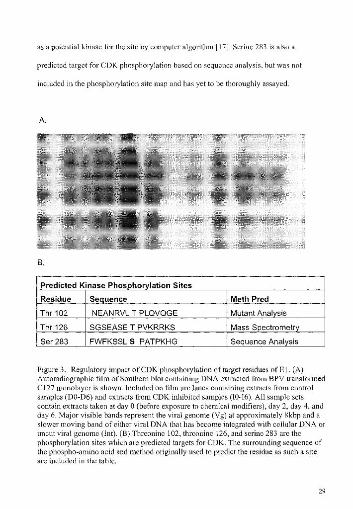
as a potential kinase for the site by computer algorithm [17]. Serine 283 is also a
predicted target for CDK phosphorylation based on sequence analysis, but was not
included in the phosphorylation site map and has yet to be thoroughly assayed.
A.
B.
Predicted Kinase Phosphorylation Sites
Residue Sequence Meth Pred
Thr102 NEANRVL T PLQVQGE Mutant Analysis
Thr126 SGSEASE T PVKRRKS Mass S_2ectrometty
Ser283 FWFKSSL S PATPKHG Sequence Analysis
Figure 3. Regulatory impact ofCDK phosphorylation oftm·get residues ofEl. (A) Autoradiographic film of Southern blot containing DNA extracted from BPV transformed C127 monolayer is shown. Included on film are lanes containing extracts from control samples (DO-D6) and extracts from CDK inhibited samples (I0-16). All sample sets contain extracts taken at day 0 (before exposure to chemical modifiers), day 2, day 4, and day 6. Major visible bands represent the viral genome (Vg) at approximately 8kbp and a slower moving band of either viral DNA that has become integrated with cellular DNA or uncut viral genome (Int). (B) Threonine 102, threonine 126, and serine 283 are the phosphorylation sites which are predicted targets for CDK. The sunounding sequence of the phospho-amino acid and method originally used to predict the residue as such a site are included in the table.
29

Regulatmy replication activity of CDK was assayed by comparison of a control set of
sample extracts with an inhibited set of extracts. The autoradiography of these extract
samples is shown in Figure 3A. Lanes DO-D6 contain the DNA extracted from the
BPV/Cl27 cells that had been maintained in complete DMEM and a small volume of
DMSO dilutent to control for the volume of inhibiting chemical added to the inhibited
set. The presence of viral DNA (V g) is detectable at the first time point extract (DO) and
accumulates over the course of the experiment, up to day 6 (D6). The band of intact
ge11ome is clear and distinct on all time points. A single band, heavier than the intact viral
genome (V g) is present in DO and most likely represents a portion of the viral DNA that
had integrated with the cellular DNA. There does appear to be a decrease in
concentration of viral DNA from D4 to D6. While the cellular environment was
controlled for and consistent with all time plates in all trials, some random differences in
cell growth and replication between each plate could be anticipated and is the most likely
explanation for the unaccountable decrease in viral DNA replication from these two time
points.
Lanes I0-16 contain the extracts from those plates exposed to the CDK inhibitor,
Roscovitine, which competitively inhibits the kinase by blocking the ATP-binding
domain. As with the control samples (DO-D6), the inhibited samples exhibit detectable
amounts of viral DNA as early as the first time point (IO). It is clear that the amount of
viral DNA increases over time, with no deviation. Comparison of the control and
inhibited samples displays the obvious difference in detectable amount of replicated viral
DNA. Extracted DNA from the CDK inhibited samples shows a consistently and
significantly lower concentration of viral DNA (V g) than the control samples. This
30

observation implicates a regulatory role for CDK in viral replication. Specifically, from
the notable decrease in viral DNA present when the kinase is inhibited, it appears that
under normal conditions in latent replication CDK phosphorylation of the BPV El
protein serves to stimulate the synthesis of viral DNA. While these results cannot
definitively confirm or deny that any target residue (T 102, T 126, or S 283) acts as a
regulatory phosphorylation site, it does provide evidence that CDK targets some residue
on the E1 protein to increase viral DNA replication.
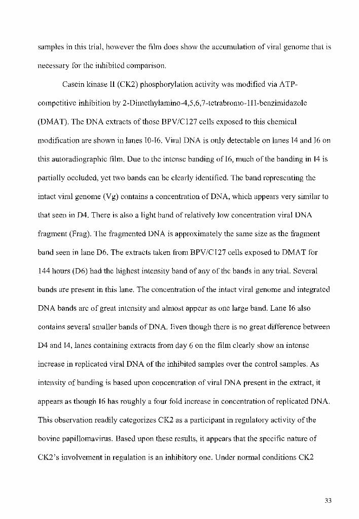
Casein Kinase II (CK2) Analysis. The enzyme CK2 has over 300 known substrates
and is a well-documented viral replication inducer [18], so it was not surprising that CK2
was the predicted kinase for so many phosphorylation sites (Fig. 4B). To study CK2's
role in BPV E1 function, cells were treated as previously described with 2-
Dimethylamino-4,5,6, 7 -tetrabromo-1H-benzimidazole (DMAT). Like the autoradiograph
of the PKC assay, the concentration of replicated viral DNA only reaches detectable
levels on the film at days four and six (D4-D6 and 14-16 ofFig-4A). The control extracts
collected at day four (D4) show a clear, distinct band of intact viral genome (V g). D4 also
contains a small band, lighter than the intact genome at approximately 6kbp (Frag) which
is most likely a portion of integrated or intact replicated genome that somehow became
fragmented. D6 has a noticeably higher concentration of viral DNA that is spilt into at
least three bands. The V g band is very subtle and barely visible on this film, especially
when compared to the denser band of integrated DNA in the D6 lane just above it. The
third D6 band is a very small portion of (intact or integrated) viral genome fragment of
only 2.2kbp. It is unclear at this time what lead to the fragmentation of the control
31
A
B.
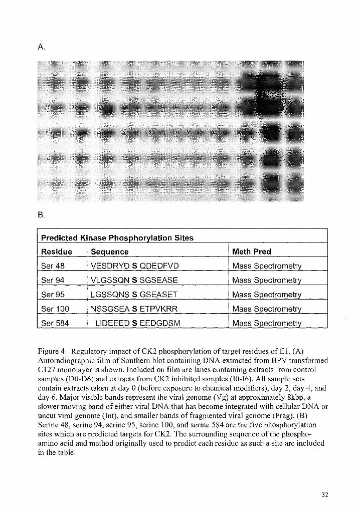
Predicted Kinase PhosQ_horylation Sites
Residue Sequence Meth Pred
Ser48 VESDRYD S QDEDFVD Mass Spectrometry
Ser94 VLGSSQN S SGSEASE Mass Spectrometry
Ser95 LGSSQNS S GSEASET Mass Spectrometry
Ser100 NSSGSEA S ETPVKRR Mass S_gectrometry
Ser584 LIDEEED S EEDGDSM Mass Spectrometry
Figure 4. Regulatory impact ofCK2 phosphorylation of target residues ofEl. (A) Autoradiographic film of Southern blot containing DNA extracted from BPV transformed C127 monolayer is shown. Included on film are lanes containing extracts from control samples (DO-D6) and extracts from CK2 inhibited samples (I0-16). All sample sets contain extracts taken at day 0 (before exposure to chemical modifiers), day 2, day 4, and day 6. Major visible bands represent the viral genome (Vg) at approximately 8kbp, a slower moving band of either viral DNA that has become integrated with cellular DNA or uncut viral genome (Int), and smaller bands of fragmented viral genome (Frag). (B) Serine 48, serine 94, serine 95, serine 100, and serine 584 are the five phosphorylation sites which are predicted targets for CK2. The surrounding sequence of the phosphoamino acid and method originally used to predict each residue as such a site are included in the table.
32
samples in this trial, however the film does show the accumulation of viral genome that is
necessary for the inhibited comparison.
Casein kinase II (CK2) phosphorylation activity was modified via ATP
competitive inhibition by 2-Dimethylamino-4,5,6, 7 -tetrabromo-1 H-benzimidazole
(DMAT). The DNA extracts of those BPV/Cl27 cells exposed to this chemical
modification are shown in lanes 10-16. Viral DNA is only detectable on lanes 14 and 16 on
this autoradiographic film. Due to the intense banding of 16, much of the banding in 14 is
partially occluded, yet two bands can be clearly identified. The band representing the
intact viral genome (V g) contains a concentration of DNA, which appears very similar to
that seen in D4. There is also a light band of relatively low concentration viral DNA
fragment (Frag). The fragmented DNA is approximately the same size as the fragment
band seen in lane D6. The extracts taken from BPV/C127 cells exposed to DMAT for
144 hours (D6) had the highest intensity band of any of the bands in any trial. Several
bands are present in this lane. The concentration of the intact viral genome and integrated
DNA bands are of great intensity and almost appear as one large band. Lane 16 also
contains several smaller bands of DNA. Even though there is no great difference between
D4 and 14, lanes containing extracts from day 6 on the film clearly show an intense
increase in replicated viral DNA of the inhibited samples over the control samples. As
intensity ofbanding is based upon concentration of viral DNA present in the extract, it
appears as though 16 has roughly a four fold increase in concentration of replicated DNA.
This observation readily categorizes CK2 as a participant in regulatory activity of the
bovine papillomavirus. Based upon these results, it appears that the specific nature of
CK2's involvement in regulation is an inhibitory one. Under normal conditions CK2
33

would therefore be expected to phosphorylate BPV's El protein and lead to a decrease in
the synthesis of the viral DNA during latent viral replication. These results are especially
encouraging for this study by showing that chemical modification of CK2 results in an
increase in viral replication.
DNA Protein Kinase (DNAPK) Analysis. The enzyme DNAPK is the predicted
phosphorylating agent of only one residue of the bovine papillomaviruses El protein,
which was detem1ined by mass spectrometry and computer algorithm (Fig. 5B). Serine
305, one of the two residues near the functional carboxyl terminus of the protein, is the
only predicted target for DNAPK phosphorylation. The autoradiographic film displaying
the extracts from the BPV/C127 plates of the DNAPK assay are shown in Figure 5A. As
with all the previous films, lanes labeled as DO-D6 contain the radio-probed time point
extracts of the control samples; those samples maintained only in complete DMEM and a
volume of DMSO equal to that of the chemical inhibitor added to the other samples.
DNA samples taken two days following the experimental set-up (D2) are the first to
shown viral DNA which had been synthesized to a detectable concentration. The
successive time point extractions collected at days 4 and 6 (D4 and D6) show a clearly
observable progressive increase in concentration. Given the significant increase in band
intensity of the intact viral genome (V g) between days two and four, it can be asserted
that the viral DNA concentration, at least, doubled. It is not as clear to assess the
concentration increase of viral DNA between D4 and D6, due to the extremely dark
banding ofboth samples. D4 appears to contain an area of radio-labeled DNA just above
the 8kbp position of the intact viral genome, and while the boundaries of two distinct
34

bands cannot be discemed, it seems apparent that D4 possesses a region of intact viral
genome close in proximity to a sample of viral DNA that has become integrated with a
portion of cellular DNA or an uncut portion of intact viral genome (1nt). The clear
distinction of these areas is further complicated in this film by the curvature in the control
bands, which is most likely due to an uneven migration of the extract samples during
electrophoresis.
The inhibited sample extracts of the DNAPK assay are contained in lanes labeled
I0-16. These samples were maintained under normal conditions as previously described,
except for the addition of an 1C50 concentration of 4,5-Dimethoxy-2-nitrobenzaldehyde
(DMNB), a highly selective competitive inhibitor ofDNAPK's ATP binding site. The
presence of replicated viral DNA was not detected in the inhibited extracts until 14. The
lane labeled as 14 possess one distinct band at 8kbp, representing the intact viral genome.
The relative intensity of the band is just slightly more dramatic than that seen in the V g
band of the D2 extracts. 16 contains a single band of viral DNA, the intensity ofwhich is
clearly increased from 14 but not quite the concentration ofD4 or D6. When comparing
the bands of viral DNA present in the control and inhibited samples, it is clear that viral
DNA is present at an earlier time point and accumulates to a much higher concentration
in the control samples than the inhibited ones. Because replication of both sample sets
unmistakably increased over time, at very different rates, the outcome of the DNAPK
assay would support prediction of the kinase's function as a regulatory enzyme in
35

A.
B.
Predicted Kinase Phosphorylation Site
Residue Sequence Meth Pred
Ser305 AQTTLNE S LQTEKFD Mass Spectrometry
Figure 5. Regulatory impact ofDNAPK phosphorylation of target residues ofEl. (A) Autoradiographic film of Southern blot containing DNA extracted from BPV transfom1ed C127 monolayer is shown. Included on film are lanes containing extracts from control samples (DO-D6) and extracts from CK2 inhibited samples (I0-16). All sample sets contain extracts taken at day 0 (before exposure to chemical modifiers), day 2, day 4, and day 6. Major visible bands represent the viral genome (Vg) at approximately 8kbp and a slower moving band of either viral DNA that has become integrated with cellular DNA or uncut viral genome (Int) (B) Serine 305 is currently the only phosphorylation site which is predicted target for DNAPK. The surrounding sequence of the phospho-amino acid and method originally used to predict the residue as such a site is included in the table.
replication mechanism. Inhibition ofDNAPK had noticeable effect on the concentration
of viral DNA synthesized, and taking in to account this decrease in replicative activity;
DNAPK could specifically be designated as a stimulatory kinase ofEl function.
36

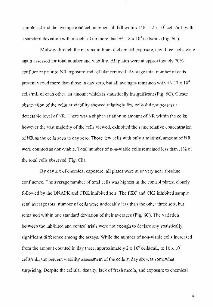
Neutral Red Viability Assay. Under control conditions the BPV/C127 cells
continuously grow and reach confluence from a 1 :20 passage of monolayer cells in 4-6
days and exhibit the characteristic fibroblast spindle shape (Fig. 6A, top left). All plates
were observed via light microscopy before addition of neutral red mixture, to ensure cells
were consistently proliferating throughout the plates and to survey the cellular
morphology of each trial's cells. There was no observable difference in visible density or
cellular shape between the control plates and any of the inhibited samples. All plates
possessed uniform distribution over the 1 Ocm diameter dishes at each time point and no
modified morphology was observed for any of the inhibited trials. The top right image in
figure 6A shows a PKC inhibited plate at day three; this image is representative of all the
inhibited plates, as there was no difference between them. The similarity seen between
the plates suggests that no significant changes in cellular maintenance or structure had
occurred due to the kinase inhibitions.
Neutral red (NR) dye was used to aid in the assay of the BPV/C127 cells'
overall health and proliferation. Neutral red uptake is a procedure commonly used to test
for chemical cyotoxicity, based upon the experimental chemical's effects on cellular
metabolism. The weakly cationic dye easily passes through the cellular membrane and is
collected inside lysosome vesicles through a process which is dependent upon intact
plasma and lysosomal membranes and active metabolism of the cell [38 and 39]. Cells
which have experienced any damage or cessation in active metabolism will not be able to
accumulate the bright red dye in the lysosomes, as an active, healthy cell would be able
to, and so can be readily distinguished.
37

A.
B.
Percent Viability of C127 Cells at Times of Chemical Exposure DayO Day3 Day6
Control 100* 99.991 99.977
CDK 100* 99.989 99.975
CK2 100* 99.989 99.965
DNAPK 100* 99.995 99.969
PKC 100* 99.997 99.967
38

C.
--.:1" < 0 ....
Total Cell Number At Time Points During Chemical Exposure
300
>< 250 1------------....1 E -II)
Ci) 200 (.)
(])
> <(
150
100 0 3 6
Time of Chemical Exposure (Days)
!•control 111CDK DCK2 DPKC
,Ill DNAPK
Figure-6. Viability and proliferative effects of chemical kinase modification on the BPV/Cl27 cells. (A) Cellular monolayer ofC127 transfected with BPV-1 genome is shown in all fields. Top left image is a control sample (Con) of cells and top right is a plate of cells post addition of the PKC inhibitor, BMI (PKC). Bottom images are the same plates exposed for four hours to lmL neutral red per lOOmL complete DMEM mixture before observation (Con NR & PKC NR). All plates were observed through light microscope at day 3 of experiment, digital images taken at IOOX magnification. Neutral red images were red enhanced for image color contrast. (B) Table includes calculated viability of cells taken at days 0, 3, and 6. All numbers reported as percent viable cells (number of viable cells/total number of cells observed). Viability was determined by each cell's ability to actively uptake the neutral red dye. * - Indicates that no cells were determined to be unviable (no non-colored cells seen). (C) Graphic representation of hemocytometer cell counts of control and inhibited samples at three different time points during experiment. Day zero counts were taken just before addition of chemical dilutent or modifier. Counts were also taken midway through (Day 3) and at the end (Day 6) of the chemical exposure.
39

The NR uptake assay was conducted as previously described with cell count,
viability, and overall morphology observed on three separate time points for control and
all types of inhibited samples. The PKC activator, TPA, was not included in this portion
of the study due to the chemicals inability to produce any differentially observable effects
from the control samples. Following each plates four hour exposure to the NR mixture,
cells were again observed through a light microscope. There was no generally detectable
difference between NR uptake of the cell plates at any time point. All viable cells that
successfully accumulated the dye exhibited an expected change in morphology. The cells
had a more swollen, rounded appearance, and a ring of red dye was seen around the
nucleus. All plates demonstrated this cellular change indicating consistent uptake of the
dye between the control and inhibited samples (Figure 6A, bottom left and bottom right
images), but closer quantitative investigation is necessary to ensure that kinase inhibition
is not significantly modifying cellular health and proliferation.
To acquire accurate cell viability and number counts, the cells were removed
from the plates and a portion of the cellular suspension was placed on a hemocytometer.
There were two plates used for each time point of each sample set and the counts were
repeated eight times for each plate to ensure statistical significance. During each count
total cell number and number of cells that had not accumulated the NR dye to a clearly
discernable level were determined. Day zero plates did 'not contain a single cell which
had not observably accumulated the dye (Fig. 6B). This was not surprising given that the
cells had just recently been passaged and so were at a low density with plenty of fresh
DMEM and no chemicals had yet been added. Total cell number was calculated for each
40
sample set and the average total cell numbers all fell within 148-152 x 104 cells/mL with
a standard deviation within each set no more than+/- 18 x 104 cells/mL (Fig. 6C).
Midway through the maximum time of chemical exposure, day three, cells were
again assessed for total number and viability. All plates were at approximately 70%
confluence prior to NR exposure and cellular removal. Average total number of cells
present varied more than those in day zero, but all averages remained with+/- 17 x 104
cells/mL of each other, an amount which is statistically insignificant (Fig. 6C). Closer
observation of the cellular viability showed relatively few cells did not possess a
detectable level ofNR. There was a slight variation in amount ofNR within the cells;
however the vast majority of the cells viewed, exhibited the same relative concentration
ofNR as the cells seen in day zero. Those few cells with only a minimal amount ofNR
were counted as non-viable. Total number of non-viable cells remained less than .1% of
the total cells observed (Fig. 6B).
By day six of chemical exposure, all plates were at or very near absolute
confluence. The average number of total cells was highest in the control plates, closely
followed by the DNAPK and CDK inhibited sets. The PKC and CK2 inhibited sample
sets' average total number of cells were noticeably less than the other three sets, but
remained within one standard deviation of their averages (Fig. 6C). The variation
between the inhibited and control trials were not enough to declare any statistically
significant difference among the assays. While the number of non-viable cells increased
from the amount counted in day three, approximately 2 x 10⁴ cells/mL, to 10 x 104
cells/mL, the percent viability assessment of the cells at day six was somewhat
surprising. Despite the cellular density, lack of fresh media, and exposure to chemical
41

inhibitors for six days, the total number of cells raised enough for the percentage of
viable cells to stay at around 99.9% for all sample plates, (Fig. 6B).
The collective results of the NR viability assay and total cell number counts,
yield excellent support for the results from the viral replication assays. Kinase inhibition
by the chemical modifiers BMI, Roscovitine, DMAT, and DMNB, do not appear to
significantly alter cellular health or proliferation of the BPV /C 127 cells. No difference in
gross cellular structure, cell viability, or cell growth was detem1ined to be considerable
en"ough to declare that a change had occuned in cellular replication. Had then been a
great difference in total cell number of any inhibited sample from the control, the
integrity of the results of that kinase inhibitor's effect on viral replication would be in
question, but because there was no significant difference in these variables, alteration of
viral replication most likely resulted from the decrease in kinase phosphorylation of the
bovine papilloma virus' E 1 protein.
DISCUSSIO~
The results of these experiments provide a clearer picture of differential
phosphorylation effects and add to the body of known information about regulation, in
general, by establishing the effects of phosphate addition to the papillomavirus
replication system. Relying upon chemical modification of specific kinases, this study
has yielded evidence which assigns a regulatory role in viral replication of the bovine
papillomavirus to the kinases PKC, CDK, CK2, and DNAPK. By analysis of the
42

concentrations of viral DNA between extracts from control and kinase inhibited
BPV/Cl27 cells, specific activities were assigned to these kinases. This data in
conjunction with the lack of impact these chemicals had upon cellular proliferation,
maintenance, and viability suggests that PKC, CDK, and DNAPK phosphorylation of the
BPV E1 protein lead to an increase in viral replication activity, while CK2
phosphorylation of this same protein causes a decrease in viral DNA replication. All
kinases assayed in this study were predicted based upon manual or computer analyzed
consensus sequence comparison of the identified phosphoresidues [2-4, 17, 25, 33]. Each
enzyme has also been reported to have some role in cell cycle regulation and/or DNA
metabolism, so it is not surprising that the results of this study implicate them as
regulators of viral replication. This study only accounts for those kinases which regulate
E 1 during the latent state of infection, when the protein would be present in the
undifferentiated cellular strata of the host. The kinases and their relative effects do not
address vegetative replication.
By predicting the kinases of five newly identified sites and two already
suspected sites the data gathered from mass spectrometric analysis served as an excellent
starting point for this study, but not every phospho-residue, and so not every possible
kinase, was definitively identified. It remains possible that some sites which are only
rarely or briefly phosphorylated were missed in the analysis [17] and of course the
kinases which recognize these sites would be overlooked. Therefore, it was of interest to
this study to include those kinases which, based upon alternative analytic means, are
predicted to phosphorylate other phospho-residues. Assays of serine 90 and serine 109
have strongly implicated these residues as phosphorylation sites [3, 33]. This data,
43

together with kinase's function in epidermal differentiation, provided enough support for
PKC's role as a regulator to include it in the kinase assays.
Inhibition of PKC in a monolayer of BPV /C 127 cells lead to a dramatic
decrease in the concentration of viral DNA that was extracted from the cells. This finding
in addition to the kinase's effects on repressed tumor suppression, rapid cell growth [15,
16], and a reported role in regulation of epithelial differentiation [3, 40], provides strong
support for the enzymes stimulatory action in viral replication. The coupling of a
regulator of skin differentiation status and viral DNA replication into the same kinase
would serve as a great means for the virus to link its lifecycle with its host replication
activity. The replication inducing activity of the kinase in this study contrasts with its
activity observed in earlier studies. Serine 90 and serine 1 09 site specific studies found
that a serine/alanine mutant of E 1, which was unable to be phosphorylated at ·those
residues, replicated more efficiently than the wild type E1 [3, 33]. Utilizing a system
similar to that used in this study, it was reported that the inability of E 1 to be
phosphorylated by PKC lead to an increase viral replication activity. What lead to the
dissimilar results of these studies is unclear at this time. However, it is quite possible that
phosphorylation of one PKC residue versus phosphorylation of two or more causes
different effects upon replication. It is important to note that the results of this study
cannot definitively assert that the kinases predicted to target the identified
phosphorylation sites are indeed acting on these sites. These results serve only as a
comprehensive analysis of the kinase effects, not the effects of any site specific
phosphate addition. A degree of overlap does exist between sequence consensuses of the
kinases, so it remains possible, although unlikely, that the kinases are targeting a site
44

other that those predicted for it or targeting a residue missed in previous assays as a
phosphorylation site.
CDK, a major regulator in cell cycle progression, has been shown to
phosphorylate threonine 102 in vitro; however mutation study of the site, like those
performed on S 90 and S 109, did not appear to significantly alter viral replication. Yet,
based upon this study's autoradiographic data, cyclin dependent kinases (CDK) appear to
donate their phosphate groups in a mechanism which promotes viral DNA replication. At
this time CDK's other predicted residues, threonine 126 and serine 283 have not been
analyzed. It may be that T 126 or S 283 site is the key replication regulator and mutation
would cause a change in viral replication like that seen in this study or that all three sites
must be phosphorylated to effect replication regulation, but further investigation of this
site is needed to determine its function. While the E 1 protein of the bovine
papillomavirus does not appear to express the CDK target domain of nuclear export, as
the human group does [21], the significant decrease in concentration of viral DNA
replicated in those cells exposed to the enzyme's inhibitor signifies that CDK
phosphorylation does have some effect on E1 activity. CDK has been reported as having
stimulatory and inhibitory functions in replication of the cellular genome [36]. The
dynamic status of this kinase may suggest that the enzyme differentially acts upon the
regulatory E1 protein, based upon concentration, cellular activity, or other metabolic
factor, in which case the results ofthe CDK assay in this study are only one facet of the
kinase's total activity.
Several characteristics of the enzyme CK2 made it of great interest to this
study; including its published effects upon replication of other viruses, high activity in
45
proliferating cells, and constitutive nature [18, 20]. This kinase has an incredibly diverse
group of known targets of both cellular and viral nature with a wide range of function.
Like CDK, it may be that this enzyme can lead to several different effects and this study
has only elucidated one of those functions in the virallifecycle. The CK2 assay clearly
indicates that phosphate addition by this kinase serves as an inhibitory factor in viral
replication. Of those residues predicted as targets for CK2, only serine 48 and serine 584
have been specifically analyzed. Mutation assay of the serine 48 residue suggested a
stimulatory role for CK2 in viral replication, where substitution of the serine amino acid
for alanine blocked phosphorylation at this site and yielded a dramatic decrease in the
concentration of viral DNA replicated [25]. The S 584 residue, found in the ATPase
domain on the carboxyl end of the E1 protein (Fig. 1), was studied by mutation assay and
results showed a clear reduction in viral replication [ 1]. This kinase activity predicted by
both mutant studies is opposite what was discovered by this study. However, as
previously mentioned the mutation analysis experiments only analyze the function of a
single residue's phosphorylation, while this study looks at the kinases' overall effects.
The three other residues, S 94, S 95, and S 100, identified as targets for this kinase have
yet to be studied. Specific study of these other residues may yield more definite
explanations of this kinase's activity.
Results of the DNAPK assay provided evidence of the stimulatory action of the
kinase's function in viral replication of the bovine papillomavirus. Inhibition of the
kinase lead to a significant decrease in concentration of viral DNA replicated that was
extracted fi·om the BPV/C127 cells. This activity is not necessarily consistent with the
lmown tumor suppressive functions ofDNAPK, but is in line with the kinase's described
46
epidermal growth factor receptor activation [29, 37]. Activity of serine 305, a residue in
the DNA binding domain ofE1 [17], to date, has not specifically been analyzed.
While the information yielded from the autoradiographic films of the kinase trials
are encouraging and certainly sheds some light on the respective regulatory roles of the
kinases studied, this data alone cannot assert that the differential concentrations of viral
DNA replicated is solely due to the alteration of E 1 protein phosphorylation by those
kinases. The very nature of the virus lifecycle further complicates this matter. All the
kinases included in this study are supplied by the host cells, which the virus takes
advantage of, and obviously the kinases' primary responsibly would be to carry out
cellular functions. Any modification of these kinases, could hypothetically lead to an
alteration of those cellular functions, such as; cellular replication, cell cycle progression,
growth, cellular maintenance, etc. Therefore, if the chemical modifications used in the
previously described experiments were causing great changes in the health or growth of
cells exposed to the inhibitors then any changes in concentration of viral DNA would
simply be an artifact of that change, and not due to changes in kinase regulation of the E 1
protein. In other words, reduction in cellular replication, growth, and overall health,
would directly lead to reduction of viral replication, which is so highly dependent upon
the cellular machinery. To support any results derived from the replication assay
experiments, any effects on cellular metabolism must be accounted for. The lack of
impact upon overall cellular health and viability as assessed by the neutral red assay
strongly dissociates the differential DNA abundance between the control and treated
samples from a simple corruption of cellular activity. Instead, such results illustrate that
47
any differences between the relative viral DNA amounts are due to the modification in
activity of the kinases and their effects upon the viral replication system.
This study utilized a cellular system similar to the native cells in the host
epithelium. The cellular monolayer of C 127 exhibited exponential growth, like the
stratum basale layer ofbovine epithelium undergoes during differentiation. Both
experimental and native systems consist ofectoderm derived mammalian cells and
possess the same kinases. There may exist some variation in replication activity of these
ki1iases between the two cells types, but effects are believed to be subtle, given the
reported consistencies of the enzyme actives between similar cells [15-21, 29]. The BPV
transformed C127 cell monolayer system used in this study has been employed
successfully by several other experiments with the papillomavirus [1-5, 33]. Our results
support a general model for regulation ofEl function and viral DNA replication by the
cellular kinases of the host organism.
Of immediate interest would be to utilize this system to gain a comprehensive
understanding of these kinases effects on viral replication of the human papilloma virus
(HPV). A phosphorylation site map ofHPV, like that ofBPV seen in figure 1, is not
available at this time, but given the similarity of human and bovine epithelium and their
native kinases, it would be reasonable to believe that comparable kinase effects would be
observed. A clear image of the exact mechanisms by which this virus uses the cellular
enzymes to regulate the replication of its viral life cycle will aid in the study and possibly
the treatment of this virus. A virus which has grave effects upon the human population,
including the approximately 70 strains of which have been attributed as a factor in the
48

development of cervical cancer, the second most common cancer in women worldwide
[11].
49

REFERE~CES
1. Lentz, M., Zanardi, T., Filzen, R., Carter, J., and Hella, M. Functional analysis of a carboxyl-tenninal phosphorylation mutant of the BPV E1 protein. J. Mol. Bioi. 316: 597-607. 2002.
2. Lentz, M. R. A carboxyl-terminal serine of the bovine papillomavirus E1 protein is phosphorylated in vivo and in vitro. Virus Res. 83: 213-219. 2002.
3. Zanardi, T. A., Stanley, C. M., Saville, B. M., Spacek, S. M., and Lentz, M. R. Modulation of bovine papillomavirus DNA replication by phosphorylation of the viral E1 protein. Virology 228: 1-10. 1997.
4. Lentz, M. R., Pak, D., Mohr, I., and Botchan, M. R. TheEl replication protein of bovine papillomavirus type-1 contains an extended nuclear localization signal that includes a p34cdc2 phosphorylation site. J. Viral. 67: 1414-1423. 1993.
5. Sun, S., T., L., Lentz, M., MacPherson, P., and Botchan, M. Identification of a 68 kilodalton nuclear ATP-binding phosphoprotein encoded by bovine papillomavirus type-1. J. Viral. 64: 5093-5105. 1990.
6. Massimi, P. Regulation of the human papillomavirus oncoproteins by differential phosphorylation. Mol. and Cell Biochem. 227(1): 137-144. 2001.
7. Wilson, V. G., West, M., Woytek, K., and Rangasamy, D. Papillomavirus E1 proteins: form, function, and features. Virus Genes 24(3): 275-290. 2002.
8. Bryan, J., Han, A., Fife, K. H., and Brown, D. R. The Human Papillomavirus Type 11 E1 and E4 Protein Is Phosphorylated in Genital Epithelium. Virology 268(2): 430-439. 2000.
9. Belyavski, M., Westerman, M., Dimichele, L., and Wilson, V. G. Perturbation of the Host Cell Cycle and DNA Replication by the Bovine Papillomavirus Replication Protein El. Virology 219(1): 206-219. 1996.
10. Zur Hausen, H. Papillomaviruses causing cancer: evasion from host-cell control in early events in carcinogenesis. J. Nat!. Cancer Inst. 92: 690-698. 2000.
11. ACS, American Cancer Society. (2003, accessed 2005, September 1). [Online]. http:/ /www.cancer.org/docroot/CRI/CRI _ 2 _lx.asp?rnav=criov&dt=8
12. Lin, B. Y., Makhov, J.D., Griffith, T. R., and Broker, L. T. Chaperone proteins abrogate inhibition of the human papillomavirus (HPV) E1 replicative helicase by the HPV E2 protein. Mol. Cell. Bioi. 22: 6592-6604. 2002.
50

13. Liu, J.-S., Kuo, S., Broker, T.R. and Chow, L. The functions of human papillomavirus type 11 E1, E2, and E2C proteins in cell-free DNA replication. J Bioi. Chem. 270: 27283-27291. 1995.
14. Masterson, P. J., Stanley, M., Lewis, A., and Romanos, M.A. A C-terminal helicase domain of the human papillomavirus E1 protein binds E2 and the DNA polymerase alpha-primase p68 subunit. J Viral. 72: 7407-7419. 1998.
15. Newton, A.C. "Protein kinase C: Structure, function and regulation." J Bioi. Chem. 270: 28485-28498 1995.
16. Kwon, Y.G. and Mendelow, M. The active site substrate specificity of protein kinase C. J Bioi. Chem. 269 (7): 4839-4844. 1994.
17. Lentz, M.R., Stevens, S.M., Raynes, J.R., and Elkhoury, N. A phosphorylation map of the bovine papillomavirus E1 helicase. Virology J 313: art 13. 2006.
18. Meggio, F. and Pinna, L. One thousand and one substrates of protein kinase CK2. FASEB J. 17: 349-362. 2003.
19. Litchfield, D. Protein kinase CK2: structure, regulation, and role in cellular decisions of life and death. Biochem. J. 369: 1-15. 2003.
20. Faust, M., and Montenarh, M. Subcellular localization of protein kinase CK2: a key to its function?. Cell Tissue Res. 301: 329-340. 2000.
21. Deng, J., Yuan Lin, B., Jin, G., Wheeler, C., Ma, T., Harper, J.W., Broker, T.R., and Chow, L.T. Cyclin/CDK Regulates the Nucleocytoplasmic Localization of the Human Papillomavirus E1 DNA Helicase. J Viral. 78(24): 13954-13965. 2004.
22. Jans, D., Xiao, C., and Lam, M. H. Nuclear targeting signal recognition: a key control point in nuclear transport? Bioessays 22: 532-544. 2000.
23. Deng, W., Jin, G., Lin, B., Van Tine, B., Broker, T., and Chow, L.T. mRNA splicing regulates human papillomavirus type 11 E1 protein production and DNA replication. J Viral. 77: 10213-10226. 2003.
24. Yang, L., Mohr, I., Lim, A., Nohaile, M., and Botchan, M .. 1993. The E1 protein of bovine papilloma virus 1 is an ATP-dependent DNA helicase. Proc. Nat!. Acad. Sci. USA. 90: 5086-5090. 1993.
25. McShan, G. D., and Wilson, V. G. Contribution ofbovine papillomavirus type 1 E1 protein residue 48 to replication function. J Gen. Viral. 81: 1995-2004. 2000.
51

26. Cueille, N., Nougarede, R., Mechali, F., Philippe, M., and Bonne-Andrea, C. Functional interaction between the bovine papillomavirus virus type 1 replicative helicase E1 and cyclin E-Cdk2. J. Viral. 72: 7255-7262. 1998.
27. Jaken, S. Protein kinase C isozymes and substrates. Curr. Opin. Cell Biol. 8 (2): 168-173. 1996.
28. ICTV, International Committee on Taxonomy of Viruses. (2002, accessed 2005, November 9). [Online]. http:/ /www.ncbi.nlm.nih.gov/ICTV db/ICTV dB/4 7020000.htm
29. Gurley, K.E. and Kemp C. J. p53 induction, cell cycle checkpoints, and apoptosis in DNAPK- deficient SCID mice. Carcinogen 17 (12): 2537-2542. 1996.
30. Baker, T.S., Newcomb, W.W., Olsen, N., Cowsert, L., Olson, C., and Brown J.C. Structures of bovine and human papillomaviruses: Analysis by cryoelectron microscopy and three-dimensional image reconstruction. Biophys. J. 60: 1445-1456. 1991.
31. Kamper, N., Day, P. M., Nowak, T., Selinka, H., Florin, L., Bolscher, J., Hilbig, L., Schiller, J.T., and Sapp, M. A membrane destabilizing peptide in capsid protein L2 is required for egress ofpapillomavirus genomes from endosomes. J. Viral. 80: 759-768. 2006.
32. Moreno, M.L. Stein, G.S., and Stein, J.L. Nucleosomal organization of a BPV minichromsome containing a human H4 histone gene. Mol. Cell Biochem. 74 (2): 173-177. 1987.
33. Zanardi, T. A. Regulation of bovine papillomavirus DNA replication by phosphorylation of the viral E1 protein. In Biochemistry and Biophysics Volume Ph.D. College Station, TX. Texas A&M. 140. 1997.
34. Waldenstrom, M., Schenstrom, K., Sollerbrant, K., and Hansson, L. Replication of bovine papillomavirus vectors in murine cells. Gene 120 (2): 175-181. 1992.
35. Joen, S., Allen-Hoffman, B., and Lambert, P. Integration of human papillomavirus type 16 into the human genome correlates with a selective growth advantage of cells. J. Viral. 69 (5): 2989-2997. 1995.
36. Henneke, G., Koundrioukoff, S., and Hubscher, U. Multiple roles for kinases in DNA replication. EMBO reports 4 (3): 252-256. 2003.
37. Friedmann, B., Caplin, M., Savic, B., Shah, T., Lord, C. J., Ashworth, A., Hartley, J., and Hochhauser, D. Interaction of the epidermal growth factor receptor and the DNAdependent protein kinase pathway following gefitinib treatment. Mol. Can. Ther. 2006 (5): 209-218. 2006.
52

38. Fieness, A. G., Walton, J., Winterbourne, D., McGlashan, D., and Hermon, J. Quantitative correlation of neutral red dye uptake with cell number in human cancer cell cultures. Cell. Bio. Int. Rep. 11 (5): 373-378. 1987.
39. Zhang, S., Lipsky, M. M., Trump, B. J., and Hsu, C. Neutral red (NR) assay for cell viability and xenobiotic-induced cytotoxicity in primary cultures of human and rat hepatocytes. Cell Bio. and Tax. 6 (2): 219-234. 1990.
40. Gherzi, R., Sparatore, B., Patrone, M., Sciutto, A., and Briata, P. Protein kinase C mRNA levels and activity in reconstituted nonnal human epidermis: relationships to cell differentiation. Biochem. Biophys. Res. Com. 184 (1): 283-291. 1992.
41. Ozbun, M.A. and Meyers, C. Temporal Usage ofMultiple Promoters during the Life Cycle of Human Papillomavirus Type 31b. J. Viral. 72 (4): 2715-2722. 1998.
53
VITA
Joshua R. Raynes
Born, Savannah, Georgia.
Manied, June 18 2004 in St. Augustine, Florida to Rachel R. Raynes.
Education
June 2000 -Graduated with honors from Nease High School. St. Augustine, Florida.
May 2004 -Graduated Summa Cum Laude with Bachelor's of Science in Biology, minor in Philosophy, from University ofNorth Florida. Jacksonville, Florida.
June 2006- Completed degree requirements for Master's of Science in Biology from University ofNorth Florida. Jacksonville, Florida.
Employment History
Aug 2005 to May 2006- Graduate teaching assistant at University of North Florida, Jacksonville, Florida.
Aug 2000 to Jan 2006- Certified Phannacy Technician at CVS pharmacy, Jacksonville, Florida.
May 2004 to Aug 2005- Research Assistant at University ofNorth Florida, Jacksonville, Florida.
June 2004 to Dec 2004- Neurology Intern at Jacksonville Neurological care center with Dr. Carlos Gama, Jacksonville, Florida.
Mar 2003 to June 2004- Patient care technician at St. Luke's Hospital Emergency Department, Jacksonville, Florida.
Publications
Lentz, M.R., Stevens, S.M., Raynes, J.R., and Elkhoury, N. A phosphorylation map of the bovine papillomavirus E1 helicase. Virology J. 313: art 13. March 2006.
54

Published Abstracts:
Shideler, T., Lambert Ferland, M-K., Raynes, J., Lentz, M. The role of cellular kinases CDK and CK2 in regulating bovine papillomavirus DNA replication. American Society for Virology Annual Meeting; Madison, WI. 2006.
Lentz, M., Raynes, J., and Stevens, S. A Phosphmylation Site Map of the Bovine Papillomavirus E 1 Helicase. American Society for Virology Annual Meeting; State College, P A. 2005.
Lentz, M., Raynes, J., Elkhoury, N., and Stevens, S. Phosphate Site Mapping of the Papillomavirus E 1 Helicase. Molecular Biology of Small DNA Tumor Viruses; Madison, WI. 2004.
55