analgorithmforthepreclinicalscreeningofanticancerdrugs...
TRANSCRIPT

International Scholarly Research NetworkISRN PharmacologyVolume 2012, Article ID 513580, 5 pagesdoi:10.5402/2012/513580
Research Article
An Algorithm for the Preclinical Screening of Anticancer DrugsEffective against Brain Tumors
Juan Sebastian Yakisich
Department of Clinical Neuroscience, Karolinska University Hospital, Karolinska Institute, SE-141 86 Stockholm, Sweden
Correspondence should be addressed to Juan Sebastian Yakisich, [email protected]
Received 23 April 2012; Accepted 11 June 2012
Academic Editors: W. Jager, T. Kumai, and T. B. Vree
Copyright © 2012 Juan Sebastian Yakisich. This is an open access article distributed under the Creative Commons AttributionLicense, which permits unrestricted use, distribution, and reproduction in any medium, provided the original work is properlycited.
The anticancer drugs screening program is a long and expensive process. It is estimated that only 5% of drugs entering clinicaltrials are approved by the FDA. Moreover, many of the drugs that enter clinical trials are often of limited use in clinical practice,and most cancers remain untreatable. Brain tumors are particularly difficult to treat due to the presence of the blood brain barrierthat limits the penetration of anticancer drugs. Additionally the isolation from most brain tumors of putative cancer stem cellsand novel models of cancer stem cell biology suggest that anticancer drugs should be delivered for prolonged time and at higherconcentrations to deplete any potential tumorigenic cell. In this paper, current concepts of cancer stem cell biology and novelconcepts of anticancer drugs screening are integrated to develop a seven-steps algorithm as a guideline for the preclinical evaluationof active compounds for the treatment of brain tumors. The flexibility of the algorithm allows the inclusion of alternative studiesto exhaustively investigate anticancer drugs and creates multiple opportunities where decisions to engage or not in early clinicaltrials can be made providing a useful tool for translational research in neurooncology.
1. Introduction
The treatment of primary and metastatic human brain tu-mors (HBTs) faces important challenges due to the presenceof the blood brain barrier that limits the diffusion of anti-cancer drugs [1]. For instance, while the serum concen-trations of etoposide after a dose of 50 mg/m2 can reachvalues >1 mg/L (∼1.7 µM) [2], the average cerebro spinalfluid (CSF) concentrations after a dose 300 mg/m2 i.v. was0.175 µM; (range, 0.066 to 2.12) [3]. As expected, etoposidealone has been of limited use for brain tumors. The isolationof cancer stem cells (CSCs) from a variety of primary braintumors including gliomas [4], oligodendrogliomas, men-ingiomas [5, 6], and other tumors that frequently metastasizeinto the brain further complicates this scenario. In particular,novel models of cancer biology support the idea that CSCsand non-CSCs can interconvert into each other [7–11].Thus, to cure cancer all malignant cells should be eliminatedat once otherwise, any surviving cell that is potentiallytumorigenic will eventually regenerate the tumor, sometimesafter long disease-free intervals. From the clinical aspect,
brain tumor treatment will require the use of drugs ordrugs combinations that should reach the brain tumoraltissue for extended periods of time at effective concentra-tions to eliminate all cancer cells. With this in mind, ourlab developed several novel concepts that include (i) theregrowth concentration zero (RC0): defined as the minimumdrug concentration that kills 100% of cancer cells preventingregrowth when cells are re-incubated in drug free media[12], (ii) the term pankiller: defined as drugs or drugscombination that deplete cancer cells [13, 14], (iii) a noveltwo phases treatment (2PT) that in vitro was shown to beeffective in eliminating all cancer cells. In this treatmentregime, cells are exposed to drugs for few weeks (first phase)that eliminates most of the cancer cells leaving few survivingcells that are eliminated by treatment with a second drug(second phase). It has the advantage that the concentrationrequired for each phase is lower than the concentrationrequired for individual drugs to deplete all cells. For instance,while 1–5 µM salinomycin alone for one week was unableto eliminate 100% of previously untreated gliomas cells,cells that survived 3-4 weeks continuous exposure to 10 mM

2 ISRN Pharmacology
hydroxyurea were eliminated with only 0.5 µM salinomycin[15].
On the other hand, there is no anticancer drug programthat really fits the needs for the screening of active com-pounds for HBTs and in practice, clinical trials are ofteninitiated with limited information that usually lead to afailure. In average, the cost of each failure for oncology drugsis estimated in the range of hundreds millions of dollars perdrug [16]. It is then imperative to develop strategies tominimize the rate of such failures and identify drugs withhigher probability to succeed in clinical trials. In this article,based in the above-mentioned current models of cancer stemcell biology and novel concepts and strategies developed inour laboratory, an integrated algorithm is proposed as ageneral guideline for a more rational screening of anticancerdrugs for HBTs.
2. Drug Screening Algorithm for Brain Tumors
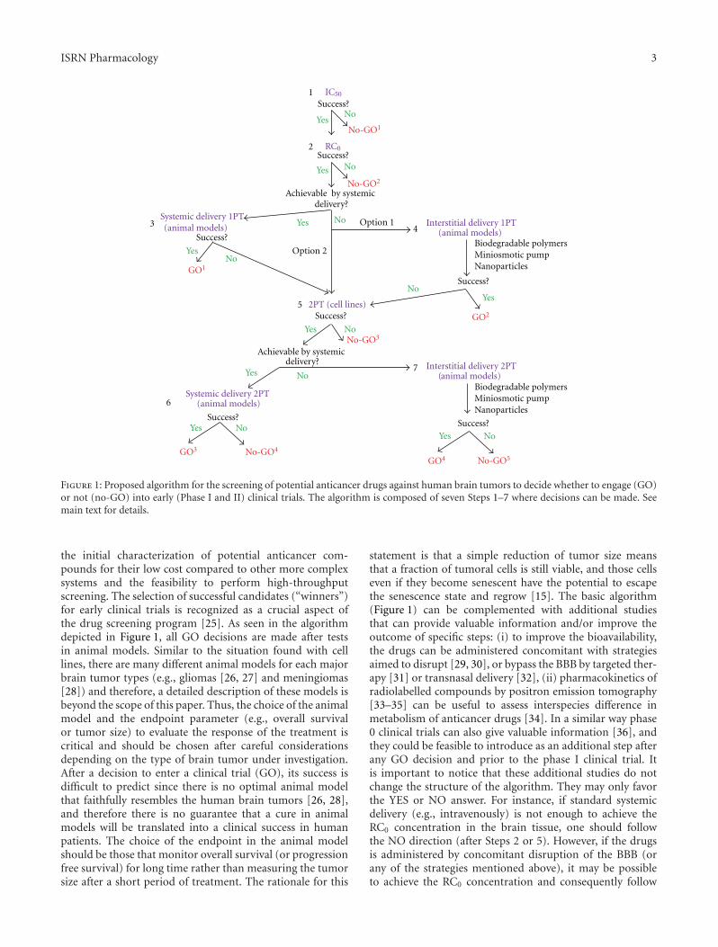
The proposed algorithm and its rationale for the screeningof new anticancer drugs for HBTs can be summarized asfollow (See Figure 1). The course to follow can be guided by asimple “Yes” or “No” response based in the success or failure,respectively, of the previous step.
Step 1. Initial Characterization of the IC50 for Specific Cancer.In this step a variety of standard primary or commercialcancer cell lines or tridimensional systems can be used. TheIC50 is a general endpoint to test the effect of drugs andgives valuable information such as potency and potentialmechanism(s) of action. The IC50 can be determined bya variety of high-throughput screening methods within 3-4 days and allows the selection of potential active drugsfrom large compound libraries. A failure (“NO”) at thisstage means that drug investigated is ineffective as anti-cancer drug, and it is not worth pursuing additional steps(no-GO1).
Step 2. Determination of the RC0 for Selected Compounds.Since there is no high-throughput assay available for thedetermination of the RC0 (that takes around 4 weeks), thisstep is rate limiting but may select compounds with thepotential to eradicate all cancer cells at once. A failure(“NO”) at this stage means that while the drug may haveshown some anticancer effect during Step 1, it will notdeplete all malignant cells as a single agent. Therefore, itshould be withdrawn from the screening process (no-GO2).During Steps 1 and 2, the pharmacokinetic of the drugsshould be studied in parallel to measure the concentrationthat actually reaches the brain tissue. If the RC0 concen-tration (and necessary exposure time) can be achieved inbrain tissue by systemic delivery (“Yes”), one should proceedwith Step 3, otherwise proceed to Step 4 (option 1) or Step 5(option 2, optional at this stage).
Step 3. Effectiveness of ADs on Animal Models Administeredby Systemic Delivery on a 1 Phase Treatment (1PT) Regime.A success at this step will lead the process to the first (GO1)decision and a failure to Step 5.
Step 4. Effectiveness of ADs on Animal Models Administeredby Interstitial Delivery on a 1 Phase Treatment (1PT) Regime.Similar to Step 3 this study will determine the effect-iveness in brain tumors and at the same time evaluate itsadverse effects. Several ways to deliver ADs locally (biode-gradable polymers, miniosmotic pump, and nanoparticles)can be evaluated at this stage. The local delivery of anti-cancer drugs by biodegradable polymers has been used inanimal models [17] and in patients [18], but its uses notwidespread. Miniosmotic pumps [19] and nanoparticles [20]are promising strategies for local delivery, but at present theiruse is limited to experimental models and they have not beentranslated into clinical use. A success (“Yes”) at this step willlead the screening process to the second (GO2) decision anda failure (“NO”) to Step 5.
Step 5. In Vitro Effectiveness of Drugs Administered in a TwoPhase Treatment (2PT) Regime. The aim of this type ofscreening is to find a drug combination that can be usedsequentially (even at lower concentrations) to eradicate alltumor cells in vitro. While the failure of this step will leadto the third decision to stop the evaluation of that particularcompound (no-GO3), the success will open the possibilityto pursue systemic delivery in animal models (Step 6) orinterstitial in animal models (Step 7) depending on whetherthe intracranial concentrations of both drugs can be achievedby systemic delivery.
Step 6. Effectiveness of ADs on Animal Models Administeredby Systemic Delivery on a 2 Phase Treatment (2PT) Regime.If successful it will lead to another opportunity to engage inclinical trials (GO3). Its failure will lead to a fourth decisionto stop the screening (no-GO4).
Step 7. Effectiveness of ADs on Animal Models Administeredby Interstitial l Delivery on a 2 Phase Treatment (2PT) Regime.If successful it will lead to the last opportunity to engage inclinical trials (GO4). Its failure will lead to a fifth decision tostop the screening (no-GO5).
3. Discussion
The term brain tumors refers to a variety of entities withunique clinicopathological characteristics [21], and it is likelythat each type of brain tumor will require a specific treatmentmodality. There is a plethora of model systems (cell linesand animal models) for those tumors that chemotherapymay play an important role and screening of new anticancerdrugs are being actively investigated. Only for gliomas, thereare several well-established cell lines commonly used inneurooncology. Barth and Kaur [22], described in detaileight rat brain tumor models (and their corresponding celllines). Mouse models of gliomas are also available [23].Therefore, at this level, the available resources are multiplefor each specific brain tumor but there is no consensus inwhich cell line is the best since in general cell cultures donot represent the complex heterogeneity of human braintumors that is the main disadvantage of cancer cell lines[24]. Despite this limitation, cell lines are very useful for
ISRN Pharmacology 3
delivery?
2PT (cell lines)
Interstitial delivery 1PT (animal models)
Success?
Yes No
YesNo
Systemic delivery 1PT
Success?
YesNo
Nanoparticles
Success?
Yes
No
No
Systemic delivery 2PT (animal models)
Yes NoSuccess?
Yes
Interstitial delivery 2PT (animal models)
Success?
Option 2
Option 1
1
2
3 4
5
7
6
delivery?
Yes No
Success?
Success?
YesNo
Yes No
IC50
No-GO1
No-GO2
No-GO3
No-GO4
RC0
Achievable by systemic
(animal models)
GO1
GO2
GO3
No-GO5GO4
Biodegradable polymersMiniosmotic pump
Nanoparticles
Biodegradable polymersMiniosmotic pump
Achievable by systemic
Figure 1: Proposed algorithm for the screening of potential anticancer drugs against human brain tumors to decide whether to engage (GO)or not (no-GO) into early (Phase I and II) clinical trials. The algorithm is composed of seven Steps 1–7 where decisions can be made. Seemain text for details.
the initial characterization of potential anticancer com-pounds for their low cost compared to other more complexsystems and the feasibility to perform high-throughputscreening. The selection of successful candidates (“winners”)for early clinical trials is recognized as a crucial aspect ofthe drug screening program [25]. As seen in the algorithmdepicted in Figure 1, all GO decisions are made after testsin animal models. Similar to the situation found with celllines, there are many different animal models for each majorbrain tumor types (e.g., gliomas [26, 27] and meningiomas[28]) and therefore, a detailed description of these models isbeyond the scope of this paper. Thus, the choice of the animalmodel and the endpoint parameter (e.g., overall survivalor tumor size) to evaluate the response of the treatment iscritical and should be chosen after careful considerationsdepending on the type of brain tumor under investigation.After a decision to enter a clinical trial (GO), its success isdifficult to predict since there is no optimal animal modelthat faithfully resembles the human brain tumors [26, 28],and therefore there is no guarantee that a cure in animalmodels will be translated into a clinical success in humanpatients. The choice of the endpoint in the animal modelshould be those that monitor overall survival (or progressionfree survival) for long time rather than measuring the tumorsize after a short period of treatment. The rationale for this
statement is that a simple reduction of tumor size meansthat a fraction of tumoral cells is still viable, and those cellseven if they become senescent have the potential to escapethe senescence state and regrow [15]. The basic algorithm(Figure 1) can be complemented with additional studiesthat can provide valuable information and/or improve theoutcome of specific steps: (i) to improve the bioavailability,the drugs can be administered concomitant with strategiesaimed to disrupt [29, 30], or bypass the BBB by targeted ther-apy [31] or transnasal delivery [32], (ii) pharmacokinetics ofradiolabelled compounds by positron emission tomography[33–35] can be useful to assess interspecies difference inmetabolism of anticancer drugs [34]. In a similar way phase0 clinical trials can also give valuable information [36], andthey could be feasible to introduce as an additional step afterany GO decision and prior to the phase I clinical trial. Itis important to notice that these additional studies do notchange the structure of the algorithm. They may only favorthe YES or NO answer. For instance, if standard systemicdelivery (e.g., intravenously) is not enough to achieve theRC0 concentration in the brain tissue, one should followthe NO direction (after Steps 2 or 5). However, if the drugsis administered by concomitant disruption of the BBB (orany of the strategies mentioned above), it may be possibleto achieve the RC0 concentration and consequently follow

4 ISRN Pharmacology
the YES direction. This algorithm is intended to improve thesuccess rate of the transition between the preclinical stageand early clinical trials (phase I and II) where toxicity andantitumor activity are the main endpoints [36]. One shouldbe aware that many drugs fail late in development (often inPhase III trials) due to unexpected issues related to safety,efficacy, and confounded outcomes [37] that can be verydifficult to predict even after successful Phase II trials. Onthe other hand, early and right decision to not engage into aclinical trial (No-GO) will save time and resources that can beused to give other compounds the chance to be evaluated asanticancer drugs. Right no-GO decisions are also importantto prevent the wrong elimination of potential useful drugsfrom the ADSP. The inclusion of the RC0 and the 2PT in thecore algorithm allows additional opportunities for a drug tobe exhaustively tested before being labeled as ineffective forHBTs. Finally, future improvements on methodologies usedat each one of the seven steps would improve the chances fora particular drug or drug combination to succeed in clinicaltrials.
4. Conclusions
The proposed algorithm combines in a rational way resultsfrom experimental studies of diverse nature (proliferationassays, pharmacokinetics, response in animal models of BTs)to create step-by-step guidelines for anticancer drug screen-ing of potential active compounds against HBTs. In partic-ular, the basic proposed algorithm is composed of 7 sevenpotential points (steps) where drugs or drugs combinationscan be experimentally tested. The progression in the algo-rithm is guided by” Yes” or “No” responses to previous resultsthat provide four opportunities where decisions can be madeto enter a clinical trial (GO1–GO4) or five opportunitieswhere the screening process should stop (no-GO1–no-GO5).Its flexibility allows the inclusion of additional studies thatcreates more opportunities to take GO or no-GO decisionsto enter clinical trials and can be a useful guideline forthe identification of active compounds and optimization oftherapeutic regimes for HBTs.
Conflict of Interests
The author declares no conflict of interests.
Acknowledgments
Research in the author’s lab is supported by grants from theSwedish Research Council and the Karolinska Institute.
References
[1] D. Fortin, “The blood-brain barrier: its influence in thetreatment of brain tumors metastases,” Current Cancer DrugTargets, vol. 12, no. 3, pp. 247–259, 2012.
[2] J. D. Hainsworth, “Extended-schedule oral etoposide in select-ed neoplasms and overview of administration and schedulingissues,” Drugs, vol. 58, no. 3, pp. 51–56, 1999.
[3] M. V. Relling, H. H. Mahmoud, C. H. Pui et al., “Etoposideachieves potentially cytotoxic concentrations in CSF of chil-dren with acute lymphoblastic leukemia,” Journal of ClinicalOncology, vol. 14, no. 2, pp. 399–404, 1996.
[4] T. Kondo, T. Setoguchi, and T. Taga, “Persistence of a smallsubpopulation of cancer stem-like cells in the C6 glioma cellline,” Proceedings of the National Academy of Sciences of theUnited States of America, vol. 101, no. 3, pp. 781–786, 2004.
[5] D. Y. Hueng, H. K. Sytwu, S. M. Huang, C. Chang, and H. I.Ma, “Isolation and characterization of tumor stem-like cellsfrom human meningiomas,” Journal of Neuro-Oncology, vol.104, no. 1, pp. 45–53, 2010.
[6] P. Rath, D. C. Miller, N. S. Litofsky et al., “Isolation andcharacterization of a population of stem-like progenitor cellsfrom an atypical meningioma,” Experimental and MolecularPathology, vol. 90, no. 2, pp. 179–188, 2011.
[7] M. Cruz, A. Siden, D. R. Tasat, and J. S. Yakisich, “Are allglioma cells cancer stem cells?” Journal of Cancer Science andTherapy, vol. 2, no. 4, pp. 100–106, 2010.
[8] M. A. Hatiboglu, J. Wei, A. S. G. Wu, and A. B. Heimberger,“Immune therapeutic targeting of glioma cancer stem cells,”Targeted Oncology, vol. 5, no. 3, pp. 217–227, 2010.
[9] D. R. Laks, K. Visnyei, and H. I. Kornblum, “Brain tumor stemcells as therapeutic targets in models of glioma,” Yonsei MedicalJournal, vol. 51, no. 5, pp. 633–640, 2010.
[10] Y. Li and J. Laterra, “Cancer stem cells: distinct entities ordynamically regulated phenotypes?” Cancer Research, vol. 72,no. 3, pp. 576–580, 2012.
[11] L. Vermeulen, F. de Sousa, E. Melo, D. J. Richel, and J. P.Medema, “The developing cancer stem-cell model: clinicalchallenges and opportunities,” The Lancet Oncology, vol. 13,no. 2, pp. 83–89, 2012.
[12] D. Avramidis, M. Cruz, A. Siden, D. R. Tasat, and J. S. Yakisich,“Regrowth Concentration Zero (RC0) as complementaryendpoint parameter to evaluate compound candidates duringpreclinical drug development for cancer treatment,” Journal ofCancer Science & Therapy, vol. 1, pp. 19–24, 2009.
[13] Z. M. Delwar, D. Avramidis, E. Follin et al., “Cytotoxic effectof menadione and sodium orthovanadate in combination onhuman glioma cells,” Investigational New Drugs. In press.
[14] M. F. Vita, N. Nagachar, D. Avramidis et al., “Pankiller effectof prolonged exposure to menadione on glioma cells: potenti-ation by vitamin C,” Investigational New Drugs, vol. 29, no. 6,pp. 1314–1320, 2010.
[15] Z. M. Delwar, D. Avramidis, A. Siden, M. Cruz, and J. S.Yakisich, “Depletion of drug-surviving glioma cells by a sec-ond phase treatment with low concentration of salinomycin,”Drugs and Therapy Studies, vol. 1, p. e7, 2011.
[16] J. A. Di Masi and H. G. Grabowski, “Economics of newoncology drug development,” Journal of Clinical Oncology, vol.25, no. 2, pp. 209–216, 2007.
[17] V. R. Recinos, B. M. Tyler, K. Bekelis et al., “Combinationof intracranial temozolomide with intracranial carmustineimproves survival when compared with either treatment alonein a rodent glioma model,” Neurosurgery, vol. 66, no. 3, pp.530–537, 2010.
[18] P. Miglierini, M. Bouchekoua, B. Rousseau, P. Dam Hieu,J. Malhaire, and O. Pradier, “Impact of the per-operatoryapplication of GLIADEL wafers (BCNU, carmustine) in com-bination with temozolomide and radiotherapy in patientswith glioblastoma multiforme: efficacy and toxicity,” ClinicalNeurology and Neurosurgery. In press.
[19] G. Xi, B. Mania-Farnell, V. Rajaram et al., “Efficacy of inter-stitial continuous vincristine infusion in a bioluminescent

ISRN Pharmacology 5
rodent intracranial tumor model,” Journal of Neuro-Oncology,vol. 106, no. 2, pp. 261–270, 2012.
[20] R. N. Saha, S. Vasanthakumar, G. Bende, and M. Snehalatha,“Nanoparticulate drug delivery systems for cancer chemother-apy,” Molecular Membrane Biology, vol. 27, no. 7, pp. 215–231,2010.
[21] D. N. Louis, H. Ohgaki, O. D. Wiestler et al., “The 2007 WHOclassification of tumours of the central nervous system,” ActaNeuropathologica, vol. 114, no. 2, pp. 97–109, 2007.
[22] R. F. Barth and B. Kaur, “Rat brain tumor models in experi-mental neuro-oncology: the C6, 9L, T9, RG2, F98, BT4C, RT-2 and CNS-1 gliomas,” Journal of Neuro-Oncology, vol. 94, no.3, pp. 299–312, 2009.
[23] E. Binello, Z.A. Qadeer, H.P. Kothari, L. Emdad, and I.M.Germano, “Stemness of the CT-2A Immunocompetent mousebrain tumor model: characterization in vitro,” Journal of Can-cer, vol. 3, pp. 166–174, 2012.
[24] H. Karlsson, M. Fryknas, R. Larsson, and P. Nygren, “Lossof cancer drug activity in colon cancer HCT-116 cells duringspheroid formation in a new 3-D spheroid cell culture system,”Experimental Cell Research, vol. 318, no. 13, pp. 1577–1585,2012.
[25] R. Goodwin, G. Giaccone, H. Calvert, M. Lobbezoo, and E.A. Eisenhauer, “Targeted agents: how to select the winners inpreclinical and early clinical studies?” European Journal ofCancer, vol. 48, no. 2, pp. 170–178, 2012.
[26] V. L. Jacobs, P. A. Valdes, W. F. Hickey, and J. A. De Leo,“Current review of in vivo GBM rodent models: emphasis onthe CNS-1 tumour model,” ASN Neuro, vol. 3, no. 3, ArticleID e00063, 2011.
[27] T. S. Jones and E. C. Holland, “Animal models for glioma drugdiscovery,” Expert Opinion on Drug Discovery, vol. 6, no. 12,pp. 1271–1283, 2011.
[28] M. Kalamarides, M. Peyre, and M. Giovannini, “Meningiomamouse models,” Journal of Neuro-Oncology, vol. 99, no. 3, pp.325–331, 2010.
[29] S. J. Madsen and H. Hirschberg, “Site-specific opening of theblood-brain barrier,” Journal of Biophotonics, vol. 3, no. 5-6,pp. 356–367, 2010.
[30] C. Y. Ting, C. H. Fan, H. L. Liu et al., “Concurrent blood-brainbarrier opening and local drug delivery using drug-carryingmicrobubbles and focused ultrasound for brain glioma treat-ment,” Biomaterials, vol. 33, no. 2, pp. 704–712, 2012.
[31] P. Zhang, L. Hu, Q. Yin, L. Feng, and Y. Li, “Transferrin-modified c[RGDfK]-paclitaxel loaded hybrid micelle forsequential blood-brain barrier penetration and glioma target-ing therapy,” Molecular Pharmacology, vol. 9, no. 6, pp. 1590–1598, 2012.
[32] T. Shingaki, D. Inoue, T. Furubayashi et al., “Transnasal deliv-ery of methotrexate to brain tumors in rats: a new strategy forbrain tumor chemotherapy,” Molecular Pharmaceutics, vol. 7,no. 5, pp. 1561–1568, 2010.
[33] O. C. Hutchinson, D. R. Collingridge, H. Barthel, P. M.Price, and E. O. Aboagye, “Pharmacokinetics of radiolabelledanticancer drugs for positron emission tomography,” CurrentPharmaceutical Design, vol. 9, no. 11, pp. 917–929, 2003.
[34] S. Osman, S. K. Luthra, F. Brady et al., “Studies on themetabolism of the novel antitumor agent [N-methyl- 11C]N-[2-(dimethylamino)ethyl]acridine-4-carboxamide in rats andhumans prior to phase I clinical trials,” Cancer Research, vol.57, no. 11, pp. 2172–2180, 1997.
[35] A. Saleem, E. O. Aboagye, J. C. Matthews, and P. M. Price,“Plasma pharmacokinetic evaluation of cytotoxic agents
radiolabelled with positron emitting radioisotopes,” Can-cer Chemotherapy and Pharmacology, vol. 61, no. 5, pp. 865–873, 2008.
[36] J. Arrondeau, H. K. Gan, A. R. Razak, X. Paoletti, and C.Le Tourneau, “Development of anti-cancer drugs,” Discoverymedicine, vol. 10, no. 53, pp. 355–362, 2010.
[37] G. J. Kelloff and C. C. Sigman, “New science-based endpointsto accelerate oncology drug development,” European Journal ofCancer, vol. 41, no. 4, pp. 491–501, 2005.
Submit your manuscripts athttp://www.hindawi.com
PainResearch and TreatmentHindawi Publishing Corporationhttp://www.hindawi.com Volume 2014
The Scientific World JournalHindawi Publishing Corporation http://www.hindawi.com Volume 2014
Hindawi Publishing Corporationhttp://www.hindawi.com
Volume 2014
ToxinsJournal of
VaccinesJournal of
Hindawi Publishing Corporation http://www.hindawi.com Volume 2014
Hindawi Publishing Corporationhttp://www.hindawi.com Volume 2014
AntibioticsInternational Journal of
ToxicologyJournal of
Hindawi Publishing Corporationhttp://www.hindawi.com Volume 2014
StrokeResearch and TreatmentHindawi Publishing Corporationhttp://www.hindawi.com Volume 2014
Drug DeliveryJournal of
Hindawi Publishing Corporationhttp://www.hindawi.com Volume 2014
Hindawi Publishing Corporationhttp://www.hindawi.com Volume 2014
Advances in Pharmacological Sciences
Tropical MedicineJournal of
Hindawi Publishing Corporationhttp://www.hindawi.com Volume 2014
Medicinal ChemistryInternational Journal of
Hindawi Publishing Corporationhttp://www.hindawi.com Volume 2014
AddictionJournal of
Hindawi Publishing Corporationhttp://www.hindawi.com Volume 2014
Hindawi Publishing Corporationhttp://www.hindawi.com Volume 2014
BioMed Research International
Emergency Medicine InternationalHindawi Publishing Corporationhttp://www.hindawi.com Volume 2014
Hindawi Publishing Corporationhttp://www.hindawi.com Volume 2014
Autoimmune Diseases
Hindawi Publishing Corporationhttp://www.hindawi.com Volume 2014
Anesthesiology Research and Practice
ScientificaHindawi Publishing Corporationhttp://www.hindawi.com Volume 2014
Journal of
Hindawi Publishing Corporationhttp://www.hindawi.com Volume 2014
Pharmaceutics
Hindawi Publishing Corporationhttp://www.hindawi.com Volume 2014
MEDIATORSINFLAMMATION
of