an automatic gastrointestinal polyp detection system in...
TRANSCRIPT
Research ArticleAn Automatic Gastrointestinal Polyp DetectionSystem in Video Endoscopy Using Fusion of Color Wavelet andConvolutional Neural Network Features
Mustain Billah,1 SajjadWaheed,1 andMohammadMotiur Rahman2
1Department of Information and Communication Technology, Mawlana Bhashani Science and Technology University,Tangail, Bangladesh2Department of Computer Science and Engineering, Mawlana Bhashani Science and Technology University, Tangail, Bangladesh
Correspondence should be addressed to Mustain Billah; [email protected]
Received 7 May 2017; Accepted 12 July 2017; Published 14 August 2017
Academic Editor: Tiange Zhuang
Copyright © 2017 Mustain Billah et al. This is an open access article distributed under the Creative Commons Attribution License,which permits unrestricted use, distribution, and reproduction in any medium, provided the original work is properly cited.
Gastrointestinal polyps are considered to be the precursors of cancer development in most of the cases. Therefore, early detectionand removal of polyps can reduce the possibility of cancer. Video endoscopy is themost used diagnosticmodality for gastrointestinalpolyps. But, because it is an operator dependent procedure, several human factors can lead to misdetection of polyps. Computeraided polyp detection can reduce polyp miss detection rate and assists doctors in finding the most important regions to payattention to. In this paper, an automatic system has been proposed as a support to gastrointestinal polyp detection. This systemcaptures the video streams from endoscopic video and, in the output, it shows the identified polyps. Color wavelet (CW) featuresand convolutional neural network (CNN) features of video frames are extracted and combined together which are used to train alinear support vector machine (SVM). Evaluations on standard public databases show that the proposed system outperforms thestate-of-the-art methods, gaining accuracy of 98.65%, sensitivity of 98.79%, and specificity of 98.52%.
1. Introduction
Themost leading cause of death in the whole world is cancer.Again, gastrointestinal cancer is the most commonly occur-ring cancer which originates from gastrointestinal polyps.Actually, gastrointestinal polyps are the abnormal growthof tissue on gastric and colonic mucosa. This growth is aslow process and in majority of the cases, before reaching alarge size, they do not produce symptoms. However, cancer ispreventable and curable, if polyps could be detected early.
Video endoscopy is the most used diagnostic modalityfor gastrointestinal polyps. In typical video endoscopy, asmall camera is entered and directed through the gastroin-testinal tract to detect and remove polyps. But typical videoendoscopy takes long period of time generating a long video.So, as an operator dependent procedure, it is not possible fora medical person to examine it with sufficient attentivenessduring such long and back-to-back endoscopy. However,accuracy of the diagnosis depends on doctor’s experience.
So, in the examination, some polyps may be undetected.This misdetection of polyps can lead to malignant tumorsin the future. Computer aided polyp detection system canreduce polyp misdetection rate and assists doctors in findingthe most important regions to be analyzed. Such system cansupport diagnosis procedure by detecting polyps, classifyingpolyps, and generating detailed report about any part thatshould be examined with more attention. Again, duration ofthis uncomfortable process for the patients and the cost ofoperation can also be reduced.
A large number of methods have been proposed andapplied for computer aided polyp detection system. Covari-ances of the second-order statistical measures over thewavelet frame transformation (CWC) of different color bandshave been used as the image features in [1] for colonoscopytumor detection with 97% specificity and 90% sensitivity. Intheir consecutive work [2], an intelligent system of SVM andcolor-texture analysis methodologies was developed havingaccuracy of 94%. Adaptive neurofuzzy-based approach for
HindawiInternational Journal of Biomedical ImagingVolume 2017, Article ID 9545920, 9 pageshttps://doi.org/10.1155/2017/9545920

2 International Journal of Biomedical Imaging
polyp detection in video capsule endoscopy (VCE) wasproposed by Kodogiannis et al. [3]. Using texture spectrumfrom different color channels, they obtained 97% sensitivityover 140 images. Alexandre et al. [4] showed the comparisonof texture based and color and position based methodsperformed in database of 4620 images and obtained areaunder the curve (AUC) value of 94.87% for the texturehistogram of RGB + XY. Combination of color and shapefeatures was used to discriminate polyp from normal regionsin [5]. About 94.20% accuracy was gained when they usedmultilayer perceptron (MLP) as the classifier. A deep con-volutional neural network based classification problem wasstudied for classifying digestive organs in wireless capsuleendoscopy in [6]. Another computer aided lesion detec-tion system based on convolutional neural network (CNN)is utilized for more features of endoscopy images in [7].They also showed comparison between CNN features andcombination of color histogram features and LBP featuresin the experiment. Features learned by CNN outperformedthe other method. Tajbakhsh et al. presented a new methodintegrating global geometric constraints of polyp and localpatterns of intensity variation across polyp boundaries [8]. In[9], CNN features have been used to improve the accuracyof colonic polyp classification with sensitivity of 95.16% andspecificity of 74.19%. A unique 3-way image presentation andconvolutional neural network based polyp detection methodhave been proposed by Tajbakhsh et al. [10]. Jia et al. used10,000WCE images for automatic bleeding detection strategy[11]. They also used convolutional neural network (CNN) forthis purpose. Ribeiro et al. suggested that features learned byCNN trained from scratch are more relevant for automatedpolyp detection system [12]. CNN derived features showgreater invariance to viewing angles and image quality factorswhen compared to the Eigen model [13]. However, fusionscheme of wavelet color-texture analysis and convolutionalneural network feature has not been reported in the literatureto the best of our knowledge.
In this paper, an automatic system has been proposedas a support to gastrointestinal polyp detection. After theendoscopy video is fed into the proposed system, it extractscolor wavelet features and convolutional neural networkfeatures from each sliding window of video frames. Fusionof all the features is fed into SVM for classifying it as polypor nonpolyp. Detected polyp window in the frame is markedand showed in the output. Proposed automatic systemdetectspolyps with an accuracy of 98.65%.
The rest of the paper is organised as follows.The proposedsystem architecture and methods used in the system aredescribed in Section 2. In Section 3, experimental results areanalyzed. Finally, the conclusions of this study are presentedin Section 4.
2. Structure and Methods
Proposed system is implemented in MATLAB 2017a. It takesvideo endoscopy in different formats such as avi, mp4,and wmv and outputs the characterized video with markedpolyps. This system is divided into some segments suchas video to preprocessed frame, frame to sliding window,
Figure 1: Original video frame.
Figure 2: Preprocessed frame.
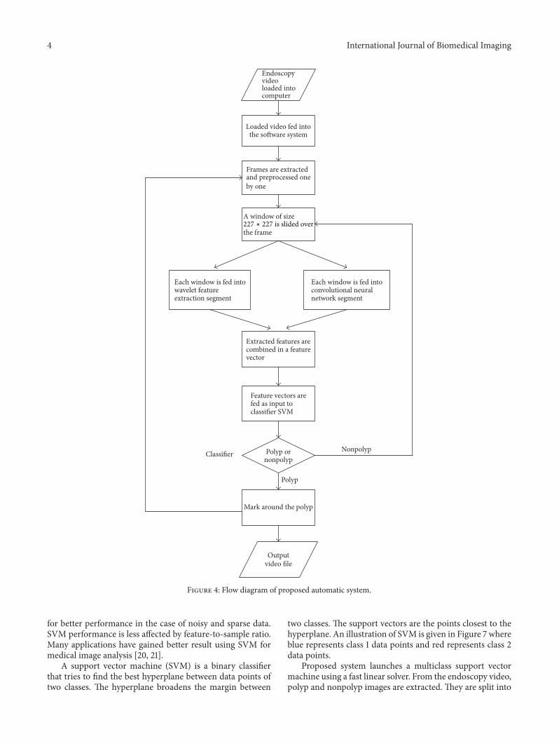
wavelet feature segment, convolution neural network seg-ment, classification segment, and output segment. All thesegments and related methods are outlined sequentially(Figure 4).
2.1. Video to Preprocessed Frame. Endoscopy video to beexamined for finding possible polyp is loaded in computer.Then the video is fed into the proposed automatic system.Actually every video is the running sequence of still imagescalled frame. Such a video frame is showed in Figure 2. Butall the regions of the original video are not significant, ratherthere are some unnecessary regions containing descriptionand other information. Examining such regions is nothingbut a waste of time. Therefore, unnecessary regions of theoriginal video frame (Figure 1) are discarded resulting inframes as in Figure 2.
2.2. Frame to Sliding Window. A window of size 227 ∗ 227is slided over the frame from left to right and top to bottom,thus generating small images (called window) from a singlevideo frame as shown in Figure 3. Each window images areconsidered to be the inputs of feature extraction segment.
2.3.Wavelet Feature Segment. The size of polyps varies in dif-ferent patients. So multiresolutional analysis such as waveletperforms better for textural analysis. But [1] suggests thatgrayscale textural features are not significant representativefor video endoscopy images. So the proposed system usescolor textural features from wavelet decomposed images.
Every RGB image has three-color channels: red, green,and blue. So input image, 𝐼 (sliding window) is decomposedinto three-color channels 𝐼𝐶, where 𝐶 = 𝑟, 𝑔, 𝑏.𝐷
6.

International Journal of Biomedical Imaging 3
Sliding window
Figure 3: A window of 227 ∗ 227 is sliding along the frame.
A 3-level and 2-dimensional discrete wavelet transforma-tion is applied on each 𝐼𝑐, generating a low resolution image𝐿𝐶𝐶𝐿
and three-detail image 𝐷𝐶𝐶𝐿, where 𝐶𝐿 = 1, 2, 3 . . . , 9 for
3-level decomposition.As textural information is localized in the middle wavelet
detailed channels original image, only the detail images for𝐶𝐿 = 4, 5, 6 are taken into account (Figure 5). So, finallytotal nine images {𝐷𝐶
𝐶𝐿} are considered for further processes,
where 𝐶𝐿 = 4, 5, 6 and 𝐶 = 𝑟, 𝑔, 𝑏.For finding information about spatial relationships of
pixels in an image, another statistical method named cooc-currence matrix is calculated over above nine images. Thesematrices are calculated in four different directions 0∘, 45∘, 90∘,and 135∘ generating 36 matrices.
In [14, 15], various statistical features were proposedamong which four statistical measures are considered inthis proposed system: correlation, energy, homogeneity, andentropy. Finally, four statistical measures for 36 matricesresult in total 144 color wavelet features.
2.4. Convolution Neural Network Segment. Each window of227∗227 size is inserted into this segment and convolutionalneural network features are extracted for the window.
A simple convolutional neural network (CNN) is asequence of layers where every layer of a CNN transformsone volume of activation to another through a differentiablefunction. CNNs apply consecutive filters to the raw pixeldata of an image to extract and learn different featuresthat can be used for classification. The architecture of atypical CNN is composed of multiple layers where each layerperforms a specific function of transforming its input intoa useful representation. There are 3 major types of layersthat are commonly observed in complex neural networkarchitectures:
(i) Convolutional layers: in this layer, convolution filtersare applied to the image. For each subregion, thelayer performs a set of mathematical operations andproduces a single value in the output feature map.Convolutional layers then typically apply a ReLUactivation function to the output, thus introducingnonlinearities into the model. ReLU, rectified linearunit, is an activation function which can be expressedmathematically: f(x) = max(0, x). A smooth approx-imation to the rectifier is the analytic function, f(x) =ln(1 + ex), called the softplus function.
(ii) Pooling layers: it downsamples the image dataextracted by the convolutional layers and reduces thedimensionality of the feature map to decrease pro-cessing time. Max pooling, a commonly used poolingalgorithm, extracts subregions of the feature mapand discards all other values keeping their maximumvalue.
(iii) Dense (fully connected) layers: this Layer performsclassification on the features extracted by the convolu-tional layers and downsampled by the pooling layers.In a dense layer, every node in the layer is connectedto every node in the preceding layer.
The CNN proposed by this work is inspired by [16]. Itcontains the following layers, parameters, and configuration(Figure 6):
(i) Input layer: sliding window image is obtained fromvideo frame of size 227 ∗ 227 ∗ 3.
(ii) Two combinations of convolutional and pooling lay-ers: first convolutional layer consists of 96 filters ofsize 11 × 11 with padding 0 and stride set to 4. Thesecond convolutional layer consists of 256 filters ofsize 5 × 5 with padding 2 and stride set to 1. Bothlayers are followed by a ReLU rectifier function. Aftereach convolutional layer, there is a max pooling layerconsisting of windows with size of 3 × 3 and stride setto 2.
(iii) Three convolutional layers and a pooling layer: thethird, fourth, and fifth convolutional layers are fol-lowed by ReLU function containing 384, 384, and256 filters, respectively. After the three convolutionallayers, there is a max pooling layer with size of 3 × 3and stride set to 2.
(iv) Fully connected layer and the output layer: in a total ofthree fully connected layers, the first and the secondfully connected layers have 4096 neurons each andthe third fully connected layer also called outputlayer has two neurons (polyp and nonpolyp). Thisoutput layer can be activated by a softmax regressionfunction.
Each layer of a CNN produces a response, or activation, toan input image. However, there are only a few layers within aCNN that are suitable for image feature extraction.The layersat the beginning of the network capture basic image features,such as edges and blobs. These “primitive” features are thenprocessed by deeper network layers, which combine the earlyfeatures to form higher level image features. These higherlevel features are better suited for recognition tasks becausethey combine all the primitive features into a richer imagerepresentation. In this system, features have been extractedfrom fully connected layer 2.
2.5. Classification Segment. Many classifiers have been usedfor computer aided medical system including linear discrim-inant analysis (LDA) [1, 17], neural networks [5, 18], adaptiveneurofuzzy inference system [3], and support vectormachine(SVM) [5, 19]. In this proposed system, SVM has been used
4 International Journal of Biomedical Imaging
Endoscopyvideoloaded intocomputer
Loaded video fed intothe software system
Frames are extractedand preprocessed oneby one
A window of size
the frame
Each window is fed intowavelet featureextraction segment
Each window is fed intoconvolutional neuralnetwork segment
Extracted features arecombined in a featurevector
Feature vectors arefed as input toclassifier SVM
Polyp ornonpolyp
Mark around the polyp
Outputvideo file
Nonpolyp
Polyp
Classifier
227 ∗ 227 is slided over
Figure 4: Flow diagram of proposed automatic system.
for better performance in the case of noisy and sparse data.SVM performance is less affected by feature-to-sample ratio.Many applications have gained better result using SVM formedical image analysis [20, 21].
A support vector machine (SVM) is a binary classifierthat tries to find the best hyperplane between data points oftwo classes. The hyperplane broadens the margin between
two classes. The support vectors are the points closest to thehyperplane. An illustration of SVM is given in Figure 7 whereblue represents class 1 data points and red represents class 2data points.
Proposed system launches a multiclass support vectormachine using a fast linear solver. From the endoscopy video,polyp and nonpolyp images are extracted. They are split into

International Journal of Biomedical Imaging 5
D4
D5D6
Figure 5: Three-level wavelet decomposition of red channel.
training and testing datasets. For all the polyp and nonpolypimages, color wavelet and CNN features are extracted. Eachimage generates 144 color wavelet features and 4096 CNNfeatures which are fused together to form the input featurevector for training SVM classifier.
After the SVM has been trained, it can be used forfurther polyp and nonpolyp classification tasks. So, usingthe extracted features of an image window (extracted fromframe), classifier gives the decision whether the window ispolyp or nonpolyp. If the window is detected as polyp it goesto the output segment; otherwise another consequent win-dow of current video frame comes under feature extractionsegment.
2.6. Output Segment. The output of classification segment isprocessed in this part to mark possible polyp region. As thesize of polyps varies in size, different portion of a polyp regionmay be marked as possible polyp like Figure 8(a). In thissituation, score values of each marker region given by SVMare assessed. After the regions with higher scores are found,their positions are averaged to find the final marker as inFigure 8(b).Then the system starts to process the next frame.An illustration of output video frames is shown in Figure 8(c).
3. Results
Though feature selection is an important factor for com-puter aided (CAD) medical video/image data analysis, dataavailability is another important issue for this purpose. Theperformance of any CAD depends on training data set.However, the strength of this proposed system is that itutilizes more than 100 standard videos from different sourcesincluding its own dataset. Most of the data have been col-lected from Department of Electronics, University of Alcala(http://www.depeca.uah.es/colonoscopy_dataset/) [16]. Anotherimportant source of data set is Endoscopic Vision Challenge(https://polyp.grand-challenge.org/databases/) [22]. Also theproposed system is assessed against standard dataset. More-over, the proposed system has been tested against humanexpert’s consulted dataset to assess its applicability in reallife. From the endoscopy videos, more than 14,000 images
are collected for training classifier, among which, one-thirdof images are polyp and the rest are nonpolyp.
3.1. Classifying Different Categories of Polyps. Whenever anyvideo is input to the system, it runs a sliding windowthrough the whole regions of the video frame. However,any region may be polyp or nonpolyp. Since there are anumber of different categories of polyps, the proposed systemis developed in such a way that it can divide the region intodifferent categories such as normal tissue, lumen, diverticula,adenoma, hyperplastic, and serrated polyp. An illustration ofdifferent types of video regions is given in Figure 9.
Hyperplastic polyps are large enough and clear, so theproposed system faces no difficulty in identifying hyper-plastic polyps. But serrated and adenomas look the same instructure, so sometimes it is difficult to differentiate them.But the proposed systemuses convolutional network features,which captures the most primitive and also the higher levelfeatures of image, thus easily classifying the video regions.Again lumen and diverticula look similar, but the proposedsystem captures the deep features of lumen regions, thusidentifying them separately. On the other hand, normaltissues have different shapes, sizes, and colors compared withpolyp images. So, it may be concluded that the proposedcomputer aided system can classify a region from a videoframe whether it is normal tissue, lumen, and diverticula orhyperplastic, adenomas, and serrated polyps.
3.2. Comparison with Other Methods. For evaluating thesystem, whole dataset is split into training and test dataset.Extracting features from training dataset, support vectormachine is trained with those features. Then features fromtest dataset are extracted and passed through the trainedclassifier.
For medical data classification, sensitivity (true positiverate) and specificity (true negative rate) aremore reliable thanaccuracy (rate of successful detection). For this system, thefollowing measures are calculated:
Sensitivity = 98.79%
Specificity = 98.52%
Accuracy = 98.65%.
(1)
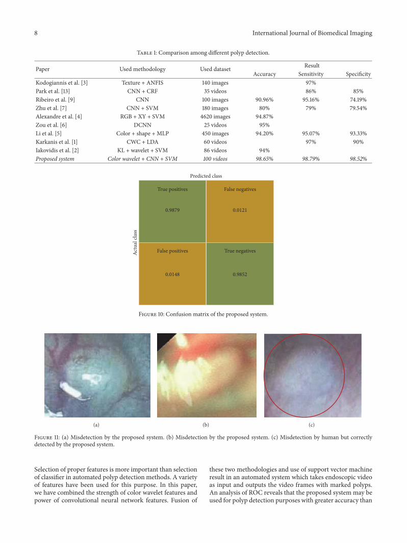
From Figure 10 and information above, it is observed thatthe proposed fusion model color wavelet features and con-volutional neural network features give much satisfactoryoutcome when choosing SVM as the classifier. A comparisonamong different polyp detection methods is showed inTable 1.
3.3. Comparison with Human Experts. Performance of theproposed approach has been compared with the diagnos-tic efficacy of human experts. Though nothing can be analternative to humans, several human factors lead to polypmisclassification. Computer aided polyp detection systemnot only can assists clinicians, but also can reduce polypmisclassification rate specialty in such cases, where polypsremain undetected for their small sizes.However, to assess the

6 International Journal of Biomedical Imaging
PolypNonpolyp
Input image Convolutionlayer 1
Convolutionlayer 2
layer 2
Convolution
Convolution
layer 3
Convolutionlayer 5
Max poolinglayer 1
layer 4
Fully
conn
ecte
d la
yer 1
Fully
conn
ecte
d la
yer 2
MaxOutput layer
96 filters of 256 filters of
384 filters
size 11 ∗ 11
size 5 ∗ 5 size 3 ∗ 3
227 ∗ 227 ∗ 3
poolinglayer 3
Maxpoolingof
Figure 6: An illustration of the proposed CNN feature extraction segment.
X2
Support vectors
Margin width
Figure 7: Linear support vector machine.
(a) (b)
Frame 1 Frame 77 Frame 146 Frame 218(c)
Figure 8: Output segment: (a) several portions are marked to be possible polyp, (b) system’s output after processing, and (c) output videoframes.

International Journal of Biomedical Imaging 7
Normaltissue
Lumen
Diverticula
Adenoma
Hyperplastic
Serrated
Figure 9: Different categories of video regions and polyps.
usability of the proposed system, images detected as polypsare fed into the system. Only two images go undetected aspolyp images as shown in Figures 11(a) and 11(b). Again,all the test data are used for assessing the system. As theproposed method gains accuracy of 98.65%, results areassessed by medical experts also. They highly appreciate thedetection and classification results. Moreover, in some cases,
images are detected as polyp which is difficult to detect forhuman experts also as shown in Figure 11(c).
4. Conclusions and Future Works
Computer aided system for automatic polyp detection is ofgreat interest nowadays as a support to the medical persons.
8 International Journal of Biomedical Imaging
Table 1: Comparison among different polyp detection.
Paper Used methodology Used dataset ResultAccuracy Sensitivity Specificity
Kodogiannis et al. [3] Texture + ANFIS 140 images 97%Park et al. [13] CNN + CRF 35 videos 86% 85%Ribeiro et al. [9] CNN 100 images 90.96% 95.16% 74.19%Zhu et al. [7] CNN + SVM 180 images 80% 79% 79.54%Alexandre et al. [4] RGB + XY + SVM 4620 images 94.87%Zou et al. [6] DCNN 25 videos 95%Li et al. [5] Color + shape + MLP 450 images 94.20% 95.07% 93.33%Karkanis et al. [1] CWC + LDA 60 videos 97% 90%Iakovidis et al. [2] KL + wavelet + SVM 86 videos 94%Proposed system Color wavelet + CNN + SVM 100 videos 98.65% 98.79% 98.52%
Predicted class
Actu
al cl
ass
True positives False negatives
False positives True negatives
0.9879
0.98520.0148
0.0121
Figure 10: Confusion matrix of the proposed system.
(a) (b) (c)
Figure 11: (a) Misdetection by the proposed system. (b) Misdetection by the proposed system. (c) Misdetection by human but correctlydetected by the proposed system.
Selection of proper features is more important than selectionof classifier in automated polyp detection methods. A varietyof features have been used for this purpose. In this paper,we have combined the strength of color wavelet features andpower of convolutional neural network features. Fusion of
these two methodologies and use of support vector machineresult in an automated system which takes endoscopic videoas input and outputs the video frames with marked polyps.An analysis of ROC reveals that the proposed system may beused for polyp detection purposes with greater accuracy than

International Journal of Biomedical Imaging 9
the state-of-the-art methods. In the future, fusion of CW andCNN features will be used for ultra sound image analysis.
Conflicts of Interest
The authors declare that there are no conflicts of interestregarding the publication of this paper.
Acknowledgments
The authors are very grateful to Dr. Md. Rabiul Hossain,Gastroenterology, Liver & Internal Medicine Specialist, forhis valuable support, suggestions, and consultancy.
References
[1] S. A. Karkanis, D. K. Iakovidis, D. E.Maroulis, D. A. Karras, andM. Tzivras, “Computer-aided tumor detection in endoscopicvideo using color wavelet features,” IEEE Transactions on Infor-mation Technology in Biomedicine, vol. 7, no. 3, pp. 141–152, 2003.
[2] D. K. Iakovidis, D. E. Maroulis, and S. A. Karkanis, “Anintelligent system for automatic detection of gastrointestinaladenomas in video endoscopy,” Computers in Biology andMedicine, vol. 36, no. 10, pp. 1084–1103, 2006.
[3] K. Vassilis and B. Maria, “An adaptive neurofuzzy approachfor the diagnosis in wireless capsule endoscopy imaging,” Inter-national Journal of Information Technology, vol. 13, no. 1, pp. 46–56, 2007.
[4] L. A. Alexandre,N.Nobre, and J. Casteleiro, “Color and positionversus texture features for endoscopic polyp detection,” inProceedings of the 1st International Conference on BioMedicalEngineering and Informatics (BMEI ’08), vol. 2, pp. 38–42,Institute of Electrical and Electronics Engineers, Sanya, China,May 2008.
[5] B. Li, Y. Fan, M. Q.-H.Meng, and L. Qi, “Intestinal polyp recog-nition in capsule endoscopy images using color and shapefeatures,” in Proceedings of the 2009 IEEE International Confer-ence on Robotics and Biomimetics (ROBIO ’09), pp. 1490–1494,Institute of Electrical and Electronics Engineers, Guilin, China,December 2009.
[6] Y. Zou, L. Li, Y. Wang, J. Yu, Y. Li, and W. J. Deng, “Classifyingdigestive organs in wireless capsule endoscopy images based ondeep convolutional neural network,” in Proceedings of the IEEEInternational Conference on Digital Signal Processing (DSP ’15),pp. 1274–1278, Institute of Electrical and Electronics Engineers,Singapore, July 2015.
[7] R. Zhu, R. Zhang, and D. Xue, “Lesion detection of endoscopyimages based on convolutional neural network features,” inProceedings of the 8th International Congress on Image andSignal Processing (CISP ’15), pp. 372–376, Institute of Electricaland Electronics Engineers, Shenyang, China, October 2015.
[8] N. Tajbakhsh, S. R. Gurudu, and J. Liang, “Automatic polypdetection using global geometric constraints and local intensityvariation patterns,” in Proceedings of the International Confer-ence on Medical Image Computing and Computer-Assisted Inter-vention (MICCAI ’14), Springer International Publishing, 2014.
[9] E. Ribeiro, A. Uhl, andM. Hafner, “Colonic polyp classificationwith convolutional neural networks,” in Proceedings of theIEEE 29th International SymposiumonComputer-BasedMedicalSystems (CBMS ’16), Institute of Electrical and ElectronicsEngineers, Dublin, Ireland, June 2016.
[10] N. Tajbakhsh, S. R. Gurudu, and J. Liang, “Automatic polypdetection in colonoscopy videos using an ensemble of con-volutional neural networks,” in Proceedings of the 12th IEEEInternational Symposium on Biomedical Imaging (ISBI ’15),Institute of Electrical and Electronics Engineers, New York, NY,USA, April 2015.
[11] X. Jia and M. Q. Meng, “A deep convolutional neural networkfor bleeding detection in Wireless Capsule Endoscopy images,”in Proceedings of the 2016 38th Annual International Conferenceof the IEEE Engineering in Medicine and Biology Society (EMBC’16), Institute of Electrical and Electronics Engineers, Orlando,Fla, USA, August 2016.
[12] E. Ribeiro, A. Uhl, G.Wimmer, andM.Hafner, “Exploring deeplearning and transfer learning for colonic polyp classification,”Computational and Mathematical Methods in Medicine, vol.2016, Article ID 6584725, 16 pages, 2016.
[13] S. Y. Park and D. Sargent, “Colonoscopic polyp detection usingconvolutional neural networks,” in Proceedings of the Interna-tional Society for Optics and Photonics, vol. 9785 of SPIEMedicalImaging, San Diego, Cali, USA, March 2016.
[14] R. M. Haralick, “Statistical and structural approaches to tex-ture,” Proceedings of the IEEE, vol. 67, no. 5, pp. 786–804, 1979.
[15] R. M. Haralick, K. Shanmugam, and I. Dinstein, “Texturalfeatures for image classification,” IEEE Transactions on Systems,Man and Cybernetics, vol. 3, no. 6, pp. 610–621, 1973.
[16] P. Mesejo, D. Pizarro, A. Abergel et al., “Computer-aided classi-fication of gastrointestinal lesions in regular colonoscopy,” IEEETransactions on Medical Imaging, vol. 35, no. 9, pp. 2051–2063,2016.
[17] D. West and V. West, “Model selection for a medical diagnosticdecision support system: a breast cancer detection case,” Artifi-cial Intelligence in Medicine, vol. 20, no. 3, pp. 183–204, 2000.
[18] W. G. Baxt, “Application of artificial neural networks to clinicalmedicine,”The Lancet, vol. 346, no. 8983, pp. 1135–1138, 1995.
[19] I. El-Naqa, Y. Yang, M. N. Wernick, N. P. Galatsanos, and R. M.Nishikawa, “A support vector machine approach for detectionof microcalcifications,” IEEE Transactions on Medical Imaging,vol. 21, no. 12, pp. 1552–1563, 2002.
[20] S. Li, J. T. Kwok, H. Zhu, and Y. Wang, “Texture classificationusing the support vectormachines,”Pattern Recognition, vol. 36,no. 12, pp. 2883–2893, 2003.
[21] S. B. Gokturk, C. Tomasi, B. Acar et al., “A statistical 3-D patternprocessing method for computer-aided detection of polyps inCT colonography,” IEEE Transactions on Medical Imaging, vol.20, no. 12, pp. 1251–1260, 2001.
[22] J. Bernal, N. Tajkbaksh, F. J. Sanchez et al., “comparative valid-ation of polyp detection methods in video colonoscopy: resultsfrom the MICCAI 2015 endoscopic vision challenge,” IEEETransactions on Medical Imaging, vol. 36, no. 6, pp. 1231–1249,2017.

RoboticsJournal of
Hindawi Publishing Corporationhttp://www.hindawi.com Volume 2014
Hindawi Publishing Corporationhttp://www.hindawi.com Volume 2014
Active and Passive Electronic Components
Control Scienceand Engineering
Journal of
Hindawi Publishing Corporationhttp://www.hindawi.com Volume 2014
International Journal of
RotatingMachinery
Hindawi Publishing Corporationhttp://www.hindawi.com Volume 2014
Hindawi Publishing Corporation http://www.hindawi.com
Journal of
Volume 201
Submit your manuscripts athttps://www.hindawi.com
VLSI Design
Hindawi Publishing Corporationhttp://www.hindawi.com Volume 201
Hindawi Publishing Corporationhttp://www.hindawi.com Volume 2014
Shock and Vibration
Hindawi Publishing Corporationhttp://www.hindawi.com Volume 2014
Civil EngineeringAdvances in
Acoustics and VibrationAdvances in
Hindawi Publishing Corporationhttp://www.hindawi.com Volume 2014
Hindawi Publishing Corporationhttp://www.hindawi.com Volume 2014
Electrical and Computer Engineering
Journal of
Advances inOptoElectronics
Hindawi Publishing Corporation http://www.hindawi.com
Volume 2014
The Scientific World JournalHindawi Publishing Corporation http://www.hindawi.com Volume 2014
SensorsJournal of
Hindawi Publishing Corporationhttp://www.hindawi.com Volume 2014
Modelling & Simulation in EngineeringHindawi Publishing Corporation http://www.hindawi.com Volume 2014
Hindawi Publishing Corporationhttp://www.hindawi.com Volume 2014
Chemical EngineeringInternational Journal of Antennas and
Propagation
International Journal of
Hindawi Publishing Corporationhttp://www.hindawi.com Volume 2014
Hindawi Publishing Corporationhttp://www.hindawi.com Volume 2014
Navigation and Observation
International Journal of
Hindawi Publishing Corporationhttp://www.hindawi.com Volume 2014
DistributedSensor Networks
International Journal of