amniovisc monograph white paper - lattice biologics · monograph . amnio-viscosupplementation:...
TRANSCRIPT
Liventa Bioscience
Viscosupplementation for Knee Osteoarthritis
MONOGRAPH
Amnio-Viscosupplementation:
Review of the Safety and Efficacy of Amniotic Fluid in the Management of Joint Pain in Osteoarthritis
Background: Osteoarthritis (degenerative joint disease – DJD) of the knee is a condition characterized by the progressive destruction of the cartilage that lines the knee joint and in the more severe cases results in bone-on-bone friction and accompanying pain, immobility and a deterioration of the activities of daily living.
In 2012 the estimated prevalence of osteoarthritis among adults in the United States, the number of individuals who had ever been told by a doctor that they had the condition was approximately 52.5 million cases (source: Centers for Disease Control and Prevention; National Statistics: National Health Interview Survey [NHIS] Arthritis Surveillance). Prevalence rates vary by the joint involved and the method of diagnosis (clinical vs. radiographic). Symptomatically, the knee is the most frequently affected joint. The prevalence of osteoarthritis of the knee is increasing rapidly because of shifting population demographics. The primary risk factors for osteoarthritis of the knee are aging, obesity, prior injury, repetitive use and female gender.
The prevalence of symptomatic knee osteoarthritis may reach 50% by the age of 75. From 2002 to 2012 the number of individuals in the US with a total knee replacement doubled from some 2 million to approximately 4 million. (Source: Systematic Review for Effectiveness of Hyaluronic Acid in the Treatment of Severe Degenerative Joint Disease of the Knee; Agency for Healthcare Research and Quality [AHRQ] Department of Health and Human Services; U.S. Government).
Osteoarthritis of the knee is usually diagnosed clinically based on pain. Radiographic evidence of osteoarthritis may precede symptomatic osteoarthritis but it correlates weakly with symptom severity.
The goals of treatment for osteoarthritis of the knee include pain relief, reduced inflammation, slowing the progression of the disease and improved mobility and function as well as health-related quality of life.
Treatment options for osteoarthritis of the knee include oral or topical analgesics, injected corticosteroids, physical therapy and exercise, weight loss, viscosupplementation using natural joint lubricants (most commonly hyaluronic acid [HA]), and partial or total arthroplasty.
Viscosupplementation, which was first used as a therapy in 1970s (trade name of Healon®) for ophthalmic use and veterinary use (Hylartil-Vet®), has become a standard of care within the continuum of care for symptomatic osteoarthritic knees.
The purpose of viscosupplements is to replenish the naturally occurring synovial fluid with a substance as close to normal, healthy synovial fluid as possible. Healthy synovial fluid, in addition to providing nourishment to the cartilage cells, also lubricates the articulating boney structures of the joint during low impact movement and shock absorption during high impact activities.
Many studies have shown that the early onset of osteoarthritis causes a deterioration of synovial fluid’s properties -- specifically elasticity and viscosity. There are various disease processes at work in an
arthritic knee including production of certain enzymes and “toxic” precursors. The result is a diseased synovial fluid which cannot perform the functions of lubrication or shock-absorption.
At every stage of the osteoarthritic disease process, the deteriorating ability of the synovial fluid can be experienced by the patient as pain, stiffness and reduced function of the joint.
The principal of viscosupplementation is to break the cycle of synovial fluid deterioration. In fact, HA viscosupplement products are not registered as drugs but rather as medical devices, like liquid bio-prosthesis.
Viscosupplementation helps an OA joint by forming a protective layer around the inflamed articulating structures, covering micro-fractures and defects, and helping to restore the lubrication and protection that healthy synovial fluid offers.
Until very recently, all viscosupplementation products were derived from either rooster combs or from bacterial fermentation processes.
This paper reviews the interim data from a new source of HA – human amniotic fluid. Human amniotic fluid has been used as a viscosupplement for ophthalmic use (Use of topical human amniotic fluid in the treatment of acute ocular alkali injuries in mice; Am. J. Ophthalmol. 2006 Aug;142(2):271-8 S.Herretes, et al).
Ocular fluid, synovial fluid and amniotic fluid are highly similar materials with equally similar biologic functions. All three fluids are designed to provide lubrication, cushioning and shock absorption within an enclosed tissue structure like the synovium or placenta. All three contain significant levels of hyaluronan as well as phosolipids, cholesterol and such inorganic compounds as sodium, potassium and magnesium.
The hypothesis is that a human derived viscosupplement with largely the same components as healthy synovial fluid would be a safe and effective viscosupplement for the management of joint pain in the osteoarthritic patient.
Objectives:
The purpose of this single arm, registry study is to assess the efficacy and safety of amnio-viscosupplementation in the management of joint pain in the osteoarthritic knee.
Methods:
In a protocol driven, single arm post-marketing Registry approved by the Western Institutional Review Board (Olympia, WA) patients with Kellgren Lawrence Grade 1-3 OA via radiologic examination were treated with a single injection of processed donated human amniotic fluid. Excluded patients were < 35 years, had BMI > 45 or had received Hyaluronic Acid injections in the previous six months, or steroid or PRP injection in the last three months. There were no threshold pain inclusion or exclusion criteria. Eligible patients were injected with 4cc of minimally processed amniotic fluid (AmnioVisc; Liventa Bioscience, West Conshohocken, PA) into the affected knee.
Primary efficacy endpoints are VAS scores and WOMAC overall and Pain, Stiffness and Difficulty (function) sub-score scales, measured during office visits at baseline and at 30, 90 and 180 days. Enrollees also filled out weekly Pain Diaries to report WOMAC Pain sub-score (5 questions) at weeks 1-4 post-treatment.
Interim Results:
To date 275 patients have completed their enrollment and had their clinical results reported. The total number of patients for whom data is available is 275 (241 with 30 day follow up data, 162 with 90 day follow-up data and 63 with 180 day follow-up data). All data was collected and analyzed by OMEGA Statistics, Murieta, CA.
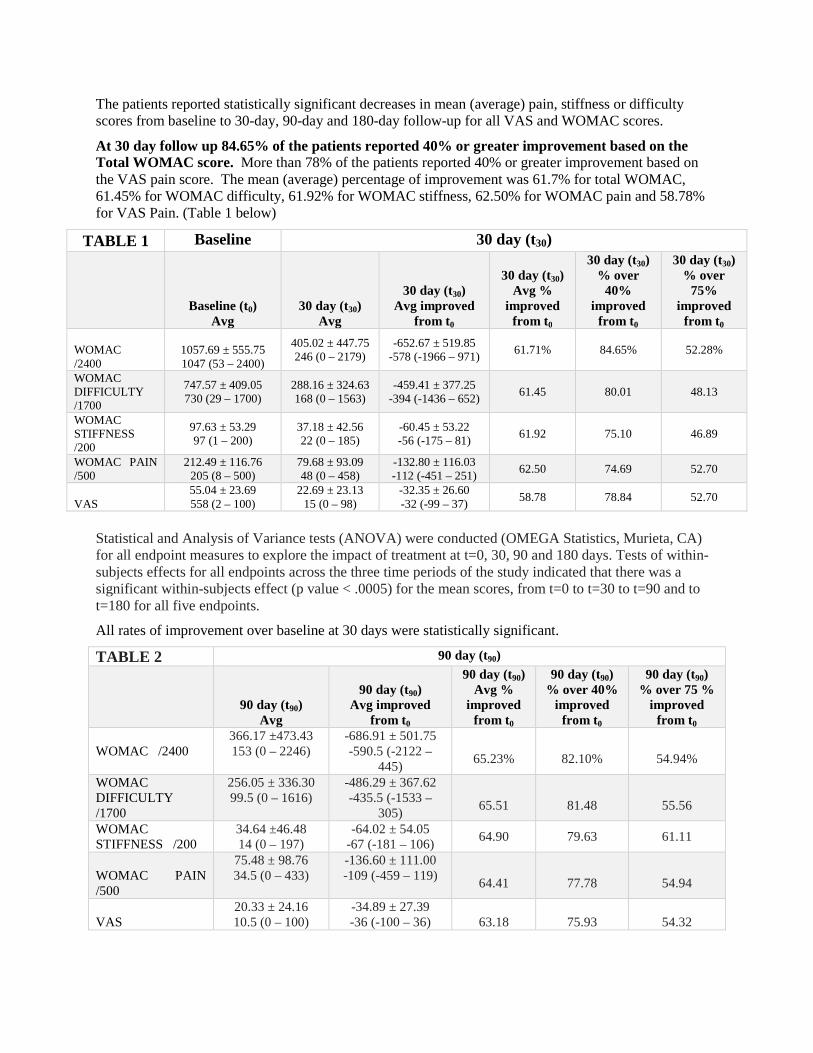
The patients reported statistically significant decreases in mean (average) pain, stiffness or difficulty scores from baseline to 30-day, 90-day and 180-day follow-up for all VAS and WOMAC scores.
At 30 day follow up 84.65% of the patients reported 40% or greater improvement based on the Total WOMAC score. More than 78% of the patients reported 40% or greater improvement based on the VAS pain score. The mean (average) percentage of improvement was 61.7% for total WOMAC, 61.45% for WOMAC difficulty, 61.92% for WOMAC stiffness, 62.50% for WOMAC pain and 58.78% for VAS Pain. (Table 1 below)
TABLE 1 Baseline 30 day (t30)
Baseline (t0) Avg
30 day (t30) Avg
30 day (t30) Avg improved
from t0
30 day (t30)
Avg % improved
from t0
30 day (t30) % over
40% improved
from t0
30 day (t30) % over
75% improved
from t0 WOMAC /2400
1057.69 ± 555.75 1047 (53 – 2400)
405.02 ± 447.75 246 (0 – 2179)
-652.67 ± 519.85 -578 (-1966 – 971)
61.71% 84.65% 52.28%
WOMAC DIFFICULTY /1700
747.57 ± 409.05 730 (29 – 1700)
288.16 ± 324.63 168 (0 – 1563)
-459.41 ± 377.25 -394 (-1436 – 652)
61.45 80.01 48.13
WOMAC STIFFNESS /200
97.63 ± 53.29 97 (1 – 200)
37.18 ± 42.56 22 (0 – 185)
-60.45 ± 53.22 -56 (-175 – 81)
61.92 75.10 46.89
WOMAC PAIN /500
212.49 ± 116.76 205 (8 – 500)
79.68 ± 93.09 48 (0 – 458)
-132.80 ± 116.03 -112 (-451 – 251)
62.50 74.69 52.70
VAS
55.04 ± 23.69 558 (2 – 100)
22.69 ± 23.13 15 (0 – 98)
-32.35 ± 26.60 -32 (-99 – 37)
58.78 78.84 52.70
Statistical and Analysis of Variance tests (ANOVA) were conducted (OMEGA Statistics, Murieta, CA) for all endpoint measures to explore the impact of treatment at t=0, 30, 90 and 180 days. Tests of within-subjects effects for all endpoints across the three time periods of the study indicated that there was a significant within-subjects effect (p value < .0005) for the mean scores, from t=0 to t=30 to t=90 and to t=180 for all five endpoints.
All rates of improvement over baseline at 30 days were statistically significant.
TABLE 2 90 day (t90)
90 day (t90)
Avg
90 day (t90)
Avg improved from t0
90 day (t90) Avg %
improved from t0
90 day (t90) % over 40%
improved from t0
90 day (t90) % over 75 %
improved from t0
WOMAC /2400
366.17 ±473.43 153 (0 – 2246)
-686.91 ± 501.75 -590.5 (-2122 –
445)
65.23%
82.10%
54.94%
WOMAC DIFFICULTY /1700
256.05 ± 336.30 99.5 (0 – 1616)
-486.29 ± 367.62 -435.5 (-1533 –
305)
65.51
81.48
55.56
WOMAC STIFFNESS /200
34.64 ±46.48 14 (0 – 197)
-64.02 ± 54.05 -67 (-181 – 106)
64.90 79.63 61.11
WOMAC PAIN /500
75.48 ± 98.76 34.5 (0 – 433)
-136.60 ± 111.00 -109 (-459 – 119)
64.41
77.78
54.94
VAS
20.33 ± 24.16 10.5 (0 – 100)
-34.89 ± 27.39 -36 (-100 – 36)
63.18
75.93
54.32

At 90 day follow up 82.10% of the patients reported 40% or greater improvement based on the Total WOMAC score. More than 75% of the patients reported 40% or greater improvement based on the VAS pain score. The mean (average) percentage of improvement was 65.23% for WOMAC/2400, 65.51% for WOMAC difficulty, 64.90% for WOMAC stiffness, 64.41% for WOMAC pain and 63.18% for VAS Pain. (Table 2 above)
All rates of improvement over baseline at 90 days were statistically significant.
TABLE 3 180 day (t180)
180 day (t180)
Avg
180 day (t180)
Avg improved from t0
180 day (t180) Avg %
improved from t0
180 day (t180) % over 40%
improved from t0
180 day (t180) % over 75 %
improved from t0
WOMAC /2400
423.76 ± 582.65 150 (0 – 2265)
-483.48 ± 624.42 -342 (-1814 –
1098)
53.29%
66.67%
50.79%
WOMAC DIFFICULTY /1700
296.13 ± 417.78 99 (0 – 1627)
-335.48 ± 452.39 -227 (-1263 – 835)
53.12
69.84
52.38
WOMAC STIFFNESS /200
39.71 ± 53.77 15 (0 – 200)
-44.60 ± 58.01 -34 (-168 – 80)
52.89
71.43
47.62
WOMAC PAIN /500
87.92 ± 118.16 31 (0 – 470)
-103.40 ± 130.91 -84 (-413 – 249)
54.05
66.67
52.38
VAS
24.56 ± 29.74 9 (0 – 100)
-26.44 ± 30.88 -23 (-89 – 41)
51.84 65.08 55.56
At 180 day follow up 66.67% of the patients reported 40% or greater improvement based on the Total WOMAC score. More than 65% of the patients reported 40% or greater improvement based on the VAS pain score. The mean (average) percentage of improvement was 53.29% for WOMAC/2400, 53.12% for WOMAC difficulty, 52.89% for WOMAC stiffness, 54.05% for WOMAC pain and 51.84% for VAS Pain. (Table 3 above)
All rates of improvement over baseline at 180 days were statistically significant. The increase in pain, stiffness and difficulty WOMAC and VAS scores between 90 and 180 days was not deemed to be statistically significant.
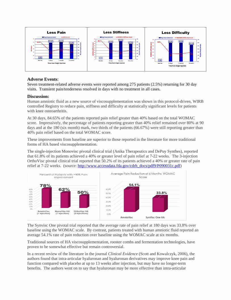
Adverse Events: Seven treatment-related adverse events were reported among 275 patients (2.5%) returning for 30 day visits. Transient pain/tenderness resolved in days with no treatment in all cases.
Discussion: Human amniotic fluid as a new source of viscosupplementation was shown in this protocol-driven, WIRB controlled Registry to reduce pain, stiffness and difficulty at statistically significant levels for patients with knee osteoarthritis.
At 30 days, 84.65% of the patients reported pain relief greater than 40% based on the total WOMAC score. Impressively, the percentage of patients reporting greater than 40% relief remained over 80% at 90 days and at the 180 (six month) mark, two thirds of the patients (66.67%) were still reporting greater than 40% pain relief based on the total WOMAC score.
These improvements from baseline are superior to those reported in the literature for more traditional forms of HA based viscosupplementation.
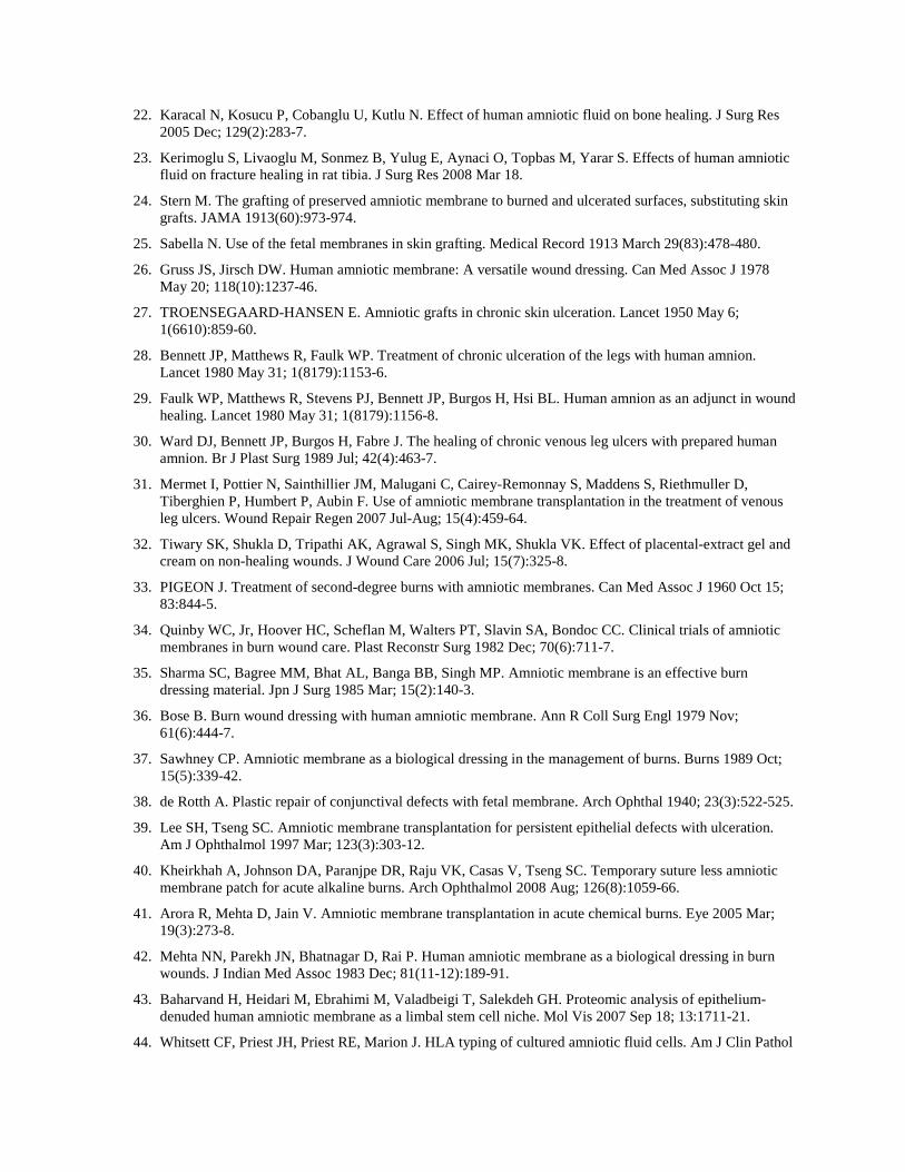
The single-injection Monovisc pivotal clinical trial (Anika Therapeutics and DePuy Synthes), reported that 61.8% of its patients achieved a 40% or greater level of pain relief at 7-22 weeks. The 3-injection OrthoVisc pivotal clinical trial reported that 50.2% of its patients achieved a 40% or greater rate of pain relief at 7-22 weeks. (source: http://www.accessdata.fda.gov/cdrh_docs/pdf9/P090031c.pdf)
The Synvisc One pivotal trial reported that the average rate of pain relief at 180 days was 33.8% over baseline using the WOMAC scale. By contrast, patients treated with human amniotic fluid reported an average 54.1% rate of pain reduction over baseline using the WOMAC scale at six months.
Traditional sources of HA viscosupplementation, rooster combs and fermentation technologies, have proven to be somewhat effective but remain controversial.
In a recent review of the literature in the journal Clinical Evidence (Scott and Kowalczyk, 2006), the authors found that intra-articular hyaluronan and hyaluronan derivatives may improve knee pain and function compared with placebo at up to 13 weeks after injection, but may have no longer-term benefits. The authors went on to say that hyaluronan may be more effective than intra-articular

corticosteroids at reducing pain at 5 to 13 weeks, although they may be as effective as each other in the shorter term. According to the review, this conclusion is based upon very low-quality evidence.
Recent guidance from the National Institute for Health and Clinical Excellence (2008) concluded that the hyaluronan viscosupplementation reduced pain up to 3 months after a series of 3 to 5 injections, although the effect size is generally small. "Given this, and the cost of the therapies together with increased clinician visits required for injections, there appears to be a poor rationale for routine clinical use."
More recently, the American Academy of Orthopedic Surgeons (2013) concluded that they "cannot recommend using hyaluronic acid for patients with symptomatic osteoarthritis of the knee." This conclusion was a strong recommendation and was based on a metaanalysis of studies that failed to show a clinically significant benefit from viscosupplementation.
By contrast, amniotic fluid derived viscosupplementation appears to offer patients higher and more durable rates of pain, stiffness and difficulty relief based on the 275 patients documented so far in this protocol-driven study.
There may be three reasons why amniotic fluid is a more effective viscosupplement than either rooster comb derived HA or HA produced by fermentation technique. Those reasons are;
1. It is human HA. Amniotic fluid has human hyaluronan and is known to be immune privileged since it lacks MHC antigens.
2. Human amniotic fluid is a homolog for human synovial fluid. The components of amniotic fluid are very similar to healthy, native synovial fluid. Both amniotic fluid and synovial fluid are ultrafiltrates from blood plasma and contain not only hyaluronan but also phospholipids, cholesterol, growth factor proteins, cytokines and nearly the same inorganic compounds.
3. Restores the correct pH levels. Human amniotic fluid has a pH level of about 7.0 which is the same as health synovial fluid. The disease of osteoarthritis tends to create an acidic and toxic environment in the knee and pH levels fall, typically, to about 3.5. Human amniotic viscosupplementation restores health pH levels which, in turn, create the conditions for the synovium to recover and potentially to slow down or even stop the disease progression.

References: 1. Balazs EA, Denlinger JL. Viscosupplementation: a new concept in the treatment of osteoarthritis. J
Rheumatology 1993;20 (suppl 39):3-9
2. Balazs EA, Denlinger JL,. Clinical uses of hyaluronan. From: The Biology of Hyaluronan (Ciba Foundation Symposium/143), (Eds. Evered D and Whelan J.) Wiley (1989), Chichester, England: 265-280
3. Barbour KE, Helmick CG, Theis KA, Murphy LB, Hootman JM, Brady TJ, Cheng YJ. Prevalence of doctor-diagnosed arthritis and arthritis-attributable activity limitation-United States, 2010-2012. Morb Mortal Wkly Rep. 2013; 62(44): 869–873. PubMed PMID: 24196662. html pdf
4. Medawar PB. Some immunological and endocrinological problems raised by the evolution of viviparity in vertebrates. Symp Soc Exp Biol 1953(7):320-338.
5. Billington WD. The immunological problem of pregnancy: 50 years with the hope of progress. A tribute to peter medawar. J Reprod Immunol 2003 Oct; 60(1):1-11.
6. Moffett A, Loke YW. The immunological paradox of pregnancy: A reappraisal. Placenta 2004 Jan; 25(1):1-8.
7. Niederkorn JY, Wang S. Immune privilege of the eye and fetus: Parallel universes? Transplantation 2005 Nov 15; 80(9):1139-44.
8. Wegmann TG. Why didn't your mother reject you? Can Med Assoc J 1980 Nov 22; 123(10):991-3.
9. Streilein JW. Unraveling immune privilege. Science 1995 Nov 17; 270(5239):1158-9.
10. Aagaard-Tillery KM, Silver R, Dalton J. Immunology of normal pregnancy. Semin Fetal Neonatal Med 2006 Oct; 11(5):279-95.
11. Veenstra van Nieuwenhoven AL, Heineman MJ, Faas MM. The immunology of successful pregnancy. Hum Reprod Update 2003 Jul-Aug; 9(4):347-57.
12. Lowry PJ. The placenta is simply a neuroendocrine parasite. J Neuroendocrinol 2008 Jun; 20(6):700-4.
13. Underwood MA, Gilbert WM, Sherman MP. Amniotic fluid: Not just fetal urine anymore. J Perinatol 2005 May; 25(5):341-8.
14. Akinbi HT, Narendran V, Pass AK, Markart P, Hoath SB. Host defense proteins in vernix caseosa and amniotic fluid. Am J Obstet Gynecol 2004 Dec; 191(6):2090-6.
15. Yoshio H, Tollin M, Gudmundsson GH, Lagercrantz H, Jornvall H, Marchini G, Agerberth B. Antimicrobial polypeptides of human vernix caseosa and amniotic fluid: Implications for newborn innate defense. Pediatr Res 2003 Feb; 53(2):211-6.
16. Espinoza J, Chaiworapongsa T, Romero R, Edwin S, Rathnasabapathy C, Gomez R, Bujold E, Camacho N, Kim YM, Hassan S, Blackwell S, Whitty J, Berman S, Redman M, Yoon BH, Sorokin Y. Antimicrobial peptides in amniotic fluid: Defensins, calprotectin and bacterial/permeability-increasing protein in patients with microbial invasion of the amniotic cavity, intra-amniotic inflammation, preterm labor and premature rupture of membranes. J Matern Fetal Neonatal Med 2003 Jan; 13(1):2-21.
17. Shepard S, Becker H, Hartmann JX. Using hyaluronic acid to create a fetal-like environment in vitro. Ann Plast Surg 1996 Jan; 36(1):65-9.
18. Trelford JD, Trelford-Sauder M. The amnion in surgery, past and present. Am J Obstet Gynecol 1979 Aug 1; 134(7):833-45.
19. Shun A, Ramsey-Stewart G. Human amnion in the treatment of chronic ulceration of the legs. Med J Aust 1983 Sep 17; 2(6):279-83.
20. Shimberg M. The use of amniotic fluid concentrate in orthopaedic conditions. J Bone Joint Surg Am 1938; 20:167-177.
21. Shukla VK, Rasheed MA, Kumar M, Gupta SK, Pandey SS. A trial to determine the role of placental extract in the treatment of chronic non-healing wounds. J Wound Care 2004 May; 13(5):177-9.
22. Karacal N, Kosucu P, Cobanglu U, Kutlu N. Effect of human amniotic fluid on bone healing. J Surg Res 2005 Dec; 129(2):283-7.
23. Kerimoglu S, Livaoglu M, Sonmez B, Yulug E, Aynaci O, Topbas M, Yarar S. Effects of human amniotic fluid on fracture healing in rat tibia. J Surg Res 2008 Mar 18.
24. Stern M. The grafting of preserved amniotic membrane to burned and ulcerated surfaces, substituting skin grafts. JAMA 1913(60):973-974.
25. Sabella N. Use of the fetal membranes in skin grafting. Medical Record 1913 March 29(83):478-480.
26. Gruss JS, Jirsch DW. Human amniotic membrane: A versatile wound dressing. Can Med Assoc J 1978 May 20; 118(10):1237-46.
27. TROENSEGAARD-HANSEN E. Amniotic grafts in chronic skin ulceration. Lancet 1950 May 6; 1(6610):859-60.
28. Bennett JP, Matthews R, Faulk WP. Treatment of chronic ulceration of the legs with human amnion. Lancet 1980 May 31; 1(8179):1153-6.
29. Faulk WP, Matthews R, Stevens PJ, Bennett JP, Burgos H, Hsi BL. Human amnion as an adjunct in wound healing. Lancet 1980 May 31; 1(8179):1156-8.
30. Ward DJ, Bennett JP, Burgos H, Fabre J. The healing of chronic venous leg ulcers with prepared human amnion. Br J Plast Surg 1989 Jul; 42(4):463-7.
31. Mermet I, Pottier N, Sainthillier JM, Malugani C, Cairey-Remonnay S, Maddens S, Riethmuller D, Tiberghien P, Humbert P, Aubin F. Use of amniotic membrane transplantation in the treatment of venous leg ulcers. Wound Repair Regen 2007 Jul-Aug; 15(4):459-64.
32. Tiwary SK, Shukla D, Tripathi AK, Agrawal S, Singh MK, Shukla VK. Effect of placental-extract gel and cream on non-healing wounds. J Wound Care 2006 Jul; 15(7):325-8.
33. PIGEON J. Treatment of second-degree burns with amniotic membranes. Can Med Assoc J 1960 Oct 15; 83:844-5.
34. Quinby WC, Jr, Hoover HC, Scheflan M, Walters PT, Slavin SA, Bondoc CC. Clinical trials of amniotic membranes in burn wound care. Plast Reconstr Surg 1982 Dec; 70(6):711-7.
35. Sharma SC, Bagree MM, Bhat AL, Banga BB, Singh MP. Amniotic membrane is an effective burn dressing material. Jpn J Surg 1985 Mar; 15(2):140-3.
36. Bose B. Burn wound dressing with human amniotic membrane. Ann R Coll Surg Engl 1979 Nov; 61(6):444-7.
37. Sawhney CP. Amniotic membrane as a biological dressing in the management of burns. Burns 1989 Oct; 15(5):339-42.
38. de Rotth A. Plastic repair of conjunctival defects with fetal membrane. Arch Ophthal 1940; 23(3):522-525.
39. Lee SH, Tseng SC. Amniotic membrane transplantation for persistent epithelial defects with ulceration. Am J Ophthalmol 1997 Mar; 123(3):303-12.
40. Kheirkhah A, Johnson DA, Paranjpe DR, Raju VK, Casas V, Tseng SC. Temporary suture less amniotic membrane patch for acute alkaline burns. Arch Ophthalmol 2008 Aug; 126(8):1059-66.
41. Arora R, Mehta D, Jain V. Amniotic membrane transplantation in acute chemical burns. Eye 2005 Mar; 19(3):273-8.
42. Mehta NN, Parekh JN, Bhatnagar D, Rai P. Human amniotic membrane as a biological dressing in burn wounds. J Indian Med Assoc 1983 Dec; 81(11-12):189-91.
43. Baharvand H, Heidari M, Ebrahimi M, Valadbeigi T, Salekdeh GH. Proteomic analysis of epithelium-denuded human amniotic membrane as a limbal stem cell niche. Mol Vis 2007 Sep 18; 13:1711-21.
44. Whitsett CF, Priest JH, Priest RE, Marion J. HLA typing of cultured amniotic fluid cells. Am J Clin Pathol

1983 Feb; 79(2):186-94.
45. COLE FR. Placental blood and placental extract in wound healing. Am J Surg 1948 Jul; 76(1):38-43.
46. Chandanwale A, Langade D, Mohod V, Sinha S, Ramteke A, Bakhshi GD, Motwani M. Comparative evaluation of human placental extract for its healing potential in surgical wounds after orthopaedic surgery: An open, randomized, comparative study. J Indian Med Assoc 2008 Jun; 106(6):405-8.
47. Akle CA, Adinolfi M, Welsh KI, Leibowitz S, McColl I. Immunogenicity of human amniotic epithelial cells after transplantation into volunteers. Lancet 1981 Nov 7; 2(8254):1003-5.
48. Adinolfi M, Akle CA, McColl I, Fensom AH, Tansley L, Connolly P, Hsi BL, Faulk WP, Travers P, Bodmer WF. Expression of HLA antigens, beta 2-microglobulin and enzymes by human amniotic epithelial cells. Nature 1982 Jan 28; 295(5847):325-7.
49. Chakraborty PD, Bhattacharyya D. Isolation of fibronectin type III like peptide from human placental extract used as wound healer. J Chromatogr B Analyt Technol Biomed Life Sci 2005 Apr 15; 818(1):67-73.
50. Shibasaki T, Odagiri E, Shizume K, Ling N. Corticotropin-releasing factor-like activity in human placental extracts. J Clin Endocrinol Metab 1982 Aug; 55(2):384-6.
51. Watanabe S, Togashi S, Takahashi N, Fukui T. L-tryptophan as an antioxidant in human placenta extract. J Nutr Sci Vitaminol (Tokyo) 2002 Feb;48(1):36-9.
52. Cheung CY. Vascular endothelial growth factor activation of intramembranous absorption: A critical pathway for amniotic fluid volume regulation. J Soc Gynecol Investig 2004 Feb;11(2):63-74.
53. Burgos H. Angiogenic and growth factors in human amnio-chorion and placenta. Eur J Clin Invest 1983 Aug;13(4):289-96.
54. Burgos H. Angiogenic factor from human term placenta. purification and partial characterization. Eur J Clin Invest 1986 Dec;16(6):486-93.
55. Ziche M, Maglione D, Ribatti D, Morbidelli L, Lago CT, Battisti M, Paoletti I, Barra A, Tucci M, Parise G, Vincenti V, Granger HJ, Viglietto G, Persico MG. Placenta growth factor-1 is chemotactic, mitogenic, and angiogenic. Lab Invest 1997 Apr;76(4):517-31.
56. Philipps AF, Holzman IR, Teng C, Battaglia FC. Tissue concentrations of free amino acids in term human placentas. Am J Obstet Gynecol 1978 Aug 15;131(8):881-7.
57. Ferretti C, Bruni L, Dangles-Marie V, Pecking AP, Bellet D. Molecular circuits shared by placental and cancer cells, and their implications in the proliferative, invasive and migratory capacities of trophoblasts. Hum Reprod Update 2007 Mar-Apr;13(2):121-41.
58. Gupta R, Chattopadhyay D. Glutamate is the chemotaxis-inducing factor in placental extracts. Amino Acids 2008 Aug 23.
59. Skerry TM, Taylor AF. Glutamate signalling in bone. Curr Pharm Des 2001 May;7(8):737-50.
60. Skerry TM, Genever PG. Glutamate signalling in non-neuronal tissues. Trends Pharmacol Sci 2001 Apr;22(4):174-81.
61. Skerry TM. The role of glutamate in the regulation of bone mass and architecture. J Musculoskelet Neuronal Interact 2008 Apr-Jun;8(2):166-73.
62. Zygmunt M, Herr F, Keller-Schoenwetter S, Kunzi-Rapp K, Munstedt K, Rao CV, Lang U, Preissner KT. Characterization of human chorionic gonadotropin as a novel angiogenic factor. J Clin Endocrinol Metab 2002 Nov;87(11):5290-6.
63. Ishikane S, Ohnishi S, Yamahara K, Sada M, Harada K, Mishima K, Iwasaki K, Fujiwara M, Kitamura S, Nagaya N, Ikeda T. Allogeneic injection of fetal membrane-derived mesenchymal stem cells induces therapeutic angiogenesis in a rat model of hind limb ischemia. Stem Cells 2008 Oct;26(10):2625-33.
64. Somerville PG. The possible use of amniotic membrane in chronic leg ulcers. Phlebologie 1982 Jan-Mar;35(1):223-9.

65. Longaker MT, Adzick NS, Hall JL, Stair SE, Crombleholme TM, Duncan BW, Bradley SM, Harrison MR, Stern R. Studies in fetal wound healing, VII. fetal wound healing may be modulated by hyaluronic acid stimulating activity in amniotic fluid. J Pediatr Surg 1990 Apr;25(4):430-3.
66. Haynes JH, Mast BA, Krummel TM, Cohen IK, Diegelmann RF. Exposure to amniotic fluid inhibits closure of open wounds in the fetal rabbit. Wound Repair Regen 1995 Oct-Dec;3(4):467-72.
67. Ozgenel GY. The influence of human amniotic fluid on the potential of rabbit ear perichondrial flaps to form cartilage tissue. Br J Plast Surg 2002 Apr;55(3):246-50.
68. Ozgenel GY, Filiz G, Ozcan M. Effects of human amniotic fluid on cartilage regeneration from free perichondrial grafts in rabbits. Br J Plast Surg 2004 Jul;57(5):423-8.
69. He Q, Li Q, Chen B, Wang Z. Repair of flexor tendon defects of rabbit with tissue engineering method. Chin J Traumatol 2002 Aug;5(4):200-8.
70. Musina RA, Bekchanova ES, Sukhikh GT. Comparison of mesenchymal stem cells obtained from different human tissues. Bull Exp Biol Med 2005 Apr;139(4):504-9.
71. Delo DM, De Coppi P, Bartsch G,Jr, Atala A. Amniotic fluid and placental stem cells. Methods Enzymol 2006;419:426-38.
72. Guillot PV, O'Donoghue K, Kurata H, Fisk NM. Fetal stem cells: Betwixt and between. Semin Reprod Med 2006 Nov;24(5):340-7.
73. Steigman SA, Fauza DO. Isolation of mesenchymal stem cells from amniotic fluid and placenta. Curr Protoc Stem Cell Biol 2007 Jun;Chapter 1:Unit 1E.2.
74. Nadri S, Soleimani M. Comparative analysis of mesenchymal stromal cells from murine bone marrow and amniotic fluid. Cytotherapy 2007;9(8):729-37.
75. Tweedell KS. New paths to pluripotent stem cells. Curr Stem Cell Res Ther 2008 Sep;3(3):151-62.
76. Caplan AI. Mesenchymal stem cells. J Orthop Res 1991 Sep;9(5):641-50.
77. Bruder SP, Fink DJ, Caplan AI. Mesenchymal stem cells in bone development, bone repair, and skeletal regeneration therapy. J Cell Biochem 1994 Nov;56(3):283-94.
78. Aggarwal S, Pittenger MF. Human mesenchymal stem cells modulate allogeneic immune cell responses. Blood 2005 Feb 15;105(4):1815-22.
79. Ryan JM, Barry FP, Murphy JM, Mahon BP. Mesenchymal stem cells avoid allogeneic rejection. J Inflamm (Lond) 2005 Jul 26;2:8.
80. Priest RE, Marimuthu KM, Priest JH. Origin of cells in human amniotic fluid cultures: Ultrastructural features. Lab Invest 1978 Aug;39(2):106-9.
81. Tyden O, Bergstrom S, Nilsson BA. Origin of amniotic fluid cells in mid-trimester pregnancies. Br J Obstet Gynaecol 1981 Mar;88(3):278-86.
82. In 't Anker PS, Scherjon SA, Kleijburg-van der Keur C, Noort WA, Claas FH, Willemze R, Fibbe WE, Kanhai HH. Amniotic fluid as a novel source of mesenchymal stem cells for therapeutic transplantation. Blood 2003 Aug 15;102(4):1548-9.
83. Tsai MS, Hwang SM, Tsai YL, Cheng FC, Lee JL, Chang YJ. Clonal amniotic fluid-derived stem cells express characteristics of both mesenchymal and neural stem cells. Biol Reprod 2006 Mar;74(3):545-51.
84. Soncini M, Vertua E, Gibelli L, Zorzi F, Denegri M, Albertini A, Wengler GS, Parolini O. Isolation and characterization of mesenchymal cells from human fetal membranes. J Tissue Eng Regen Med 2007 Jul-Aug;1(4):296-305.
85. De Coppi P, Bartsch G,Jr, Siddiqui MM, Xu T, Santos CC, Perin L, Mostoslavsky G, Serre AC, Snyder EY, Yoo JJ, Furth ME, Soker S, Atala A. Isolation of amniotic stem cell lines with potential for therapy. Nat Biotechnol 2007 Jan;25(1):100-6.
86. Kim J, Lee Y, Kim H, Hwang KJ, Kwon HC, Kim SK, Cho DJ, Kang SG, You J. Human amniotic fluid-

derived stem cells have characteristics of multipotent stem cells. Cell Prolif 2007 Feb;40(1):75-90.
87. Roubelakis MG, Pappa KI, Bitsika V, Zagoura D, Vlahou A, Papadaki HA, Antsaklis A, Anagnou NP. Molecular and proteomic characterization of human mesenchymal stem cells derived from amniotic fluid: Comparison to bone marrow mesenchymal stem cells. Stem Cells Dev 2007 Dec;16(6):931-52.
88. You Q, Cai L, Zheng J, Tong X, Zhang D, Zhang Y. Isolation of human mesenchymal stem cells from third-trimester amniotic fluid. Int J Gynaecol Obstet 2008 Nov;103(2):149-52.
89. Sessarego N, Parodi A, Podesta M, Benvenuto F, Mogni M, Raviolo V, Lituania M, Kunkl A, Ferlazzo G, Bricarelli FD, Uccelli A, Frassoni F. Multipotent mesenchymal stromal cells from amniotic fluid: Solid perspectives for clinical application. Haematologica 2008 Mar;93(3):339-46.
90. Wulf GG, Viereck V, Hemmerlein B, Haase D, Vehmeyer K, Pukrop T, Glass B, Emons G, Trumper L. Mesengenic progenitor cells derived from human placenta. Tissue Eng 2004 Jul-Aug;10(7-8):1136-47.
91. Matikainen T, Laine J. Placenta--an alternative source of stem cells. Toxicol Appl Pharmacol 2005 Sep 1;207(2 Suppl):544-9.
92. Miao Z, Jin J, Chen L, Zhu J, Huang W, Zhao J, Qian H, Zhang X. Isolation of mesenchymal stem cells from human placenta: Comparison with human bone marrow mesenchymal stem cells. Cell Biol Int 2006 Sep;30(9):681-7.
93. Alviano F, Fossati V, Marchionni C, Arpinati M, Bonsi L, Franchina M, Lanzoni G, Cantoni S, Cavallini C, Bianchi F, Tazzari PL, Pasquinelli G, Foroni L, Ventura C, Grossi A, Bagnara GP. Term amniotic membrane is a high throughput source for multipotent mesenchymal stem cells with the ability to differentiate into endothelial cells in vitro. BMC Dev Biol 2007 Feb 21;7:11.
94. Battula VL, Bareiss PM, Treml S, Conrad S, Albert I, Hojak S, Abele H, Schewe B, Just L, Skutella T, Buhring HJ. Human placenta and bone marrow derived MSC cultured in serum-free, b-FGF-containing medium express cell surface frizzled-9 and SSEA-4 and give rise to multilineage differentiation. Differentiation 2007 Apr;75(4):279-91.