altered responsiveness of medial prefrontal cortex neurons to glutamate and dopamine after...
TRANSCRIPT
Short Communication
Altered Responsiveness of MedialPrefrontal Cortex Neurons to Glutamate
and Dopamine after Withdrawal fromRepeated Amphetamine Treatment
JAYMS D. PETERSON1, MARINA E. WOLF1 AND FRANCIS J. WHITE2
1Department of Neuroscience, Finch University of Health Sciences/The Chicago Medical School,North Chicago, IL USA
2Cellular and Molecular Pharmacology, Finch University of Health Sciences/The Chicago Medical School,North Chicago, IL USA
KEY WORDS behavioral sensitization; excitatory amino acids
ABSTRACT Using in vivo extracellular single-cell recording and microiontophoresis,we compared the responsiveness of medial prefrontal cortex (mPFC) neurons (layersV-VI) to dopamine and glutamate in rats that had received repeated amphetamine orsaline injections. Neurons from amphetamine-pretreated rats showed increased respon-siveness to glutamate and decreased responsiveness to dopamine after three days ofwithdrawal, suggesting that the mPFC is transiently hyperexcitable during amphet-amine withdrawal. Synapse 36:342–344, 2000. Published 2000 Wiley-Liss, Inc.†
Behavioral sensitization, defined as the augmenta-tion of behavioral responses to psychomotor stimulantsduring and after their repeated administration, pro-vides an animal model for the intensification of drugcraving in human addicts (Robinson and Berridge,1993). Sensitization appears to be initiated by drugactions in the midbrain ventral tegmental area (VTA),origin of the mesocorticolimbic dopamine (DA) system(Kalivas and Stewart, 1991). Many lines of evidencesuggest that a transient increase in VTA DA cell activ-ity is an obligatory step in a cascade of neuroadapta-tions leading to persistent behavioral sensitization(White, 1996; White and Kalivas, 1998).
Excitatory amino acids (EAAs) also play a criticalrole in sensitization. The activity of glutamate-contain-ing neurons originating in the medial prefrontal cortex(mPFC) and projecting to the VTA appears to be re-quired for the induction of sensitization, since it isprevented by systemic or intra-VTA administration ofglutamate receptor antagonists or by prior excitotoxiclesions of the mPFC (Wolf, 1998). It is possible thatstimulant-induced adaptations in the mPFC contributeto the increased activity of VTA dopamine (DA) neu-rons implicated in the initiation process. We have re-ported that levels of the AMPA receptor subunit GluR1are transiently increased in PFC after repeated am-phetamine administration (Lu et al., 1997; Lu andWolf, 1999). In addition, we have demonstrated a tran-
sient decrease in responsiveness of mPFC neurons toiontophoretic dopamine after repeated cocaine admin-istration (White et al., 1995). The purpose of thepresent study was to determine if the responsiveness ofmPFC neurons to glutamate and dopamine is alteredafter withdrawal from repeated amphetamine admin-istration.
Adult male Sprague-Dawley rats (Harlan, Indianap-olis, Ind., USA) weighing 250-350 g were used. Allprocedures were performed in strict compliance withthe National Research Council Guide for the Care andUse of Laboratory animals (National Academy Press1996). Rats received 5 daily injections of saline or 5.0mg/kg amphetamine sulfate (i.p.). Electrophysiologicalrecordings from mPFC neurons were performed threedays after the last injection. Rats were anesthetizedwith urethane (1.7 g/kg) one hr prior to being mountedin a stereotaxic frame. Stereotaxic coordinates werechosen to target layers V-VI of prelimbic and infralim-bic PFC, which receive dense innervation from meso-cortical DA neurons. Coordinates (relative to bregmaand cortical surface in mm) were: A 3.2-3.4, L 0.7-0.9, V2.5-4. Carbon fiber seven-barrel extracellular elec-
Correspondence to: Francis J. White, Ph.D. FUHS/The Chicago Medical SchoolDepartment of Cellular and Molecular Pharmacology, 3333 Green Bay RoadNorth Chicago, IL 60064-3095. Email [email protected]
Received 14 May 1999; Accepted 24 August 1999.
SYNAPSE 36:342–344 (2000)
Published 2000 WILEY-LISS, INC. †This article is a US governmentwork and, as such, is in the public domain in the United States of America.

trodes (Kation Scientific, Novato, CA) were used forthese experiments. One side barrel was filled with 2 MNaCl solution for automatic current balancing whereasthe remaining side barrels contained L-glutamatemonosodium salt (100mM, pH 8) or dopamine HCl (10mM, pH 4). We limited our sample to cells with a basalfiring rate between 2.5 and 12.5 HZ to ensure thatanimals had roughly equivalent levels of anesthesia,since the level of anesthesia is an important determi-nant of PFC neuronal activity. Each neuron was re-corded for 3–5 minutes to establish a stable base-linefiring rate. Glutamate was then applied with a nega-tive current for 60 seconds followed by a 60 seconds offperiod to allow the neuron to return to baseline firing.At the end of the recovery period, the current applied tothe drug barrel was doubled up to 32 nA. After the lastglutamate current, cells were allowed to return to base-line firing at which time a second 3–5 minute baselinewas recorded. DA was then applied with a positivecurrent up to 64 nA. DA always followed glutamate dueto the short duration of the effects of glutamate and thelonger duration effects of DA. The experimenter wasalways blind to the drug pretreatment of the rat. Oneto three neurons were recorded per rat. All electrodeplacements were verified histologically. Only neuronsfrom the deep layers (V-VI) of the prelimbic and infra-limbic mPFC were included in our study. Details ofprocedures for single unit recording and microionto-phoresis are described elsewhere (Henry and White,1995). Statistical comparisons between amphetamineand saline treated groups were performed by repeatedmeasures analysis of variance (ANOVA). Post hoc anal-ysis comparing groups at each glutamate or dopaminecurrent was performed with a Univariate F-Test.
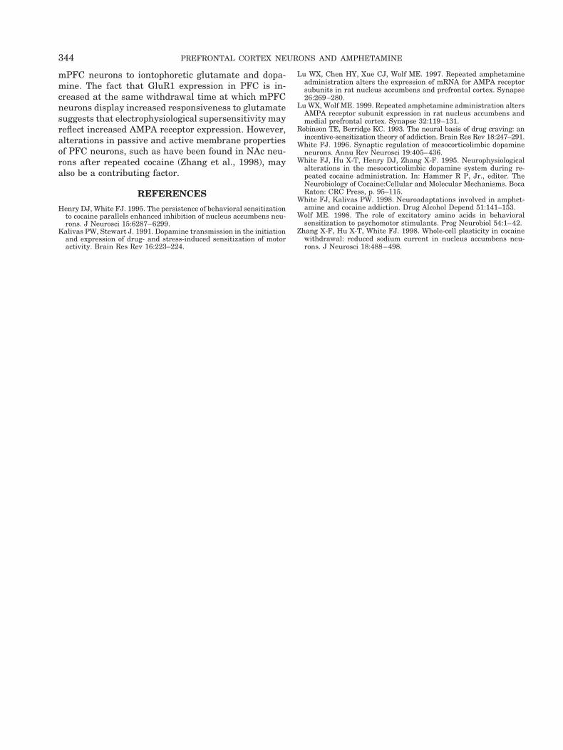
In saline-pretreated (control) rats, glutamate pro-duced a current dependent increase in the firing rate ofmPFC neurons. Currents greater than 32 nA wereavoided because they often produced apparent depolar-ization block, thereby disrupting subsequent DA test-ing. Neurons from amphetamine pretreated rats dem-onstrated a significantly greater response to lowercurrents of glutamate as compared to saline controls(Fig. 1A). Iontophoretic application of DA caused acurrent-dependent inhibition of mPFC neuronal firingrates in control rats, and this effect was significantlydecreased in amphetamine pre-treated rats, at least athigh DA currents (Fig. 1B).
The increased sensitivity to iontophoretic glutamateduring amphetamine withdrawal suggests that mPFCneurons have become hyper-responsive to glutamater-gic inputs. This, in turn, would be expected to increasethe activity of mPFC projection neurons, includingthose innervating the VTA. This may account, in part,for the increase in VTA DA cell activity that is believedto represent an obligatory step in the induction of be-havioral sensitization (Henry and White, 1995; White,1996). At high iontophoretic currents, DA exerts inhib-
itory effects on mPFC neurons. Accordingly, the am-phetamine-induced decrease in responsiveness to DAwould produce disinhibition and perhaps combine withincreased responsiveness to glutamate to promote ahyperactive state of the mPFC. Given that sensitiza-tion requires NMDA transmission within the VTA(above), it is possible that increased activity of PFCafferents to VTA DA neurons results in NMDA-depen-dent events in the VTA that may be similar to LTP.
Further research will be required to elucidate themechanism(s) underlying altered responsiveness of
Fig. 1. In vivo extracellular recordings performed from deep layer(V-VI) mPFC neurons in rats injected with saline or amphetamine forfive consecutive days followed by a three day withdrawal. mPFCneurons exhibited increased responsiveness to the excitatory effectsglutamate and decreased responsiveness to the inhibitory effects ofhigh iontophoretic DA currents. A. Microiontophoretic application ofglutamate caused a current-dependent increase in the firing of mPFCneurons. The excitatory response to glutamate in the amphetamine-pretreated rats (n 5 10 cells) was increased significantly at lower butnot higher currents (between groups factor, F1,18 5 4.87, p 5 0.041;*p,0.05) as compared to saline-pretreated rats (n 5 10 cells). B.Microiontophoretic application of DA caused a current-dependent de-crease in the firing of mPFC neurons. ANOVA revealed a non-signif-icant between groups factor (F1,13 5 1.96, p 5 0.185) but a significantgroups x current interaction (F7,91 5 2.62, p 5 0.017) indicating thatthe inhibitory response to DA in amphetamine-pretreated rats (n 5 9cells) was decreased significantly as the current was increased (*p ,0.05; # p 5 0.06).
J.D. PETERSON ET AL. 343
mPFC neurons to iontophoretic glutamate and dopa-mine. The fact that GluR1 expression in PFC is in-creased at the same withdrawal time at which mPFCneurons display increased responsiveness to glutamatesuggests that electrophysiological supersensitivity mayreflect increased AMPA receptor expression. However,alterations in passive and active membrane propertiesof PFC neurons, such as have been found in NAc neu-rons after repeated cocaine (Zhang et al., 1998), mayalso be a contributing factor.
REFERENCES
Henry DJ, White FJ. 1995. The persistence of behavioral sensitizationto cocaine parallels enhanced inhibition of nucleus accumbens neu-rons. J Neurosci 15:6287–6299.
Kalivas PW, Stewart J. 1991. Dopamine transmission in the initiationand expression of drug- and stress-induced sensitization of motoractivity. Brain Res Rev 16:223–224.
Lu WX, Chen HY, Xue CJ, Wolf ME. 1997. Repeated amphetamineadministration alters the expression of mRNA for AMPA receptorsubunits in rat nucleus accumbens and prefrontal cortex. Synapse26:269–280.
Lu WX, Wolf ME. 1999. Repeated amphetamine administration altersAMPA receptor subunit expression in rat nucleus accumbens andmedial prefrontal cortex. Synapse 32:119–131.
Robinson TE, Berridge KC. 1993. The neural basis of drug craving: anincentive-sensitization theory of addiction. Brain Res Rev 18:247–291.
White FJ. 1996. Synaptic regulation of mesocorticolimbic dopamineneurons. Annu Rev Neurosci 19:405–436.
White FJ, Hu X-T, Henry DJ, Zhang X-F. 1995. Neurophysiologicalalterations in the mesocorticolimbic dopamine system during re-peated cocaine administration. In: Hammer R P, Jr., editor. TheNeurobiology of Cocaine:Cellular and Molecular Mechanisms. BocaRaton: CRC Press, p. 95–115.
White FJ, Kalivas PW. 1998. Neuroadaptations involved in amphet-amine and cocaine addiction. Drug Alcohol Depend 51:141–153.
Wolf ME. 1998. The role of excitatory amino acids in behavioralsensitization to psychomotor stimulants. Prog Neurobiol 54:1–42.
Zhang X-F, Hu X-T, White FJ. 1998. Whole-cell plasticity in cocainewithdrawal: reduced sodium current in nucleus accumbens neu-rons. J Neurosci 18:488–498.
344 PREFRONTAL CORTEX NEURONS AND AMPHETAMINE