aha scientific statement - idsociety.org · heart association committee on rheumatic fever,...
TRANSCRIPT

AHA Scientific Statement
1435
Background—Infective endocarditis is a potentially lethal disease that has undergone major changes in both host and pathogen. The epidemiology of infective endocarditis has become more complex with today’s myriad healthcare-associated factors that predispose to infection. Moreover, changes in pathogen prevalence, in particular a more common staphylococcal origin, have affected outcomes, which have not improved despite medical and surgical advances.
Methods and Results—This statement updates the 2005 iteration, both of which were developed by the American Heart Association under the auspices of the Committee on Rheumatic Fever, Endocarditis, and Kawasaki Disease, Council on Cardiovascular Disease of the Young. It includes an evidence-based system for diagnostic and treatment recommendations used by the American College of Cardiology and the American Heart Association for treatment recommendations.
Conclusions—Infective endocarditis is a complex disease, and patients with this disease generally require management by a team of physicians and allied health providers with a variety of areas of expertise. The recommendations provided in this document are intended to assist in the management of this uncommon but potentially deadly infection. The clinical variability and complexity in infective endocarditis, however, dictate that these recommendations be used to support and not supplant decisions in individual patient management. (Circulation. 2015;132:1435-1486. DOI: 10.1161/CIR.0000000000000296.)
Key Words: AHA Scientific Statements ◼ anti-infective agents ◼ echocardiography ◼ endocarditis ◼ infection
(Circulation. 2015;132:1435-1486. DOI: 10.1161/CIR.0000000000000296.)© 2015 American Heart Association, Inc.
Circulation is available at http://circ.ahajournals.org DOI: 10.1161/CIR.0000000000000296
The American Heart Association makes every effort to avoid any actual or potential conflicts of interest that may arise as a result of an outside relationship or a personal, professional, or business interest of a member of the writing panel. Specifically, all members of the writing group are required to complete and submit a Disclosure Questionnaire showing all such relationships that might be perceived as real or potential conflicts of interest.
This statement was approved by the American Heart Association Science Advisory and Coordinating Committee on May 12, 2015, and the American Heart Association Executive Committee on June 12, 2015. A copy of the document is available at http://my.americanheart.org/statements by selecting either the “By Topic” link or the “By Publication Date” link. To purchase additional reprints, call 843-216-2533 or e-mail [email protected].
The American Heart Association requests that this document be cited as follows: Baddour LM, Wilson WR, Bayer AS, Fowler VG Jr, Tleyjeh IM, Rybak MJ, Barsic B, Lockhart PB, Gewitz MH, Levison ME, Bolger AF, Steckelberg JM, Baltimore RS, Fink AM, O’Gara P, Taubert KA; on behalf of the American Heart Association Committee on Rheumatic Fever, Endocarditis, and Kawasaki Disease of the Council on Cardiovascular Disease in the Young, Council on Clinical Cardiology, Council on Cardiovascular Surgery and Anesthesia, and Stroke Council. Infective endocarditis in adults: diagnosis, antimicrobial therapy, and management of complications: a scientific statement for healthcare professionals from the American Heart Association. Circulation. 2015;132:1435–1486.
Expert peer review of AHA Scientific Statements is conducted by the AHA Office of Science Operations. For more on AHA statements and guidelines development, visit http://my.americanheart.org/statements and select the “Policies and Development” link.
Permissions: Multiple copies, modification, alteration, enhancement, and/or distribution of this document are not permitted without the express permission of the American Heart Association. Instructions for obtaining permission are located at http://www.heart.org/HEARTORG/General/Copyright-Permission-Guidelines_UCM_300404_Article.jsp. A link to the “Copyright Permissions Request Form” appears on the right side of the page.
Infective Endocarditis in Adults: Diagnosis, Antimicrobial Therapy, and Management of Complications
A Scientific Statement for Healthcare Professionals From the American Heart Association
Endorsed by the Infectious Diseases Society of America
Larry M. Baddour, MD, FAHA, Chair; Walter R. Wilson, MD; Arnold S. Bayer, MD; Vance G. Fowler, Jr, MD, MHS; Imad M. Tleyjeh, MD, MSc;
Michael J. Rybak, PharmD, MPH; Bruno Barsic, MD, PhD; Peter B. Lockhart, DDS; Michael H. Gewitz, MD, FAHA; Matthew E. Levison, MD; Ann F. Bolger, MD, FAHA;
James M. Steckelberg, MD; Robert S. Baltimore, MD; Anne M. Fink, PhD, RN; Patrick O’Gara, MD, FAHA; Kathryn A. Taubert, PhD, FAHA; on behalf of the American Heart
Association Committee on Rheumatic Fever, Endocarditis, and Kawasaki Disease of the Council on Cardiovascular Disease in the Young, Council on Clinical Cardiology, Council on Cardiovascular
Surgery and Anesthesia, and Stroke Council
Infective endocarditis (IE) is an uncommon infectious dis-ease with an annual incidence ranging from 3 to 7 per
100 000 person-years in the most contemporary population
surveys.1–3 Although relatively rare, IE continues to be char-acterized by increased morbidity and mortality and is now the third or fourth most common life-threatening infection
by guest on February 21, 2017http://circ.ahajournals.org/
Dow
nloaded from
by guest on February 21, 2017http://circ.ahajournals.org/
Dow
nloaded from
by guest on February 21, 2017http://circ.ahajournals.org/
Dow
nloaded from
by guest on February 21, 2017http://circ.ahajournals.org/
Dow
nloaded from
by guest on February 21, 2017http://circ.ahajournals.org/
Dow
nloaded from

1436 Circulation October 13, 2015
syndrome, after sepsis, pneumonia, and intra-abdominal abscess. Globally, in 2010, IE was associated with 1.58 mil-lion disability-adjusted life-years or years of healthy life lost as a result of death and nonfatal illness or impairment.4
Epidemiological surveys from France and the International Collaboration on Endocarditis have confirmed that the epide-miological profile of IE has changed substantially. Although the overall IE incidence has remained stable,1,2,5–9 the incidence of IE caused by Staphylococcus aureus has increased, and S aureus is now the most common causative organism in most of the industrialized world. The emergence of S aureus IE is due in part to the increasing importance of healthcare contact as a leading risk associated with infection. Characteristics of IE patients have also shifted toward an increased mean patient age, a higher proportion of prosthetic valves and other car-diac devices, and a decreasing proportion of rheumatic heart disease. Moreover, the proportion of IE patients undergoing surgery has increased over time to reach ≈50%.1,10,11
In addition to these temporal epidemiological changes, major new findings from multiple diagnostic, prognostic, and therapeutic studies have been published since the last iteration of the American Heart Association (AHA) statement on diagnosis and management of IE complications was published in 2005.12 For example, the rapid detection of pathogens from valve tissue from patients undergoing surgery for IE by polymerase chain reaction (PCR) has been validated. Moreover, diagnostic inno-vations have emerged through new imaging techniques such as 3-dimensional (3D) echocardiography, “head-to-toe” multislice computed tomography (CT), and cardiac magnetic resonance imaging (MRI). Furthermore, the role of cerebral MRI and magnetic resonance angiography in the diagnosis and manage-ment of IE has been better defined in several studies. In addi-tion, several risk stratification models for quantifying morbidity and mortality in IE patients overall and particularly in those undergoing valve surgeries have been developed and validated. Finally, daptomycin has been evaluated in the treatment of S aureus bacteremia and IE in a randomized, controlled trial.13 Several rigorously conducted observational studies11,14–16 and a randomized, controlled trial17 have examined the impact and timing of valve surgery in IE management. In addition, updated international management guidelines have been published.18,19
The present AHA IE Writing Committee conducted com-prehensive and focused reviews of the literature published between January 2005 and October 2013 to update the previous version of the guidelines. Literature searches of the PubMed/MEDLINE databases were undertaken to identify pertinent articles. Searches were limited to the English language. The major search terms included endocarditis, infective endocardi-tis, infectious endocarditis, intracardiac, valvular, mural, infec-tion, diagnosis, bacteremia, case definition, epidemiology, risks, demographics, injection drug use, echocardiography, microbiology, culture-negative, therapy, antibiotic, antifungal, antimicrobial, antimicrobial resistance, adverse drug effects, drug monitoring, outcome, meta-analysis, complications, abscess, heart failure, embolic events, stroke, conduction abnormalities, survival, pathogens, organisms, treatment, sur-gery, indications, valve replacement, valve repair, ambulatory care trials, and prevention. In addition, the present statement includes a new section, Surgical Therapy. This work addresses
primarily IE in adults; a more detailed review of the unique features of IE in children is available in another statement from the AHA Committee on Rheumatic Fever, Endocarditis, and Kawasaki Disease.20 The committee also published state-ments on endocarditis that complicates electrophysiological (pacemakers, intracardiac defibrillators),21 ventricular assist, and other nonvalvular cardiac devices.22
Evidence-Based System for Diagnostic and Treatment Recommendations
The writing group was charged with the task of performing an evidence-based assessment of the data and providing a class of recommendation and a level of evidence for each recom-mendation according to the American College of Cardiology/AHA classification system (http://circ.ahajournals.org/ manual/manual_IIstep6.shtml). The class of recommendation is an estimate of the size of the treatment effect, considering risks versus benefits, in addition to evidence or agreement that a given treatment or procedure is or is not useful or effective or in some situations may cause harm. The level of evidence is an estimate of the certainty or precision of the treatment effect. The Writing Group reviewed and assessed the strength of evidence supporting each recommendation with the level of evidence ranked as A, B, or C according to the specific defini-tions included in Table 1. For certain conditions for which data were either unavailable or inadequate, recommendations were based on expert consensus and clinical experience, and these were ranked as Level of Evidence C. The scheme for the class of recommendations and levels of evidence is summarized in Table 1, which also provides suggested phrases for writing recommendations within each class of recommendation.
DiagnosisThe diagnosis of IE is straightforward in the minority of patients who present with a consistent history and classic oslerian manifestations: sustained bacteremia or fungemia, evidence of active valvulitis, peripheral emboli, and immu-nological vascular phenomena. In most patients, however, the “textbook” history and physical examination findings may be few or absent. Cases with limited manifestations of IE may occur early during IE, particularly among patients who are injection drug users (IDUs), in whom IE is often the result of acute S aureus infection of right-sided heart valves. Acute IE may evolve too quickly for the development of immunologi-cal vascular phenomena, which are more characteristic of the later stages of the more insidious subacute form of untreated IE. In addition, valve lesions in right-sided IE usually do not create the peripheral emboli and immunological vascular phe-nomena that can result from left-sided valvular involvement. Right-sided IE, however, can cause septic pulmonary emboli.
The variability in clinical presentation of IE and the importance of early accurate diagnosis require a diagnostic strategy that is both sensitive for disease detection and spe-cific for its exclusion across all forms of the disease. In 1994, Durack and colleagues23 from the Duke University Medical Center proposed a diagnostic schema that stratified patients with suspected IE into 3 categories: definite, possible, and rejected cases (Tables 2 and 3).
by guest on February 21, 2017http://circ.ahajournals.org/
Dow
nloaded from

Baddour et al Infective Endocarditis in Adults 1437
A diagnosis of IE with the original Duke criteria was based on the presence of either major or minor clinical cri-teria (Tables 2 and 3). The Duke criteria gave diagnostic weight to bacteremia with staphylococci or enterococci only, on the basis of the location of acquisition and with-out an apparent primary focus; these types of bacteremia have the highest risk of being associated with IE.23,25,26 The Duke criteria incorporated echocardiographic find-ings into the diagnostic strategy (Tables 2 and 3; see the Echocardiography section). Six common but less specific findings of IE were included as minor criteria in the original Duke schema (Tables 2 and 3).
In the mid to late 1990s, direct analyses of the Duke crite-ria were made in 12 major studies27–38 including nearly 1700 patients composed of geographically and clinically diverse groups (adult, pediatric, and older adult [≥60 years of age] patients; patients from the community; IDU and non-IDU patients; and those with both native and prosthetic valves). The studies27–38 confirmed the high sensitivity and specificity of the Duke criteria and the diagnostic utility of echocardiography in identifying clinically definite cases. Moreover, a retrospective study of 410 patients showed good agreement (72%–90%) between the Duke criteria and clinical assessment by infec-tious disease experts blinded to underlying IE risk factors.39
Table 1. Applying Classification of Recommendations and Level of Evidence
A recommendation with Level of Evidence B or C does not imply that the recommendation is weak. Many important clinical questions addressed in the guidelines do not lend themselves to clinical trials. Although randomized trials are unavailable, there may be a very clear clinical consensus that a particular test or therapy is useful or effective.
*Data available from clinical trials or registries about the usefulness/efficacy in different subpopulations, such as sex, age, history of diabetes, history of prior myocardial infarction, history of heart failure, and prior aspirin use.
†For comparative effectiveness recommendations (Class I and IIa; Level of Evidence A and B only), studies that support the use of comparator verbs should involve direct comparisons of the treatments or strategies being evaluated.
by guest on February 21, 2017http://circ.ahajournals.org/
Dow
nloaded from

1438 Circulation October 13, 2015
Several refinements have been made to both the major and minor Duke criteria. In the original Duke criteria, bacteremia resulting from S aureus or enterococci was considered to fulfill a major criterion only if it was community acquired because ample literature suggested that this parameter was an important surro-gate marker for underlying IE.27 However, an increasing number of more contemporary studies documented IE in patients experi-encing nosocomial staphylococcal bacteremia. For example, of 59 consecutive patients with S aureus IE, 45.8% had nosocomial infections, and 50.8% had a removable focus of infection.39 In an analysis of 262 patients at the Duke University Medical Center who had hospital-acquired S aureus bacteremia, 34 (13%) were subsequently diagnosed with definite IE. Therefore, the modi-fied Duke criteria (Tables 2 and 3) recommend the inclusion of S aureus bacteremia as a major criterion, regardless of whether the infection is hospital acquired (with or without a removable source of infection) or community acquired.24
Specific serological data have been included in the Duke IE diagnostic schema to establish the pathogenic agents of culture-negative IE more precisely (ie, as a surrogate for positive blood cultures). These serological criteria would be applied in circumstances in which the pathogenic organism is slow growing in routine blood cultures (eg, Brucella spe-cies) or requires special blood culture media (eg, Bartonella species, Legionella species, Tropheryma whipplei, fungi, and Mycobacterium species) or in which the organism is not culturable (eg, Coxiella burnetii, the agent of Q fever). For example, in the original Duke criteria, a positive serology for Q fever was considered a minor microbiological criterion. Subsequently, Fournier et al40 studied 20 pathologically con-firmed cases of Q fever IE. When the original Duke criteria were used, 4 of the 20 patients were classified as having pos-sible IE. When Q fever serological results and a single blood culture positive for C burnetii were considered to be a major criterion, however, each of these 4 cases was reclassified from
possible IE to definite IE. On the basis of these data, specific serological data as a surrogate marker for positive blood cul-tures have now been included in the Duke criteria. Thus, an anti–phase I immunoglobulin G antibody titer ≥1:800 or a single blood culture positive for C burnetii should be a major criterion in the modified Duke schema.24
Serological tests and PCR-based testing for other diffi-cult-to-cultivate organisms such as Bartonella quintana or Tropheryma whippelii also have been discussed as future major criteria. At present, there are significant methodologi-cal problems associated with proposing antibody titers that are positive for Bartonella and Chlamydia species or PCR-based testing for T whippelii as a major criterion in the Duke schema. For example, IE caused by Bartonella and Chlamydia species often are indistinguishable in serological test results because of cross-reactions.41 Low sensitivity is a major limitation of PCR unless cardiac valvular tissue is available for testing.42–45 Few centers provide timely PCR-based testing for these rare causes of IE. Therefore, the inclusion of these assays as major criteria should be deferred until the serodiagnostic and PCR approaches can be standardized and validated in a sufficient number of cases of these rare types of IE, the aforementioned technical problems are resolved, and the availability of such assays becomes more widespread.
The expansion of minor criteria to include elevated eryth-rocyte sedimentation rate or C-reactive protein, the presence of newly diagnosed clubbing, splenomegaly, and microscopic hematuria also has been proposed. In a study of 100 consecu-tive cases of pathologically proven native valve IE (NVE), inclusion of these additional parameters with the existing Duke minor criteria resulted in a 10% increase in the fre-quency of cases being deemed clinically definite, with no loss of specificity. The major limitations of the erythrocyte sedi-mentation rate and C-reactive protein are that they are non-specific and particularly challenging to interpret in patients with comorbid conditions. These additional parameters have not been formally integrated into the modified Duke criteria,24 however, which are universally accepted.
One minor criterion from the original Duke schema, “echocardiogram consistent with IE but not meeting major criterion,” was re-evaluated. This criterion originally was used in cases in which nonspecific valvular thickening was detected by transthoracic echocardiography (TTE). In a reanalysis of patients in the Duke University database (containing records collected prospectively on >800 cases of definite and possible IE since 1984), this echocardiographic criterion was used in only 5% of cases and was never used in the final analysis of any patient who underwent transesophageal echocardiogra-phy (TEE). Therefore, this minor criterion was eliminated in the modified Duke criteria.24
Finally, adjustment of the Duke criteria to require a mini-mum of 1 major plus 1 minor criterion or 3 minor criteria as a “floor” to designate a case as possible IE (as opposed to “findings consistent with IE that fall short of ‘definite’ but not ‘rejected’ ”) has been incorporated into the modified criteria to reduce the proportion of patients assigned to the IE possible category. This approach was used in a series of patients ini-tially categorized as possible IE by the original Duke criteria.
Table 2. Definition of IE According to the Modified Duke Criteria*
Definite IE
Pathological criteria
Microorganisms demonstrated by culture or histological examination of a vegetation, a vegetation that has embolized, or an intracardiac abscess specimen; or pathological lesions; vegetation or intracardiac abscess confirmed by histological examination showing active endocarditis
Clinical criteria
2 Major criteria, 1 major criterion and 3 minor criteria, or 5 minor criteria
Possible IE
1 Major criterion and 1 minor criterion, or 3 minor criteria
Rejected
Firm alternative diagnosis explaining evidence of IE; or resolution of IE syndrome with antibiotic therapy for ≤4 d; or no pathological evidence of IE at surgery or autopsy with antibiotic therapy for ≤4 d; or does not meet criteria for possible IE as above
IE indicates infective endocarditis.Modifications appear in boldface.*These criteria have been universally accepted and are in current use.Reprinted from Li et al24 by permission of the Infectious Diseases Society of
America. Copyright © 2000, the Infectious Diseases Society of America.
by guest on February 21, 2017http://circ.ahajournals.org/
Dow
nloaded from

Baddour et al Infective Endocarditis in Adults 1439
With the guidance of the “diagnostic floor,” a number of these cases were reclassified as rejected for IE.24
Follow-up in these reclassified patients documented the specificity of this diagnostic schema because no patients developed IE during the subsequent 12 weeks of observation.
Thus, on the basis of the weight of clinical evidence involving nearly 2000 patients in the current literature, it appears that patients suspected of having IE should be clini-cally evaluated, with the modified Duke criteria as the primary diagnostic schema. It should be pointed out that the Duke cri-teria were originally developed to facilitate epidemiological and clinical research efforts so that investigators could com-pare and contrast the clinical features and outcomes of various case series of patients. Extending these criteria to the clinical practice setting has been somewhat more difficult. It should
also be emphasized that full application of the Duke criteria requires detailed clinical, microbiological, radiological, and echocardiographic queries. Because IE is a heterogeneous disease with highly variable clinical presentations, the use of these criteria alone will never suffice. Criteria changes that add sensitivity often do so at the expense of specificity and vice versa. The Duke criteria are meant to be a guide for diag-nosing IE and must not replace clinical judgment. Clinicians may appropriately and wisely decide whether or not to treat an individual patient, regardless of whether the patient meets or fails to meet the criteria for definite or possible IE by the Duke criteria. We believe, however, that the modifications of the Duke criteria (Tables 2 and 3) will help investigators who wish to examine the clinical and epidemiological features of IE and will serve as a guide for clinicians struggling with dif-ficult diagnostic problems. These modifications require fur-ther validation among patients who are hospitalized in both community-based and tertiary care hospitals, with particular attention to longer-term follow-up of patients rejected as hav-ing IE because they did not meet the minimal floor criteria for possible IE.
The diagnosis of IE must be made as soon as possible to initiate appropriate empirical antibiotic therapy and to iden-tify patients at high risk for complications who may be best managed by early surgery. In cases with a high suspicion of IE based on either the clinical picture or the patient’s risk fac-tor profile such as injection drug use, another focus of car-diovascular infection, including catheter-related bloodstream infections caused by S aureus, or a history of previous IE, the presumption of IE often is made before blood culture results are available. Identification of vegetations and incremental valvular insufficiency with echocardiography often com-pletes the diagnostic criteria for IE and affects the duration of therapy. Although the use of case definitions to establish a diagnosis of IE should not replace clinical judgment,46 the recently modified Duke criteria24 have been useful in both epidemiological and clinical trials and in individual patient management. Clinical, echocardiographic, and microbiologi-cal criteria (Tables 2 and 3) are used routinely to support a diagnosis of IE, and they do not rely on histopathological confirmation of resected valvular material or arterial embolus. If suggestive features are absent, then a negative echocardio-gram should prompt a more thorough search for alternative sources of fever and sepsis. In light of these important func-tions, at least 3 sets of blood cultures obtained from separate venipuncture sites should be obtained, with the first and last samples drawn at least 1 hour apart. In addition, echocardiog-raphy should be performed expeditiously in patients suspected of having IE.
Recommendations
1. At least 3 sets of blood cultures obtained from dif-ferent venipuncture sites should be obtained, with the first and last samples drawn at least 1 hour apart (Class I; Level of Evidence A).
2. Echocardiography should be performed expedi-tiously in patients suspected of having IE (Class I; Level of Evidence A).
Table 3. Definition of Terms Used in the Modified Duke Criteria for the Diagnosis of IE*
Major criteria
Blood culture positive for IE
Typical microorganisms consistent with IE from 2 separate blood cultures: Viridans streptococci, Streptococcus bovis, HACEK group, Staphylococcus aureus; or community-acquired enterococci in the absence of a primary focus, or microorganisms consistent with IE from persistently positive blood cultures defined as follows: at least 2 positive cultures of blood samples drawn >12 h apart or all 3 or a majority of ≥4 separate cultures of blood (with first and last sample drawn at least 1 h apart)
Single positive blood culture for Coxiella burnetii or anti–phase 1 IgG antibody titer ≥1:800
Evidence of endocardial involvement
Echocardiogram positive for IE (TEE recommended for patients with prosthetic valves, rated at least possible IE by clinical criteria, or complicated IE [paravalvular abscess]; TTE as first test in other patients) defined as follows: oscillating intracardiac mass on valve or supporting structures, in the path of regurgitant jets, or on implanted material in the absence of an alternative anatomic explanation; abscess; or new partial dehiscence of prosthetic valve or new valvular regurgitation (worsening or changing or pre-existing murmur not sufficient)
Minor criteria
Predisposition, predisposing heart condition, or IDU
Fever, temperature >38°C
Vascular phenomena, major arterial emboli, septic pulmonary infarcts, mycotic aneurysm, intracranial hemorrhage, conjunctival hemorrhages, and Janeway lesions
Immunological phenomena: glomerulonephritis, Osler nodes, Roth spots, and rheumatoid factor
Microbiological evidence: positive blood culture but does not meet a major criterion as noted above (excludes single positive cultures for coagulase-negative staphylococci and organisms that do not cause endocarditis) or serological evidence of active infection with organism consistent with IE
Echocardiographic minor criteria eliminated
HACEK indicates Haemophilus species, Aggregatibacter species, Cardiobacterium hominis, Eikenella corrodens, and Kingella species; IDU, injection drug use; IE, infective endocarditis; IgG, immunoglobulin G; TEE transesophageal echocardiography; and TTE, transthoracic echocardiography.
Modifications appear in boldface.*These criteria have been universally accepted and are in current use. Reprinted from Li et al24 by permission of the Infectious Diseases Society of
America. Copyright © 2000, the Infectious Diseases Society of America.
by guest on February 21, 2017http://circ.ahajournals.org/
Dow
nloaded from

1440 Circulation October 13, 2015
EchocardiographyEchocardiography is central to the diagnosis and management of patients with IE. As previously stated (Table 3), echocar-diographic evidence of an oscillating intracardiac mass or vegetation, an annular abscess, prosthetic valve partial dehis-cence, and new valvular regurgitation are major criteria in the diagnosis of IE.
Both TTE and TEE are done in many patients with IE dur-ing initial evaluation and subsequent follow-up and provide complementary information. Therefore, TTE should be done initially in all cases of suspected IE (Figure). If any circum-stances preclude the securing of optimal echocardiographic windows, including chronic obstructive lung disease, previous thoracic or cardiovascular surgery, morbid obesity, or other conditions, then TEE should be performed as soon as pos-sible after TTE. When TTE is negative and clinical suspicion remains low, then other clinical entities should be considered. If TTE shows vegetations but the likelihood of complications is low, then subsequent TEE is unlikely to alter initial medi-cal management. On the other hand, if clinical suspicion of IE or its complications is high (eg, prosthetic valve or new atrioventricular block), then a negative TTE will not definitely rule out IE or its potential complications, and TEE should be performed first. Investigation in adults has shown TEE to be significantly more sensitive than TTE for the detection of veg-etations and abscesses.47 In the setting of a prosthetic valve, transthoracic images are greatly hampered by the structural components of the prosthesis and are inadequate for assess-ment of the perivalvular area where those infections often start.48 Although cost-effectiveness calculations suggest that TEE should be the first examination in adults with suspected
IE (Table 4), particularly in the setting of staphylococcal bac-teremia,49,50 many patients are not candidates for immediate TEE because of having eaten within the preceding 6 hours or because the patients are in institutions that cannot provide 24-hour TEE services. When TEE is not clinically possible or must be delayed, early TTE should be performed without delay. Although TTE will not definitively exclude vegeta-tions or abscesses, it will allow identification of very-high-risk patients, establish the diagnosis in many, and guide early treatment decisions. Although interesting results suggest that there may be a high negative predictive value of TTE in some patients,51 further work is needed to better define the subgroup of patients with bloodstream infection caused by S aureus who need only TTE to evaluate for IE.
Many findings identified by TEE also can be detected on TTE. Concurrent TTE images can serve as a baseline for rapid and noninvasive comparison of vegetation size, valvular insufficiency, or change in abscess cavities during the course of the patient’s treatment should clinical dete-rioration occur. For tricuspid vegetations or abnormalities of the right ventricular outflow tract, visualization may be enhanced by choosing TTE rather than TEE.52 Finally, many cardiologists believe TTE is superior to TEE for quantifying hemodynamic dysfunction manifested by valvular regurgi-tation, ventricular dysfunction, and elevated left and right ventricular filling pressures and pulmonary artery pressure. These echocardiographic findings can occur in patients who have no heart failure symptoms.
Both TEE and TTE may produce false-negative results if vegetations are small or have embolized.53 Even TEE may miss initial perivalvular abscesses, particularly when the study is performed early in the patient’s illness.54 In such cases, the
Figure. An approach to the diagnostic use of echocardiography (echo). Rx indicates prescription; TEE, transesophageal echocardiography; and TTE, transthoracic echocardiography. *For example, a patient with fever and a previously known heart murmur and no other stigmata of infective endocarditis (IE). †High initial patient risks include prosthetic heart valves, many congenital heart diseases, previous endocarditis, new murmur, heart failure, or other stigmata of endocarditis. ‡High-risk echocardiographic features include large or mobile vegetations, valvular insufficiency, suggestion of perivalvular extension, or secondary ventricular dysfunction (see text). Modified from Baddour et al.12 Copyright © 2005, American Heart Association, Inc.
by guest on February 21, 2017http://circ.ahajournals.org/
Dow
nloaded from

Baddour et al Infective Endocarditis in Adults 1441
incipient abscess may be seen only as nonspecific perivalvu-lar thickening, which on repeat imaging across several days may become more recognizable as it expands and develops a cavity. Similarly, perivalvular fistulas and pseudoaneurysms develop over time, and negative early TEE images do not exclude the potential for their development.
False-positive results from TEE or TTE studies may occur when valvular abnormalities are seen that may not be related to a current infection. Previous scarring, severe myxomatous change, and even normal structures such as Lambl excres-cences may be indistinguishable from active changes in the valves. As echocardiographic technology improves with higher frequencies and refined beam-forming technology, subtle findings continue to be recognized and may add to the category of indeterminate findings. One approach to minimiz-ing confusion from these latter structures is to exploit the high frame rates that are often available with current equipment to improve temporal resolution and to clearly visualize rap-idly moving structures such as microcavities from prosthetic valves or fibrillar components.
Several echocardiographic features identify patients at high risk for a complicated course or with a need for surgery (Table 5). These features include large (>10 mm in diameter) vegetations, severe valvular insufficiency, abscess cavities or pseudoaneurysms, valvular perforation or dehiscence, and evidence of decompensated heart failure.21 The ability of echo-cardiographic features to predict embolic events is limited.55–57
The greatest risk of embolic complications appears to occur with large (≥10 mm) vegetations on the anterior mitral leaf-let.58 Vegetation size and mobility may be taken into account, along with bacteriological factors and other indications for surgery, when considering early surgery to avoid emboliza-tion, although mobility characteristics alone should not be the principal driver as a surgical indication.59
Recommendation
1. TTE should be performed in all cases of suspected IE (Class I; Level of Evidence B).
Repeat EchocardiographyIf the initial TTE images are negative and the diagnosis of IE is still being considered, then TEE should be performed as soon as possible (Table 4). Among patients with an initially positive TTE and a high risk for intracardiac complications, including perivalvular extension of infection, TEE should be obtained as soon as possible. Repeating the TEE in 3 to 5 days (or sooner if clinical findings change) after an initial negative result is recommended when clinical suspicion of IE persists.60 In some cases, vegetations may reach a detectable size in the interval, or abscess cavities or fistulous tracts may become evident. An interval increase in vegetation size on serial echocardiography despite the administration of appro-priate antibiotic therapy has serious implications and has been associated with an increased risk of complications and the need for surgery.60 Repeat TEE should be done when a patient with an initially positive TEE develops worrisome clinical features during antibiotic therapy. These features, including unexplained progression of heart failure symptoms, change in cardiac murmurs, and new atrioventricular block or arrhyth-mia, should prompt emergent evaluation by TEE if possible.
Recommendations
1. TEE should be done if initial TTE images are nega-tive or inadequate in patients for whom there is an ongoing suspicion for IE or when there is concern for intracardiac complications in patients with an initial positive TTE (Class I; Level of Evidence B).
2. If there is a high suspicion of IE despite an initial negative TEE, then a repeat TEE is recommended in 3 to 5 days or sooner if clinical findings change (Class I; Level of Evidence B).
3. Repeat TEE should be done after an initially posi-tive TEE if clinical features suggest a new develop-ment of intracardiac complications (Class I; Level of Evidence B).
Intraoperative EchocardiographyPreoperative surgical planning for patients with IE will ben-efit from echocardiographic delineation of the mechanisms of valvular dysfunction or regions of myocardial abscess forma-tion (Table 5). The use of aortic homografts is facilitated by preoperative estimates of annular size, which allow the selec-tion of appropriately sized donor tissues.61,62 Intraoperatively, echocardiographic goals include assessment of not only
Table 4. Use of Echocardiography During Diagnosis and Treatment of Endocarditis
Early
Echocardiography as soon as possible (<12 h after initial evaluation)
TEE preferred; obtain TTE views of any abnormal findings for later comparison
TTE if TEE is not immediately available
TTE may be sufficient in small children
Repeat echocardiography
TEE after positive TTE as soon as possible in patients at high risk for complications
TEE 3–5 d after initial TEE if suspicion exists without diagnosis of IE or with worrisome clinical course during early treatment of IE
Intraoperative
Prepump
Identification of vegetations, mechanism of regurgitation, abscesses, fistulas, and pseudoaneurysms
Postpump
Confirmation of successful repair of abnormal findings
Assessment of residual valve dysfunction
Elevated afterload if necessary to avoid underestimating valve insufficiency or presence of residual abnormal flow
Completion of therapy
Establish new baseline for valve function and morphology and ventricular size and function
TTE usually adequate; TEE or review of intraoperative TEE may be needed for complex anatomy to establish new baseline
TEE indicates transesophageal echocardiography; and TTE, transthoracic echocardiography. by guest on February 21, 2017
http://circ.ahajournals.org/D
ownloaded from

1442 Circulation October 13, 2015
the obviously dysfunctional valve but also the other valves and contiguous structures. Post– cardiopulmonary bypass images should confirm the adequacy of the repair or replace-ment and document the successful closure of fistulous tracts. Perivalvular leaks related to technical factors should be docu-mented to avoid later confusion about whether such leaks are the result of recurrent infection. During postpump imaging, it is often necessary to augment afterload to reach representative ambulatory levels to avoid underestimation of regurgitant jet size and significance and to ensure that abnormal communi-cations were closed.63 Afterload augmentation, however, may not mimic actual “awake physiology” and may still lead occa-sionally to an inaccurate evaluation of the awake postopera-tive hemodynamic state.
Echocardiography at the Completion of TherapyAll patients who have experienced an episode of IE remain at increased risk for recurrent infection indefinitely. Many believe that it is extremely important for the future care of these patients to establish a new baseline for valvular morphology, including the presence of vegetations and valvular insufficiency, once treatment has been completed. Documentation of heart rate, heart rhythm, and blood pres-sure at the time of echocardiographic study is important because changes in these conditions may explain future differences in valvular insufficiency independent of pathol-ogy (Table 4). TTE is reasonable for this evaluation because spectral Doppler interrogation for functionality metrics is more thorough than TEE. TEE, however, may be mer-ited to define the new baseline in some patients with poor acoustic windows or complicated anatomy such as after extensive debridement and reconstruction. Although intra-operative postpump TEE views may be adequate for this
new baseline, they should be reviewed for adequacy and repeated if necessary. Some patients will have significant valvular dysfunction at the end of otherwise successful antimicrobial treatment that will require eventual valvular surgery. Posttreatment echocardiography can guide both medical management and the discussion of the appropriate timing of such interventions.
Recommendation
1. TTE at the time of antimicrobial therapy comple-tion to establish baseline features is reasonable (Class IIa; Level of Evidence C).
3D Echocardiography and Other Imaging ModalitiesAlthough newer imaging modalities are undergoing pre-liminary evaluation, echocardiography will continue to be pivotal in patients with IE for the foreseeable future. In this regard, early investigations64,65 of 3D TEE have demonstrated advantages over 2-dimensional TEE (which is routinely used) to better detect and delineate vegetations and to identify IE complications and their relationships with surrounding struc-tures. Unfortunately, the lower temporal and lateral resolu-tion with 3D echocardiography compared with 2-dimensional echocardiography leads to an overestimation of vegetation size and technically challenging visualization of fast-moving structures.
Although cardiac CT is used principally to evaluate great vessels and coronary artery disease, there may be a role for this tool66–68 in cases of IE in which definitive evidence of IE and its complications is not secured with TEE. Moreover, coronary CT angiography can provide coronary artery evalu-ation in patients who are to undergo cardiac surgery for IE complications. In addition, this methodology may be useful in head-to-toe preoperative screening, including evaluation for central nervous system (CNS) lesions, and in intra-abdominal lesions (eg, silent splenic abscesses). Limitations include the associated exposure to radiation, nephrotoxicity associated with contrast dye, and relative lack of sensitivity in 1 study to demonstrate valve perforations.67
MRI has had a major impact on IE diagnosis and manage-ment, especially as a tool to detect cerebral embolic events, many of which are clinically silent.69 Indications for the rou-tine use of MRI and magnetic resonance angiography in IE management, however, are not well established. Comments related to mycotic or infectious aneurysms are provided in a later section of this document.
More study is needed to define the utility of 18F-fluoro-deoxyglucose positron emission tomography/CT in the diag-nosis and management of IE. In a prospective study of 25 IE cases, 18F-fluorodeoxyglucose positron emission tomography/CT was useful in identifying peripheral embolization in 11 patients and in detecting IE extracardiac manifestations in 7 patients who did not demonstrate any clinical manifestations of IE.70
The use of multimodality imaging in IE may increase in the future as the risks and benefits of each diagnostic tool are defined.71
Table 5. Clinical and Echocardiographic Features That Suggest Potential Need for Surgical Intervention
Vegetation
Persistent vegetation after systemic embolization
Anterior mitral leaflet vegetation, particularly with size >10 mm*
≥1 Embolic events during first 2 wk of antimicrobial therapy*
Increase in vegetation size despite appropriate antimicrobial therapy*†
Valvular dysfunction
Acute aortic or mitral insufficiency with signs of ventricular failure†
Heart failure unresponsive to medical therapy†
Valve perforation or rupture†
Perivalvular extension
Valvular dehiscence, rupture, or fistula†
New heart block†‡
Large abscess or extension of abscess despite appropriate antimicrobial therapy†
See text for a more complete discussion of indications for surgery based on vegetation characterizations.
*Surgery may be required because of risk of embolization.†Surgery may be required because of heart failure or failure of medical
therapy.‡Echocardiography should not be the primary modality used to detect or
monitor heart block.
by guest on February 21, 2017http://circ.ahajournals.org/
Dow
nloaded from

Baddour et al Infective Endocarditis in Adults 1443
Antimicrobial TherapyTherapeutic PrinciplesThe primary goal of antibiotic treatment is to eradicate infection, including sterilizing vegetations, although the unique character-istics of infected vegetations can pose a variety of challenges. These characteristics include focal infection with high bacterial density, slow rate of bacterial growth within biofilms, and low microorganism metabolic activity.72 Host characteristics such as impaired immunity also contribute to challenges in thera-peutics. In addition, antibiotics may fail to eradicate infection as a result of increased binding of the drug to serum proteins, perturbations of antibiotic penetration into the vegetation, and unique antibiotic pharmacokinetic/pharmacodynamic (PK/PD) features. Therefore, prolonged, parenteral, bactericidal therapy is required for attempted infection cure.
Inoculum EffectThe effect of high bacterial densities on antimicrobial activ-ity is called the inoculum effect in which certain groups of antimicrobials commonly used to treat IE such as β-lactams and glycopeptides (and, to a lesser extent, lipopeptides such as daptomycin) are less active against highly dense bacterial populations.73–75 Therefore, the effective mini-mum inhibitory concentration (MIC) at the site of infection with bacterial densities of 108 to 1011 colony-forming units per 1 g tissue can be much higher than anticipated by in vitro susceptibility tests that use a standard inoculum (105.5 colony-forming units per milliliter). In addition, bacteria that are otherwise killed at low densities by bactericidal antibiotics such as penicillins can be relatively resistant to or tolerant of their bactericidal effect in dense populations. An inoculum effect has been demonstrated with penicillin versus streptococci in both in vitro and animal models. For example, the curative dose of penicillin for streptococcal infections in animal models has been shown to increase markedly with the number of organisms inoculated and the duration of the infection, presumably because of the interim increase in the number of organisms in the infected host.76 In addition, the stationary growth-phase conditions make it less likely that bacterial cell wall–active antibiotics (β-lactams and glycopeptides) are optimally effective.77–79 Stationary-phase organisms have been associated with a loss of penicillin-binding proteins that are the active tar-get sites required for β-lactam antibacterial activity. This loss of penicillin-binding proteins during stationary-phase growth may be responsible in part for the inoculum effect observed in vivo and may account for the failure of penicil-lin in both experimental and human cases of severe strep-tococcal infections.80 Importantly, fluoroquinolones and aminoglycoside antibiotics are less affected by the size of the inoculum because of their different mechanisms of bac-tericidal activity.81,82
An inoculum effect also occurs with β-lactamase–susceptible β-lactam antibiotics versus β-lactamase–producing bacteria, presumably because more β-lactamase is present in denser β-lactamase–producing bacterial populations, as observed in vitro with some enterococci,83 S aureus,84 and Gram-negative bacilli85; in animal models of experimental IE86,87; and clinically.88
High inocula are also more likely to have antibiotic-resis-tant subpopulations that can emerge in the setting of antibiotic therapy. For example, in an in vitro PD model, the activity of vancomycin against heterogeneous vancomycin-intermediate S aureus (hVISA) and non-hVISA isolates was reduced in the presence of a high inoculum amount (108 colony-forming units per milliliter).75
Bactericidal DrugsData from animal models of IE and clinical investigations support the need for bactericidal antibiotics to sterilize veg-etations in IE with high bacterial densities.89 For enterococci, bactericidal activity can be achieved by the combination of certain β-lactam antibiotics (eg, penicillin, ampicillin, and piperacillin) with an aminoglycoside. The bactericidal effect achieved by a combination of antibacterial drugs that alone only inhibit bacterial growth is called synergy. The rate of bactericidal activity against some other organisms can also be enhanced by a combination of a β-lactam antibiotic plus an aminoglycoside.
Duration of Antimicrobial TherapyThe duration of therapy in IE must be sufficient to ensure complete eradication of microorganisms within vegetations. Prolonged therapy is necessary because of the high bacterial densities within vegetations and the relatively slow bacteri-cidal activity of some antibiotics such as β-lactams and van-comycin. When the bactericidal activity is known to be more rapid or the likely vegetation bacterial burden is lower, then the clinician may prescribe a shorter duration of antimicro-bial therapy in unique instances. Combination therapy with penicillin or ceftriaxone and an aminoglycoside for 2 weeks is highly effective in viridans group streptococci (VGS) IE90 in very select patients with uncomplicated infection. Both β-lactam therapy alone and combination therapy with nafcil-lin and an aminoglycoside for only 2 weeks have been effec-tive in patients with uncomplicated right-sided IE caused by S aureus91; monotherapy with a β-lactam would be selected for use in cases of uncomplicated IE.92
Of interest, right-sided vegetations tend to have lower bacterial densities, which may result from host defense mech-anisms, including polymorphonuclear activity or platelet-derived antibacterial cationic peptides.90,91,93
Drug PenetrationThe penetration of antibiotics is a significant issue in the treatment of IE because cardiac vegetations, which are com-posed of layers of fibrin and platelets, pose a considerable mechanical barrier between the antibiotic and the embedded targeted microorganisms.94,95 The efficacy of antimicrobial drugs varies, depending on the degree of penetration into the vegetation, pattern of distribution within the vegetation, and vegetation size.96,97 Patterns of diffusion differ by class of anti-biotic, which may have implications for therapeutic outcomes in patients being treated for IE.98–100
PK/PD and Dosing Implications in IEIn the design of dose regimens for the treatment of IE, it is important to fully optimize the PK/PD parameter for the selected antibiotic to increase the likelihood of success
by guest on February 21, 2017http://circ.ahajournals.org/
Dow
nloaded from

1444 Circulation October 13, 2015
and to decrease the potential for developing resistance.101 Antibiotic PK/PD is related to both PK and microorganism susceptibility to the drug.102 With the use of in vitro and in vivo evaluations, antibiotics are categorized on the basis of whether they possess concentration-dependent or time-dependent effects on microorganisms and on the basis of 4 common PK/PD parameters that predict antibiotic efficacy: the ratio of the maximum serum concentration to the MIC, the ratio of the area under the 24-hour plasma concentra-tion-time curve to the MIC (AUC
24/MIC), the duration of
time that the serum concentration exceeds the MIC, and the duration of the postantibiotic effect.101,103 More detailed discussion of the calculation of these parameters has been given previously.100
Whereas both the ratio of maximum serum concentra-tion to MIC and the AUC
24/MIC ratio have been shown to
predict efficacy as the optimized PD parameters for ami-noglycoside, fluoroquinolone, and daptomycin therapy, the AUC
24/MIC is the optimized PD activity for glycopeptides
such as vancomycin, teicoplanin, telavancin, oritavancin, and lipopeptides such as daptomycin. β-Lactam efficacy, in contrast, is best predicted by the percent duration of time that the serum concentration exceeds the MIC.102 For peni-cillins and cephalosporins to achieve a bacteriostatic effect in a murine model, the time the free drug must exceed the MIC is 35% to 40% of the dosing interval, whereas a bac-tericidal response requires 60% to 70% of the dosing inter-val.104 Two retrospective studies examined the continuous infusion of 2 β-lactams (cefazolin and oxacillin) for meth-icillin-sensitive S aureus (MSSA) infections, including IE, with results supporting continuous infusion of these drugs. More study is needed, however, before a strong recommen-dation can be made.105,106
For concentration-dependent antibiotics such as amino-glycosides and fluoroquinolones, a ratio of maximum serum concentration to MIC of >10 was associated with improved efficacy in patients with Gram-negative pneumonia, whereas an AUC
24/MIC >125 was associated with an improved clini-
cal efficacy for ciprofloxacin against infections caused by Pseudomonas aeruginosa.107,108 Liu et al109 demonstrated that the minimal AUC
24/MIC requirement for daptomycin with an
80% kill efficacy in a S aureus infection mouse model was ≈250, which would be easily achieved by the recommended dose of 6 mg·kg−1·d−1 for complicated bacteremia, including right-sided IE.
Some experts have recommended daptomycin doses of 8 to 10 mg·kg−1·d−1 for the treatment of complicated methicillin-resistant S aureus (MRSA) bacteremia, particularly IE. This recommendation is based on the concentration-dependent properties of daptomycin, improved efficacy for infections caused by organisms with reduced susceptibility to dapto-mycin, and an attempt to reduce the emergence of resistance to daptomycin after vancomycin therapy.110 The evidence for these recommendations has come largely from in vitro PK/PD models using high-inoculum–simulated endocardial vegeta-tions with S aureus111 and enterococci and from animal mod-els of IE.112
With regard to vancomycin, an AUC24
/MIC ≥400 is rec-ommended as the targeted PK/PD parameter for patients
with serious S aureus infections.112 In an evaluation of 320 MRSA patients with complicated bacteremia, including IE, Kullar et al113 demonstrated that an AUC
24/MIC >421 was
significantly associated with improved patient outcomes. This AUC
24/MIC ratio was associated with trough serum
concentrations >15 mg/L, attainable if the vancomycin MIC was <1 mg/L.
Antimicrobial Treatment PerspectivesIn many cases, the initial therapy of IE is empirical; typi-cally, results of blood cultures are monitored for hours to days until a pathogen is identified. During this time, empiri-cal antimicrobial therapy is administered with the expectation that the regimen will be revised once a pathogen is defined and susceptibility results are obtained. The selection of an optimal empiric regimen is usually broad and is based on fac-tors that relate to patient characteristics, prior antimicrobial exposures and microbiological findings, and epidemiological features. Therefore, infectious diseases consultation should occur at the time of empirical therapy initiation to help define a regimen114,115 because the selection of a regimen is highly variable. In this regard, please refer the Culture-Negative Endocarditis section of this statement and the related Table 6 for additional details.
Results of clinical efficacy studies support the use of most treatment regimens described in these guidelines. Other recommendations listed in this section are based largely on in vitro data and consensus opinion and include the following management considerations. It is reasonable for the counting of days for the duration of therapy to begin on the first day on which blood cultures are negative in cases in which blood cultures were initially positive. It is reasonable to obtain 2 sets of blood cultures every 24 to 48 hours until bloodstream infection is cleared. However, if a patient undergoes valve surgery and the resected valve tissue is culture positive or a perivalvular abscess is found, then an entire course of antimicrobial therapy is reasonable after valve surgery. If the resected tissue is culture negative, then it may be reasonable for the duration of postoperative treatment given less the number of days of treatment admin-istered for native valve infection before valve replacement. This, however, has been challenged by retrospectively col-lected data from 2 different medical centers116,117 that sug-gest that 2 weeks of antibiotic therapy may be sufficient in patients who undergo valve surgery and have negative valve tissue cultures, particularly in IE cases caused by VGS or Streptococcus gallolyticus (bovis). Whether a 2-week treatment course would be sufficient after valve sur-gery in patients with positive valve cultures either was not addressed in 1 survey116 or included only 5 patients in the other.117 Histopathological evidence of bacteria with valve tissue Gram staining in patients with negative tissue cul-tures can represent killed organisms and is not a factor in defining the length of therapy after valve surgery.110
For patients with NVE who undergo valve resection with prosthetic valve replacement or repair with an annuloplasty ring, there is a lack of consensus as to whether the postopera-tive treatment regimen should be one that is recommended for prosthetic valve treatment rather than one that is recommended
by guest on February 21, 2017http://circ.ahajournals.org/
Dow
nloaded from

Baddour et al Infective Endocarditis in Adults 1445
for native valve treatment. In regimens that contain combi-nation antimicrobial therapy, it is reasonable to administer agents at the same time or temporally close together to maxi-mize the synergistic killing effect on an infecting pathogen.
Recommendations
1. Infectious diseases consultation should be obtained to define an optimal empirical treatment regimen at the time of initiation of antimicrobial therapy (Class I; Level of Evidence B).
2. It is reasonable that the counting of days for the duration of antimicrobial therapy begin on the first day on which blood cultures are negative in cases in which blood cultures were initially positive (Class IIa; Level of Evidence C).
3. It is reasonable to obtain at least 2 sets of blood cul-tures every 24 to 48 hours until bloodstream infec-tion has cleared (Class IIa; Level of Evidence C).
4. If operative tissue cultures are positive, then an entire antimicrobial course is reasonable after valve surgery (Class IIa; Level of Evidence B).
5. If operative tissue cultures are negative, it may be reasonable to count the number of days of anti-microbial therapy administered before surgery in the overall duration of therapy (Class IIb; Level of Evidence C).
6. It is reasonable to time the administration of antimi-crobial therapy at the same time or temporally close together for regimens that include >1 antimicrobial agent (Class IIa; Level of Evidence C).
Dog or cat exposure Bartonella sp
Pasteurella sp
Capnocytophaga sp
Contact with contaminated milk or infected farm animals Brucella sp
Coxiella burnetii
Erysipelothrix sp
Homeless, body lice Bartonella sp
AIDS Salmonella sp
S pneumoniae
S aureus
Pneumonia, meningitis S pneumoniae
Solid organ transplantation S aureus
Aspergillus fumigatus
Enterococcus sp
Candida sp
Gastrointestinal lesions S gallolyticus (bovis)
Enterococcus sp
Clostridium septicum
HACEK indicates Haemophilus species, Aggregatibacter species, Cardiobacterium hominis, Eikenella corrodens, and Kingella species; IDU, injection drug use; and VGS, viridans group streptococci.
Table 6. Epidemiological Clues That May be Helpful in Defining the Etiological Diagnosis of Culture-Negative Endocarditis
Epidemiological Feature Common Microorganism
IDU S aureus, including community-acquired oxacillin-resistant strains
Coagulase-negative staphylococci
β-Hemolytic streptococci
Fungi
Aerobic Gram-negative bacilli, including Pseudomonas aeruginosa
Polymicrobial
Indwelling cardiovascular medical devices
S aureus
Coagulase-negative staphylococci
Fungi
Aerobic Gram-negative bacilli
Corynebacterium sp
Genitourinary disorders, infection, and manipulation, including pregnancy, delivery, and abortion
Enterococcus sp
Group B streptococci (S agalactiae)
Listeria monocytogenes
Aerobic Gram-negative bacilli
Neisseria gonorrhoeae
Chronic skin disorders, including recurrent infections
S aureus
β-Hemolytic streptococci
Poor dental health, dental procedures
VGS
Nutritionally variant streptococci
Abiotrophia defectiva
Granulicatella sp
Gemella sp
HACEK organisms
Alcoholism, cirrhosis Bartonella sp
Aeromonas sp
Listeria sp
S pneumoniae
β-Hemolytic streptococci
Burn S aureus
Aerobic Gram-negative bacilli, including P aeruginosa
Fungi
Diabetes mellitus S aureus
β-Hemolytic streptococci
S pneumoniae
Early (≤1 y) prosthetic valve placement
Coagulase-negative staphylococci
S aureus
Aerobic Gram-negative bacilli
Fungi
Corynebacterium sp
Legionella sp
Late (>1 y) prosthetic valve placement
Coagulase-negative staphylococci
S aureus
Viridans group streptococci
Enterococcus species
Fungi
Corynebacterium sp
(Continued )
Table 6. Continued
Epidemiological Feature Common Microorganism
by guest on February 21, 2017http://circ.ahajournals.org/
Dow
nloaded from

1446 Circulation October 13, 2015
Overview of VGS, Streptococcus gallolyticus (Formerly Known as Streptococcus bovis), Abiotrophia defectiva, and
Granulicatella SpeciesVGS are common pathogenic agents in community-acquired NVE in patients who are not IDUs. The taxonomy of VGS is evolving. The species that most commonly cause IE are S sanguis, S oralis (mitis), S salivarius, S mutans, and Gemella morbillorum (formerly called S morbillorum). Members of the S anginosus group (S intermedius, angi-nosus, and constellatus) also have been referred to as the S milleri group, and this has caused some confusion. In contrast to other α-hemolytic streptococcal species, the S anginosus group tends to form abscesses and to cause hematogenously disseminated infection (eg, myocardial and visceral abscesses, septic arthritis, and vertebral osteomy-elitis). In addition, although the S anginosus group usually is sensitive to penicillin, some strains may exhibit variable penicillin resistance. The recommendations that follow are intended to assist clinicians in selecting appropriate antimi-crobial therapy for patients with IE caused by VGS and S gallolyticus (bovis, a nonenterococcal penicillin-susceptible group D Streptococcus). S gallolyticus (bovis) expresses the group D antigen, but it can be distinguished from group D Enterococcus by appropriate biochemical tests. Patients with either S gallolyticus (bovis) bacteremia or IE should undergo a colonoscopy to determine whether malignancy or other mucosal lesions are present.
Certain VGS have biological characteristics that may com-plicate diagnosis and therapy. A defectiva and Granulicatella species (G elegans, G adiacens, G paraadiacens, and G balaenopterae), formerly known as nutritionally variant strep-tococci, are detected by automated blood culture systems but may yield pleomorphic forms by Gram stain and will not grow on subculture unless chocolate agar or other media supple-mented with pyridoxal or cysteine is used.
Treatment regimens outlined for VGS, A. defectiva, and Granulicatella species are subdivided into categories based on penicillin MIC data.
Native Valve
Highly Penicillin-Susceptible VGS and S gallolyticus (bovis) (MIC ≤0.12 µg/mL)Bacteriological cure rates ≥ 98% may be anticipated in patients who complete 4 weeks of therapy with parenteral penicillin or ceftriaxone for IE caused by highly penicillin-susceptible VGS or S gallolyticus (bovis)118,119 (Table 7). Ampicillin is a reasonable alternative to penicillin and has been used when penicillin is not available because of supply deficiencies.
The addition of gentamicin sulfate to penicillin exerts a synergistic killing effect in vitro on VGS and S gallolyticus (bovis). The combination of penicillin or ceftriaxone with gentamicin results in synergistic killing in animal models of VGS or S gallolyticus (bovis) experimental IE. In selected patients, treatment with a 2-week regimen with either penicil-lin or ceftriaxone combined with an aminoglycoside resulted in cure rates that are similar to those after monotherapy with penicillin or ceftriaxone administered for 4 weeks.83,120 Studies
performed in Europe, South America, and the United States demonstrated that the combination of once-daily ceftriaxone with either netilmicin or gentamicin administered once daily was equivalent in efficacy to 2 weeks of therapy with peni-cillin with an aminoglycoside administered in daily divided doses.83,120 The 2-week regimen of penicillin or ceftriaxone combined with single daily-dose gentamicin is reasonable for uncomplicated cases of IE caused by highly penicillin-suscep-tible VGS or S gallolyticus (bovis) in patients at low risk for adverse events caused by gentamicin therapy (Table 7). This 2-week regimen is not recommended for patients with known extracardiac infection or those with a creatinine clearance of <20 mL/min.
Although the two, 4-week ß-lactam–containing regi-mens shown in Table 7 produce similar outcomes, each regi-men has advantages and disadvantages. Monotherapy with either penicillin or ceftriaxone for 4 weeks avoids the use of gentamicin, which is potentially ototoxic and nephrotoxic. Compared with penicillin, the advantage of once-daily cef-triaxone is its simplicity for use in therapy administered to outpatients.118,121 Both penicillin and ceftriaxone are overall well tolerated but, like all antimicrobials, have the potential for causing adverse drug events; some of the more common ones include rash, fever, diarrhea, and neutropenia. Liver function abnormalities can be seen with ceftriaxone use and are sometimes associated with “sludging” of drug in the gallbladder.122
For patients who are unable to tolerate penicillin or ceftriax-one, vancomycin is a reasonably effective alternative. Prolonged intravenous use of vancomycin may be complicated by throm-bophlebitis, rash, fever, neutropenia, and rarely ototoxic reac-tions. The likelihood of “red man” syndrome is reduced with an infusion of vancomycin over ≥1 hour. Desired trough vancomy-cin levels should range between 10 and 15 µg/mL.
Recommendations
1. Both aqueous crystalline penicillin G and ceftriax-one are reasonable options for a 4-week treatment duration (Class IIa; Level of Evidence B).
2. A 2-week treatment regimen that includes gentami-cin is reasonable in patients with uncomplicated IE, rapid response to therapy, and no underlying renal disease (Class IIa; Level of Evidence B).
3. Vancomycin for a 4-week treatment duration is a reasonable alternative in patients who cannot tol-erate penicillin or ceftriaxone therapy (Class IIa; Level of Evidence B).
4. The desired trough vancomycin level should range between 10 and 15 µg/mL (Class I; Level of Evidence C).
Relatively Penicillin-Resistant VGS and S gallolyticus (bovis) (MIC >0.12–<0.5 µg/mL)Penicillin resistance in vitro occurs among some strains of VGS and S gallolyticus (bovis). To date, however, the num-ber of IE cases that have been reported as a result of VGS or S gallolyticus (bovis) strains that harbor any degree of penicil-lin resistance is small.123–126 Therefore, it is difficult to define the optimal treatment strategies for this group of patients.
by guest on February 21, 2017http://circ.ahajournals.org/
Dow
nloaded from

Baddour et al Infective Endocarditis in Adults 1447
Table 8 shows regimens for treatment of NVE caused by rel-atively penicillin-resistant strains (MIC >0.12–<0.5 µg/mL). For patients with VGS or S gallolyticus (bovis) IE caused by these relatively resistant strains, it is reasonable to admin-ister penicillin for 4 weeks, together with single daily-dose gentamicin for the first 2 weeks of treatment. Ampicillin is a reasonable alternative to penicillin if shortages of penicillin exist.
If the isolate is ceftriaxone susceptible, then ceftriaxone alone may be considered (Class IIb; Level of Evidence C). Vancomycin alone may be a reasonable alternative if the patient is intolerant of β-lactam therapy (Class IIb; Level of Evidence C). Consultation with an infectious diseases special-ist is encouraged in both of these scenarios.
Recommendations
1. It is reasonable to administer penicillin for 4 weeks with single daily-dose gentamicin for the first 2 weeks of therapy (Class IIa; Level of Evidence B).
2. If the isolate is ceftriaxone susceptible, then ceftri-axone alone may be considered (Class IIb; Level of Evidence C).
3. Vancomycin alone may be a reasonable alternative in patients who are intolerant of β-lactam therapy (Class IIa; Level of Evidence C).
A defectiva and Granulicatella Species and VGS With a Penicillin MIC ≥0.5 µg/mLThe determination of antimicrobial susceptibilities of A defec-tiva and Granulicatella species (both formerly known as nutri-tionally variant streptococci) is often technically difficult, and the results may not be accurate. Moreover, IE caused by these microorganisms is uncommon and has been more difficult to cure microbiologically compared with IE caused by a strain of non–nutritionally variant VGS.127 For these reasons, in patients with IE caused by A defectiva and Granulicatella species, it is reasonable to administer a combination regimen that includes ampicillin (12 g/d in divided doses) or penicillin (18–30 mil-lion U/D in divided doses or by continuous infusion) plus gen-tamicin (3 mg·kg−1·d−1 in 2–3 divided doses) with infectious diseases consultation to determine length of therapy. Findings from an animal model of experimental endocarditis suggest that if vancomycin is chosen for use in patients intolerant of penicillin or ampicillin, then the addition of gentamicin is not needed.128 Ceftriaxone combined with gentamicin may be a reasonable alternative treatment option125,126 for VGS isolates that are susceptible to ceftriaxone on the basis of the Clinical and Laboratory Standards Institute definition and are resistant to penicillin (MIC ≥0.5 µg/mL, as defined in this statement). Currently, there is no reported clinical experience with the combination of ampicillin plus ceftriaxone for IE caused by these organisms.
Table 7. Therapy of NVE Caused by Highly Penicillin-Susceptible VGS and Streptococcus gallolyticus (bovis)
Regimen Dose* and Route Duration, wkStrength of
Recommendation Comments
Aqueous crystalline penicillin G sodium
12–18 million U/24 h IV either continuously or in 4 or 6 equally divided doses
4 Class IIa; Level of Evidence B
Preferred in most patients >65 y or patients with impairment of eighth cranial nerve function or renal function.
Ampicillin 2 g IV every 4 h is a reasonable alternative to penicillin if a penicillin shortage exists.
Or
Ceftriaxone sodium 2 g/24 h IV/IM in 1 dose 4 Class IIa; Level of Evidence B
Aqueous crystalline penicillin G sodium
12–18 million U/24 h IV either continuously or in 6 equally divided doses
2 Class IIa; Level of Evidence B
2-wk regimen not intended for patients with known cardiac or extracardiac abscess or for those with creatinine clearance of <20 mL/min, impaired eighth cranial nerve function, or Abiotrophia, Granulicatella, or Gemella spp infection; gentamicin dose should be adjusted to achieve peak serum concentration of 3–4 μg/mL and trough serum concentration of <1 μg/mL when 3 divided doses are used; there are no optimal drug concentrations for single daily dosing.†
Or
Ceftriaxone sodium 2 g/24 h IV or IM in 1 dose 2 Class IIa; Level of Evidence B
Plus
Gentamicin sulfate‡ 3 mg/kg per 24 h IV or IM in 1 dose 2
Vancomycin hydrochloride§ 30 mg/kg per 24 h IV in 2 equally divided doses
4 Class IIa; Level of Evidence B
Vancomycin therapy is reasonable only for patients unable to tolerate penicillin or ceftriaxone; vancomycin dose should be adjusted to a trough concentration range of 10–15 μg/mL.
IM indicates intramuscular; IV, intravenous; NVE, native valve infective endocarditis; and VGS, viridans group streptococci. Minimum inhibitory concentration is ≤0.12 μg/mL. The subdivisions differ from Clinical and Laboratory Standards Institute–recommended break points that are used to define penicillin susceptibility.
*Doses recommended are for patients with normal renal function.†Data for once-daily dosing of aminoglycosides for children exist, but no data for treatment of IE exist.‡Other potentially nephrotoxic drugs (eg, nonsteroidal anti-inflammatory drugs) should be used with caution in patients receiving gentamicin therapy. Although it
is preferred that gentamicin (3 mg/kg) be given as a single daily dose to adult patients with endocarditis caused by viridans group streptococci, as a second option, gentamicin can be administered daily in 3 equally divided doses.
§Vancomycin dosages should be infused during the course of at least 1 hour to reduce the risk of histamine-release “red man” syndrome.
by guest on February 21, 2017http://circ.ahajournals.org/
Dow
nloaded from

1448 Circulation October 13, 2015
Recommendations
1. It is reasonable to treat patients with IE caused by A defectiva, Granulicatella species, and VGS with a penicillin MIC ≥0.5 µg/mL with a combination of ampicillin or penicillin plus gentamicin as done for enterococcal IE with infectious diseases con-sultation (Class IIa; Level of Evidence C).
2. If vancomycin is used in patients intolerant of ampi-cillin or penicillin, then the addition of gentamicin is not needed (Class III; Level of Evidence C).
3. Ceftriaxone combined with gentamicin may be a reasonable alternative treatment option for VGS isolates with a penicillin MIC ≥0.5 µg/mL that are susceptible to ceftriaxone (Class IIb; Level of Evidence C).
Prosthetic Valve or Valvular Prosthetic Material
Endocarditis of Prosthetic Valves or Other Prosthetic Material Caused by VGS and S gallolyticus (bovis)For patients with IE complicating prosthetic valves or other prosthetic material caused by a highly penicillin-susceptible strain (MIC ≤0.12 µg/mL), it is reasonable to administer 6 weeks of therapy with penicillin or cef-triaxone with or without gentamicin for the first 2 weeks (Table 9). It is reasonable to administer 6 weeks of therapy with a combination of penicillin or ceftriaxone and gen-tamicin in patients with IE caused by a strain that is rela-tively or highly resistant to penicillin MIC >0.12 µg/mL. Vancomycin is useful only for patients who are unable to tolerate penicillin, ceftriaxone, or gentamicin. Ampicillin is an acceptable alternative to penicillin if shortages of penicillin exist.
Recommendations
1. Aqueous crystalline penicillin G or ceftriaxone for 6 weeks with or without gentamicin for the first 2 weeks is reasonable (Class IIa; Level of Evidence B).
2. It is reasonable to extend gentamicin to 6 weeks if the MIC is >0.12 µg/mL for the infecting strain (Class IIa; Level of Evidence C).
3. Vancomycin can be useful in patients intolerant of penicillin, ceftriaxone, or gentamicin (Class IIa; Level of Evidence B).
Streptococcus pneumoniae, Streptococcus pyogenes, and Groups B, C, F, and
G β-Hemolytic StreptococciIE caused by these streptococci is uncommon. There are few published reports of large case series evaluating management strategies for IE caused by these microorganisms. Results of logistic regression analysis of clinical variables from cases of pneumococcal IE demonstrated the potential value of valve replacement in preventing early death in 1 investigation.129 For patients with NVE caused by highly penicillin-susceptible S pneumoniae, it is reasonable to administer 4 weeks of anti-microbial therapy with penicillin, cefazolin, or ceftriaxone. Vancomycin is reasonable only for patients who are unable to tolerate β-lactam therapy. Six weeks of therapy is reasonable for patients with prosthetic valve endocarditis (PVE).
Pneumococci with intermediate penicillin resistance (MIC >0.1–1.0 µg/mL) or high penicillin resistance (MIC ≥2.0 µg/mL) are recovered uncommonly from patients with bacteremia.130 Moreover, cross-resistance of pneumococci to other antimicrobial agents such as cephalosporins, mac-rolides, fluoroquinolones, carbapenems, and even vanco-mycin is increasing in frequency. In 1 multicenter study131 with a relatively large number of patients with IE caused by
Table 8. Therapy of NVE Caused by Strains of VGS and Streptococcus gallolyticus (bovis) Relatively Resistant to Penicillin
Regimen Dose* and Route Duration, wkStrength of
Recommendation Comments
Aqueous crystalline penicillin G sodium
24 million U/24 h IV either continuously or in 4–6 equally divided doses
4 Class IIa; Level of Evidence B
It is reasonable to treat patients with IE caused penicillin-resistant (MIC ≥0.5 μg/mL) VGS strains with a combination of ampicillin or penicillin plus gentamicin as done for enterococcal IE with infectious diseases consultation (Class IIa; Level of Evidence C). Ampicillin 2 g IV every 4 h is a reasonable alternative to penicillin if a penicillin shortage exists.
Plus
Gentamicin sulfate† 3 mg/kg per 24 h IV or IM in 1 dose 2 Ceftriaxone may be a reasonable alternative treatment option for VGS isolates that are susceptible to ceftriaxone (Class IIb; Level of Evidence C).
Vancomycin hydrochloride‡ 30 mg/kg per 24 h IV in 2 equally divided doses
4 Class IIa; Level of Evidence C
Vancomycin therapy is reasonable only for patients unable to tolerate penicillin or ceftriaxone therapy.
IE indicates infective endocarditis; IM, intramuscular; IV, intravenous; MIC, minimum inhibitory concentration; NVE, native valve infective endocarditis; and VGS, viridans group streptococci. MIC is >0.12 to <0.5 μg/mL for penicillin. The subdivisions differ from Clinical and Laboratory Standards Institute–recommended break points that are used to define penicillin susceptibility.)
*Doses recommended are for patients with normal renal function.†See Table 7 for appropriate dose of gentamicin. Although it is preferred that gentamicin (3 mg/kg) be given as a single daily dose to adult patients with endocarditis
caused by viridans group streptococci, as a second option, gentamicin can be administered daily in 3 equally divided doses.‡See Table 7 for appropriate dosage of vancomycin.
by guest on February 21, 2017http://circ.ahajournals.org/
Dow
nloaded from

Baddour et al Infective Endocarditis in Adults 1449
S pneumoniae resistant to penicillin (MIC, 0.1–4 µg/mL), patients were evaluated and compared with 39 patients who were infected with penicillin-susceptible strains. Several key observations were made. Infection by penicillin-resis-tant strains did not worsen prognosis. High-dose penicillin or a third-generation cephalosporin is reasonable in patients with penicillin-resistant IE without meningitis. In patients with IE and meningitis, high doses of cefotaxime are rea-sonable. If the isolate is resistant (MIC ≥2 µg/mL) to cefo-taxime, then the addition of vancomycin and rifampin may be considered. Ceftriaxone may be considered instead of cefotaxime in the previous recommendations. These find-ings are based on current levels of resistance, and increasing MICs could dictate revisions in future treatment selections. Accordingly, the treatment of patients with pneumococcal IE should be coordinated in consultation with an infectious diseases specialist.
For S pyogenes IE, penicillin G administered intrave-nously for 4 to 6 weeks is reasonable treatment on the basis of limited published data. Ceftriaxone is a reasonable alternative to penicillin. Vancomycin is reasonable only for patients who are unable to tolerate a β-lactam antibiotic.
In general, strains of group B, C, F, and G streptococci are slightly more resistant to penicillin than are strains of group
A streptococci. In these patients, the addition of gentamicin to penicillin or to ceftriaxone for at least the first 2 weeks of a 4- to 6-week course of antimicrobial therapy for group B, C, and G streptococcal IE may be considered.132,133 There is a clini-cal impression134,135 that early cardiac surgical intervention has improved overall survival rates among treated patients with β-hemolytic streptococcal IE compared with patients treated decades ago. Because of the relative infrequency of IE caused by these microorganisms, consultation with an infectious dis-eases specialist during treatment is recommended.
Recommendations
1. Four weeks of antimicrobial therapy with penicillin, cefazolin, or ceftriaxone is reasonable for IE caused by S pneumoniae; vancomycin can be useful for patients intolerant of β-lactam therapy (Class IIa; Level of Evidence C).
2. Six weeks of therapy is reasonable for PVE caused by S pneumoniae (Class IIa; Level of Evidence C).
3. High-dose penicillin or a third-generation cepha-losporin is reasonable in patients with IE caused by penicillin-resistant S pneumoniae without men-ingitis; if meningitis is present, then high doses of
Table 9. Therapy for Endocarditis Involving a Prosthetic Valve or Other Prosthetic Material Caused by VGS and Streptococcus gallolyticus (bovis)
Regimen Dose* and Route Duration, wkStrength of
Recommendation Comments
Penicillin-susceptible strain (≤0.12 μg/mL)
Aqueous crystalline penicillin G sodium
24 million U/24 h IV either continuously or in 4–6 equally divided doses
6 Class IIa; Level of Evidence B
Penicillin or ceftriaxone together with gentamicin has not demonstrated superior cure rates compared with monotherapy with penicillin or ceftriaxone for patients with highly susceptible strain; gentamicin therapy should not be administered to patients with creatinine clearance <30 mL/min.
Or
Ceftriaxone 2 g/24 h IV or IM in 1 dose 6 Class IIa; Level of Evidence B
With or without
Gentamicin sulfate† 3 mg/kg per 24 h IV or IM in 1 dose
2 Ampicillin 2 g IV every 4 h is a reasonable alternative to penicillin if a penicillin shortage exists.
Vancomycin hydrochloride‡
30 mg/kg per 24 h IV in 2 equally divided doses
6 Class IIa; Level of Evidence B
Vancomycin is reasonable only for patients unable to tolerate penicillin or ceftriaxone.
Penicillin relatively or fully resistant strain (MIC >0.12 μg/mL)
Aqueous crystalline penicillin sodium
24 million U/24 h IV either continuously or in 4–6 equally divided doses
6 Class IIa; Level of Evidence B
Ampicillin 2 g IV every 4 h is a reasonable alternative to penicillin if a penicillin shortage exists.
Or
Ceftriaxone 2 g/24 h IV/IM in 1 dose 6 Class IIa; Level of Evidence B
Plus
Gentamicin sulfate 3 mg/kg per 24 h IV/IM in 1 dose 6
Vancomycin hydrochloride 30 mg/kg per 24 h IV in 2 equally divided doses
6 Class IIa; Level of Evidence B
Vancomycin is reasonable only for patients unable to tolerate penicillin or ceftriaxone.
IM indicates intramuscular; IV, intravenous; MIC indicates minimum inhibitory concentration; and VGS, viridans group streptococci.*Doses recommended are for patients with normal renal function.†See Table 7 for appropriate dose of gentamicin. Although it is preferred that gentamicin (3 mg/kg) be given as a single daily dose to adult patients with endocarditis
resulting from VGS, as a second option, gentamicin can be administered daily in 3 equally divided doses.‡See text and Table 7 for appropriate dose of vancomycin.
by guest on February 21, 2017http://circ.ahajournals.org/
Dow
nloaded from

1450 Circulation October 13, 2015
cefotaxime (or ceftriaxone) are reasonable (Class IIa; Level of Evidence C).
4. The addition of vancomycin and rifampin to cefo-taxime (or ceftriaxone) may be considered in patients with IE caused by S pneumoniae that are resistant to cefotaxime (MIC >2 µg/mL) (Class IIb; Level of Evidence C).
5. Because of the complexities of IE caused by S pneu-moniae, consultation with an infectious diseases spe-cialist is recommended (Class I; Level of Evidence C).
6. For IE caused by S pyogenes, 4 to 6 weeks of therapy with aqueous crystalline penicillin G or ceftriaxone is reasonable; vancomycin is reasonable only in patients intolerant of β-lactam therapy (Class IIa; Level of Evidence C).
7. For IE caused by group B, C, or G streptococci, the addition of gentamicin to aqueous crystalline peni-cillin G or ceftriaxone for at least the first 2 weeks of a 4- to 6-week treatment course may be considered (Class IIb; Level of Evidence C).
8. Consultation with an infectious diseases specialist to guide treatment is recommended in patients with IE caused by β-hemolytic streptococci (Class I; Level of Evidence C).
StaphylococciIE may be caused by staphylococci that are coagulase positive (S aureus) or coagulase negative (S epidermidis, S lugdunen-sis, and various other species). Although coagulase-positive staphylococci were traditionally believed to cause primarily NVE and coagulase-negative staphylococci (CoNS) were associated with PVE, considerable overlap now exists. For example, in a multicenter, prospective, observational investi-gation involving >1000 consecutive patients with definite IE from >20 countries, S aureus was the most common cause of PVE (25.8% of 214 cases), whereas 64 cases of NVE (8%) resulted from CoNS.136 In addition, the prevalence of CoNS NVE appears to be increasing.137 Thus, it is important to con-sider both pathogen groups when a patient with suspected IE has a preliminary blood culture that suggests staphylococci by Gram stain interpretation.
S aureusS aureus is the most common cause of IE in much of the devel-oped world.6–8 Data from >70 million hospitalizations in the United States suggest that rates of S aureus IE have increased significantly relative to other causes of IE.3 This increase is primarily a consequence of healthcare contact (eg, intra-vascular catheters, surgical wounds, indwelling prosthetic devices, hemodialysis)6,8,9 and is especially prevalent in North America.6,138,139 Increasing rates of oxacillin-resistant S aureus or MRSA isolates in both hospital and community settings and the recovery of clinical S aureus isolates both partially and fully138,139 resistant to vancomycin have complicated the treatment of S aureus IE. An increasing body of evidence suggests an association between high (but still susceptible on the basis of the Clinical and Laboratory Standards Institute definition) vancomycin MICs in S aureus and worse clinical outcome in both MRSA infections treated with vancomycin140
and MRSA bacteremia treated with antistaphylococcal peni-cillins.141 Importantly, this association between higher vanco-mycin MIC in infecting MSSA and worse clinical outcomes among patients treated with antistaphylococcal penicillins (not vancomycin) was externally validated in a large cohort of patients with MSSA IE.142 These data suggest that host- or pathogen-specific factors, rather than higher MICs of the infecting pathogen to vancomycin, contribute to the poor out-comes in these patients (because the latter patients were not treated with a glycopeptide).
In non-IDUs, S aureus IE involves primarily the left side of the heart and is associated with mortality rates ranging from 25% to 40%. S aureus IE in IDUs often involves the tricuspid valve. Cure rates for right-sided S aureus IE in IDUs are high (>85%) and may be achieved with relatively short courses of either parenteral or oral treatment (2–4 weeks; see below). Complicated IE manifested, for example, by deep tis-sue abscesses or osteoarticular infection may require more prolonged therapy.
Coagulase-Negative StaphylococciAs noted above, in addition to their importance in PVE, CoNS now cause a significant but relatively small proportion of NVE cases.2 Risk factors for CoNS IE are similar to those for S aureus and include typical risk factors associated with exten-sive healthcare contact. Of interest, data suggest that the over-all outcomes for patients with CoNS IE and S aureus IE are similar.137 Most CoNS are resistant to methicillin. These resis-tant organisms are particularly prominent among patients with healthcare-associated staphylococcal IE. Methicillin-resistant strains also are clinically resistant to cephalosporins and car-bapenems, although this fact is not always reflected accurately in the results of standard in vitro tests.
An important subset of patients with CoNS IE has been identified: those with infection caused by S lugdunensis. This species of CoNS tends to cause a substantially more virulent form of IE, with a high rate of perivalvular extension of infec-tion and metastatic infection. This organism is uniformly sus-ceptible in vitro to most antibiotics.143–145 Most experts believe that IE caused by this organism can be treated with standard regimens based on the in vitro susceptibility profiles of the strain. The patient also should be monitored carefully for the development of periannular extension or extracardiac spread of infection. Although microbiological differentiation of S lugdunensis requires specific biochemical assays, the poor outcomes associated with S lugdunensis underscore the impor-tance of performing these specialized assays. Initial screening can be done with pyrrolidonyl aminopeptidase hydrolysis test-ing, and isolates that test positive should be further identified by a multisubstrate identification system, matrix-assisted laser desorption ionization–time of flight, or other methods, includ-ing PCR.146,147
Recommendation
1. Ongoing vigilance for IE complications, including perivalvular extension of infection and extracardiac foci of infection, is reasonable (Class IIa; Level of Evidence C).
by guest on February 21, 2017http://circ.ahajournals.org/
Dow
nloaded from

Baddour et al Infective Endocarditis in Adults 1451
IE Caused by Staphylococci in the Absence of Prosthetic Valves or Other Prosthetic Material
Right-Sided IE in IDUsThe addition of gentamicin to nafcillin or oxacillin has tradi-tionally been a standard approach for the treatment of right-sided IE. For example, in IDUs with uncomplicated right-sided S aureus IE (no evidence of renal failure, extrapulmonary metastatic infections, aortic or mitral valve involvement, meningitis, or infection by MRSA), combined short-course (2 weeks) β-lactam plus aminoglycoside therapy was highly effective in several studies.9,138–141 In 1 study, 92 patients pro-vided such combination therapy had excellent outcomes, even HIV-infected patients and those who had large tricuspid valve vegetations (>10 mm in diameter). In contrast, short-course regimens with glycopeptides (teicoplanin or vancomycin) plus gentamicin appeared to be less effective for right-sided S aureus IE caused by either MSSA or MRSA strains.91 These glycopeptides may be less effective because of limited bacte-ricidal activity, poor penetration into vegetations, or increased drug clearance among IDUs.
A growing body of evidence suggests that the addition of adjunctive aminoglycoside therapy not only is unnecessary for patients with uncomplicated right-sided native valve S aureus IE but may cause harm. For example, 1 study showed that a 2-week monotherapy regimen of intravenous cloxacillin was equivalent to cloxacillin plus gentamicin administered for 2 weeks.92 In 2006, the US Food and Drug Administration (FDA) approved the use of daptomycin (6 mg·kg−1·d−1) for the treatment of S aureus bacteremia and right-sided S aureus IE.13 In a reg-istrational open-label, multinational, clinical trial for the treat-ment of S aureus bacteremia or right-sided IE comparing the efficacy of daptomycin monotherapy with therapy that included low-dose (1 mg/kg IV every 8 hours or adjusted on the basis of renal function) gentamicin for the first 4 days, patients did equally well in either treatment arm. In the predefined subgroup of those with MRSA bacteremia, daptomycin demonstrated a 44.4% success rate compared with 31.8% for standard therapy; this difference was not statistically significant (absolute differ-ence, 12.6%, 95% confidence interval, −7.4 to 32.6; P=0.28). Of note, in a post hoc analysis of this landmark clinical trial,148 the addition of even such low-dose, short-course gentamicin in 1 arm of the study was significantly associated with renal toxic-ity, which occurred early and often, and the clinical association between gentamicin dose and duration was minimal.
Thus, current evidence suggests that either parenteral β-lactam or daptomycin short-course therapy is adequate for the treatment of uncomplicated MSSA right-sided IE. In contrast, glycopeptide therapy for MRSA right-sided IE may require more prolonged treatment regimens. For both MSSA and MRSA infections, use of adjunctive gentamicin for the treatment of S aureus bacteremia or right-sided NVE is discouraged
Recommendation
1. Gentamicin is not recommended for treatment of right-sided staphylococcal NVE (Class III; Level of Evidence B).
In patients for whom parenteral antibiotic therapy is problem-atic, oral treatment may be a reasonable option. Two studies have evaluated the use of predominantly oral 4-week antibiotic regimens (featuring ciprofloxacin plus rifampin) for the ther-apy of uncomplicated right-sided MSSA IE in IDUs.149,150 In each study, including one in which >70% of patients were HIV seropositive,149 cure rates were >90%. However, the relatively high rate of quinolone resistance among contemporary S aureus strains has made this alternative treatment strategy problematic.
IE in Non-IDUsOlder anecdotal case reports in non-IDUs with S aureus IE suggested that the use of combined gentamicin-methicillin therapy may be of benefit in patients who fail to respond to monotherapy with methicillin.151 This issue was addressed in a multicenter, prospective trial comparing nafcillin alone for 6 weeks with nafcillin plus gentamicin (for the initial 2 weeks) in the treatment of predominantly left-sided IE caused by S aureus.152 Nafcillin-gentamicin therapy reduced the duration of bacteremia by ≈1 day compared with nafcillin monotherapy. However, combination therapy did not reduce mortality or the frequency of cardiac complications. Furthermore, combina-tion therapy increased the frequency of gentamicin-associated nephrotoxicity. As noted above,148 the risk of clinically sig-nificant nephrotoxicity with even short courses of adjunctive low-dose gentamicin for S aureus bacteremia and right-sided IE can be substantial. In addition, gentamicin should not be used with vancomycin in patients with MRSA NVE because of the nephrotoxicity risk.13,142 In cases of brain abscess com-plicating MSSA IE, nafcillin is the preferred agent rather than cefazolin, which has inadequate blood-brain barrier penetra-bility. If the patient cannot tolerate nafcillin therapy, then van-comycin should be used.
Vancomycin is often included with cefazolin as empirical coverage for patients with IE caused by S aureus while await-ing susceptibility results. An analysis of the literature, however, compared the use of empirical combination of vancomycin and antistaphylococcal β-lactam therapy with vancomycin alone and demonstrated the superiority of β-lactam–containing regi-mens over vancomycin monotherapy for bacteremic MSSA infections, including IE.153 This differential outcome included studies in which there was an early shift from empirical van-comycin to β-lactam therapy as soon as blood cultures yielded MSSA (not MRSA). The meta-analysis included small, retro-spective studies, however, which limits support for initial com-bination therapy by some experts. Therefore, the usefulness of empiric combination therapy in patients with S aureus bactere-mia until oxacillin susceptibility is known is uncertain.
Although the large majority of staphylococci are resis-tant to penicillin, occasional strains remain susceptible. Unfortunately, the current laboratory screening procedures for detecting penicillin susceptibility may not be reliable. Therefore, IE caused by these organisms should be treated with regimens outlined for MSSA that includes nafcillin (or equivalent antistaphylococcal penicillin) as an option rather than penicillin (Table 10).
There are no evidence-based data that demonstrate the most appropriate duration of nafcillin therapy for treat-ment of left-sided NVE caused by MSSA. For patients with
by guest on February 21, 2017http://circ.ahajournals.org/
Dow
nloaded from

1452 Circulation October 13, 2015
uncomplicated infection, 6 weeks of therapy is recommended. For patients with complications of IE such as perivalvular abscess ormation and septic metastatic complications, at least 6 weeks of nafcillin is recommended.
Currently, defining the optimal therapy for NVE attribut-able to MRSA is challenging. Historically, vancomycin has been used and is recommended. As outlined in the Therapy of MSSA IE in Patients Allergic to or Intolerant of β-Lactams section below, daptomycin may be a reasonable alternative to daptomycin for left-sided NVE caused by MRSA on the basis of limited data in a prospective, randomized trial; a multina-tional, prospective cohort investigation of the use of high-dose (≈9 mg/kg per dose) daptomycin; and a multicenter, retro-spective, observational study that included daptomycin at ≥8 mg/kg per dose.13,110,153 Selection of daptomycin dosing should be assisted by infectious diseases consultation.
At this time, additional study of ceftaroline is needed to define its role, if any, in the treatment of left-sided NVE caused by MRSA.
Recommendations
1. Gentamicin should not be used for treatment of NVE caused by MSSA or MRSA (Class III; Level of Evidence B).
2. In cases of brain abscess resulting from MSSA IE, nafcillin should be used instead of cefazolin; vanco-mycin should be given in cases of nafcillin intoler-ance (Class I; Level of Evidence C).
3. The usefulness of empirical combination ther-apy with vancomycin plus an antistaphylococcal β-lactam antibiotic in patients with S aureus bac-teremia until oxacillin susceptibility is known is uncertain (Class IIb; Level of Evidence B).
4. IE caused by staphylococci that are penicillin sus-ceptible should be treated with antistaphylococcal β-lactam antibiotics rather than aqueous crystalline
penicillin G because clinical laboratories are not able to detect penicillin susceptibility (Class I; Level of Evidence B).
5. Six weeks of nafcillin (or equivalent antistaphylo-coccal penicillin) is recommended for uncompli-cated left-sided NVE caused by MSSA; at least 6 weeks of nafcillin (or equivalent antistaphylococcal penicillin) is recommended for complicated left-sided NVE caused by this organism (Class I; Level of Evidence C).
6. Daptomycin may be a reasonable alternative to vancomycin for treatment of left-sided IE resulting from MRSA (Class IIb; Level of Evidence B).
7. Selection of daptomycin dosing should be assisted by infectious diseases consultation (Class I; Level of Evidence C).
Therapy of MSSA IE in Patients Allergic to or Intolerant of β-LactamsTherapy for MSSA IE in patients truly unable to tolerate ß-lactams is problematic. One decision analysis study con-cluded that patients with a questionable history of immediate-type hypersensitivity to penicillins in the context of IE caused by MSSA should be skin tested before starting antibiotic therapy.154 However, the limited availability of standardized skin test reagents makes testing impractical. Instead, most experts endorse one of the published standard desensitiza-tion protocols. For patients with a well-defined history of nonanaphylactoid reactions to penicillins (eg, simple skin rash), a first-generation cephalosporin such as cefazolin is reasonable. Although cefazolin may be more susceptible to β-lactamase–mediated hydrolysis than nafcillin155 and less effective in the treatment of MSSA experimental IE,156 the clinical significance of these observations is unknown. Many experts regularly use cefazolin for S aureus IE instead of naf-cillin because of drug tolerability and cost, for MSSA IE in
Table 10. Therapy for NVE Caused by Staphylococci
Regimen Dose* and Route Duration, wkStrength of
Recommendation Comments
Oxacillin-susceptible strains
Nafcillin or oxacillin 12 g/24 h IV in 4–6 equally divided doses 6 Class I; Level of Evidence C
For complicated right-sided IE and for left-sided IE; for uncomplicated right-sided IE, 2 wk (see text).
For penicillin-allergic (nonanaphylactoid type) patients
Consider skin testing for oxacillin-susceptible staphylococci and questionable history of immediate-type hypersensitivity to penicillin.
Cefazolin* 6 g/24 h IV in 3 equally divided doses 6 Class I; Level of Evidence B
Cephalosporins should be avoided in patients with anaphylactoid-type hypersensitivity to β-lactams; vancomycin should be used in these cases.
Oxacillin-resistant strains
Vancomycin§ 30 mg/kg per 24 h IV in 2 equally divided doses
6 Class I; Level of Evidence C
Adjust vancomycin dose to achieve trough concentration of 10–20 μg/mL (see text for vancomycin alternatives).
Daptomycin ≥8 mg/kg/dose 6 Class IIb; Level of Evidence B
Await additional study data to define optimal dosing.
IE indicates infective endocarditis; IV, intravenous; and NVE, native valve infective endocarditis.*Doses recommended are for patients with normal renal function.§For specific dosing adjustment and issues concerning vancomycin, see Table 7 footnotes.
by guest on February 21, 2017http://circ.ahajournals.org/
Dow
nloaded from

Baddour et al Infective Endocarditis in Adults 1453
penicillin-intolerant patients, or to facilitate outpatient antibi-otic administration.
Vancomycin is often recommended as an alternative to β-lactam therapy for MSSA IE. As outlined above, β-lactam allergy evaluation should be conducted in every case in which vancomycin is considered for use because poorer outcomes related to vancomycin therapy for a variety of MSSA infec-tions are well recognized.147
Clindamycin has been associated with IE relapse and is not recommended.157 For MSSA IE in patients with anaphy-lactoid-type β-lactam allergy who exhibit either a suboptimal response to vancomycin or vancomycin allergy, β-lactam desensitization should be considered as noted above.158
Daptomycin is a reasonable alternative to vancomycin for adults in the treatment of S aureus NVE. In the above-noted multinational trial13 of S aureus bacteremia and right-sided IE, this agent (at 6 mg·kg−1·d−1) was noninferior to standard ther-apy with vancomycin or an antistaphylococcal penicillin plus low-dose, short-course gentamicin. Importantly, the small number (n=18; 9 in each arm) of patients with left-sided IE enrolled in the trial prevented meaningful conclusions on the comparative efficacy of daptomycin in this infection. For this reason, the FDA indication for daptomycin explicitly omitted left-sided IE. However, in an observational study, high-dose daptomycin (≈9 mg/kg per dose) for treatment of left-sided IE was as effective as standard-of-care therapy and cleared MRSA bacteremia significantly faster than did standard-of-care treatment.159
The emergence of organisms with decreased susceptibility to daptomycin was observed in ≈5% of daptomycin-treated patients. All of these patients needed but for a variety of rea-sons did not receive surgical intervention for debridement of deep-seated infections or left-sided IE. As indicated, the FDA-approved dose of daptomycin for S aureus bacteremia and right-sided IE is currently 6 mg/kg IV once daily. Some experts recommend higher doses of daptomycin at 8 to 10 mg/kg for complicated infections, including left-sided IE (these doses are not approved by the FDA).109 This recommenda-tion is based in part on evidence suggesting that higher-dose daptomycin may reduce the likelihood of treatment-emergent resistance, is generally well tolerated, and is not associated with excess toxicities. Whether this higher dosing strategy prevents treatment-emergent resistance of daptomycin is still not answered.
Daptomycin is inhibited by pulmonary surfactant160 and thus is contraindicated in the treatment of S aureus pneu-monia acquired via the aspiration route. In the registrational trial,13 however, this agent performed as well as vancomycin or β-lactams in treating septic pulmonary emboli caused by S aureus, reflecting the distinct pathogenesis of this syndrome as opposed to traditional pneumonia.
Recommendations
1. Cefazolin is reasonable in patients with a well-defined history of nonanaphylactoid reactions to penicillins (Class IIa; Level of Evidence B).
2. Allergy evaluation for tolerance to β-lactam therapy should be done in every case in which vancomycin is
considered for treatment of MSSA IE (Class I; Level of Evidence B).
3. Clindamycin is not recommended as a result of an increased IE relapse rate (Class III; Level of Evidence B).
4. Daptomycin is a reasonable alternative to vancomy-cin for NVE caused by MSSA (Class IIa; Level of Evidence B).
Additional or Adjunctive TherapiesAs discussed above, combination therapy with gentamicin therapy in S aureus NVE is discouraged because of the rela-tively high rates of intrinsic gentamicin resistance, a lack of clear-cut efficacy, and documented toxicity issues.148,152,161
Although most staphylococci are highly susceptible to rifampin, resistance develops rapidly when this agent is used alone. The in vivo efficacy of rifampin in combination with nafcillin, oxacillin, vancomycin, trimethoprim/sulfamethoxa-zole, or aminoglycosides is highly variable. Moreover, use of rifampin as adjunct therapy for S aureus NVE has been associ-ated with higher rates of adverse events (primarily hepatotox-icity) and a significantly lower survival rate.162 Thus, routine use of rifampin is not recommended for treatment of staphy-lococcal NVE. Of note, a prospective trial in patients with IE caused by MRSA failed to demonstrate that the addition of rifampin to vancomycin either enhanced survival or reduced the duration of bacteremia compared with treatment with vancomycin alone.163 Rifampin is often used in native valve S aureus IE when this infection is complicated by involvement of selected anatomic sites where rifampin penetrates effec-tively (eg, bone, joint, cerebrospinal fluid).164
No standard therapies exist for the treatment of S aureus IE caused by isolates that are not susceptible to vancomy-cin. Classification of these isolates has become complex and includes designations of reduced susceptibility (hVISA), intermediate resistance (VISA), and high-level resistance (VRSA). To date, the limited number of patients reported to have IE caused by these isolates precludes specific treatment recommendations. Thus, these infections should be managed in conjunction with an infectious diseases consultant.
Although Markowitz et al165 showed that trimethoprim-sulfamethoxazole was inferior to vancomycin in the treat-ment of invasive S aureus infections, it is sometimes used in salvage situations. Interestingly, all treatment failures with trimethoprim-sulfamethoxazole occurred in patients infected with MSSA in that report,165 whereas patients with MRSA infection were uniformly cured. The efficacy of trimethoprim-sulfamethoxazole and other folate antagonists may be attenu-ated by thymidine release from damaged host cells (eg, at sites of tissue damage such as abscesses).166 In an in vitro study,167 the addition of trimethoprim-sulfamethoxazole to daptomy-cin was rapidly bactericidal for a daptomycin-nonsusceptible strain compared with daptomycin monotherapy. The com-bination of daptomycin and a β-lactam antibiotic has been reported to be effective in treating a limited number of patients with persistent MRSA bacteremia.168 The potential effective-ness of this combination may be due in part to the capacity of the β-lactam agent to alter the surface charge of the organism
by guest on February 21, 2017http://circ.ahajournals.org/
Dow
nloaded from
1454 Circulation October 13, 2015
in a nonbactericidal mechanism, allowing enhanced surface binding of daptomycin.169–171 Linezolid was reported to be effective in the treatment of persistent MRSA bacteremia,172 but this study had important study design weaknesses.173 Patient outcomes with linezolid therapy for S aureus left-sided IE have generally been poor.174–176 Quinupristin-dalfopristin177 and telavancin178 have been used successfully as salvage ther-apy in selected patients with MRSA IE who clinically failed vancomycin therapy.
Ceftaroline received FDA registrational indications for acute bacterial skin and soft tissue infections caused by both MRSA and MSSA, as well as community-acquired pneu-monia caused by MSSA. Several case series suggest that it may have utility in complicated S aureus infections, including IE.179–181 These promising observations should be verified with appropriately designed clinical studies before ceftaroline can be recommended for widespread use in such off-label settings.
Recommendations
1. Routine use of rifampin is not recommended for treatment of staphylococcal NVE (Class III; Level of Evidence B).
2. IE caused by vancomycin-resistant staphylococci (hVISA, VISA, or VRSA) should be managed in conjunction with an infectious diseases consultant (Class I; Level of Evidence C).
IE Caused by Staphylococci in the Presence of Prosthetic Valves or Other Prosthetic Material
Coagulase-Negative StaphylococciCoNS that cause PVE usually are methicillin resistant, particu-larly when IE develops within 1 year after surgery.182 Unless susceptibility to methicillin can be demonstrated conclusively, it should be assumed that the organism is methicillin resistant, and treatment should be planned accordingly. Experimental IE models caused by methicillin-resistant staphylococci demon-strated that vancomycin combined with rifampin and gentami-cin is the optimal regimen, and limited clinical reports support this approach.183 The dosing of rifampin is done by convention and is not based on PK data. Vancomycin and rifampin are rec-ommended for a minimum of 6 weeks, with the use of gen-tamicin limited to the first 2 weeks of therapy (Table 11). If the organism is resistant to gentamicin, then an aminoglycoside to which it is susceptible should be substituted for gentamicin. Some authorities recommend delaying the initiation of rifampin therapy for several days to allow adequate penetration of van-comycin into the cardiac vegetations in an attempt to prevent treatment-emergent resistance to rifampin. If the organism is resistant to all available aminoglycosides, such adjunctive treatment should be omitted. In this situation, if the organism is susceptible to a fluoroquinolone, animal studies of therapy for foreign-body infection suggest that a fluoroquinolone can be used instead of gentamicin.184 Thus, although clinical data are not available to support the practice, selection for fluoroquino-lone resistance during treatment can occur, and prevalent fluo-roquinolone resistance among CoNS will limit its use, it may reasonable to use a fluoroquinolone in this setting.
PVE, particularly when onset is within 12 months of cardiac valve implantation and an aortic valve prosthesis is involved, is frequently complicated by perivalvular or myo-cardial abscesses or valvular dysfunction.136 Surgery is often required in these patients and may be lifesaving. As noted above, CoNS may become resistant to rifampin during therapy for PVE. Because of the potential for changes in the patterns of antibiotic susceptibility during therapy, organisms recov-ered from surgical specimens or blood from patients who have had a bacteriological relapse should be carefully retested for complete antibiotic susceptibility profiles.
Although published data on combinations of antimicrobial therapy are limited, we suggest that PVE caused by oxacillin-susceptible CoNS should be treated with nafcillin or oxacillin plus rifampin in combination with gentamicin for the first 2 weeks of therapy. A first-generation cephalosporin or vanco-mycin may be substituted for nafcillin/oxacillin for patients who are truly allergic to penicillin.
Recommendations
1. Vancomycin and rifampin are recommended for a minimum of 6 weeks, with the use of gentamicin lim-ited to the first 2 weeks of therapy (Class I; Level of Evidence B).
2. If CoNS are resistant to gentamicin, then an amino-glycoside to which they are susceptible may be con-sidered (Class IIb; Level of Evidence C).
3. If CoNS are resistant to all aminoglycosides, then substitution with a fluoroquinolone may be consid-ered if the isolate is susceptible to a fluoroquinolone (Class IIb; Level of Evidence C).
4. Organisms recovered from surgical specimens or blood from patients who have had a bacteriological relapse should be carefully retested for complete antibiotic susceptibility profiles (Class I; Level of Evidence C).
S aureusBecause of the high mortality rate associated with S aureus PVE,136 combination antimicrobial therapy is recommended (Table 11). The use of combination therapy is based not on studies of in vitro synergy but rather on the efficacy of this therapy for treatment of CoNS PVE, as well as the results of treatment of experimental IE and infected devices. In animal studies, rifampin appears to be key in the complete steriliza-tion of foreign bodies infected by MRSA.184,185
For infection caused by MSSA, nafcillin or oxacillin together with rifampin is suggested; with MRSA, vancomycin and rifampin should be used. Gentamicin should be adminis-tered for the initial 2 weeks of therapy with either β-lactam or vancomycin-containing regimens. If a strain is resistant to gentamicin, then a fluoroquinolone may be used if the strain is susceptible. Early cardiac surgical interventions play an important role in maximizing outcomes in S aureus PVE,186 especially in the presence of heart failure.11
Recommendations
1. Combination antimicrobial therapy is recom-mended (Class I; Level of Evidence C).
by guest on February 21, 2017http://circ.ahajournals.org/
Dow
nloaded from

Baddour et al Infective Endocarditis in Adults 1455
2. Gentamicin should be administered for the initial 2 weeks of therapy with either β-lactam or vancomycin-containing regimens (Class I; Level of Evidence C).
EnterococciAlthough there are >15 species within the Enterococcus genus, E faecalis and E faecium are the major species isolated from clinical sources in IE patients. Enterococci are the third leading cause of IE and account for ≈10% of cases in non-IDUs. E faecalis causes ≈97% of cases of IE; E faecium, ≈1% to 2%; and other species, ≈1%.
Regimens recommended for enterococcal IE are shown in Tables 12 through 15. Enterococci should be routinely tested in vitro for susceptibility to penicillin or ampicillin and van-comycin (MIC determination) and for high-level resistance to gentamicin to predict synergistic interactions (see below). Because of the striking increase in resistance of enterococci to vancomycin, aminoglycosides, and penicillin, additional susceptibility tests may be necessary to identify alternative antimicrobial regimens. For strains of enterococci resistant to β-lactams, vancomycin, or aminoglycosides, it is reason-able to test for susceptibility in vitro to daptomycin and line-zolid. Linezolid is bacteriostatic in vitro against enterococci, whereas daptomycin is bactericidal in vitro in susceptible strains. Although rarely identified, β-lactamase–producing enterococci may account for relapse of infection. Routine screening for β-lactamase production is not sensitive enough, and specialized testing will be needed for detection.
Compared with VGS and β-hemolytic streptococci, enterococci are relatively resistant to penicillin, ampicillin, and vancomycin. These streptococci usually are killed by monotherapy with these antimicrobials, whereas enterococci
are inhibited but not killed. Killing of susceptible strains of enterococci requires the synergistic action of penicillin, ampi-cillin, or vancomycin in combination with either gentamicin or streptomycin.
Enterococci are relatively impermeable to aminogly-cosides. High concentrations of aminoglycosides in the extracellular environment are required to achieve sufficient concentrations of the drug at the site of the ribosomal target within the bacterial cell for bactericidal activity. These con-centrations are higher than can be achieved safely in patients; however, cell wall–active agents such as penicillin, ampicillin, and vancomycin raise the permeability of the enterococcal cell so that a bactericidal effect can be achieved by relatively low concentrations of an aminoglycoside. If an enterococcal strain is resistant to the cell wall–active agent or high concentrations of an aminoglycoside (500 µg/mL gentamicin or 1000 µg/mL streptomycin), then the combination of an aminoglycoside and the cell wall–active agent will not result in bactericidal activity in vitro or in vivo (ie, in experimental IE models), nor will it predictably produce a microbiological cure in human enterococcal IE.
Recommendations
1. Enterococci should be tested routinely in vitro for susceptibility to penicillin and vancomycin (MIC determination) and for high-level resistance to gen-tamicin to predict synergistic interactions (Class I; Level of Evidence A).
2. In vitro susceptibility to daptomycin and linezolid should be obtained for strains that are resistant to β-lactams, vancomycin, or aminoglycosides (Class I; Level of Evidence C).
Table 11. Therapy for Endocarditis Involving a Prosthetic Valve or Other Prosthetic Material Caused by Staphylococci
Regimen Dose* and Route Duration, wkStrength of
Recommendation Comments
Oxacillin-susceptible strains
Nafcillin or oxacillin 12 g/24 h IV in 6 equally divided doses ≥6 Class I; Level of Evidence B
Vancomycin should be used in patients with immediate-type hypersensitivity reactions to β-lactam antibiotics (see Table 5 for dosing guidelines); cefazolin may be substituted for nafcillin or oxacillin in patients with non–immediate-type hypersensitivity reactions to penicillins.
Plus
Rifampin 900 mg per 24 h IV or orally in 3 equally divided doses
≥6
Plus
Gentamicin† 3 mg/kg per 24 h IV or IM in 2 or 3 equally divided doses
2
Oxacillin-resistant strains
Vancomycin 30 mg/kg 24 h in 2 equally divided doses ≥6 Class I; Level of Evidence B
Adjust vancomycin to a trough concentration of 10–20 μg/mL.(see text for gentamicin alternatives) Plus
Rifampin 900 mg/24 h IV/PO in 3 equally divided doses
≥6
Plus
Gentamicin 3 mg/kg per 24 h IV/IM in 2 or 3 equally divided doses
2
IM indicates intramuscular; and IV, intravenous.*Doses recommended are for patients with normal renal function.†Gentamicin should be administered in close proximity to vancomycin, nafcillin, or oxacillin dosing. See Table 7 for appropriate dose of gentamicin.
by guest on February 21, 2017http://circ.ahajournals.org/
Dow
nloaded from

1456 Circulation October 13, 2015
Role of Aminoglycosides in the Treatment of Patients With Enterococcal IE: Special ConsiderationsAminoglycoside-containing regimens have been a cornerstone of antimicrobial therapy for enterococcal IE187 and have been recommended as standard therapy in previous (1995) AHA guidelines.188 Since the publication of the latest (2005) AHA statement on antimicrobial therapy of patients with IE,12 the frequency of aminoglycoside-resistant strains of enterococci has increased significantly. In addition, a number of studies have been published on the dosing of aminoglycosides, the duration of aminoglycoside therapy, and the possible role of non–aminoglycoside-containing regimens for the treatment of E faecalis IE.189–191
Approximately 97% of cases of enterococcal IE are caused by E faecalis, and the majority of these remain susceptible to β-lactams and vancomycin, but aminoglycoside resistance is increasing in frequency. In the study by Gavaldà et al,190 approximately half of the patients had IE caused by high-level aminoglycoside-resistant strains of E faecalis. In the study by Fernández-Hidalgo et al,191 26% of the 272 patients had high-level aminoglycoside-resistant strains of E faecalis. Therefore, aminoglycoside-containing regimens would not be effective therapy for these patients.
A number of factors should be considered in the selec-tion of aminoglycoside-containing regimens. Compared with other patients with IE, in general, patients with enterococcal IE are older; are often debilitated; may have complicated, underlying urological conditions, including pre-existing renal failure; may have healthcare-associated infections; and have significant other underlying comorbidities common in older age groups.192 In these patients, gentamicin-associated neph-rotoxicity may significantly complicate a “standard” 4- to 6-week course of therapy and could result in serious, possibly life-threatening, complications such as renal failure requiring
hemodialysis. In these situations, the potential risk of attempt-ing to complete a 4- to 6-week course of gentamicin therapy may exceed the benefit.193
In patients with VGS IE treated with multiple divided doses of gentamicin, single daily-dose therapy with genta-micin resulted in similar response rates and was well tol-erated (see treatment of VGS IE above). Studies of single daily dosing of gentamicin compared with divided doses in enterococcal experimental IE and in humans have yielded conflicting results. These results may reflect different PK of aminoglycosides in animals compared with humans. Studies in humans of the dosing interval of gentamicin were not controlled or standardized. Dosing of gentamicin ranged from once daily to 3 times daily; therefore, the data were insufficient to compare the efficacy of once-daily doses with divided doses. Until more convincing data demonstrate that once-daily dosing of gentamicin is as effective as multiple dosing, in patients with normal renal function, gentami-cin should be administered in daily multiple divided doses (total, ≈3 mg·kg−1·d−1) rather than a daily single dose to patients with enterococcal IE. In patients with normal renal function, it is reasonable to administer gentamicin every 8 hours with the dose adjusted to achieve a 1-hour serum con-centration of ≈3 µg/mL and a trough concentration of <1 µg/mL. Increasing the dose of gentamicin in these patients did not result in enhanced efficacy but did increase the risk of nephrotoxicity.194
Many patients with enterococcal IE are managed in a nontertiary care facility, and the laboratory may not have the capability for rapid determination of serum gentamicin concentrations or may not have a clinical pharmacist avail-able to assist in optimal dosing adjustments. These and other factors have prompted studies to evaluate the efficacy of non–gentamicin-containing regimens for the treatment of
Table 12. Therapy for Endocarditis Involving a Native or Prosthetic Valve or Other Prosthetic Material Resulting From Enterococcus Species Caused by Strains Susceptible to Penicillin and Gentamicin in Patients Who Can Tolerate β-Lactam Therapy*
Regimen Dose† and Route Duration, wkStrength of
Recommendation Comments
Either Class IIa; Level of Evidence B
Native valve: 4-wk therapy recommended for patients with symptoms of illness <3 mo; 6-wk therapy recommended for native valve symptoms >3 mo and for patients with prosthetic valve or prosthetic material. Recommended for patients with creatinine clearance >50 mL/min.
Ampicillin sodium 2 g IV every 4 h 4–6
4–6 Class IIa; Level of Evidence B
Or
Aqueous penicillin G sodium
18–30 million U/24 h IV either continuously or in 6 equally divided doses
4–6
Plus
Gentamicin sulfate‡ 3 mg/kg ideal body weight in 2–3 equally divided doses
Or
Double β-lactam Ampicillin 2 g IV every 4 h 6
Class IIa; Level of Evidence B
Recommended for patients with initial creatinine clearance <50 mL/min or who develop creatinine clearance <50 mL/min during therapy with gentamicin-containing regimen.
Plus
Ceftriaxone 2 g IV every 12 h 6
IV indicates intravenous. *For patients unable to tolerate a β-lactam, see Table 14.†Doses recommended are for patients with normal renal and hepatic function.‡Dose of gentamicin should be adjusted to achieve a peak serum concentration of 3 to 4 µg/mL and a trough concentration of <1 µg/mL.
by guest on February 21, 2017http://circ.ahajournals.org/
Dow
nloaded from

Baddour et al Infective Endocarditis in Adults 1457
enterococcal IE.195 The decision of whether to use an ami-noglycoside-containing regimen must be individualized for each patient. The rationale and recommendations for spe-cific aminoglycoside-containing regimens for the treatment of enterococcal IE based on in vitro susceptibilities are dis-cussed in the following groups of patients and in Tables 12 through 15.
Recommendations
1. Gentamicin should be administered in daily mul-tiple divided doses (total, ≈3 mg·kg−1·d−1) rather than a single daily dose to patients with enterococ-cal IE and normal renal function (Class I; Level of Evidence B).
2. It is reasonable to administer gentamicin every 8 hours with the dose adjusted to achieve a 1-hour serum con-centration of ≈3 µg/mL and a trough concentration of <1 µg/mL (Class IIa; Level of Evidence B).
Enterococcal Endocarditis Susceptible to Penicillin, Vancomycin, and AminoglycosidesAntimicrobial regimens outlined in Table 12 are reasonable for treatment of patients with IE caused by these organisms. In a prospective study, the duration of antimicrobial therapy in native valve E faecalis IE was based on the duration of infec-tion before diagnosis and onset of effective therapy.196 Patients with <3 months’ duration of symptoms were treated success-fully with 4 weeks of antimicrobial therapy. Patients with ≥3 months’ duration of symptoms were successfully treated with 6 weeks of therapy. The duration of therapy for NVE is based on this work, and the regimens that may be considered are listed in Table 12. In patients with PVE, 6 weeks of antimicro-bial therapy is reasonable.
Patients with preexisting mild (creatinine clearance, 30–50 mL/min) or severe (creatinine clearance, <30 mL/min) renal failure may not be able to safely complete a 4- to 6-week course of gentamicin therapy because of gentamicin-associated nephrotoxicity. Alternative regimens that should be considered include the use of streptomycin instead of gentamicin, short-course gentamicin therapy (2–3 weeks), and use of a non–ami-noglycoside-containing double–β-lactam regimen. The risks and benefits of the alternative regimens are as follows.
Streptomycin TherapyAlthough there are no published studies comparing the effi-cacy of regimens containing streptomycin or gentamicin, a similar cure rate was reported in a single noncomparative study.197 The main advantage is that streptomycin is less neph-rotoxic than gentamicin. There are several disadvantages of using streptomycin-containing regimens, including a lack of familiarity among clinicians with streptomycin, a higher risk of ototoxicity, which may not be reversible, and drug avail-ability limitations. In addition, most laboratories do not rou-tinely perform serum streptomycin assays and may not have access to a clinical pharmacist to assist in dosing adjustments. Streptomycin use should be avoided in patients with creati-nine clearance <50 mL/min. If the strain of enterococcus is susceptible to both gentamicin and streptomycin, it is reason-able to use gentamicin rather than streptomycin for therapy.
When gentamicin therapy is not an option, then a double–β-lactam regimen (see later section) is reasonable.
Short-Course (≈2-Week) Gentamicin TherapyOlaison and Schadewitz189 in Sweden reported a 5-year pro-spective study of 78 cases of enterococcal IE treated with a β-lactam and an aminoglycoside. The older age of these patients was a factor in their inability to tolerate prolonged aminoglycoside therapy. The median duration of aminogly-coside therapy was 15 days, and the microbiological cure and survival rates were similar to those for patients who received longer courses of gentamicin therapy. The major advantage of short-course aminoglycoside therapy is reduced risk of aminoglycoside-associated nephrotoxicity. The disadvan-tage is that this is a single nonrandomized, noncomparative study. The results of a Danish pilot study185 that represented a “before and after” study, which was based on 2007 guidelines that recommended a 2-week treatment course of gentamicin for enterococcal IE in combination with β-lactam therapy for 4 to 6 weeks, confirmed the results seen in the Swedish investigation.181
Double–β-Lactam Regimens for E faecalis IEMost strains of E faecalis are inhibited but not killed in vitro by penicillin or ampicillin, with MICs usually 2 to 4 µg/mL penicillin; ampicillin MICs are usually 1 dilution lower. Cephalosporins and antistaphylococcal penicillins (oxacillin, nafcillin) have minimal or no in vitro activity against entero-cocci. The in vitro activity of carbapenems is variable, with imipenem being most active.
Because there are few therapeutic alternatives to amino-glycoside-containing regimens, combinations of β-lactams were tested in vitro and in animal models of enterococcal experimental IE. The combination of ampicillin and imipe-nem acted synergistically in vitro and was effective therapy of multidrug-resistant enterococcal experimental IE.198 This study led to additional studies of experimental IE that dem-onstrated that the combination of ampicillin-ceftriaxone was effective therapy for gentamicin-susceptible or high-level gentamicin-resistant E faecalis experimental IE.199 The likely mechanism of double–β-lactam combinations against entero-cocci is saturation of different penicillin-binding proteins. These in vitro and in vivo studies provided the rationale for double–β-lactam therapeutic trials in humans with E faecalis IE caused by gentamicin-susceptible or high-level gentami-cin-resistant strains. A large, multicenter study by Spanish and Italian investigators compared ampicillin-ceftriaxone with ampicillin-gentamicin therapy of E faecalis IE.191 Patients with high-level aminoglycoside-resistant strains were not treated with ampicillin-gentamicin. A smaller study by this group compared ceftriaxone-ampicillin therapy of aminogly-coside-susceptible with high-level aminoglycoside-resistant E faecalis IE.190 Both of these studies had significant limitations: They were observational, largely retrospective, and nonran-domized; the regimens were not standardized among the dif-ferent centers; discontinuation of gentamicin therapy was at the discretion of the investigators and not always the result of gentamicin-associated nephrotoxicity; and the serum con-centrations of gentamicin were not assessed or reported in all study sites.
by guest on February 21, 2017http://circ.ahajournals.org/
Dow
nloaded from

1458 Circulation October 13, 2015
Despite these limitations, these 2 studies provide impor-tant data. First, these are the largest series of E faecalis IE reported to date, 43 patients in 1 study190 and 272 in the other study.191 Second, high-level aminoglycoside-resistant E faeca-lis IE treated with ampicillin-ceftriaxone therapy was present in 50% of the patients in the smaller study and 33% of patients in the larger study. Third, none of the patients in either study developed nephrotoxicity with ampicillin-ceftriaxone ther-apy, whereas 20 of 87 (23%) ampicillin-gentamicin–treated patients developed nephrotoxicity (P<0.001). Fourth, in the larger study, the median age was 70 years in both treatment groups; however, patients in the ampicillin-ceftriaxone group were generally sicker and had more comorbid conditions (eg, chronic renal failure [P=0.004], neoplasm [P=0.015], and nosocomial acquisition of infection [P=0.006]). Fifth, in 1 study, PVE was present in 59 (37%) and 30 (34%) of patients treated with ampicillin-ceftriaxone and ampicillin-gentamicin, respectively, with similar success rates. Sixth, in the larger study, there were no significant differences between ampicillin-ceftriaxone and ampicillin-gentamicin in the need for surgery, complications (except for fewer cases of renal fail-ure in the ampicillin-ceftriaxone group), relapse, or mortality. Finally, the overall microbiological cure and success rates for ampicillin-ceftriaxone therapy in both studies were similar to rates in previously reported studies in patients treated with aminoglycoside-containing regimens.190,191
The major advantages of the ampicillin-ceftriaxone regi-men are the lower risk of nephrotoxicity and the lack of need for measuring aminoglycoside serum concentrations. The potential disadvantage is the possibility of hypersensitivity reactions to 2 separate β-lactams. Because it would likely not be possible to discriminate between hypersensitivities related to ampicillin or to ceftriaxone, both drugs might have to be discontinued with substitution of vancomycin-gentamicin therapy. At this time, the writing group does not have a prefer-ence for one regimen over the other but rather advocates an individualized approach to regimen selection for each patient.
Recommendations
1. Therapy that includes either ampicillin or aqueous crystalline penicillin G plus gentamicin or ampicil-lin plus ceftriaxone is reasonable (Class IIa; Level of Evidence B).
2. Either 4 or 6 weeks of therapy is reasonable for NVE, depending on the duration of IE symptoms before the initiation of therapy if ampicillin or peni-cillin plus gentamicin is used (Class IIa; Level of Evidence B).
3. Six weeks of therapy is reasonable if ampicillin plus ceftriaxone is selected as the treatment regimen, regardless of symptom duration (Class IIa; Level of Evidence B).
4. Six weeks of antimicrobial therapy is reasonable for PVE (Class IIa; Level of Evidence B).
5. Streptomycin should be avoided in patients with creatinine clearance <50 mL/min (Class III; Level of Evidence B).
6. If the strain of Enterococcus is susceptible to both gentamicin and streptomycin, it is reasonable to use
gentamicin rather than streptomycin for therapy (Class IIa; Level of Evidence C).
7. When gentamicin therapy is not an option, then a double–β-lactam regimen (see later section) is rea-sonable (Class IIa; Level of Evidence B).
E faecalis IE Susceptible to Penicillin, Resistant to Aminoglycosides, or Gentamicin Resistant and Streptomycin SusceptibleAminoglycoside resistance in enterococci is most commonly the result of the acquisition of plasmid-mediated aminoglyco-side-modifying enzymes. E faecalis strains resistant to high levels of gentamicin are resistant to most other aminoglyco-sides, although some of them are susceptible to streptomycin. The regimens for E faecalis IE with strains that are penicil-lin-susceptible and aminoglycoside-resistant are shown in Table 13. Ceftriaxone-ampicillin therapy is reasonable and is given for 6 weeks. The rationale for double–β-lactam therapy is outlined above.
For gentamicin-resistant and streptomycin-susceptible E faecalis, ampicillin-ceftriaxone is reasonable. The 2005 AHA document12 recommended streptomycin for patients with gentamicin-resistant strains of enterococci. The limitations of streptomycin use are summarized above. The total number of cases published in the European studies far exceeds the rela-tively small number of reported streptomycin-treated patients with enterococcal IE. Although there are no published data comparing ampicillin-ceftriaxone with streptomycin-con-taining regimens, we believe that ampicillin-ceftriaxone is reasonable for these patients. Disadvantages of streptomycin-containing regimens are outlined above.
Recommendations
1. Ceftriaxone-ampicillin combination therapy is reasonable for IE caused by aminoglycoside-resistant enterococcal strains (Class IIa; Level of Evidence B).
2. For gentamicin-resistant and streptomycin-suscep-tible Enterococcus species, ampicillin-ceftriaxone combination therapy is reasonable (Class IIa; Level of Evidence B).
Vancomycin Therapy for Enterococcal IE in Patients Unable to Tolerate β-Lactams or Patients With E faecalis Resistant to PenicillinThe regimens that are reasonable for these patients are shown in Table 14. Vancomycin should be administered only if a patient is unable to tolerate penicillin or ampicillin. Combinations of pen-icillin or ampicillin with gentamicin are preferable to combined vancomycin-gentamicin because of the potential increased risk of ototoxicity and nephrotoxicity with the vancomycin-genta-micin combination. Moreover, combinations of penicillin or ampicillin and gentamicin are more active than combinations of vancomycin and gentamicin in vitro and in animal models of experimental IE. It is reasonable that patients with NVE receive 6 weeks of vancomycin-gentamicin therapy and that patients with PVE receive at least 6 weeks of therapy.
by guest on February 21, 2017http://circ.ahajournals.org/
Dow
nloaded from

Baddour et al Infective Endocarditis in Adults 1459
Rarely, strains of E faecalis produce an inducible β-lactamase. These β-lactamase–producing strains are susceptible to ampicil-lin-sulbactam and to vancomycin. Intrinsic penicillin resistance is uncommon in E faecalis but is common in E faecium. It is reasonable to treat patients with E faecalis IE caused by strains that are intrinsically resistant to penicillin with a combination of vancomycin plus gentamicin. Recommendations for treatment of IE caused by these strains are shown in Table 14.
Recommendations
1. Vancomycin should be administered only if a patient is unable to tolerate penicillin or ampicillin (Class I; Level of Evidence B).
2. It is reasonable that patients with NVE receive 6 weeks of vancomycin-gentamicin therapy and that patients with PVE receive at least 6 weeks of ther-apy (Class IIa; Level of Evidence B).
3. Patients with E faecalis IE caused by strains that are intrinsically resistant to penicillin should be treated with a combination of vancomycin plus gentamicin (Class I; Level of Evidence B).
Enterococcal Endocarditis Resistant to Penicillin, Aminoglycosides, and VancomycinThe rapid emergence of vancomycin-resistant enterococci has become a global issue of major clinical importance. Most of these strains are E faecium, and as many as 95% of strains
express multidrug resistance to vancomycin, aminoglyco-sides, and penicillins. Only about 3% of E faecalis strains are multidrug resistant, and many vancomycin-resistant E faecalis are penicillin susceptible. Fortunately, E faecium IE is uncom-mon. Most of the reports of multidrug-resistant E faecium IE are single case reports, reports of a small number of collected cases, or cases reported in new drug trials.200
Enterococci are considered to be resistant to vancomycin if MICs are >4 µg/mL. Linezolid and daptomycin are the only 2 antimicrobial agents currently available in the United States that may be useful for the treatment of multidrug-resistant E faecium IE. Quinupristin-dalfopristin may be active in vitro but only against strains of E faecium and is inactive against E faecalis. Quinupristin-dalfopristin is rarely used because of severe side effects, including intractable muscle pain. Tigecycline is active in vitro against some strains of multidrug-resistant enterococci, but there are minimal published data on its use clinically. The same can be said for tedizolid, which has been released.
Table 15 lists possible therapeutic options for the treat-ment of multidrug-resistant enterococcal IE. These patients should be managed by specialists in infectious diseases, car-diology, cardiovascular surgery, clinical pharmacy, and, if necessary, pediatrics. Antimicrobial regimens are discussed as follows.
LinezolidLinezolid is a synthetic drug that is the first member of the oxazolidinone class. It acts by inhibiting ribosomal protein
Table 13. Therapy for Endocarditis Involving a Native or Prosthetic Valve or Other Prosthetic Material Resulting From Enterococcus species Caused by a Strain Susceptible to Penicillin and Resistant to Aminoglycosides or Streptomycin-Susceptible Gentamicin-Resistant in Patients Able to Tolerate β-Lactam Therapy*
Regimen Dose† and Route Duration, wkStrength of
Recommendation Comments
Double β-lactam Ampicillin
2 g IV every 4 h
6
Class IIa; Level of Evidence B
Double β-lactam is reasonable for patients with normal or impaired renal function abnormal cranial nerve VIII function or if the laboratory is unable to provide rapid results of streptomycin serum concentration; native valve infection with symptoms of infection <3-mo duration may be treated for 4 wk with the streptomycin-containing regimen. PVE, NVE with symptoms >3 mo, or treatment with a double β-lactam regimen require a minimum of 6 wk of therapy.
Plus
Ceftriaxone 2 g IV every 12 h
Alternative for streptomycin susceptible/gentamicin resistant
Either 4–6 Class IIa; Level of Evidence B
Use is reasonable only for patients with availability of rapid streptomycin serum concentrations. Patients with creatinine clearance <50 mL/min or who develop creatinine clearance <50 mL/min during treatment should be treated with double–β- lactam regimen. Patients with abnormal cranial nerve VIII function should be treated with double–β-lactam regimen.
Ampicillin sodium 2 g IV every 4 h
Or
Aqueous penicillin G sodium
18–30 million U/24 h IV either continuously or in 6 equally divided doses
Plus
Streptomycin sulfate‡ 15 mg/kg ideal body weight per 24h IV or IM in 2 equally divided doses
IM indicates intramuscular; IV, intravenous; NVE, native valve infective endocarditis; and PVE, prosthetic valve infective endocarditis.*For patients unable to tolerate a β-lactam, see Table 14.†Doses recommended for patients with normal renal and hepatic function.‡Streptomycin dose should be adjusted to obtain a serum peak concentration of 20 to 35 µg/mL and a trough concentration of <10 µg/mL.
by guest on February 21, 2017http://circ.ahajournals.org/
Dow
nloaded from
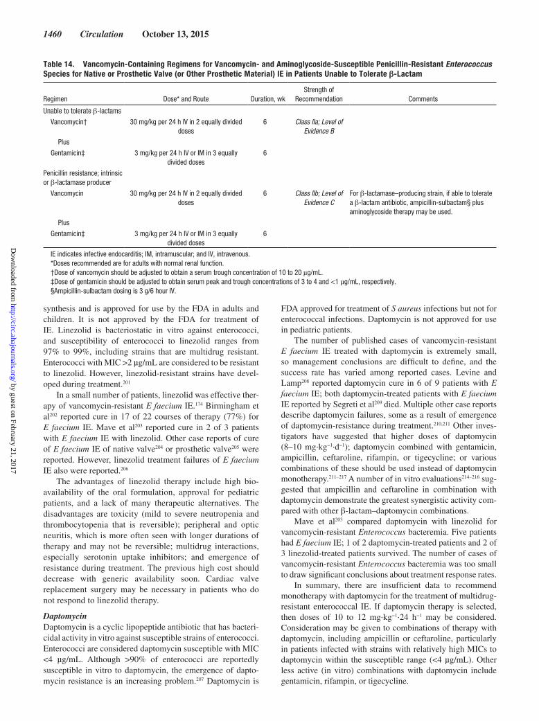
1460 Circulation October 13, 2015
synthesis and is approved for use by the FDA in adults and children. It is not approved by the FDA for treatment of IE. Linezolid is bacteriostatic in vitro against enterococci, and susceptibility of enterococci to linezolid ranges from 97% to 99%, including strains that are multidrug resistant. Enterococci with MIC >2 µg/mL are considered to be resistant to linezolid. However, linezolid-resistant strains have devel-oped during treatment.201
In a small number of patients, linezolid was effective ther-apy of vancomycin-resistant E faecium IE.174 Birmingham et al202 reported cure in 17 of 22 courses of therapy (77%) for E faecium IE. Mave et al203 reported cure in 2 of 3 patients with E faecium IE with linezolid. Other case reports of cure of E faecium IE of native valve204 or prosthetic valve205 were reported. However, linezolid treatment failures of E faecium IE also were reported.206
The advantages of linezolid therapy include high bio-availability of the oral formulation, approval for pediatric patients, and a lack of many therapeutic alternatives. The disadvantages are toxicity (mild to severe neutropenia and thrombocytopenia that is reversible); peripheral and optic neuritis, which is more often seen with longer durations of therapy and may not be reversible; multidrug interactions, especially serotonin uptake inhibitors; and emergence of resistance during treatment. The previous high cost should decrease with generic availability soon. Cardiac valve replacement surgery may be necessary in patients who do not respond to linezolid therapy.
DaptomycinDaptomycin is a cyclic lipopeptide antibiotic that has bacteri-cidal activity in vitro against susceptible strains of enterococci. Enterococci are considered daptomycin susceptible with MIC <4 µg/mL. Although >90% of enterococci are reportedly susceptible in vitro to daptomycin, the emergence of dapto-mycin resistance is an increasing problem.207 Daptomycin is
FDA approved for treatment of S aureus infections but not for enterococcal infections. Daptomycin is not approved for use in pediatric patients.
The number of published cases of vancomycin-resistant E faecium IE treated with daptomycin is extremely small, so management conclusions are difficult to define, and the success rate has varied among reported cases. Levine and Lamp208 reported daptomycin cure in 6 of 9 patients with E faecium IE; both daptomycin-treated patients with E faecium IE reported by Segreti et al209 died. Multiple other case reports describe daptomycin failures, some as a result of emergence of daptomycin-resistance during treatment.210,211 Other inves-tigators have suggested that higher doses of daptomycin (8–10 mg·kg−1·d−1); daptomycin combined with gentamicin, ampicillin, ceftaroline, rifampin, or tigecycline; or various combinations of these should be used instead of daptomycin monotherapy.211–217 A number of in vitro evaluations214–216 sug-gested that ampicillin and ceftaroline in combination with daptomycin demonstrate the greatest synergistic activity com-pared with other β-lactam–daptomycin combinations.
Mave et al203 compared daptomycin with linezolid for vancomycin-resistant Enterococcus bacteremia. Five patients had E faecium IE; 1 of 2 daptomycin-treated patients and 2 of 3 linezolid-treated patients survived. The number of cases of vancomycin-resistant Enterococcus bacteremia was too small to draw significant conclusions about treatment response rates.
In summary, there are insufficient data to recommend monotherapy with daptomycin for the treatment of multidrug-resistant enterococcal IE. If daptomycin therapy is selected, then doses of 10 to 12 mg·kg−1·24 h−1 may be considered. Consideration may be given to combinations of therapy with daptomycin, including ampicillin or ceftaroline, particularly in patients infected with strains with relatively high MICs to daptomycin within the susceptible range (<4 µg/mL). Other less active (in vitro) combinations with daptomycin include gentamicin, rifampin, or tigecycline.
Table 14. Vancomycin-Containing Regimens for Vancomycin- and Aminoglycoside-Susceptible Penicillin-Resistant Enterococcus Species for Native or Prosthetic Valve (or Other Prosthetic Material) IE in Patients Unable to Tolerate β-Lactam
Regimen Dose* and Route Duration, wkStrength of
Recommendation Comments
Unable to tolerate β-lactams
Vancomycin† 30 mg/kg per 24 h IV in 2 equally divided doses
6 Class IIa; Level of Evidence B
Plus
Gentamicin‡ 3 mg/kg per 24 h IV or IM in 3 equally divided doses
6
Penicillin resistance; intrinsic or β-lactamase producer
Vancomycin 30 mg/kg per 24 h IV in 2 equally divided doses
6 Class IIb; Level of Evidence C
For β-lactamase–producing strain, if able to tolerate a β-lactam antibiotic, ampicillin-sulbactam§ plus aminoglycoside therapy may be used.
Plus
Gentamicin‡ 3 mg/kg per 24 h IV or IM in 3 equally divided doses
6
IE indicates infective endocarditis; IM, intramuscular; and IV, intravenous.*Doses recommended are for adults with normal renal function.†Dose of vancomycin should be adjusted to obtain a serum trough concentration of 10 to 20 µg/mL.‡Dose of gentamicin should be adjusted to obtain serum peak and trough concentrations of 3 to 4 and <1 µg/mL, respectively.§Ampicillin-sulbactam dosing is 3 g/6 hour IV.
by guest on February 21, 2017http://circ.ahajournals.org/
Dow
nloaded from

Baddour et al Infective Endocarditis in Adults 1461
Recommendations
1. Patients with IE attributable to Enterococcus spe-cies resistant to penicillin, aminoglycosides, and vancomycin should be managed by specialists in infectious diseases, cardiology, cardiovascular sur-gery, clinical pharmacy, and, if necessary, pediatrics (Class I; Level of Evidence C).
2. If daptomycin therapy is selected, then doses of 10 to 12 mg·kg−1·24 h−1 may be considered (Class IIb; Level of Evidence C).
3. Combination therapy with daptomycin and ampi-cillin or ceftaroline may be considered, especially in patients with persistent bacteremia or enterococcal strains with high MICs (ie, 3 µg/mL) to daptomy-cin within the susceptible range (Class IIb; Level of Evidence C).
HACEK MicroorganismsIE caused by fastidious Gram-negative bacilli of the HACEK group (HACEK indicates Haemophilus species, Aggregatibacter species, Cardiobacterium hominis, Eikenella corrodens, and Kingella species) accounts for ≈5% to 10% of community-acquired NVE in patients who are not IDUs.218 These microorganisms grow slowly in standard blood cul-ture media, and recovery may require prolonged incubation. Typically, only a small fraction of blood culture bottles in patients with HACEK IE demonstrate growth. Bacteremia caused by HACEK microorganisms in the absence of an obvi-ous focus of infection is highly suggestive of IE even without typical physical findings of IE.
Previously, the HACEK group of microorganisms was uniformly susceptible to ampicillin. However, β-lactamase–producing strains of HACEK are appearing with increased frequency; rarely, resistance to ampicillin can occur in β-lactamase–negative strains.219 Moreover, difficulty in per-forming antimicrobial susceptibility testing as a result of failure of growth in in vitro susceptibility testing is common-place. In 1 survey, 60% of isolates did not grow adequately in control wells, and no valid in vitro susceptibility results were available.219 Therefore, unless growth is adequate for in vitro screening, then HACEK microorganisms should be consid-ered ampicillin resistant, and penicillin and ampicillin should not be used to treat patients with IE in these cases. Almost all strains of the HACEK group are susceptible to ceftriaxone (or
other third- or fourth-generation cephalosporins). Ceftriaxone has commonly been used to treat HACEK IE220 and is reason-able for treatment (Table 16). The duration of therapy for NVE of 4 weeks is reasonable; for PVE, the duration of therapy of 6 weeks is reasonable. Gentamicin is no longer recommended because of its nephrotoxicity risks.
The HACEK group is usually susceptible in vitro to fluo-roquinolones.206 On the basis of these susceptibility data, a flu-oroquinolone (ciprofloxacin, levofloxacin, or moxifloxacin) may be considered as an alternative agent in patients unable to tolerate ceftriaxone (or other third- or fourth-generation cephalosporins) therapy. There are only a few case reports of HACEK IE treated with a fluoroquinolone, however. In addi-tion, ampicillin-sulbactam may be considered a treatment option, although HACEK resistance to this agent in vitro has been described.219 Accordingly, patients with HACEK IE who cannot tolerate ceftriaxone therapy should be treated in con-sultation with an infectious diseases specialist.
Recommendations1. Unless growth is adequate in vitro to obtain suscep-
tibility testing results, HACEK microorganisms are considered ampicillin resistant, and penicillin and ampicillin should not be used for the treatment of patients with IE (Class III; Level of Evidence C).
2. Ceftriaxone is reasonable for treatment of HACEK IE (Class IIa; Level of Evidence B).
3. The duration of therapy for HACEK NVE of 4 weeks is reasonable (Class IIa; Level of Evidence B); for HACEK PVE, the duration of therapy of 6 weeks is reasonable (Class IIa; Level of Evidence C).
4. Gentamicin is not recommended because of its nephrotoxicity risks (Class III; Level of Evidence C).
5. A fluoroquinolone (ciprofloxacin, levofloxacin, or moxifloxacin) may be considered an alternative agent for patients unable to tolerate ceftriaxone (or other third- or fourth-generation cephalosporins) (Class IIb; Level of Evidence C).
6. Ampicillin-sulbactam may be considered a treat-ment option for HACEK IE (Class IIb; Level of Evidence C).
7. Patients with HACEK IE who do not tolerate cef-triaxone therapy should be treated in consultation with an infectious diseases specialist (Class I; Level of Evidence C).
Table 15. Therapy for Endocarditis Involving a Native or Prosthetic Valve or Other Prosthetic Material Resulting From Enterococcus Species Caused by Strains Resistant to Penicillin, Aminoglycosides, and Vancomycin
Regimen Dose* and Route Duration, wkStrength of
Recommendation Comments
Linezolid 600 mg IV or orally every 12 h >6 Class IIb; Level of Evidence C
Linezolid use may be associated with potentially severe bone marrow suppression, neuropathy, and numerous drug interactions. Patients with IE caused by these strains should be treated by a care team including specialists in infectious diseases, cardiology, cardiac surgery, clinical pharmacy, and, in children, pediatrics. Cardiac valve replacement may be necessary for cure.
Or
Daptomycin 10–12 mg/kg per dose >6 Class IIb; Level of Evidence C
IE indicates infective endocarditis, and IV, intravenous.*Doses recommended are for patients with normal renal and hepatic function.
by guest on February 21, 2017http://circ.ahajournals.org/
Dow
nloaded from

1462 Circulation October 13, 2015
Non-HACEK Gram-Negative BacilliIE caused by non-HACEK Gram-negative aerobic bacilli (Enterobacteriaceae and Pseudomonas species) is rare. In 1 large multinational database221 that included 2761 patients seen in 61 hospitals in 28 countries, only 49 cases (1.8%) were attributable to non-HACEK Gram-negative aerobic bacilli. It is noteworthy that healthcare exposure was asso-ciated with the development of IE caused by this group of organisms in 57% of patients. In contrast, IDU, a prominent risk factor for the development of this IE syndrome in ear-lier years, was recognized in only 4% of cases in the mul-tinational survey that included cases seen between 2000 and 2005. Escherichia coli and Pseudomonas aeruginosa accounted for 51% of cases, and 59% had PVE. Although management included cardiac surgery in 51% of cases, the in-hospital mortality rate was 24%.
Despite the very rare occurrence of IE caused by Salmonella species in North America, this syndrome deserves specific mention because it occurs with some frequency in other geo-graphic areas.222 Salmonella species have a proclivity to infect cardiovascular structures in adults. Therefore, all patients with bloodstream infection resulting from Salmonella species should be evaluated for complicating cardiovascular infec-tions, including IE, myocarditis, pericarditis, and endarteritis. Although many serotypes have been implicated, most cases are caused by S choleraesuis, S typhimurium, and S enteritidis.222
Cardiac surgery in combination with prolonged courses of combined antibiotic therapy is reasonable for most patients with IE caused by non-HACEK Gram-negative aerobic bacilli, particularly in the setting of left-sided valvular involvement.
Prospective trial data are lacking to define the optimal antimicrobial regimen for the treatment of IE caused by non-HACEK Gram-negative aerobic bacilli. Input from special-ists in infectious diseases who are experienced in the medical management of IE should be obtained to define an antibiotic
regimen in each case. This is particularly important in IE caused by non-HACEK Gram-negative aerobic bacilli for sev-eral reasons. First, as stated previously, healthcare exposure is commonly seen in these cases; thus, multidrug resistance often characterizes these pathogens. Second, therapy may include agents with increased toxicity risks such as aminogly-cosides (given in high dosages) and colistin. Third, because regimens that include >1 agent are often selected for use, the risks of drug-drug interactions and increased drug-related adverse events are problematic. Fourth, mortality is high in these infections, and medical-surgical approaches are often required for optimal management and favorable outcomes.
Combination antibiotic therapy with a β-lactam (penicillins, cephalosporins, or carbapenems) and either an aminoglycoside or fluoroquinolone for 6 weeks is reasonable.221 Consultation with an infectious diseases expert in IE should be sought because of the various mechanisms of antibiotic resistance that can be found in the non-HACEK Gram-negative aerobic bacilli. For example, several of these bacteria may harbor “inducible β-lactamases” that could require supplemental laboratory screening, in addition to routine in vitro susceptibility testing.
Medical therapy may be successful in right-sided P aerugi-nosa IE in 50% to 75% of cases. If the disease is refractory to anti-biotics, then partial tricuspid valvulectomy or “vegetectomy”223 without valve replacement is indicated.224 Typically, these patients have been IDUs, and because of their high recidivism risk, avoid-ance of placement of prosthetic valves is desirable.
Recommendations
1. Cardiac surgery is reasonable in combination with prolonged courses of combined antibiotic therapy for most patients with IE caused by non-HACEK Gram-negative aerobic bacilli, particularly P aeru-ginosa (Class IIb; Level of Evidence B).
Table 16. Therapy for Endocarditis Involving a Native or Prosthetic Valve or Other Prosthetic Material Caused by HACEK Microorganisms
Regimen Dose and Route Duration, wkStrength of
Recommendation Comments
Ceftriaxone sodium* 2 g/24 h IV or IM in 1 dose 4, NVE;6, PVE
Class IIa; Level of Evidence B
Preferred therapy: cefotaxime or another third- or fourth-generation cephalosporin may be substituted.
Or
Ampicillin sodium 2 g IV every 4 h Class IIa; Level of Evidence B
Ampicillin sodium may be an option if the growth of the isolate is sufficient to permit in vitro susceptibility results. Or
Ciprofloxacin† 1000 mg/24 h orally or 800 mg/24 h IV in 2 equally divided doses
Class IIb; Level of Evidence C
Fluoroquinolone therapy‡ may be considered for patients unable to tolerate cephalosporin and ampicillin therapy; levofloxacin or moxifloxacin may be substituted; fluoroquinolones generally is not recommended for patients <18 y old. Treatment for 6 wk is reasonable in patients with PVE (Class IIa; Level of Evidence C).
HACEK indicates Haemophilus species, Aggregatibacter species, Cardiobacterium hominis, Eikenella corrodens, and Kingella species; IM, intramuscular; IV, intravenous; NVE, native valve infective endocarditis; and PVE, prosthetic valve infective endocarditis.
*Patients should be informed that intramuscular injection of ceftriaxone is painful.†Dose recommended for patients with normal renal function.‡Fluoroquinolones are highly active in vitro against HACEK microorganisms. Published data on the use of fluoroquinolones for endocarditis caused by HACEK are minimal.
by guest on February 21, 2017http://circ.ahajournals.org/
Dow
nloaded from

Baddour et al Infective Endocarditis in Adults 1463
2. Combination antibiotic therapy with a β-lactam (penicillins, cephalosporins, or carbapenems) and either an aminoglycoside or fluoroquinolone for 6 weeks is reasonable (Class IIa; Level of Evidence C).
3. Consultation with an infectious diseases expert in IE should be sought because of the various mechanisms of antibiotic resistance that can be found in the non-HACEK Gram-negative aerobic bacilli (Class I; Level of Evidence C).
Culture-Negative EndocarditisPositive blood cultures are a major diagnostic criterion in IE and key to identifying an pathogenic agent and an optimal antimicrobial regimen.225,226 Continuous bacteremia and a high frequency of positive blood cultures are typical hallmarks of this infection. The intensity of bacteremia may not be great, however, with <50 colony-forming units per 1 mL blood detected in the majority of patients in an investigation.227
Failure to culture microorganisms that cause IE can be a major problem that complicates diagnosis and timely, effec-tive treatment. Although most previous studies have put the frequency of blood culture–negative IE at 5% to 10%, a European study of IE that included 820 cases indicated that ≈20% of patients with confirmed IE had all negative blood cultures.228 This may be attributable to inadequate microbio-logical techniques, infection with highly fastidious bacteria or fungi, noncultivatable agents, or the previous administration of antimicrobial agents before blood cultures were obtained. Administration of antimicrobial agents to IE patients before blood cultures are obtained reduces the recovery rate of bac-teria by 35% to 40%.228–232 The antimicrobial susceptibility of the organism, the dose, and the duration and nature of pre-vious antimicrobial therapy together determine the length of time that blood cultures will remain negative.232 IE patients with blood cultures that are initially negative after only a few days of antibiotic therapy may have positive blood cultures after several days without antibiotics. The blood cultures of patients who receive longer courses of high-dose bactericidal antimicrobials may remain negative for weeks.
Selection of medical therapy for patients with culture-negative IE is difficult. On the one hand, there is a need to provide empirical antimicrobials for all likely pathogens. On the other hand, certain therapeutic agents, including amino-glycosides, have potentially toxic effects that dictate limita-tion or avoidance of use if at all possible. Moreover, some of the laboratory-based diagnostic techniques to define fas-tidious or unusual pathogens are not available in most clinical laboratories and require considerable time for completion of testing if specimens are sent to a referral laboratory.233 During this period, patients are often treated empirically for the more common bacterial causes of IE, which can result in exposure to potentially toxic therapy that could be avoided with earlier pathogen identification.
An evaluation of epidemiological factors (Table 6), history of prior infections including cardiovascular infections, expo-sure to antimicrobials, clinical course, severity, and extracar-diac sites of infection of the current infection should be done in all IE cases. During the period between the collection of blood cultures and the determination of a pathogen or if blood
cultures are ultimately deemed culture negative, empirical therapy is generally required. Consultation with an infectious diseases specialist to define the most appropriate choice of therapy is recommended. Collection of additional clinical and laboratory data often dictates subsequent revisions in initial empirical therapy that will be administered over the treatment course.
For patients with acute (days) clinical presentations of native valve infection, coverage for S aureus, β-hemolytic streptococci, and aerobic Gram-negative bacilli is reasonable. Empirical coverage could include vancomycin and cefepime as an initial regimen. For patients with a subacute (weeks) presentation of NVE, empirical coverage of S aureus, VGS, HACEK, and enterococci is reasonable. One treatment option could include vancomycin and ampicillin-sulbactam to pro-vide some coverage for these organisms. Subsequent regimen revision can be done when a pathogen is recovered from blood cultures.
For patients with culture-negative PVE, coverage for staphylococci, enterococci, and aerobic Gram-negative bacilli is reasonable if the onset of symptoms is within 1 year of prosthetic valve placement. A regimen could include vanco-mycin, rifampin, gentamicin, and cefepime. If symptom onset is >1 year after valve placement, then IE is more likely to be caused by staphylococci, VGS, and enterococci, and antibiotic therapy for these potential pathogens is reasonable. One initial treatment option could include vancomycin and ceftriaxone.
If subsequent blood culture results or other laboratory methodologies define a pathogen, then empirical therapy should be revised to focused therapy that is recommended for the specific pathogen identified.
True culture-negative IE can be caused by uncommon or rare pathogens that do not grow in routinely used blood culture systems.234–237 The organisms that have garnered the most attention are Bartonella species, Chlamydia species, C burnetii, Brucella species, Legionella species, Tropheryma whipplei, Candida, and non-Candida fungi (particularly Aspergillus species). The last 2 groups of organisms are espe-cially relevant to PV recipients. With the use of special diag-nostic techniques, Bartonella species, C burnetii, and Brucella species have been identified in the majority of cases of cul-ture-negative IE caused by fastidious organisms. Additional laboratory screening is required to identify the causes of culture-negative IE.233 In some cases, serological and special blood culture techniques can be helpful. In other cases, tissue (usually valve) screening is required. Diagnostic methods for resected valve tissue include microbiological, histopathologi-cal, and molecular techniques, the last of which includes gene amplification with PCR methods. Unfortunately, most clinical laboratories do not perform molecular screening, and speci-mens must be sent to reference laboratories.
The most prevalent pathogen among these uncommon causes of culture-negative IE in this group has varied glob-ally according to published data.236 Incidence data from population-based surveys for IE caused by these organisms are lacking in the United States. In PVE cases, the timing of infection onset can also be important in defining pathogens.235 Limitations such as referral bias and sampling bias may have affected the findings.235–237
by guest on February 21, 2017http://circ.ahajournals.org/
Dow
nloaded from

1464 Circulation October 13, 2015
Results of a large prospective analysis of referred samples from culture-negative IE performed by a well-recognized reference laboratory deserve additional comment.236 First, there was identification of a pathogen in 62.7% of 759 cases; in 2.5%, a noninfectious origin (see below) was confirmed. Second, serological results were positive in 47.7% of cases, primarily for Coxiella and Bartonella species infection. Third, PCR identified a pathogen in two thirds of the valves studied. Fourth, no cause was defined in 35% of cases.
Treatment of the wide variety of microorganisms that cause culture-negative IE without prior antibiotic exposure has been described anecdotally, and regimens of choice are based on limited data and can be found in other publications.
Noninfectious causes of valvular vegetations can produce a syndrome similar to culture-negative IE. Perhaps the one that has received the most attention is anti-phospholipid anti-body (APA) syndrome,238 which has been described as both a primary and a secondary syndrome and is associated with the presence of APA. In its secondary form, the APA syndrome has been linked to autoimmune disorders, particularly sys-temic lupus erythematosus, and malignancies. Sterile valvular vegetations form and embolize, clinically mimicking in many respects culture-negative IE. The mitral valve is most often affected, and valvular regurgitation is the predominant func-tional abnormality seen in APA syndrome with complicating valvular involvement. To complicate matters, the APA syn-drome may develop secondary to IE.239
Numerous other causes of noninfective vegetative endo-carditis can mimic IE. These can be categorized into 4 groups230: neoplasia associated (atrial myxoma, marantic endocarditis, neoplastic disease, and carcinoid), autoimmune associated (rheumatic carditis, systemic lupus erythematosus, polyarteritis nodosa, and Behçet disease), postvalvular surgery (thrombus, stitch, or other postsurgery changes), and miscel-laneous (eosinophilic heart disease, ruptured mitral chordae, and myxomatous degeneration).
Recommendations
1. An evaluation of epidemiological factors, history of prior infections including cardiovascular infections, exposure to antimicrobials, clinical course, sever-ity, and extracardiac sites of infection of the current infection should be performed in all culture-nega-tive endocarditis cases (Class I; Level of Evidence C).
2. Consultation with an infectious diseases specialist to define the most appropriate choice of therapy in patients with culture-negative endocarditis is rec-ommended (Class I; Level of Evidence C).
3. For patients with acute (days) clinical presentations of native valve infection, coverage for S aureus, β-hemolytic streptococci, and aerobic Gram-negative bacilli is reasonable (Class IIa; Level of Evidence C).
4. For patients with a subacute (weeks) presenta-tion of NVE, coverage of S aureus, VGS, HACEK, and enterococci is reasonable (Class IIa; Level of Evidence C).
5. For patients with culture-negative PVE, cover-age for staphylococci, enterococci, and aerobic
Gram-negative bacilli is reasonable if onset of symp-toms is within 1 year of prosthetic valve placement (Class IIa; Level of Evidence C).
6. If symptom onset is >1 year after valve placement, then IE is more likely to be caused by staphylococci, VGS, and enterococci, and antibiotic therapy for these potential pathogens is reasonable (Class IIa; Level of Evidence C).
7. If subsequent blood culture results or other labora-tory methodologies define a pathogen, then empiri-cal therapy should be revised to focused therapy that is recommended for the specific pathogen iden-tified (Class I; Level of Evidence C).
FungiFungal IE is rare but can develop in a wide range of patients.240,241 The well-recognized risk factors associated with fungal IE (eg, IDU and immunocompromised state) have become less prevalent compared with the presence of a car-diovascular device, including central venous catheters, perma-nent pacemakers and defibrillators, and prosthetic valves.240–246
Fungal IE has been recognized as a cause of early PVE, but a case series from a single medical center demonstrated that 43% of these cases had symptom onset >1 years after pros-thetic valve placement.242 In contrast to the expected older age predilection for the development of IE, patients with fungal IE have been younger, which was somewhat unanticipated, con-sidering the low prevalence of IDU among the cohort. Candida and Aspergillus species account for the large majority of fun-gal IE, and Candida-related IE is much more common than Aspergillus-related disease.240,241 Blood cultures are ultimately positive in most cases caused by the former pathogen, whereas they are rarely positive in cases caused by the latter fungus. Thus, Aspergillus is a cause of culture-negative IE, and when this occurs, it is usually in a patient with a prosthetic cardiac valve.240 A variety of other fungi, including endemic mycoses, can rarely cause IE and can involve both native and prosthetic valves. Noncardiac sites of metastatic infection often compli-cate fungal IE; this can include, for example, endophthalmitis in patients with candidal IE, which may require both systemic and intraocular antifungal therapy. Further guidelines are available from the Infectious Diseases Society of America for additional management aspects of several of the fungal patho-gens (http://www.idsociety.org/IDSA_Practice_Guidelines/).
Despite aggressive combined medical and surgical inter-ventions and a younger cohort, mortality rates for fungal IE are unacceptably high. The survival rate for patients with mold-related IE is <20%. Historically, 2 treatment doctrines have prevailed in fungal IE despite the lack of prospective tri-als conducted to define the most appropriate therapy: Fungal IE is a “stand-alone indication” for surgical replacement of an infected valve; and amphotericin B, a fungicidal agent, is the initial drug of choice for fungal IE. Because of the alarming mortality rate associated with fungal IE and the availability of newer antifungal drugs, in particular fungicidal drugs like the echinocandins, a re-evaluation of these principles seems in order. If done, however, this will probably be based on anec-dotal experience and expert opinion rather than on clinical trial data because of the rarity of the syndrome.
by guest on February 21, 2017http://circ.ahajournals.org/
Dow
nloaded from

Baddour et al Infective Endocarditis in Adults 1465
A 2-phase treatment of fungal IE has evolved. The initial or induction phase consists of control of infection. Treatment includes a combination of a parenteral antifungal agent, usu-ally an amphotericin B–containing product, and valve sur-gery. Valve surgery should be done in most cases of fungal IE. Results of a meta-analysis that included 879 cases of Candida IE demonstrated a marked reduction in death (prevalence odds ratio, 0.56; 95% confidence interval, 0.16–1.99) among those who underwent adjunctive valve surgery.244 In addition, patients who were treated with combination therapy including amphotericin B and flucytosine had reduced mortality com-pared with those who received antifungal monotherapy.
Antifungal therapy usually is given for >6 weeks. After completion of this initial therapy, long-term (lifelong) sup-pressive therapy with an oral azole is reasonable.243,244,246 Suppressive therapy has been used in 2 populations. First, because of the high relapse rate of fungal IE and the prolonged delay (years in some cases) in relapse, oral azoles have been administered after combined medical and surgical induction therapy. In a second population with fungal IE, lifelong oral antifungal suppressive therapy has been given to patients who respond clinically to induction medical therapy but are not deemed appropriate surgical candidates for valve replacement for attempted infection cure. Anecdotal case series243,245 indi-cate that IE has been successfully suppressed for months to years. A meta-analysis that included 64 reported patients with Candida IE who did not undergo valve surgery because they were deemed to be unacceptable surgical candidates supports the notion that fluconazole suppressive therapy is useful; 20 of 21 patients (95%) who were ultimately treated with long-term suppressive therapy survived during follow-up, which was ≥6 months.244
Recommendations
1. Valve surgery should be done in most cases of fungal IE (Class I; Level of Evidence B).
2. After completion of initial parenteral therapy, life-long suppressive therapy with an oral azole is rea-sonable (Class IIa; Level of Evidence B).
Surgical ManagementThere is a prevailing opinion that valve surgery is crucial for optimal therapy in selected patients with complicated IE.247–249 In a systematic review5 of 15 population-based IE investiga-tions from 7 countries, after adjustment for country, the pro-portion of IE cases undergoing valve surgery increased 7% per decade (95% confidence interval, −0.4% to 14%; P=0.06) between 1969 and 2000. In surveys involving population-based1 and international multicenter cohorts,10,11 ≈50% of both NVE and PVE patients undergo valve surgery during the active phase of IE (during initial hospitalization before com-pletion of a full therapeutic course of antibiotics).
Although valve replacement surgery has served as an important option in the management of individual IE cases, only 1 small, randomized trial17 has been performed to date to examine the role of valve surgery in the management of IE. In this trial, 76 patients with left-sided NVE, severe valve regur-gitation without heart failure, and vegetations >10 mm were
assigned to early surgery within 48 hours or to conventional treatment. Although the authors17 report a reduction in the composite outcome of in-hospital deaths and embolic events with early surgery (3% versus 23%), the differences between the 2 groups were driven by a significant decrease in embolic events with early surgery. Thus, firm conclusions cannot be drawn from this trial on the effect of early surgery on mor-tality, given the small sample size of the study. In addition, patients in this trial were young and had limited comorbidity based on a EuroSCORE, a calculated risk of surgical mortality (http://www.euroscore.org), a low prevalence of S aureus IE, and lower mortality compared with most contemporary patient cohorts. Moreover, many patients had signs of embolization (a Class IIa indication for surgery) before randomization. Data from nonrandomized trials from a single-center experience250 and an international collaboration251 support the notion252 that early valve surgery may not be beneficial in all patients with native or PVE caused by S aureus.
Over several decades, expert panels have relied on data from observational studies to make recommendations on the indications for early surgery. Despite the availability of new studies, the indications for surgery have not changed appre-ciably over time.18 Considering that observational studies are prone to bias and confounding, researchers have used regres-sion analysis and calculated propensity scores to adjust for prognostically important baseline differences between surgi-cal and medical patients.14,253 Studies14,15,136,249,253–256 examin-ing the association between valve surgery and outcome in left-sided IE using propensity score analysis, however, have demonstrated conflicting results, likely because of the use of different analytical approaches.
Until 2007, none of the published studies adjusted for sur-vivor bias, which occurs because patients who live longer are more likely to undergo surgery than those who die early. A cor-relation between longer survival and surgery may be wrongly interpreted as evidence that surgical treatment improves sur-vival.257 Since 2007, at least 3 studies15,16,258 have documented the effect of survivor bias on the association between surgery and mortality in IE patients. When adjusted for survivor bias, analyses have shown either a statistical loss of benefit of early surgery or findings indicating that the surgical intervention may actually result in harm.
Between 2007 and 2013, at least 6 observational stud-ies10,11,14–16,259 that adjusted for selection bias, confounding, and survivor bias were conducted. Three studies that included 2 cohorts of patients with NVE and PVE and 1 cohort with NVE showed an association between early surgery and lower mortality in IE patients in general or in specific subgroups of patients such as those with heart failure or paravalvular com-plications. Only 1 study examined the role of valve surgery in PVE.259 After adjustment for differences in clinical char-acteristics and survival bias, early valve replacement was not associated with lower mortality compared with medical therapy in the overall cohort. Subgroup analysis indicated a lower in-hospital and 1-year mortality with early surgery only in the group of patients with the highest surgical propensity. Table 17 summarizes the characteristics of rigorously con-ducted observational studies that support the role of surgery in IE management.
by guest on February 21, 2017http://circ.ahajournals.org/
Dow
nloaded from

1466 Circulation October 13, 2015
Indications for SurgeryDecisions on surgical intervention are complex and depend on many clinical and prognostic factors257–262 that vary among patients, including infecting organism, vegetation size, presence of perivalvular infection, presence of embolism or heart failure, age, noncardiac comorbidities, and available surgical expertise. There is a paucity of evidence available to define the optimal timing of valve surgery. Decisions on the indication and timing of surgical intervention should be determined by a multispecialty team with expertise in cardiology, imaging, cardiothoracic sur-gery, and infectious diseases.261 Recommendations for early sur-gery in patients with recurrent emboli and persistent vegetations have generally been enacted after clinical events. Whether recur-rent, asymptomatic emboli detected on advanced imaging stud-ies should influence decision making should be considered on an individual basis. Risk stratification models such as the Society of Thoracic Surgeons Endocarditis Score are available to predict morbidity and mortality risks in IE patients after valve surgery and to assist in decision making and patient counseling.260
Early Valve Surgery in Left-Sided NVE: Recommendations
1. Early surgery (during initial hospitalization and before completion of a full course of antibiotics) is indicated in patients with IE who present with valve dysfunction resulting in symptoms or signs of heart failure (Class I; Level of Evidence B).
2. Early surgery should be considered particularly in patients with IE caused by fungi or highly resistant organisms (eg, vancomycin-resistant Enterococcus, multidrug-resistant Gram-negative bacilli) (Class I; Level of Evidence B).
3. Early surgery is indicated in patients with IE com-plicated by heart block, annular or aortic abscess, or destructive penetrating lesions (Class I; Level of Evidence B).
4. Early surgery is indicated for evidence of persistent infection (manifested by persistent bacteremia or fever lasting >5–7 days and provided that other sites of infection and fever have been excluded) after the start of appropriate antimicrobial therapy (Class I; Level of Evidence B).
5. Early surgery is reasonable in patients who present with recurrent emboli and persistent or enlarging vegetations despite appropriate antibiotic therapy (Class IIa; Level of Evidence B).
6. Early surgery is reasonable in patients with severe valve regurgitation and mobile vegetations >10 mm (Class IIa, Level of Evidence B).
7. Early surgery may be considered in patients with mobile vegetations >10 mm, particularly when involving the anterior leaflet of the mitral valve and associated with other relative indications for sur-gery (Class IIb; Level of Evidence C).
Early Valve Surgery in PVE: Recommendations
1. Early surgery is indicated in patients with symp-toms or signs of heart failure resulting from valve
dehiscence, intracardiac fistula, or severe prosthetic valve dysfunction (Class I; Level of Evidence B).
2. Early surgery should be done in patients who have persistent bacteremia despite appropriate antibi-otic therapy for 5 to 7 days in whom other sites of infection have been excluded (Class I; Level of Evidence B).
3. Early surgery is indicated when IE is compli-cated by heart block, annular or aortic abscess, or destructive penetrating lesions (Class I; Level of Evidence B).
4. Early surgery is indicated in patients with PVE caused by fungi or highly resistant organisms (Class I; Level of Evidence B).
5. Early surgery is reasonable for patients with PVE who have recurrent emboli despite appropriate anti-biotic treatment (Class IIa; Level of Evidence B).
6. Early surgery is reasonable for patients with relaps-ing PVE (Class IIa; Level of Evidence C).
7. Early surgery may be considered in patients with mobile vegetations >10 mm (Class IIb; Level of Evidence C).
Valve Surgery in Patients With Right-Sided IEAlthough outcomes are better for patients with right-sided IE compared with patients with left-sided infection, surgi-cal intervention is occasionally considered in the former group. Because many of the patients with right-sided IE develop infection as a result of IDU (see the Right-Sided IE in IDUs section), the general approach is to treat these patients
Table 17. Direct Evidence Supporting an Association Between Valve Surgery and Lower Mortality From Observational Studies: Level of Evidence B
Study Mortality IE Group PE vs SA
Lalani et al10 In-hospital mortality NVE PE
Bannay et al15 5-y mortality NVE+PVE PE
Kiefer et al11 In-hospital and 1-y mortality
CHF (NVE+PVE) PE
Lalani et al10 In-hospital mortality Paravalvular complications (NVE)
SA
Bannay et al15 5-y mortality Intracardiac abscess (NVE+PVE)
SA
Lalani et al10 In-hospital mortality Systemic embolization (NVE)
SA
Bannay et al15 5-y mortality Systemic embolization (NVE+PVE)
SA
Lalani et al10 In-hospital mortality S aureus (NVE) SA
Bannay et al15 5-y mortality CHF (NVE+PVE) SA
Lalani et al259 In-hospital and 1-y mortality
PVE with the highest propensity to undergo
surgery
SA
CHF indicates congestive heart failure; NVE, native valve infective endocarditis; PE, primary end point; PVE, prosthetic valve infective endocarditis; and SA, subgroup analysis.
All studies have adjusted for selection and survivor bias and confounding. Valve surgery was performed during the active phase of the disease (during initial hospitalization before completion of a full therapeutic course of antibiotics).
by guest on February 21, 2017http://circ.ahajournals.org/
Dow
nloaded from

Baddour et al Infective Endocarditis in Adults 1467
medically and to avoid placement of valve prostheses because of the subsequent risk of device infection with continued IDU. Surgical intervention is reasonable for patients with the fol-lowing complications: right heart failure secondary to severe tricuspid regurgitation with poor response to medical therapy, sustained infection caused by difficult-to-treat organisms (ie, fungi, multidrug resistant bacteria) or lack of response to appropriate antimicrobial therapy, and tricuspid valve vegeta-tions that are ≥20 mm in diameter and recurrent pulmonary embolism despite antimicrobial therapy. Valve repair rather than replacement should be performed when feasible. If valve replacement is performed, then an individualized choice of prosthesis by the surgeon is reasonable.263,264
Recommendations
1. Surgical intervention is reasonable for patients with certain complications (Class IIa; Level of Evidence C).
2. Valve repair rather than replacement should be per-formed when feasible (Class I; Level of Evidence C).
3. If valve replacement is performed, then an individu-alized choice of prosthesis by the surgeon is reason-able (Class IIa; Level of Evidence C).
4. It is reasonable to avoid surgery when possible in patients who are IDUs (Class IIa; Level of Evidence C).
Valve Surgery in Patients With Prior Emboli/Hemorrhage/StrokeThe timing of valve surgery in IE patients with stroke remains controversial. Stroke is an independent risk factor for post-operative mortality in IE patients. After stroke, neurological deterioration can occur as a result of hemorrhagic transforma-tion with anticoagulation during cardiopulmonary bypass or exacerbation of cerebral ischemia attributable to hypotension during cardiac surgery. The risk of intracranial hemorrhage is dependent on several factors, including extent and size of infarction, whether it is ischemic or hemorrhagic, and the exact timing of surgery.
One clinical quandary is whether early valve surgery can be safely performed within 7 days after a stroke or if it is better to postpone surgery for at least 1 week. No randomized trials have addressed this conundrum. The high rates of postoperative mor-bidity and mortality seen in earlier studies265–267 have resulted in a reluctance to refer patients with IE and acute stroke for imme-diate valve surgery. However, these initial studies included a limited number of patients, and risk adjustments were not per-formed. The largest early series of operated patients with cere-bral complications included 181 patients.267 Hospital mortality rates as a function of the interval between evidence of cerebral infarction to cardiac surgery were 66.3% when surgery was per-formed within 24 hours of stroke and gradually decreased every week to 7.0% with surgery >4 weeks after stroke.
Investigations have suggested better outcomes for IE patients with ischemic stroke who undergo early cardiac sur-gery.268–272 Ruttmann et al270 analyzed 65 patients who under-went cardiac surgery after cardioembolic (embolic) stroke (median time, 4 days; range, 0–38 days). Surgery in this
time frame was not associated with worse patient outcomes. Fifty of the 61 patients (81.9%) with CT-verified preopera-tive stroke survived cardiac surgery. Latency between the neurological event and cardiac surgery was not a significant factor with respect to the perioperative neurological compli-cation rate or the postoperative neurological recovery rate. Full neurological recovery was achieved in 70% of 50 stroke patients. Other studies6–8 suggest that the risk of neurologi-cal deterioration during cardiac surgery after a stroke is lower than previously assumed, particularly in patients with silent cerebrovascular emboli.
The first study to evaluate the timing of surgery after stroke in IE that included a risk adjustment for differences in patient characteristics comprised 198 patients.273 Fifty-eight patients who underwent surgery within 1 week of stroke were compared with 140 patients who underwent surgery ≥8 days after stroke. Hospital mortality was numerically but not significantly higher in the early surgery group (22.4% versus 12%). After adjustment for other risk factors such as age, paravalvular abscess, and heart failure, the risk of hos-pital mortality remained nonsignificantly higher in the early surgery group (odds ratio, 2.308; 95% confidence interval, 0.942–5.652). Differences in 1-year mortality were less pro-nounced, with an adjusted hazard ratio of 1.138 (95% con-fidence interval, 0.802–1.650). In-hospital mortality in the early surgery group was comparable to that of the medically treated patients.
After hemorrhagic stroke, the risk of exacerbation by sur-gery is prohibitively high in the first month but can extend beyond 1 month in some patients, possibly because of the presence of undetected mycotic aneurysms (MAs). In a mul-ticenter study of patients with hemorrhagic stroke, mortality was higher when surgery was performed within 4 weeks of the hemorrhagic event compared with later surgery (75% versus 40%, respectively).274
These data support the following recommendations: Valve surgery may be performed in IE patients with stroke or sub-clinical cerebral emboli without delay if intracranial hemor-rhage has been excluded by imaging studies and neurological damage is not severe (ie, coma). In patients with major isch-emic stroke or intracranial hemorrhage, it is reasonable to delay valve surgery for at least 4 weeks.
Recommendations
1. Valve surgery may be considered in IE patients with stroke or subclinical cerebral emboli and residual vegetation without delay if intracranial hemorrhage has been excluded by imaging studies and neurolog-ical damage is not severe (ie, coma) (Class IIb; Level of Evidence B).
2. In patients with major ischemic stroke or intra-cranial hemorrhage, it is reasonable to delay valve surgery for at least 4 weeks (Class IIa; Level of Evidence B).
Risk of EmbolizationSystemic embolization occurs in 22% to 50% of cases of IE.55,57,274–277 Rates can be higher if noninvasive imaging,
by guest on February 21, 2017http://circ.ahajournals.org/
Dow
nloaded from
1468 Circulation October 13, 2015
including MRI and CT scanning, is routinely done to detect asymptomatic (silent) emboli. Emboli often involve major arterial beds, including the brain, lungs, coronary arteries, spleen, bowel, and extremities. Up to 65% of embolic events involve the CNS, and >90% of CNS emboli lodge in the distri-bution of the middle cerebral artery.277 The highest incidence of embolic complications is seen with mitral valve IE (and more with anterior rather than posterior mitral leaflet involve-ment) and with IE caused by S aureus, Candida, and HACEK organisms.
Emboli can occur before diagnosis, during therapy, or after therapy is completed, although most emboli occur within the first 2 to 4 weeks of antimicrobial therapy.58,278 Of note, 2 inde-pendent studies have confirmed that the rate of embolic events decreases dramatically during and after the first 2 to 3 weeks of successful antibiotic therapy. In a study from 1991, the embolic rate dropped from 13 to <1.2 embolic events per 1000 patient-days during that time.58 Vilacosta et al278 confirmed the reduced frequency of embolization after 2 weeks of therapy. Moreover, the latter study reemphasized the increased risk of embolization with increasing vegetation size during therapy, mitral valve involvement, and staphylococcal pathogenesis. In a survey that included the International Collaboration on Endocarditis cohort, Dickerman and colleagues280 focused on the incidence of stroke in a multicenter IE population and demonstrated that acute stroke rates fell significantly after initiation of antibiotic therapy regardless of valve involved or pathogen identified. Moreover, only 3.1% of the cohort suf-fered stroke after the first week of antimicrobial therapy; this finding has led to the opinion that stroke prevention as a sole indication for valve surgery after 1 week of appropriate antibi-otic therapy is not warranted.
Prediction of individual patient risk for embolization is extremely difficult. Many studies have attempted to use echocardiography to identify a high-risk subset of IE patients who might benefit from early surgery to avoid embolization. Several studies with TTE have demonstrated a trend toward higher embolic rates with left-sided vegetations >1 cm in diameter.55 De Castro and colleagues276 compared TTE with multiplane TEE and found that neither technique was help-ful in defining embolic risk in patients with vegetations. In a study57 based on TEE, mitral vegetations >1 cm in diameter were associated with the greatest frequency of embolism. The association was strengthened when the analysis was limited to those patients who had not yet experienced a clinical embolic event. Another prospective TEE study, however, found no clear correlation of vegetation size with embolization.59 Nevertheless, the same investigators later reported the results of a new prospective study of 118 patients who underwent TEE and found that, on multivariable analysis, risk factors associated with embolic risk included vegetation size >10 mm and mitral valve involvement.56 Overall, these data are com-patible with previous observations that indicate that, in gen-eral, mitral vegetations of any size are associated with a higher risk of embolization (25%) than aortic vegetations (10%). As noted above, the highest embolic risk (37%) has been seen in the subset of patients with mitral vegetations attached to the anterior rather than the posterior mitral leaflet.59,63 This sug-gests that the mechanical effects of broad and abrupt leaflet
excursion, occurring twice per heartbeat, may contribute to the propensity of a vegetation in this location to fragment and embolize.
In another study, the effect of vegetation size on embolic potential was dependent on the infecting organism, with large vegetations independently predicting embolic events only in the setting of streptococcal IE.60 In contrast, as confirmed above by Vilacosta et al,278 staphylococcal or fungal IE appears to carry a high incidence rate of embolization independently of vegetation size.
Prognosis based on echocardiographic findings was exam-ined in a large, multicenter, prospective investigation. On the basis of TEE findings in 384 consecutive adult patients with definite IE, vegetation length >15 mm was a predictor of 1-year mortality (adjusted relative risk, 1.8; 95% confidence interval, 1.10–2.82; P=0.02) in multivariable analysis.281
The role of echocardiography in predicting embolic events has been controversial. In 1 survey282 that included 4 echocar-diographers who were blinded to clinical data, interobserver agreement was mixed on the characterization of vegetations. Agreement was high for the presence of vegetation (98%) and involved site (97%); interobserver agreement was consider-ably less for vegetation size (73%), mobility (57%), shape (37%), and attachment (40%). However, all of the series that included >100 patients who underwent TEE showed a positive relationship between embolic events and vegetation size. Moreover, the study with the largest number of patients (n=176) that assessed the value of TEE and included silent embolism detected by CT scanning demonstrated that the risk of embolic events was highly related to vegetation size and mobility but not to other known risk factors associated with embolism in IE.283 The conflicting results on the relationship between echocardiography and embolic risk can be explained at least partially by the poor standardization of diagnostic cri-teria for IE in older series, inclusion or not of silent embolism, inclusion or not of previous embolism, echocardiographic method used, lack of focus on future embolic events after TEE, and sample size.
An increase in vegetation size over 4 to 8 weeks of therapy as documented by TEE appears to predict embolic events.282 In addition, a second, albeit infrequent, peak of late embolic events has been observed to occur 15 to 30 weeks after the diagnosis of IE and has been associated with nonhealing veg-etations (failure of a vegetation to stabilize or diminish in size) as defined by echocardiography.63
The traditional indication for valvular surgery for IE to avoid embolization has been ≥2 major embolic events.284 This criterion is arbitrary and excludes cutaneous embolization, which is common, or embolism occurring before the insti-tution of therapy, which is common among IE patients who develop embolic events. Because of the observed decreases in embolic risk during the first 2 weeks of antibiotic therapy, the benefit of surgery in avoiding catastrophic embolic events is greatest early in the treatment course of IE. Early surgi-cal intervention may preclude a primary or recurrent major embolic event but exposes the patient to both immediate and lifelong risks of valve replacement if the valve cannot be pri-marily repaired. At this time, the strategy for surgical inter-vention to avoid systemic embolization in IE remains specific
by guest on February 21, 2017http://circ.ahajournals.org/
Dow
nloaded from

Baddour et al Infective Endocarditis in Adults 1469
to the individual patient, with benefit being greatest in the early phase of IE when embolic rates are highest and other predictors of a complicated course (ie, recurrent embolization; heart failure; aggressive, antibiotic-resistant organisms; or PVE) are present (Table 5). The benefits of early surgery were demonstrated in the prospective, randomized trial17 that was discussed earlier in this document. Because of several limita-tions of that trial, additional study is needed before routine application of early surgery solely to reduce embolic risk can be strongly advocated.
Embolic events are important prognostic indicators of IE outcomes. In 1 analysis, an embolic event was 1 of 4 early pre-dictors of in-hospital death caused by IE.285 Other independent predictors of death by logistic regression modeling among 267 consecutive patients with definite or possible IE by modified Duke criteria were diabetes mellitus, S aureus infection, and Acute Physiology And Chronic Health Evaluation (APACHE) II score.
Another controversial topic is whether imaging to detect emboli should be performed in all IE patients. The current paradigm includes dedicated, anatomic imaging if there are signs or symptoms suggestive of an embolic event. There is less agreement on imaging, which can pose risks because con-trast material is usually required, in patients without symp-toms or signs of emboli, some of whom may have silent or subclinical events. In particular, should MRI of the brain be obtained in all IE patients because cerebral emboli are so com-monplace? As previously mentioned in an earlier section (3D Echocardiography and Other Imaging Modalities), some have advocated this strategy in all patients who are to undergo valve surgery to identify those who may harbor embolic lesions that could pose a higher risk of intracranial bleeding with cardio-pulmonary bypass and heparin administration used for cardiac surgery.
AnticoagulationAnticoagulation in IE patients is controversial, particularly in mechanical valve IE.286 Some authorities recommend con-tinuation of anticoagulant therapy in patients with mechani-cal valve IE. However, the general advice is to discontinue all forms of anticoagulation in patients with mechanical valve IE who have experienced a CNS embolic event for at least 2 weeks.286 This time should allow for thrombus organization and prevent the acute hemorrhagic transformation of embolic lesions. Reintroduction of anticoagulation in these patients should be done with great caution, beginning with intravenous unfractionated heparin titrated to an activated partial thrombo-plastin time range of 50 to 70 seconds and transitioning care-fully to adjusted dose warfarin. The novel oral anticoagulants are not approved for use with either mechanical valves or bio-prosthetic valves when risk factors for thromboembolism exist (eg, atrial fibrillation).
The benefit of therapeutic anticoagulation has never been demonstrated convincingly in patients with NVE. In part related to findings that demonstrated a salutary effect of intra-venous aspirin therapy in established experimental S aureus IE,287 a randomized trial compared oral aspirin 325 mg/d with placebo in 115 IE patients.288 No significant benefit was observed in aspirin-treated patients in terms of vegetation
resolution and embolic events. Moreover, there was a trend toward more bleeding episodes in the aspirin-treated patients. Aspirin levels, a critical correlate of antimicrobial efficacy in an animal model, were not monitored in this study.289
Retrospective, observational studies290–296 have examined the impact, if any, of long-term antiplatelet therapy before the onset of IE on infection-related outcomes. Findings from these investigations have been mixed in terms of IE-related outcomes. Until definitive data are available, the initiation of aspirin or other antiplatelet agents as adjunctive therapy in IE is not recommended. In contrast, the continuation of long-term antiplatelet therapy at the time of development of IE with no bleeding complications may be considered.
Recommendations
1. Discontinuation of all forms of anticoagulation in patients with mechanical valve IE who have expe-rienced a CNS embolic event for at least 2 weeks is reasonable (Class IIa; Level of Evidence C).
2. Initiation of aspirin or other antiplatelet agents as adjunctive therapy in IE is not recommended (Class III; Level of Evidence B).
3. The continuation of long-term antiplatelet therapy at the time of development of IE with no bleeding complications may be considered (Class IIb; Level of Evidence B).
Periannular Extension of InfectionExtension of IE beyond the valve annulus predicts a higher mortality rate, more frequent development of heart fail-ure, and more frequent need for cardiac surgery.284,297,298 Perivalvular cavities form when annular infections break through and spread into contiguous tissue. In aortic NVE, this generally occurs through the weakest portion of the annulus, which is near the membranous septum and atrio-ventricular node.299 The anatomic vulnerability of this area explains both why abscesses occur in this location and why heart block is a frequent sequela.300 Periannular exten-sion is common, occurring in 10% to 40% of all NVE and complicating aortic IE more commonly than mitral or tri-cuspid IE.301–304 Periannular infection is of even greater con-cern with PVE, occurring in 56% to 100% of patients.298,302 Perivalvular abscesses are particularly common with pros-thetic valves because the annulus, rather than the leaflet, is the usual primary site of infection, especially in early PVE and on bioprosthetic valves.302
Under the influence of systemic intravascular pressures, abscesses may progress to fistulous tracts that create intracar-diac or pericardial shunts. The mortality rate was 41% in a series304 of patients with aorto-cavitary fistulization despite surgical intervention in 87%. Multivariate analysis demon-strated that factors associated with an increased risk of death included moderate to severe heart failure, PVE, and urgent or emergency surgical intervention. In some cases, progressive periannular infection totally disrupts the ventricular-aortic continuity or the mitral-aortic trigone. Such structural lesions and intracardiac fistulas may be catastrophic; even if their hemodynamic impact is tolerated, these lesions will not heal
by guest on February 21, 2017http://circ.ahajournals.org/
Dow
nloaded from

1470 Circulation October 13, 2015
with medical treatment alone and require urgent operative intervention.
Clinical parameters for the diagnosis of perivalvular extension of IE are inadequate. Persistent bacteremia or fever, recurrent emboli, heart block, heart failure, or a new patho-logical murmur in a patient with IE on appropriate antibiotics may suggest extension.303 Only aortic valve involvement and current IDU have been prospectively identified as independent risk factors for perivalvular abscess.297 On ECG, new atrio-ventricular block has a positive predictive value of 88% for abscess formation but low (45%) sensitivity.298
Patients at risk for perivalvular extension of IE require prompt evaluation. The size of vegetations is not helpful for predicting perivalvular extension.297 The sensitivity of TTE for detecting perivalvular abscess is low (18% to 63% in pro-spective and retrospective studies, respectively).305,306 TEE dramatically improves the sensitivity for defining periannular extension of IE (76% to 100%) while retaining excellent spec-ificity (95%) and positive and negative predictive values (87% and 89%, respectively).54,307 When combined with spectral and color Doppler techniques, TEE can demonstrate the distinc-tive flow patterns of fistulas and pseudoaneurysms and can rule out communications from unruptured abscess cavities. Because of these combined capabilities, TEE is recommended for the initial assessment of any patient suspected of having perivalvular extension of IE.
A small number of patients with periannular extension of infection or myocardial abscess may be treated successfully without surgical intervention.307,308 These patients potentially include those who have smaller (<1 cm) abscesses and who do not have complications such as heart block, echocardiographic evidence of progression of abscess during therapy, valvular dehiscence, or insufficiency. Such patients should be monitored closely with serial TEE; TEE should be repeated at intervals of 2, 4, and 8 weeks after completion of antimicrobial therapy.
Surgery for patients with perivalvular extension of IE is directed toward eradication of the infection and correction of hemodynamic abnormalities. Drainage of abscess cavities, excision of necrotic tissue, and closure of fistulous tracts often accompany valve replacement or repair surgery.309 Although valve replacement is usually required, its successful performance may be compromised by extensive destruction of the periannular supporting tissues. Under these conditions, human aortic homo-grafts, when available, can be used to replace the damaged aor-tic valve and to reconstruct the damaged aorta.310,311 Homografts have a constant but low incidence rate of IE.312 Some groups have advocated the use of stentless or mini-stented aortic valve prostheses with debridement in the same clinical scenario, par-ticularly if homografts are not readily available.313
Recommendation
1. TEE is recommended for the initial assessment of any patient suspected of having perivalvular exten-sion of IE (Class I; Level of Evidence B).
Metastatic Foci of InfectionSimilar to embolic complications, metastatic foci of infec-tion frequently occur in IE and can greatly affect management
strategies, in particular timing of valve surgery, duration of antimicrobial therapy, and need for invasive interventions (usually surgical or interventional radiological drainage). Much like embolic events, metastatic foci of infection either can remain asymptomatic or may cause major clinical signs or symptoms. In the latter case, sustained fever can be a valu-able clue, particularly when bloodstream infection has been cleared or in cases when bloodstream infection persists despite adequate antimicrobial coverage. In addition, distinguish-ing bland infarction caused by an embolus from a metastatic focus (abscess) sometimes can be difficult. In patients who are symptomatic, a diagnostic evaluation including radiologi-cal, ultrasonographic, and invasive procedures such as joint aspiration for both diagnostic and therapeutic reasons is rec-ommended. Invasive procedures such as percutaneous drain-age of soft tissue or organ abscess may be needed. Surgical intervention, as mentioned above, may be required for radi-cal infection cure. For example, splenic abscesses generally require splenectomy or a drainage procedure because the usefulness of antimicrobial therapy is in preventing disease extension in the spleen and treating systemic infection rather than eliminating abscesses.314 Whether percutaneous aspira-tion or drainage of splenic abscesses can be performed safely and effectively should be decided by an experienced team of clinicians.
Identification and management of metastatic foci of infec-tion are critically important in patients who require valve surgery. When feasible, all invasive procedures for the initial management of metastatic foci of infection should be done before valve surgery to reduce the likelihood of infecting a placed prosthetic valve or annuloplasty ring.
Cerebrovascular imaging may be considered in all patients with left-sided IE who have no CNS signs or symptoms (see the Intracranial MAs section below). There are currently no other recommendations for routinely evaluating all patients with IE for metastatic foci of infection, although many clinicians rec-ommend such routine screening for all cases of S aureus IE. Rather, a directed workup is advocated on the basis of local-izing signs or symptoms. Depending on the site of interest, the choice of diagnostic procedure (eg, CT, MRI, ultrasonogra-phy) varies, and the selection should be individualized for each patient. The choice of procedure may require consultation with experts. It is strongly recommended that a discussion of which laboratory (microbiology, pathology including cytology) stud-ies will be needed once tissue or fluid aspirate specimens are available takes place before an invasive procedure is performed.
Recommendation
1. The choice of diagnostic procedure (eg, CT, MRI, ultrasonography) varies and the selection should be individualized for each patient (Class I; Level of Evidence C).
Mycotic AneurysmsMAs are uncommon complications of IE that result from sep-tic embolization of vegetations to the arterial vasa vasorum or the intraluminal space, with subsequent spread of infec-tion through the intima and outward through the vessel wall.
by guest on February 21, 2017http://circ.ahajournals.org/
Dow
nloaded from

Baddour et al Infective Endocarditis in Adults 1471
Arterial branching points favor the impaction of emboli and are the most common sites of development of MAs. MAs caused by IE occur most frequently in the intracranial arteries, followed by the visceral arteries and the arteries of the upper and lower extremities.
A detailed analysis of the complex management of MAs has been included in a separate AHA Scientific Statement that addresses vascular infections and is pending publication; please refer to this document for additional information.
Intracranial MAsIntracranial MAs (ICMAs) represent a relatively small but extremely dangerous subset of neurological complications. The overall mortality rate among IE patients with ICMAs is 60%. Among patients with unruptured ICMAs, the mortality rate is 30%; in patients with ruptured ICMAs, the mortality rate approaches 80%.315,316 The reported occurrence of ICMAs in 1.2% to 5% of cases316–320 is probably underestimated because some ICMAs remain asymptomatic and resolve with antimicrobial therapy. Streptococci and S aureus account for 50% and 10% of cases, respectively,317,318 and ICMAs are seen with increased frequency among IDUs with IE.318 The dis-tal middle cerebral artery branches are most often involved, especially the bifurcations. Multiple ICMAs occur in 20% of cases319; mortality rates are similar for multiple and single distal ICMAs. The mortality rate for patients with proximal ICMAs is >50%.321
The clinical presentation of patients with ICMAs is highly variable. Patients may develop severe headache, altered sen-sorium, or focal neurological deficits such as hemianopsia or cranial neuropathies. Neurological signs and symptoms are nonspecific and may suggest a mass lesion or an embolic event.315,317 Some ICMAs leak slowly before rupture and pro-duce mild meningeal irritation. Frequently, the spinal fluid in these patients is sterile, and it usually contains erythrocytes, leukocytes, and elevated protein. In other patients, there are no clinically recognized premonitory findings before sudden subarachnoid or intraventricular hemorrhage.315
Symptomatic cerebral emboli frequently but not invari-ably precede the finding of an ICMA.315 Therefore, imaging procedures to detect ICMAs are indicated in IE patients with localized or severe headaches; “sterile” meningitis, especially if erythrocytes or xanthochromia is present; or focal neurologi-cal signs. Several imaging modalities can be used to identify ICMAs; currently, there is no preferred initial imaging study that can be recommended.322 Techniques include cardiac (mul-tislice) CT angiography with 3D reconstruction, digital subtrac-tion angiography, and magnetic resonance angiography with 3D reconstruction. In cases when there is a high clinical suspi-cion of ICMAs and a negative initial screening with 1 of these modalities, then conventional angiography is reasonable to per-form. Cerebral MRI may be considered in all patients with left-sided IE who have no CNS signs or symptoms. MRI findings may assist in subsequent medical and surgical management.322
Recommendations
1. Cerebrospinal imaging should be performed to detect ICMA or CNS bleeding in all patients with
IE or contiguous spread of infection who develop severe, localized headache, neurological deficits, or meningeal signs (Class I; Level of Evidence B).
2. Cerebrovascular imaging may be considered in all patients with left-sided IE who have no CNS signs or symptoms (Class IIb; Level of Evidence C).
3. CT angiography, magnetic resonance angiography, or digital subtraction angiography is reasonable as an initial imaging test for detection of ICMA (Class IIa, Level of Evidence B).
4. Conventional angiography for detection of sus-pected ICMA is reasonable in patients with negative CT angiography, magnetic resonance angiography, or digital subtraction angiography (Class IIa; Level of Evidence B).
Extracranial MAsIntrathoracic or intra-abdominal MAs often are asymptomatic until leakage or rupture occurs. Presumably, most extracranial MAs will rupture if not excised. The appearance of a tender, pulsatile mass in a patient with IE suggests an extracranial MA. Hematemesis, hematobilia, and jaundice suggest rupture of a hepatic artery MA; arterial hypertension and hematuria sug-gest rupture of a renal MA; and massive bloody diarrhea sug-gests the rupture of an extracranial MA into the small or large bowel. Either CT scanning or multislice CT angiography with 3D reconstruction is indicated for initial imaging. TEE is useful in identifying MAs of the sinus of Valsalva and thoracic aorta.
Recommendations
1. Either CT scanning or multislice CT angiography with 3D reconstruction is indicated for initial imag-ing (Class I; Level of Evidence B).
2. TEE is useful in identifying MAs of the sinus of Valsalva and thoracic aorta (Class I; Level of Evidence B).
Outpatient TherapyOutpatient parenteral antibiotic therapy (OPAT) is efficacious, safe, and cost-effective for a variety of infections,323–325 includ-ing IE that requires prolonged parenteral therapy in hospital-ized patients who otherwise no longer require inpatient care but do require continued parenteral antimicrobial therapy. Antibiotic regimens recommended for IE vary widely and often require ≥4 weeks of therapy, generally given by the intra-venous route. Absorption of orally administered antimicrobial agents may be unreliable, and such a strategy is generally not recommended as sole therapy for IE. Several other aspects of OPAT such as drug stability at room temperature; frequency of drug dosing; access to ancillary equipment, including ambula-tory pumps; insurance coverage; and whether the patient has a history of IDU can all affect the ultimate use of OPAT.
The timing for transition from inpatient antibiotic therapy to OPAT and patient exclusion criteria have been critically evaluated by Andrews and von Reyn.326 These guidelines are based on the local availability of medical care in the outpa-tient setting and risk factors and timing of potential adverse outcomes that would be best managed in the inpatient setting.
by guest on February 21, 2017http://circ.ahajournals.org/
Dow
nloaded from
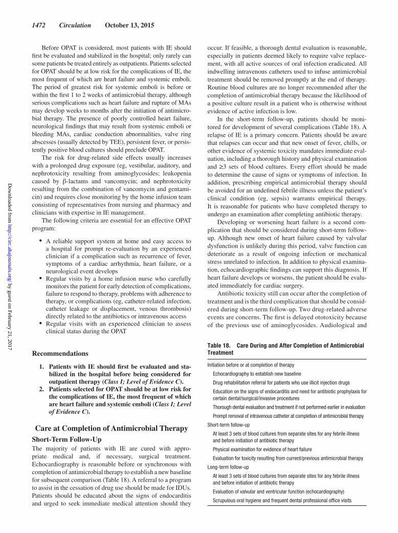
1472 Circulation October 13, 2015
Before OPAT is considered, most patients with IE should first be evaluated and stabilized in the hospital; only rarely can some patients be treated entirely as outpatients. Patients selected for OPAT should be at low risk for the complications of IE, the most frequent of which are heart failure and systemic emboli. The period of greatest risk for systemic emboli is before or within the first 1 to 2 weeks of antimicrobial therapy, although serious complications such as heart failure and rupture of MAs may develop weeks to months after the initiation of antimicro-bial therapy. The presence of poorly controlled heart failure, neurological findings that may result from systemic emboli or bleeding MAs, cardiac conduction abnormalities, valve ring abscesses (usually detected by TEE), persistent fever, or persis-tently positive blood cultures should preclude OPAT.
The risk for drug-related side effects usually increases with a prolonged drug exposure (eg, vestibular, auditory, and nephrotoxicity resulting from aminoglycosides; leukopenia caused by β-lactams and vancomycin; and nephrotoxicity resulting from the combination of vancomycin and gentami-cin) and requires close monitoring by the home infusion team consisting of representatives from nursing and pharmacy and clinicians with expertise in IE management.
The following criteria are essential for an effective OPAT program:
• A reliable support system at home and easy access to a hospital for prompt re-evaluation by an experienced clinician if a complication such as recurrence of fever, symptoms of a cardiac arrhythmia, heart failure, or a neurological event develops
• Regular visits by a home infusion nurse who carefully monitors the patient for early detection of complications, failure to respond to therapy, problems with adherence to therapy, or complications (eg, catheter-related infection, catheter leakage or displacement, venous thrombosis) directly related to the antibiotics or intravenous access
• Regular visits with an experienced clinician to assess clinical status during the OPAT
Recommendations
1. Patients with IE should first be evaluated and sta-bilized in the hospital before being considered for outpatient therapy (Class I; Level of Evidence C).
2. Patients selected for OPAT should be at low risk for the complications of IE, the most frequent of which are heart failure and systemic emboli (Class I; Level of Evidence C).
Care at Completion of Antimicrobial TherapyShort-Term Follow-UpThe majority of patients with IE are cured with appro-priate medical and, if necessary, surgical treatment. Echocardiography is reasonable before or synchronous with completion of antimicrobial therapy to establish a new baseline for subsequent comparison (Table 18). A referral to a program to assist in the cessation of drug use should be made for IDUs. Patients should be educated about the signs of endocarditis and urged to seek immediate medical attention should they
occur. If feasible, a thorough dental evaluation is reasonable, especially in patients deemed likely to require valve replace-ment, with all active sources of oral infection eradicated. All indwelling intravenous catheters used to infuse antimicrobial treatment should be removed promptly at the end of therapy. Routine blood cultures are no longer recommended after the completion of antimicrobial therapy because the likelihood of a positive culture result in a patient who is otherwise without evidence of active infection is low.
In the short-term follow-up, patients should be moni-tored for development of several complications (Table 18). A relapse of IE is a primary concern. Patients should be aware that relapses can occur and that new onset of fever, chills, or other evidence of systemic toxicity mandates immediate eval-uation, including a thorough history and physical examination and ≥3 sets of blood cultures. Every effort should be made to determine the cause of signs or symptoms of infection. In addition, prescribing empirical antimicrobial therapy should be avoided for an undefined febrile illness unless the patient’s clinical condition (eg, sepsis) warrants empirical therapy. It is reasonable for patients who have completed therapy to undergo an examination after completing antibiotic therapy.
Developing or worsening heart failure is a second com-plication that should be considered during short-term follow-up. Although new onset of heart failure caused by valvular dysfunction is unlikely during this period, valve function can deteriorate as a result of ongoing infection or mechanical stress unrelated to infection. In addition to physical examina-tion, echocardiographic findings can support this diagnosis. If heart failure develops or worsens, the patient should be evalu-ated immediately for cardiac surgery.
Antibiotic toxicity still can occur after the completion of treatment and is the third complication that should be consid-ered during short-term follow-up. Two drug-related adverse events are concerns. The first is delayed ototoxicity because of the previous use of aminoglycosides. Audiological and
Table 18. Care During and After Completion of Antimicrobial Treatment
Initiation before or at completion of therapy
Echocardiography to establish new baseline
Drug rehabilitation referral for patients who use illicit injection drugs
Education on the signs of endocarditis and need for antibiotic prophylaxis for certain dental/surgical/invasive procedures
Thorough dental evaluation and treatment if not performed earlier in evaluation
Prompt removal of intravenous catheter at completion of antimicrobial therapy
Short-term follow-up
At least 3 sets of blood cultures from separate sites for any febrile illness and before initiation of antibiotic therapy
Physical examination for evidence of heart failure
Evaluation for toxicity resulting from current/previous antimicrobial therapy
Long-term follow-up
At least 3 sets of blood cultures from separate sites for any febrile illness and before initiation of antibiotic therapy
Evaluation of valvular and ventricular function (echocardiography)
Scrupulous oral hygiene and frequent dental professional office visits
by guest on February 21, 2017http://circ.ahajournals.org/
Dow
nloaded from

Baddour et al Infective Endocarditis in Adults 1473
vestibular toxicity can develop despite the maintenance of appropriate serum drug concentrations during treatment. For patients receiving long-term aminoglycosides, particu-larly those with underlying renal or otic disorders, serial audiograms may be considered during therapy if feasible and available. No tools are routinely available for monitor-ing vestibular function, and patients should be told to report the onset of any symptoms of vestibular toxicity during or after treatment.
The second antibiotic-related adverse event is Clostridium difficile infection. Onset of diarrhea can be delayed as long as 4 weeks after the last dose of antibiotic. The hope is that prompt recognition and treatment of this infectious complica-tion will diminish the likelihood of severe complications.
Recommendations
1. Echocardiography is reasonable before or synchro-nous with completion of antimicrobial therapy to establish a new baseline for subsequent compari-son (Class IIa; Level of Evidence C).
2. A referral to a program to assist in the cessation of drug use should be made for IDUs (Class I; Level of Evidence C).
3. Patients should be educated about the signs of endocarditis and urged to seek immediate medi-cal attention should they develop (Class I; Level of Evidence C).
4. A thorough dental evaluation is reasonable, espe-cially in patients deemed likely to require valve replacement, with all active sources of oral infec-tion eradicated (Class IIa; Level of Evidence C).
5. Routine blood cultures are not recommended after the completion of antimicrobial therapy because the likelihood of a positive culture result in a patient who is otherwise without evidence of active infection is low (Class III; Level of Evidence C).
6. All indwelling intravenous catheters used to infuse antimicrobial treatment should be removed promptly at the end of therapy (Class I; Level of Evidence C).
7. For patients receiving long-term aminoglycosides, particularly those with underlying renal or otic disorders, serial audiograms may be considered during therapy if available (Class IIb; Level of Evidence C).
8. In the short-term follow-up, patients should be monitored for the development of several com-plications, including IE relapse and heart failure (Class I; Level of Evidence C).
9. Patients should be aware that relapses can occur and that new onset of fever, chills, or other evidence of systemic toxicity mandates immediate evalu-ation, including a thorough history and physical examination and ≥3 sets of blood cultures (Class I; Level of Evidence C).
10. Because of concerns for IE relapse, a thorough evalua-tion should be done to determine the cause of infection signs and symptoms (Class I; Level of Evidence C).
11. Empirical antimicrobial therapy for suspected infection should be avoided unless the patient’s
clinical condition (eg, sepsis) warrants it (Class III; Level of Evidence C).
12. It is reasonable to have patients who have completed therapy and do not have symptoms of systemic tox-icity undergo an examination after completing anti-biotic therapy (Class IIa; Level of Evidence C).
13. Developing or worsening heart failure is a common complication that should be monitored for during short-term follow-up (Class I; Level of Evidence C).
14. If heart failure develops or worsens, the patient should be evaluated immediately for cardiac sur-gery (Class I; Level of Evidence B).
15. Antibiotic toxicity still can occur after the comple-tion of treatment and is a complication that should be considered during short-term follow-up (Class I; Level of Evidence C).
16. No tools are routinely available for monitoring vestibular function, and patients should be told to report the onset of any symptoms of vestibular toxicity during or after treatment (Class I; Level of Evidence C).
Long-Term Follow-UpMonths to years after completion of medical therapy for IE, patients should have ongoing observation for and education about recurrent infection and delayed onset of worsening valve dysfunction (Table 18). Daily dental hygiene should be stressed, with serial evaluations by a dentist who is familiar with this patient population. Patients should be questioned about symptoms of heart failure, and a thorough physi-cal examination should be done. Additional evaluation with echocardiography is indicated in selected patients with posi-tive findings from history and physical examination. Patients should be instructed to seek immediate medical evaluation for persistent fever (Table 18). This is necessary because IE can mimic a variety of febrile illnesses. Blood cultures should be obtained. Antibiotic therapy should not be initiated for treat-ment of undefined febrile illnesses without blood cultures being obtained first. Antibiotics prescribed for nonspecific or unproved febrile syndromes are a major cause of (blood) culture-negative IE, and this practice should be strongly discouraged.
Recommendations
1. Months to years after completion of medical ther-apy for IE, patients should have ongoing observa-tion for and education about recurrent infection and delayed onset of worsening valve dysfunction (Class I; Level of Evidence C).
2. Daily dental hygiene should be stressed, with serial evaluations by a dentist who is familiar with this patient population (Class I; Level of Evidence C).
3. Patients should be questioned about symptoms of heart failure, and a thorough physical examination should be done (Class I; Level of Evidence C).
4. Additional evaluations with echocardiography should be obtained in selected patients with posi-tive findings from history and physical examina-tion (Class I; Level of Evidence C).
by guest on February 21, 2017http://circ.ahajournals.org/
Dow
nloaded from

1474 Circulation October 13, 2015
5. Patients should be instructed to seek immediate medical evaluation for fever, and blood cultures should be obtained (Class I; Level of Evidence C).
6. Antimicrobial therapy should not be initiated for the treatment of undefined febrile illnesses unless the patient’s condition (eg, sepsis) warrants it (Class III; Level of Evidence C).
Dental ManagementA large, prospective study demonstrated a strong associa-tion between 3 indexes of oral hygiene and gingival disease and the incidence of bacteremia from IE-related species.327 Poor oral hygiene results in gingivitis, which often leads to periodontitis, and it is likely that these 2 periodontal dis-eases are associated with community-acquired IE. Current evidence suggests that poor oral hygiene and periodon-tal diseases, not dental office procedures, are likely to be responsible for the vast majority of cases of IE that originate in the mouth.328
Regardless of the source of infection, inpatients with IE should be thoroughly evaluated by a dentist familiar with the potential role of the mouth in these cases. The optimal timing for this evaluation may be after the patient’s cardiac status has stabilized and early enough that all invasive dental procedures can be accomplished during intravenous antibiotic therapy. The clinical examination should rule out periodontal inflammation and pocketing around the teeth and caries that will eventually result in pulpal infection. A full series of intra-oral radiographs is required for the identification of caries and periodontal disease (eg, bone loss, tooth fractures). All of this is aimed at reducing the incidence and magnitude of bactere-mia from any manipulation of the gingival tissues, including normal daily events such as brushing teeth and chewing food. Treatment invariably involves a thorough dental cleaning by a hygienist who will review with patients the importance of maintaining scrupulous oral hygiene.
Dental disease is almost entirely preventable if patients are compliant with 4 measures. First, the cause of both peri-odontal disease and caries is bacterial plaque accumulation on teeth, and prevention is dependent on keeping teeth free of plaque. Second, patients must understand that dietary mea-sures are critically important in preventing the formation of plaque, especially in areas on the teeth that are difficult to keep clean. The degree to which sugar and other refined carbohydrates are eliminated from the diet will have a major impact on the growth of pathogenic bacterial species, some of which are responsible for IE. Third, routine follow-up with their family dentist is necessary for close monitoring of oral hygiene and the early identification and eradication of oral disease. Finally, the daily use of a high-concentration fluori-dated toothpaste will help to ensure that the acid from plaque does not decalcify tooth structures and result in caries. A focus on all 4 measures should help to reduce the incidence of bac-teremia and the risk for recurrent IE.
Recommendations
1. Inpatients with IE should be thoroughly evaluated by a dentist to identify and eliminate oral diseases that predispose to bacteremia and may therefore contribute to the risk for recurrent IE (Class I; Level of Evidence C).
2. The clinical examination should focus on periodon-tal inflammation and pocketing around teeth and caries that may result in pulpal infection and subse-quent abscess (Class I; Level of Evidence C).
3. A full series of intraoral radiographs will allow the identification of caries and periodontal disease and other disease (eg, tooth fractures) not evident from the physical examination. This should occur when the patient is able to travel to a dental facility (Class I; Level of Evidence C).
by guest on February 21, 2017http://circ.ahajournals.org/
Dow
nloaded from

Baddour et al Infective Endocarditis in Adults 1475
Writing Group Disclosures
Writing Group Member Employment Research Grant
Other Research Support
Speakers’ Bureau/
Honoraria Expert WitnessOwnership
InterestConsultant/
Advisory Board Other
Larry M. Baddour
Mayo Clinic None None None None None None None
Robert S. Baltimore
Yale University School of Medicine
None None None None None None None
Bruno Barsic Hospital for Infectious Diseases, School of
Medicine Zagreb
None None Astellas*; Pfizer*; MSD*
None None None None
Arnold S. Bayer LA Biomedical Research Institute Infectious
Diseases
Astellas†; Cubist†; NIH/NIAID*; Theravance*
None None Johnson Graffe et al*; Morrow,
Kidman, Tinker et al*; Galloway, Lucchese et al*; MES Solutions*; Hoffman, Sheffield et al*; Clifford Law*
None None None
Ann F. Bolger UCSF None None None None None None None
Anne M. Fink University of Illinois at Chicago
None None None None None None None
Vance G. Fowler, Jr. Duke University CDC†; Cerexa/Forest†; MedImmune†; NIH†;
FDA†; Cubist*
None None Witness in case involving group B Streptococcal
Vertebral osteomyelitis†
None MedImmune*; Novartis†; The
Medicines Comapy*; Novadigm*;
Debiopharm*; Cerexa*;Affinium*;
Tetraphase*; Bayer*; Theravance*;
Cubist*; Genetech*; Basilea*
None
Michael H. Gewitz New York Medical College
None None None None None None None
Matthew E. Levison
Drexel University College of Medicine
None None None None None Merck Manual† None
Peter B. Lockhart Carolinas Medical Center None None None None None None None
Patrick O’Gara Brigham and Women’s Hospital
None None None None None None None
Michael J. Rybak Wayne State University Cubist*; Forest*; MDCH*; NIH*;
Actavis*; Theravance*
None Cubist*; Forest*; Novartis*; Actavis*;
The Medicines Company*
None None Cubist*; Forest*; Theravance*; Actavis†; The
Medicines Company*
None
James M. Steckelberg
Mayo Clinic None None None None None None None
Kathryn A. Taubert
American Heart Association
None None None None None None None
Imad M. Tleyjeh King Fahd Medical City None None None None None None None
Walter R. Wilson Mayo Clinic None None None None None None None
This table represents the relationships of writing group members that may be perceived as actual or reasonably perceived conflicts of interest as reported on the Disclosure Questionnaire, which all members of the writing group are required to complete and submit. A relationship is considered to be “significant” if (a) the person receives $10 000 or more during any 12-month period, or 5% or more of the person’s gross income; or (b) the person owns 5% or more of the voting stock or share of the entity, or owns $10 000 or more of the fair market value of the entity. A relationship is considered to be “modest” if it is less than “significant” under the preceding definition.
*Modest.†Significant.
Disclosures
by guest on February 21, 2017http://circ.ahajournals.org/
Dow
nloaded from

1476 Circulation October 13, 2015
References 1. Duval X, Delahaye F, Alla F, Tattevin P, Obadia JF, Le Moing V, Doco-
Lecompte T, Celard M, Poyart C, Strady C, Chirouze C, Bes M, Cambau E, Iung B, Selton-Suty C, Hoen B; AEPEI Study Group. Temporal trends in infective endocarditis in the context of prophylaxis guideline modifi-cations: three successive population-based surveys. J Am Coll Cardiol. 2012;59:1968–1976. doi: 10.1016/j.jacc.2012.02.029.
2. Correa de Sa DD, Tleyjeh IM, Anavekar NS, Schultz JC, Thomas JM, Lahr BD, Bachuwar A, Pazdernik M, Steckelberg JM, Wilson WR, Baddour LM. Epidemiological trends of infective endocarditis: a population-based study in Olmsted County, Minnesota [published correction appears in Mayo Clin Proc. 2010;85:772]. Mayo Clin Proc. 2010;85:422–426.
3. Federspiel JJ, Stearns SC, Peppercorn AF, Chu VH, Fowler VG Jr. Increasing US rates of endocarditis with Staphylococcus aureus: 1999-2008. Arch Intern Med. 2012;172:363–365. doi: 10.1001/archinternmed.2011.1027.
4. Murray CJ, Vos T, Lozano R, Naghavi M, Flaxman AD, Michaud C, Ezzati M, Shibuya K, Salomon JA, Abdalla S, Aboyans V, Abraham J, Ackerman I, Aggarwal R, Ahn SY, Ali MK, Alvarado M, Anderson HR, Anderson LM, Andrews KG, Atkinson C, Baddour LM, Bahalim AN, Barker-Collo S, Barrero LH, Bartels DH, Basáñez MG, Baxter A, Bell ML, Benjamin EJ, Bennett D, Bernabé E, Bhalla K, Bhandari B, Bikbov B, Bin Abdulhak A, Birbeck G, Black JA, Blencowe H, Blore JD, Blyth F, Bolliger I, Bonaventure A, Boufous S, Bourne R, Boussinesq M, Braithwaite T, Brayne C, Bridgett L, Brooker S, Brooks P, Brugha TS, Bryan-Hancock C, Bucello C, Buchbinder R, Buckle G, Budke CM, Burch M, Burney P, Burstein R, Calabria B, Campbell B, Canter CE, Carabin H, Carapetis J, Carmona L, Cella C, Charlson F, Chen H, Cheng AT, Chou D, Chugh SS, Coffeng LE, Colan SD, Colquhoun S, Colson KE, Condon J, Connor MD, Cooper LT, Corriere M, Cortinovis M, de Vaccaro KC, Couser W, Cowie BC, Criqui MH, Cross M, Dabhadkar KC, Dahiya M, Dahodwala N, Damsere-Derry J, Danaei G, Davis A, De Leo D, Degenhardt L, Dellavalle R, Delossantos A, Denenberg J, Derrett S, Des Jarlais DC, Dharmaratne SD, Dherani M, Diaz-Torne C, Dolk H, Dorsey ER, Driscoll T, Duber H, Ebel B, Edmond K, Elbaz A, Ali SE, Erskine H, Erwin PJ, Espindola P, Ewoigbokhan SE, Farzadfar F, Feigin V, Felson DT, Ferrari A, Ferri CP, Fèvre EM, Finucane MM, Flaxman S, Flood L, Foreman K, Forouzanfar MH, Fowkes FG, Fransen M, Freeman MK, Gabbe BJ, Gabriel SE, Gakidou E, Ganatra HA, Garcia B, Gaspari F, Gillum RF, Gmel G, Gonzalez-Medina D, Gosselin R, Grainger R, Grant B, Groeger J, Guillemin F, Gunnell D, Gupta R, Haagsma J, Hagan H, Halasa YA, Hall W, Haring D, Haro JM, Harrison JE, Havmoeller R, Hay RJ, Higashi H, Hill C, Hoen B, Hoffman H, Hotez PJ, Hoy D, Huang JJ, Ibeanusi SE, Jacobsen KH, James SL, Jarvis D, Jasrasaria R, Jayaraman S, Johns N, Jonas JB, Karthikeyan G, Kassebaum N, Kawakami N, Keren A, Khoo JP, King CH, Knowlton LM, Kobusingye O, Koranteng A, Krishnamurthi R, Laden F, Lalloo R, Laslett LL, Lathlean T, Leasher JL, Lee YY, Leigh J, Levinson D, Lim SS, Limb E, Lin JK, Lipnick M, Lipshultz SE, Liu W, Loane M, Ohno SL, Lyons R, Mabweijano J, MacIntyre MF, Malekzadeh R, Mallinger L, Manivannan S, Marcenes W, March L, Margolis DJ, Marks GB, Marks R, Matsumori A, Matzopoulos R, Mayosi BM, McAnulty JH, McDermott MM, McGill N, McGrath J, Medina-Mora ME, Meltzer M, Mensah GA, Merriman TR, Meyer AC, Miglioli V, Miller M, Miller TR, Mitchell PB, Mock C, Mocumbi AO,
Moffitt TE, Mokdad AA, Monasta L, Montico M, Moradi-Lakeh M, Moran A, Morawska L, Mori R, Murdoch ME, Mwaniki MK, Naidoo K, Nair MN, Naldi L, Narayan KM, Nelson PK, Nelson RG, Nevitt MC, Newton CR, Nolte S, Norman P, Norman R, O’Donnell M, O’Hanlon S, Olives C, Omer SB, Ortblad K, Osborne R, Ozgediz D, Page A, Pahari B, Pandian JD, Rivero AP, Patten SB, Pearce N, Padilla RP, Perez-Ruiz F, Perico N, Pesudovs K, Phillips D, Phillips MR, Pierce K, Pion S, Polanczyk GV, Polinder S, Pope CA 3rd, Popova S, Porrini E, Pourmalek F, Prince M, Pullan RL, Ramaiah KD, Ranganathan D, Razavi H, Regan M, Rehm JT, Rein DB, Remuzzi G, Richardson K, Rivara FP, Roberts T, Robinson C, De Leòn FR, Ronfani L, Room R, Rosenfeld LC, Rushton L, Sacco RL, Saha S, Sampson U, Sanchez-Riera L, Sanman E, Schwebel DC, Scott JG, Segui-Gomez M, Shahraz S, Shepard DS, Shin H, Shivakoti R, Singh D, Singh GM, Singh JA, Singleton J, Sleet DA, Sliwa K, Smith E, Smith JL, Stapelberg NJ, Steer A, Steiner T, Stolk WA, Stovner LJ, Sudfeld C, Syed S, Tamburlini G, Tavakkoli M, Taylor HR, Taylor JA, Taylor WJ, Thomas B, Thomson WM, Thurston GD, Tleyjeh IM, Tonelli M, Towbin JA, Truelsen T, Tsilimbaris MK, Ubeda C, Undurraga EA, van der Werf MJ, van Os J, Vavilala MS, Venketasubramanian N, Wang M, Wang W, Watt K, Weatherall DJ, Weinstock MA, Weintraub R, Weisskopf MG, Weissman MM, White RA, Whiteford H, Wiebe N, Wiersma ST, Wilkinson JD, Williams HC, Williams SR, Witt E, Wolfe F, Woolf AD, Wulf S, Yeh PH, Zaidi AK, Zheng ZJ, Zonies D, Lopez AD, AlMazroa MA, Memish ZA. Disability-adjusted life years (DALYs) for 291 diseases and injuries in 21 regions, 1990-2010: a systematic analysis for the Global Burden of Disease Study 2010 [pub-lished correction appears in Lancet. 2013;381:628]. Lancet. 2012;380:2197–2223. doi: 10.1016/S0140-6736(12)61689-4.
5. Tleyjeh IM, Abdel-Latif A, Rahbi H, Scott CG, Bailey KR, Steckelberg JM, Wilson WR, Baddour LM. A systematic review of population-based studies of infective endocarditis. Chest. 2007;132:1025–1035. doi: 10.1378/chest.06-2048.
6. Fowler VG Jr, Miro JM, Hoen B, Cabell CH, Abrutyn E, Rubinstein E, Corey GR, Spelman D, Bradley SF, Barsic B, Pappas PA, Anstrom KJ, Wray D, Fortes CQ, Anguera I, Athan E, Jones P, van der Meer JT, Elliott TS, Levine DP, Bayer AS; ICE Investigators. Staphylococcus aureus endocarditis: a consequence of medical progress [published correction appears in JAMA. 2005;294:900]. JAMA. 2005;293:3012–3021. doi: 10.1001/jama.293.24.3012.
7. Selton-Suty C, Célard M, Le Moing V, Doco-Lecompte T, Chirouze C, Iung B, Strady C, Revest M, Vandenesch F, Bouvet A, Delahaye F, Alla F, Duval X, Hoen B; AEPEI Study Group. Preeminence of Staphylococcus aureus in infective endocarditis: a 1-year population-based survey. Clin Infect Dis. 2012;54:1230–1239. doi: 10.1093/cid/cis199.
8. Murdoch DR, Corey GR, Hoen B, Miró JM, Fowler VG Jr, Bayer AS, Karchmer AW, Olaison L, Pappas PA, Moreillon P, Chambers ST, Chu VH, Falcó V, Holland DJ, Jones P, Klein JL, Raymond NJ, Read KM, Tripodi MF, Utili R, Wang A, Woods CW, Cabell CH; International Collaboration on Endocarditis-Prospective Cohort Study (ICE-PCS) Investigators. Clinical presentation, etiology, and outcome of infec-tive endocarditis in the 21st century: the International Collaboration on Endocarditis-Prospective Cohort Study. Arch Intern Med. 2009;169:463–473. doi: 10.1001/archinternmed.2008.603.
9. Benito N, Miró JM, de Lazzari E, Cabell CH, del Río A, Altclas J, Commerford P, Delahaye F, Dragulescu S, Giamarellou H, Habib G,
Reviewer Disclosures
Reviewer EmploymentResearch
Grant
Other Research Support
Speakers’ Bureau/Honoraria
Expert Witness
Ownership Interest
Consultant/ Advisory Board Other
Giovanni Di Salvo King Faisal Hospital and Research Center (Saudi Arabia)
None None None None None None None
Franklin D. Lowy Columbia University None None None None None None UptoDate*
Stanford T. Shulman
Lurie Children’s Hospital, Children’s Memorial
Hospital, Northwestern University Medical School
None None None None None None None
This table represents the relationships of reviewers that may be perceived as actual or reasonably perceived conflicts of interest as reported on the Disclosure Questionnaire, which all reviewers are required to complete and submit. A relationship is considered to be “significant” if (a) the person receives $10 000 or more during any 12-month period, or 5% or more of the person’s gross income; or (b) the person owns 5% or more of the voting stock or share of the entity, or owns $10 000 or more of the fair market value of the entity. A relationship is considered to be “modest” if it is less than “significant” under the preceding definition.
*Modest.
by guest on February 21, 2017http://circ.ahajournals.org/
Dow
nloaded from

Baddour et al Infective Endocarditis in Adults 1477
Kamarulzaman A, Kumar AS, Nacinovich FM, Suter F, Tribouilloy C, Venugopal K, Moreno A, Fowler VG Jr; ICE-PCS (International Collaboration on Endocarditis Prospective Cohort Study) Investigators. Health care-associated native valve endocarditis: importance of non-nosocomial acquisition [published correction appears in Ann Intern Med. 2010;152:480]. Ann Intern Med. 2009;150:586–594. doi: 10.7326/0003-4819-150-9-200905050-00004
10. Lalani T, Cabell CH, Benjamin DK, Lasca O, Naber C, Fowler VG Jr, Corey GR, Chu VH, Fenely M, Pachirat O, Tan RS, Watkin R, Ionac A, Moreno A, Mestres CA, Casabé J, Chipigina N, Eisen DP, Spelman D, Delahaye F, Peterson G, Olaison L, Wang A; International Collaboration on Endocarditis-Prospective Cohort Study (ICE-PCS) Investigators. Analysis of the impact of early surgery on in-hospital mortality of native valve endocarditis: use of propensity score and instrumental variable methods to adjust for treatment-selection bias. Circulation. 2010;121:1005–1013. doi: 10.1161/CIRCULATIONAHA.109.864488.
11. Kiefer T, Park L, Tribouilloy C, Cortes C, Casillo R, Chu V, Delahaye F, Durante-Mangoni E, Edathodu J, Falces C, Logar M, Miró JM, Naber C, Tripodi MF, Murdoch DR, Moreillon P, Utili R, Wang A. Association between valvular surgery and mortality among patients with infective endocarditis complicated by heart failure. JAMA. 2011;306:2239–2247. doi: 10.1001/jama.2011.1701.
12. Baddour LM, Wilson WR, Bayer AS, Fowler VG Jr, Bolger AF, Levison ME, Ferrieri P, Gerber MA, Tani LY, Gewitz MH, Tong DC, Steckelberg JM, Baltimore RS, Shulman ST, Burns JC, Falace DA, Newburger JW, Pallasch TJ, Takahashi M, Taubert KA. Infective endocarditis: diagno-sis, antimicrobial therapy, and management of complications: a state-ment for healthcare professionals from the Committee on Rheumatic Fever, Endocarditis, and Kawasaki Disease, Council on Cardiovascular Disease in the Young, and the Councils on Clinical Cardiology, Stroke, and Cardiovascular Surgery and Anesthesia, American Heart Association [published correction appears in Circulation. 2007;115:e408; Circulation. 2007;116:e547; Circulation. 2005;112:2373; Circulation. 2008;118:e497]. Circulation. 2005;111:e394–e434. doi: 10.1161/CIRCULATIONAHA.105.165564.
13. Fowler VG Jr, Boucher HW, Corey GR, Abrutyn E, Karchmer AW, Rupp ME, Levine DP, Chambers HF, Tally FP, Vigliani GA, Cabell CH, Link AS, DeMeyer I, Filler SG, Zervos M, Cook P, Parsonnet J, Bernstein JM, Price CS, Forrest GN, Fätkenheuer G, Gareca M, Rehm SJ, Brodt HR, Tice A, Cosgrove SE; S aureus Endocarditis and Bacteremia Study Group. Daptomycin versus standard therapy for bacteremia and endocarditis caused by Staphylococcus aureus. N Engl J Med. 2006;355:653–665. doi: 10.1056/NEJMoa053783.
14. Tleyjeh IM, Ghomrawi HM, Steckelberg JM, Hoskin TL, Mirzoyev Z, Anavekar NS, Enders F, Moustafa S, Mookadam F, Huskins WC, Wilson WR, Baddour LM. The impact of valve surgery on 6-month mortality in left-sided infective endocarditis. Circulation. 2007;115:1721–1728. doi: 10.1161/CIRCULATIONAHA.106.658831.
15. Bannay A, Hoen B, Duval X, Obadia JF, Selton-Suty C, Le Moing V, Tattevin P, Iung B, Delahaye F, Alla F; AEPEI Study Group. The impact of valve surgery on short- and long-term mortality in left-sided infective endocarditis: do differences in methodological approaches explain previous conflicting results? Eur Heart J. 2011;32:2003–2015. doi: 10.1093/eurheartj/ehp008.
16. Sy RW, Bannon PG, Bayfield MS, Brown C, Kritharides L. Survivor treat-ment selection bias and outcomes research: a case study of surgery in infective endocarditis. Circ Cardiovasc Qual Outcomes. 2009;2:469–474. doi: 10.1161/CIRCOUTCOMES.109.857938.
17. Kang DH, Kim YJ, Kim SH, Sun BJ, Kim DH, Yun SC, Song JM, Choo SJ, Chung CH, Song JK, Lee JW, Sohn DW. Early surgery versus conven-tional treatment for infective endocarditis. N Engl J Med. 2012;366:2466–2473. doi: 10.1056/NEJMoa1112843.
18. Habib G, Hoen B, Tornos P, Thuny F, Prendergast B, Vilacosta I, Moreillon P, de Jesus Antunes M, Thilen U, Lekakis J, Lengyel M, Müller L, Naber CK, Nihoyannopoulos P, Moritz A, Zamorano JL; ESC Committee for Practice Guidelines. Guidelines on the prevention, diagnosis, and treat-ment of infective endocarditis (new version 2009): the Task Force on the Prevention, Diagnosis, and Treatment of Infective Endocarditis of the European Society of Cardiology (ESC): endorsed by the European Society of Clinical Microbiology and Infectious Diseases (ESCMID) and the International Society of Chemotherapy (ISC) for Infection and Cancer. Eur Heart J. 2009;30:2369–2413. doi: 10.1093/eurheartj/ehp285.
19. Gould FK, Denning DW, Elliott TS, Foweraker J, Perry JD, Prendergast BD, Sandoe JA, Spry MJ, Watkin RW; Working Party of the British Society for Antimicrobial Chemotherapy. Guidelines for the diagnosis and antibiotic treatment of endocarditis in adults: a report of the Working Party
of the British Society for Antimicrobial Chemotherapy [published correc-tion appears in J Antimicrob Chemother. 2011;67:1304]. J Antimicrob Chemother. 2012;67:269–289. doi: 10.1093/jac/dkr450.
20. Baltimore RS, Gewitz M, Baddour LM, Beerman LB, Jackson MA, Lockhart PB, Pahl E, Pike NA, Schutze GE, Shulman ST, Willoughby R Jr; on behalf of the American Heart Association Rheumatic Fever, Endocarditis, and Kawasaki Disease Committee of the Council on Cardiovascular Disease in the Young and the Council on Cardiovascular and Stroke Nursing. Infective endocarditis in childhood: 2015 update: a scientific statement from the American Heart Association [published online ahead of print September 14, 2015]. Circulation. doi: 10.1161/CIR.0000000000000298.
21. Baddour LM, Epstein AE, Erickson CC, Knight BP, Levison ME, Lockhart PB, Masoudi FA, Okum EJ, Wilson WR, Beerman LB, Bolger AF, Estes NA 3rd, Gewitz M, Newburger JW, Schron EB, Taubert KA; on behalf of the American Heart Association Rheumatic Fever, Endocarditis, and Kawasaki Disease Committee; Council on Cardiovascular Disease in Young; Council on Cardiovascular Surgery and Anesthesia; Council on Cardiovascular Nursing; Council on Clinical Cardiology; and Interdisciplinary Council on Quality of Care. Update on cardiovascu-lar implantable electronic device infections and their management: a scientific statement from the American Heart Association. Circulation. 2010;121:458–477. doi: 10.1161/CIRCULATIONAHA.109.192665.
22. Baddour LM, Bettmann MA, Bolger AF, Epstein AE, Ferrieri P, Gerber MA, Gewitz MH, Jacobs AK, Levison ME, Newburger JW, Pallasch TJ, Wilson WR, Baltimore RS, Falace DA, Shulman ST, Tani LY, Taubert KA; AHA. Nonvalvular cardiovascular device-related infections. Circulation. 2003;108:2015–2031. doi: 10.1161/01.CIR.0000093201.57771.47.
23. Durack DT, Lukes AS, Bright DK. New criteria for diagnosis of infec-tive endocarditis: utilization of specific echocardiographic findings: Duke Endocarditis Service. Am J Med. 1994;96:200–209.
24. Li JS, Sexton DJ, Mick N, Nettles R, Fowler VG Jr, Ryan T, Bashore T, Corey GR. Proposed modifications to the Duke criteria for the diag-nosis of infective endocarditis. Clin Infect Dis. 2000;30:633–638. doi: 10.1086/313753.
25. Bayer AS, Lam K, Ginzton L, Norman DC, Chiu CY, Ward JI. Staphylococcus aureus bacteremia: clinical, serologic, and echocardio-graphic findings in patients with and without endocarditis. Arch Intern Med. 1987;147:457–462.
26. Fowler VG Jr, Olsen MK, Corey GR, Woods CW, Cabell CH, Reller LB, Cheng AC, Dudley T, Oddone EZ. Clinical identifiers of complicated Staphylococcus aureus bacteremia. Arch Intern Med. 2003;163:2066–2072. doi: 10.1001/archinte.163.17.2066.
27. Bayer AS, Ward JI, Ginzton LE, Shapiro SM. Evaluation of new clini-cal criteria for the diagnosis of infective endocarditis. Am J Med. 1994;96:211–219.
28. Hoen B, Selton-Suty C, Danchin N, Weber M, Villemot JP, Mathieu P, Floquet J, Canton P. Evaluation of the Duke criteria versus the Beth Israel criteria for the diagnosis of infective endocarditis. Clin Infect Dis. 1995;21:905–909.
29. Dodds GA, Sexton DJ, Durack DT, Bashore TM, Corey GR, Kisslo J. Negative predictive value of the Duke criteria for infective endocarditis. Am J Cardiol. 1996;77:403–407.
30. Arguello EA, Varini S, Romorini A, Elizari A, Clara L, Casabe H. Infective endocarditis in the Argentine Republic [abstract 152]. Paper presented at: Third International Symposium on Modern Concepts in Endocarditis; July 13–15, 1995; Boston, MA.
31. Kanavos K, Antoniadou A, Venetis C, Gezerlis P, Giamarellou H. Retrospective analysis of Duke’s criteria in 60 cases of infective endo-carditis in Greece [abstract 138]. Paper presented at: Third International Symposium on Modern Concepts in Endocarditis; July 13–15, 1995; Boston, MA.
32. Del Pont JM, De Cicco LT, Vartalitis C, Ithurralde M, Gallo JP, Vargas F, Gianantonio CA, Quirós RE. Infective endocarditis in children: clinical analyses and evaluation of two diagnostic criteria. Pediatr Infect Dis J. 1995;14:1079–1086.
33. Gagliardi JP, Nettles RE, McCarty DE, Sanders LL, Corey GR, Sexton DJ. Native valve infective endocarditis in elderly and younger adult patients: comparison of clinical features and outcomes with use of the Duke criteria and the Duke Endocarditis Database. Clin Infect Dis. 1998;26:1165–1168.
34. Heiro M, Nikoskelainen J, Hartiala JJ, Saraste MK, Kotilainen PM. Diagnosis of infective endocarditis: sensitivity of the Duke vs von Reyn criteria. Arch Intern Med. 1998;158:18–24.
by guest on February 21, 2017http://circ.ahajournals.org/
Dow
nloaded from

1478 Circulation October 13, 2015
35. Nettles RE, McCarty DE, Corey GR, Li J, Sexton DJ. An evaluation of the Duke criteria in 25 pathologically confirmed cases of prosthetic valve endocarditis. Clin Infect Dis. 1997;25:1401–1403.
36. Stockheim JA, Chadwick EG, Kessler S, Amer M, Abdel-Haq N, Dajani AS, Shulman ST. Are the Duke criteria superior to the Beth Israel criteria for the diagnosis of infective endocarditis in children? Clin Infect Dis. 1998;27:1451–1456.
37. Hoen B, Béguinot I, Rabaud C, Jaussaud R, Selton-Suty C, May T, Canton P. The Duke criteria for diagnosing infective endocarditis are specific: analysis of 100 patients with acute fever or fever of unknown origin. Clin Infect Dis. 1996;23:298–302.
38. Sekeres MA, Abrutyn E, Berlin JA, Kaye D, Kinman JL, Korzeniowski OM, Levison ME, Feldman RS, Strom BL. An assessment of the useful-ness of the Duke criteria for diagnosing active infective endocarditis. Clin Infect Dis. 1997;24:1185–1190.
39. Fowler VG Jr, Sanders LL, Kong LK, McClelland RS, Gottlieb GS, Li J, Ryan T, Sexton DJ, Roussakis G, Harrell LJ, Corey GR. Infective endocar-ditis due to Staphylococcus aureus: 59 prospectively identified cases with follow-up. Clin Infect Dis. 1999;28:106–114. doi: 10.1086/515076.
40. Fournier PE, Casalta JP, Habib G, Messana T, Raoult D. Modification of the diagnostic criteria proposed by the Duke Endocarditis Service to permit improved diagnosis of Q fever endocarditis. Am J Med. 1996;100:629–633.
41. Raoult D, Fournier PE, Drancourt M, Marrie TJ, Etienne J, Cosserat J, Cacoub P, Poinsignon Y, Leclercq P, Sefton AM. Diagnosis of 22 new cases of Bartonella endocarditis [published correction appears in Ann Intern Med. 1997;127:249]. Ann Intern Med. 1996;125:646–652.
42. Hamed KA, Dormitzer PR, Su CK, Relman DA. Haemophilus parainflu-enzae endocarditis: application of a molecular approach for identification of pathogenic bacterial species. Clin Infect Dis. 1994;19:677–683.
43. Podglajen I, Bellery F, Poyart C, Coudol P, Buu-Hoï A, Bruneval P, Mainardi JL. Comparative molecular and microbiologic diagnosis of bac-terial endocarditis. Emerg Infect Dis. 2003;9:1543–1547. doi: 10.3201/eid0912.030229.
44. Goldenberger D, Künzli A, Vogt P, Zbinden R, Altwegg M. Molecular diagnosis of bacterial endocarditis by broad-range PCR amplification and direct sequencing. J Clin Microbiol. 1997;35:2733–2739.
45. Lamas CC, Eykyn SJ. Suggested modifications to the Duke criteria for the clinical diagnosis of native valve and prosthetic valve endo-carditis: analysis of 118 pathologically proven cases. Clin Infect Dis. 1997;25:713–719.
46. Berlin JA, Abrutyn E, Strom BL, Kinman JL, Levison ME, Korzeniowski OM, Feldman RS, Kaye D. Assessing diagnostic criteria for active infec-tive endocarditis. Am J Cardiol. 1994;73:887–891.
47. Reynolds HR, Jagen MA, Tunick PA, Kronzon I. Sensitivity of transtho-racic versus transesophageal echocardiography for the detection of native valve vegetations in the modern era. J Am Soc Echocardiogr. 2003;16:67–70. doi: 10.1067/mje.2003.43.
48. Daniel WG, Mügge A, Grote J, Hausmann D, Nikutta P, Laas J, Lichtlen PR, Martin RP. Comparison of transthoracic and transesophageal echocar-diography for detection of abnormalities of prosthetic and bioprosthetic valves in the mitral and aortic positions. Am J Cardiol. 1993;71:210–215.
49. Heidenreich PA, Masoudi FA, Maini B, Chou TM, Foster E, Schiller NB, Owens DK. Echocardiography in patients with suspected endocarditis: a cost-effectiveness analysis. Am J Med. 1999;107:198–208.
50. Lindner JR, Case RA, Dent JM, Abbott RD, Scheld WM, Kaul S. Diagnostic value of echocardiography in suspected endocarditis: an evaluation based on the pretest probability of disease. Circulation. 1996;93:730–736.
51. Barton TL, Mottram PM, Stuart RL, Cameron JD, Moir S. Transthoracic echocardiography is still useful in the initial evaluation of patients with suspected infective endocarditis: evaluation of a large cohort at a ter-tiary referral center. Mayo Clin Proc. 2014;89:799–805. doi: 10.1016/j.mayocp.2014.02.013.
52. San Román JA, Vilacosta I, Zamorano JL, Almería C, Sánchez-Harguindey L. Transesophageal echocardiography in right-sided endocarditis. J Am Coll Cardiol. 1993;21:1226–1230.
53. Bayer AS. Infective endocarditis. Clin Infect Dis. 1993;17:313–320; quiz 321.
54. Daniel WG, Mügge A, Martin RP, Lindert O, Hausmann D, Nonnast-Daniel B, Laas J, Lichtlen PR. Improvement in the diagnosis of abscesses associated with endocarditis by transesophageal echocardiography. N Engl J Med. 1991;324:795–800. doi: 10.1056/NEJM199103213241203.
55. Sanfilippo AJ, Picard MH, Newell JB, Rosas E, Davidoff R, Thomas JD, Weyman AE. Echocardiographic assessment of patients with infectious
endocarditis: prediction of risk for complications. J Am Coll Cardiol. 1991;18:1191–1199.
56. Rohmann S, Erbel R, Görge G, Makowski T, Mohr-Kahaly S, Nixdorff U, Drexler M, Meyer J. Clinical relevance of vegetation localization by transoesophageal echocardiography in infective endocarditis. Eur Heart J. 1992;13:446–452. doi: http://dx.doi.org.
57. Mügge A, Daniel WG, Frank G, Lichtlen PR. Echocardiography in infec-tive endocarditis: reassessment of prognostic implications of vegetation size determined by the transthoracic and the transesophageal approach. J Am Coll Cardiol. 1989;14:631–638. doi: 10.1016/0735-1097(89)90104-6.
58. Steckelberg JM, Murphy JG, Ballard D, Bailey K, Tajik AJ, Taliercio CP, Giuliani ER, Wilson WR. Emboli in infective endocarditis: the prognos-tic value of echocardiography. Ann Intern Med. 1991;114:635–640. Doi: 10.7326/0003-4819-114-8-635.
59. Erbel R, Rohmann S, Drexler M, Mohr-Kahaly S, Gerharz CD, Iversen S, Oelert H, Meyer J. Improved diagnostic value of echocardiography in patients with infective endocarditis by transoesophageal approach: a pro-spective study. Eur Heart J. 1988;9:43–53.
60. Rohmann S, Erbel R, Darius H, Görge G, Makowski T, Zotz R, Mohr-Kahaly S, Nixdorff U, Drexler M, Meyer J. Prediction of rapid versus prolonged healing of infective endocarditis by monitoring vegetation size. J Am Soc Echocardiogr. 1991;4:465–474.
61. Sabik JF, Lytle BW, Blackstone EH, Marullo AG, Pettersson GB, Cosgrove DM. Aortic root replacement with cryopreserved allograft for prosthetic valve endocarditis. Ann Thorac Surg. 2002;74:650–659.
62. Brecker SJ, Jin XY, Yacoub MH. Anatomical definition of aortic root abscesses by transesophageal echocardiography: planning a surgical strat-egy using homograft valves. Clin Cardiol. 1995;18:353–359.
63. Konstadt SN, Louie EK, Shore-Lesserson L, Black S, Scanlon P. The effects of loading changes on intraoperative Doppler assessment of mitral regurgitation. J Cardiothorac Vasc Anesth. 1994;8:19–23.
64. Hansalia S, Biswas M, Dutta R, Hage FG, Hsiung MC, Nanda NC, Singh P, Manda J, Kesanolla SK, Wei J, Yin WH. The value of live/real time three-dimensional transesophageal echocardiography in the assessment of valvular vegetations. Echocardiography. 2009;26:1264–1273. doi: 10.1111/j.1540-8175.2009.01042.x.
65. Liu YW, Tsai WC, Lin CC, Hsu CH, Li WT, Lin LJ, Chen JH. Usefulness of real-time three-dimensional echocardiography for diagnosis of infective endocarditis. Scand Cardiovasc J. 2009;43:318–323. doi: 10.1080/14017430902737940.
66. Fagman E, Perrotta S, Bech-Hanssen O, Flinck A, Lamm C, Olaison L, Svensson G. ECG-gated computed tomography: a new role for patients with suspected aortic prosthetic valve endocarditis. Eur Radiol. 2012;22:2407–2414. doi: 10.1007/s00330-012-2491-5.
67. Feuchtner GM, Stolzmann P, Dichtl W, Schertler T, Bonatti J, Scheffel H, Mueller S, Plass A, Mueller L, Bartel T, Wolf F, Alkadhi H. Multislice computed tomography in infective endocarditis: comparison with trans-esophageal echocardiography and intraoperative findings. J Am Coll Cardiol. 2009;53:436–444. doi: 10.1016/j.jacc.2008.01.077.
68. Gahide G, Bommart S, Demaria R, Sportouch C, Dambia H, Albat B, Vernhet-Kovacsik H. Preoperative evaluation in aortic endocarditis: findings on cardiac CT. AJR Am J Roentgenol. 2010;194:574–578. doi: 10.2214/AJR.08.2120.
69. Duval X, Iung B, Klein I, Brochet E, Thabut G, Arnoult F, Lepage L, Laissy JP, Wolff M, Leport C; IMAGE (Resonance Magnetic Imaging at the Acute Phase of Endocarditis) Study Group. Effect of early cerebral magnetic resonance imaging on clinical decisions in infective endocardi-tis: a prospective study. Ann Intern Med. 2010;152:497–504, W175. doi: 10.7326/0003-4819-152-8-201004200-00006.
70. Van Riet J, Hill EE, Gheysens O, Dymarkowski S, Herregods MC, Herijgers P, Peetermans WE, Mortelmans L. (18)F-FDG PET/CT for early detection of embolism and metastatic infection in patients with infective endocarditis. Eur J Nucl Med Mol Imaging. 2010;37:1189–1197. doi: 10.1007/s00259-010-1380-x.
71. Thuny F, Gaubert JY, Jacquier A, Tessonnier L, Cammilleri S, Raoult D, Habib G. Imaging investigations in infective endocarditis: current approach and perspectives. Arch Cardiovasc Dis. 2013;106:52–62. doi: 10.1016/j.acvd.2012.09.004.
72. Durack DT, Beeson PB. Experimental bacterial endocarditis, II: survival of a bacteria in endocardial vegetations. Br J Exp Pathol. 1972;53:50–53.
73. LaPlante KL, Rybak MJ. Impact of high-inoculum Staphylococcus aureus on the activities of nafcillin, vancomycin, linezolid, and daptomycin, alone and in combination with gentamicin, in an in vitro pharmacody-namic model. Antimicrob Agents Chemother. 2004;48:4665–4672. doi: 10.1128/AAC.48.12.4665-4672.2004.
by guest on February 21, 2017http://circ.ahajournals.org/
Dow
nloaded from

Baddour et al Infective Endocarditis in Adults 1479
74. Arhin FF, Sarmiento I, Parr TR Jr, Moeck G. Activity of oritavancin and comparators in vitro against standard and high inocula of Staphylococcus aureus. Int J Antimicrob Agents. 2012;39:159–162. doi: 10.1016/j.ijantimicag.2011.09.017.
75. Rose WE, Leonard SN, Rossi KL, Kaatz GW, Rybak MJ. Impact of inoc-ulum size and heterogeneous vancomycin-intermediate Staphylococcus aureus (hVISA) on vancomycin activity and emergence of VISA in an in vitro pharmacodynamic model. Antimicrob Agents Chemother. 2009;53:805–807. doi: 10.1128/AAC.01009-08.
76. Eagle H. The effect of the size of the inoculum and the age of the infection on the curative dose of penicillin in experimental infections with streptococci, pneumococci, and Treponema pallidum. J Exp Med. 1949;90:595–607.
77. Lunde CS, Rexer CH, Hartouni SR, Axt S, Benton BM. Fluorescence microscopy demonstrates enhanced targeting of telavancin to the divi-sion septum of Staphylococcus aureus. Antimicrob Agents Chemother. 2010;54:2198–2200. doi: 10.1128/AAC.01609-09.
78. Pogliano J, Pogliano N, Silverman JA. Daptomycin-mediated reorga-nization of membrane architecture causes mislocalization of essential cell division proteins. J Bacteriol. 2012;194:4494–4504. doi: 10.1128/JB.00011-12.
79. Eng RH, Padberg FT, Smith SM, Tan EN, Cherubin CE. Bactericidal effects of antibiotics on slowly growing and nongrowing bacteria. Antimicrob Agents Chemother. 1991;35:1824–1828.
80. Stevens DL, Yan S, Bryant AE. Penicillin-binding protein expression at different growth stages determines penicillin efficacy in vitro and in vivo: an explanation for the inoculum effect. J Infect Dis. 1993;167:1401–1405.
81. LaPlante KL, Woodmansee S. Activities of daptomycin and vancomy-cin alone and in combination with rifampin and gentamicin against biofilm-forming methicillin-resistant Staphylococcus aureus isolates in an experimental model of endocarditis. Antimicrob Agents Chemother. 2009;53:3880–3886. doi: 10.1128/AAC.00134-09.
82. Mizunaga S, Kamiyama T, Fukuda Y, Takahata M, Mitsuyama J. Influence of inoculum size of Staphylococcus aureus and Pseudomonas aerugi-nosa on in vitro activities and in vivo efficacy of fluoroquinolones and carbapenems. J Antimicrob Chemother. 2005;56:91–96. doi: 10.1093/jac/dki163.
83. Murray BE, Church DA, Wanger A, Zscheck K, Levison ME, Ingerman MJ, Abrutyn E, Mederski-Samoraj B. Comparison of two beta-lacta-mase-producing strains of Streptococcus faecalis. Antimicrob Agents Chemother. 1986;30:861–864.
84. Sabath LD, Garner C, Wilcox C, Finland M. Effect of inoculum and of beta-lactamase on the anti-staphylococcal activity of thirteen penicillins and cephalosporins. Antimicrob Agents Chemother. 1975;8:344–349.
85. Johnson CC, Livornese L, Gold MJ, Pitsakis PG, Taylor S, Levison ME. Activity of cefepime against ceftazidime-resistant Gram-negative bacilli using low and high inocula. J Antimicrob Chemother. 1995;35:765–773.
86. Bayer AS, Hirano L, Yih J. Development of beta-lactam resis-tance and increased quinolone MICs during therapy of experimental Pseudomonas aeruginosa endocarditis. Antimicrob Agents Chemother. 1988;32:231–235.
87. Ingerman M, Pitsakis PG, Rosenberg A, Hessen MT, Abrutyn E, Murray BE, Levison ME. Beta-lactamase production in experimental endocardi-tis due to aminoglycoside-resistant Streptococcus faecalis. J Infect Dis. 1987;155:1226–1232.
88. Jimenez-Lucho VE, Saravolatz LD, Medeiros AA, Pohlod D. Failure of therapy in pseudomonas endocarditis: selection of resistant mutants. J Infect Dis. 1986;154:64–68.
89. Hunter TH. Speculations on the mechanism of cure of bacterial endocar-ditis. J Am Med Assoc. 1950;144:524–527.
90. Francioli P, Ruch W, Stamboulian D. Treatment of streptococcal endocar-ditis with a single daily dose of ceftriaxone and netilmicin for 14 days: a prospective multicenter study. Clin Infect Dis. 1995;21:1406–1410.
91. Chambers HF, Miller RT, Newman MD. Right-sided Staphylococcus aureus endocarditis in intravenous drug abusers: two-week combination therapy. Ann Intern Med. 1988;109:619–624.
92. Ribera E, Gómez-Jimenez J, Cortes E, del Valle O, Planes A, Gonzalez-Alujas T, Almirante B, Ocaña I, Pahissa A. Effectiveness of cloxacil-lin with and without gentamicin in short-term therapy for right-sided Staphylococcus aureus endocarditis: a randomized, controlled trial. Ann Intern Med. 1996;125:969–974.
93. Bayer AS, Cheng D, Yeaman MR, Corey GR, McClelland RS, Harrel LJ, Fowler VG Jr. In vitro resistance to thrombin-induced platelet microbici-dal protein among clinical bacteremic isolates of Staphylococcus aureus correlates with an endovascular infectious source. Antimicrob Agents Chemother. 1998;42:3169–3172.
94. Carbon C, Crémieux AC, Fantin B. Pharmacokinetic and pharmacody-namic aspects of therapy of experimental endocarditis. Infect Dis Clin North Am. 1993;7:37–51.
95. Cremieux AC, Maziere B, Vallois JM, Ottaviani M, Azancot A, Raffoul H, Bouvet A, Pocidalo JJ, Carbon C. Evaluation of antibiotic diffusion into cardiac vegetations by quantitative autoradiography. J Infect Dis. 1989;159:938–944.
96. Crémieux AC, Mazière B, Vallois JM, Ottaviani M, Bouvet A, Pocidalo JJ, Carbon C. Ceftriaxone diffusion into cardiac fibrin vegetation: quali-tative and quantitative evaluation by autoradiography. Fundam Clin Pharmacol. 1991;5:53–60.
97. Cremieux AC, Saleh-Mghir A, Vallois JM, Maziere B, Muffat-Joly M, Devine C, Bouvet A, Pocidalo JJ, Carbon C. Efficacy of temafloxacin in experimental Streptococcus adjacens endocarditis and autoradio-graphic diffusion pattern of [14C]temafloxacin in cardiac vegetations. Antimicrob Agents Chemother. 1992;36:2216–2221.
98. Fantin B, Leclercq R, Ottaviani M, Vallois JM, Maziere B, Duval J, Pocidalo JJ, Carbon C. In vivo activities and penetration of the two com-ponents of the streptogramin RP 59500 in cardiac vegetations of experi-mental endocarditis. Antimicrob Agents Chemother. 1994;38:432–437.
99. Mertes PM, Jehl F, Burtin P, Dopff C, Pinelli G, Villemot JP, Monteil H, Dureux JB. Penetration of ofloxacin into heart valves, myocardium, mediastinal fat, and sternal bone marrow in humans. Antimicrob Agents Chemother. 1992;36:2493–2496.
100. Nicolau DP, Freeman CD, Nightingale CH, Coe CJ, Quintiliani R. Minocycline versus vancomycin for treatment of experimental endocar-ditis caused by oxacillin-resistant Staphylococcus aureus. Antimicrob Agents Chemother. 1994;38:1515–1518.
101. Rybak MJ. Pharmacodynamics: relation to antimicrobial resistance. Am J Med. 2006;119(suppl 1):S37–S44. doi: 10.1016/j.amjmed.2006.04.001.
102. Drusano GL. Antimicrobial pharmacodynamics: critical interactions of “bug and drug.” Nat Rev Microbiol. 2004;2:289–300. doi: 10.1038/nrmicro862.
103. Craig WA. Pharmacokinetic/pharmacodynamic parameters: rationale for antibacterial dosing of mice and men. Clin Infect Dis. 1998;26:1–10; quiz 11–12.
104. Forrest A, Nix DE, Ballow CH, Goss TF, Birmingham MC, Schentag JJ. Pharmacodynamics of intravenous ciprofloxacin in seriously ill patients. Antimicrob Agents Chemother. 1993;37:1073–1081.
105. Hughes DW, Frei CR, Maxwell PR, Green K, Patterson JE, Crawford GE, Lewis JS 2nd. Continuous versus intermittent infusion of oxacillin for treatment of infective endocarditis caused by methicillin-susceptible Staphylococcus aureus. Antimicrob Agents Chemother. 2009;53:2014–2019. doi: 10.1128/AAC.01232-08.
106. Li J, Echevarria KL, Hughes DW, Cadena JA, Bowling JE, Lewis JS 2nd. Comparison of cefazolin versus oxacillin for treatment of complicated bacteremia caused by methicillin-susceptible Staphylococcus aureus. Antimicrob Agents Chemother. 2014;58:5117–5124. doi: 10.1128/AAC.02800-14.
107. Kashuba AD, Nafziger AN, Drusano GL, Bertino JS Jr. Optimizing aminoglycoside therapy for nosocomial pneumonia caused by Gram-negative bacteria. Antimicrob Agents Chemother. 1999;43:623–629.
108. Louie A, Kaw P, Liu W, Jumbe N, Miller MH, Drusano GL. Pharmacodynamics of daptomycin in a murine thigh model of Staphylococcus aureus infection. Antimicrob Agents Chemother. 2001;45:845–851. doi: 10.1128/AAC.45.3.845-851.2001.
109. Liu C, Bayer A, Cosgrove SE, Daum RS, Fridkin SK, Gorwitz RJ, Kaplan SL, Karchmer AW, Levine DP, Murray BE, Rybak MJ, Talan DA, Chambers HF. Clinical practice guidelines by the Infectious Diseases Society of America for the treatment of methicillin-resistant Staphylococcus aureus infections in adults and children: executive sum-mary. Clin Infect Dis. 2011;52:285–292. doi: 10.1093/cid/cir034.
110. Rose WE, Leonard SN, Sakoulas G, Kaatz GW, Zervos MJ, Sheth A, Carpenter CF, Rybak MJ. Daptomycin activity against Staphylococcus aureus following vancomycin exposure in an in vitro pharmacody-namic model with simulated endocardial vegetations. Antimicrob Agents Chemother. 2008;52:831–836. doi: 10.1128/AAC.00869-07.
111. Chambers HF, Basuino L, Diep BA, Steenbergen J, Zhang S, Tattevin P, Alder J. Relationship between susceptibility to daptomycin in vitro and activity in vivo in a rabbit model of aortic valve endocarditis. Antimicrob Agents Chemother. 2009;53:1463–1467. doi: 10.1128/AAC.01307-08.
112. Rybak M, Lomaestro B, Rotschafer JC, Moellering R Jr, Craig W, Billeter M, Dalovisio JR, Levine DP. Therapeutic monitoring of vanco-mycin in adult patients: a consensus review of the American Society of Health-System Pharmacists, the Infectious Diseases Society of America,
by guest on February 21, 2017http://circ.ahajournals.org/
Dow
nloaded from

1480 Circulation October 13, 2015
and the Society of Infectious Diseases Pharmacists [published correction appears in Am J Health Syst Pharm. 2009;66:887]. Am J Health Syst Pharm. 2009;66:82–98. doi: 10.2146/ajhp080434.
113. Kullar R, Davis SL, Levine DP, Rybak MJ. Impact of vancomycin expo-sure on outcomes in patients with methicillin-resistant Staphylococcus aureus bacteremia: support for consensus guidelines suggested targets. Clin Infect Dis. 2011;52:975–981. doi: 10.1093/cid/cir124.
114. Bai AD, Showler A, Burry L, Steinberg M, Ricciuto DR, Fernandes T, Chiu A, Raybardhan S, Science M, Fernando E, Tomlinson G, Bell CM, Morris AM. Impact of infectious disease consultation on quality of care, mortality, and length of stay in Staphylococcus aureus bacte-remia: results from a large multicenter cohort study. Clin Infect Dis. 2015;60:1451–1461. doi: 10.1093/cid/civ120.
115. Yamamoto S, Hosokawa N, Sogi M, Inakaku M, Imoto K, Ohji G, Doi A, Iwabuchi S, Iwata K. Impact of infectious diseases service consultation on diagnosis of infective endocarditis. Scand J Infect Dis. 2012;44:270–275. doi: 10.3109/00365548.2011.638317.
116. Morris AJ, Drinković D, Pottumarthy S, MacCulloch D, Kerr AR, West T. Bacteriological outcome after valve surgery for active infective endo-carditis: implications for duration of treatment after surgery. Clin Infect Dis. 2005;41:187–194. doi: 10.1086/430908.
117. Muñoz P, Giannella M, Scoti F, Predomingo M, Puga D, Pinto A, Roda J, Marin M, Bouza E; Group for the Management of Infective Endocarditis of the Gregorio Marañón Hospital (GAME). Two weeks of postsurgi-cal therapy may be enough for high-risk cases of endocarditis caused by Streptococcus viridans or Streptococcus bovis. Clin Microbiol Infect. 2012;18:293–299. doi: 10.1111/j.1469-0691.2011.03594.x.
118. Francioli P, Etienne J, Hoigné R, Thys JP, Gerber A. Treatment of streptococ-cal endocarditis with a single daily dose of ceftriaxone sodium for 4 weeks: efficacy and outpatient treatment feasibility. JAMA. 1992;267:264–267.
119. Wilson WR. Ceftriaxone sodium therapy of penicillin G-susceptible streptococcal endocarditis. JAMA. 1992;267:279–280.
120. Sexton DJ, Tenenbaum MJ, Wilson WR, Steckelberg JM, Tice AD, Gilbert D, Dismukes W, Drew RH, Durack DT. Ceftriaxone once daily for four weeks compared with ceftriaxone plus gentamicin once daily for two weeks for treatment of endocarditis due to penicillin-susceptible streptococci: Endocarditis Treatment Consortium Group. Clin Infect Dis. 1998;27:1470–1474.
121. Francioli PB. Ceftriaxone and outpatient treatment of infective endocar-ditis. Infect Dis Clin North Am. 1993;7:97–115.
122. Bickford CL, Spencer AP. Biliary sludge and hyperbilirubinemia associated with ceftriaxone in an adult: case report and review of the literature. Pharmacotherapy. 2005;25:1389–1395. doi: 10.1592/phco.2005.25.10.1389.
123. Prabhu RM, Piper KE, Baddour LM, Steckelberg JM, Wilson WR, Patel R. Antimicrobial susceptibility patterns among viridans group strepto-coccal isolates from infective endocarditis patients from 1971 to 1986 and 1994 to 2002. Antimicrob Agents Chemother. 2004;48:4463–4465. doi: 10.1128/AAC.48.11.4463-4465.2004.
124. Knoll B, Tleyjeh IM, Steckelberg JM, Wilson WR, Baddour LM. Infective endocarditis due to penicillin-resistant viridans group strepto-cocci. Clin Infect Dis. 2007;44:1585–1592. doi: 10.1086/518174.
125. Shelburne SA 3rd, Greenberg SB, Aslam S, Tweardy DJ. Successful ceftriaxone therapy of endocarditis due to penicillin non-susceptible viridans streptococci. J Infect. 2007;54:e99–e101. doi: 10.1016/j.jinf.2006.05.010.
126. Fujitani S, Rowlinson MC, George WL. Penicillin G-resistant viridans group streptococcal endocarditis and interpretation of the American Heart Association’s guidelines for the treatment of infective endocarditis. Clin Infect Dis. 2008;46:1064–1066. doi: 10.1086/529199.
127. Roberts R. Streptococcal endocarditis: the viridans and beta-hemolytic streptococci. In: Kaye D, ed. Infective Endocarditis. New York, NY: Raven Press; 1992:191–208.
128. Bouvet A, Cremieux AC, Contrepois A, Vallois JM, Lamesch C, Carbon C. Comparison of penicillin and vancomycin, individually and in com-bination with gentamicin and amikacin, in the treatment of experimental endocarditis induced by nutritionally variant streptococci. Antimicrob Agents Chemother. 1985;28:607–611.
129. Lefort A, Mainardi JL, Selton-Suty C, Casassus P, Guillevin L, Lortholary O. Streptococcus pneumoniae endocarditis in adults: a mul-ticenter study in France in the era of penicillin resistance (1991-1998): the Pneumococcal Endocarditis Study Group. Medicine (Baltimore). 2000;79:327–337.
130. Yu VL, Chiou CC, Feldman C, Ortqvist A, Rello J, Morris AJ, Baddour LM, Luna CM, Snydman DR, Ip M, Ko WC, Chedid MB, Andremont
A, Klugman KP; International Pneumococcal Study Group. An interna-tional prospective study of pneumococcal bacteremia: correlation with in vitro resistance, antibiotics administered, and clinical outcome. Clin Infect Dis. 2003;37:230–237. doi: 10.1086/377534.
131. Martínez E, Miró JM, Almirante B, Aguado JM, Fernandez-Viladrich P, Fernandez-Guerrero ML, Villanueva JL, Dronda F, Moreno-Torrico A, Montejo M, Llinares P, Gatell JM; Spanish Pneumococcal Endocarditis Study Group. Effect of penicillin resistance of Streptococcus pneu-moniae on the presentation, prognosis, and treatment of pneumococ-cal endocarditis in adults. Clin Infect Dis. 2002;35:130–139. doi: 10.1086/341024.
132. Smyth EG, Pallett AP, Davidson RN. Group G streptococcal endocardi-tis: two case reports, a review of the literature and recommendations for treatment. J Infect. 1988;16:169–176.
133. Baddour LM. Infective endocarditis caused by beta-hemolytic strepto-cocci: the Infectious Diseases Society of America’s Emerging Infections Network. Clin Infect Dis. 1998;26:66–71.
134. Lefort A, Lortholary O, Casassus P, Selton-Suty C, Guillevin L, Mainardi JL; β-Hemolytic Streptococci Infective Endocarditis Study Group. Comparison between adult endocarditis due to beta-hemolytic strepto-cocci (serogroups A, B, C, and G) and Streptococcus milleri: a multi-center study in France. Arch Intern Med. 2002;162:2450–2456.
135. Sambola A, Miro JM, Tornos MP, Almirante B, Moreno-Torrico A, Gurgui M, Martinez E, Del Rio A, Azqueta M, Marco F, Gatell JM. Streptococcus agalactiae infective endocarditis: analysis of 30 cases and review of the literature, 1962-1998. Clin Infect Dis. 2002;34:1576–1584. doi: 10.1086/340538.
136. Wang A, Athan E, Pappas PA, Fowler VG Jr, Olaison L, Paré C, Almirante B, Muñoz P, Rizzi M, Naber C, Logar M, Tattevin P, Iarussi DL, Selton-Suty C, Jones SB, Casabé J, Morris A, Corey GR, Cabell CH; International Collaboration on Endocarditis-Prospective Cohort Study Investigators. Contemporary clinical profile and outcome of pros-thetic valve endocarditis. JAMA. 2007;297:1354–1361. doi: 10.1001/jama.297.12.1354.
137. Chu VH, Woods CW, Miro JM, Hoen B, Cabell CH, Pappas PA, Federspiel J, Athan E, Stryjewski ME, Nacinovich F, Marco F, Levine DP, Elliott TS, Fortes CQ, Tornos P, Gordon DL, Utili R, Delahaye F, Corey GR, Fowler VG Jr; International Collaboration on Endocarditis-Prospective Cohort Study Group. Emergence of coagulase-negative staphylococci as a cause of native valve endocarditis. Clin Infect Dis. 2008;46:232–242. doi: 10.1086/524666.
138. Chang S, Sievert DM, Hageman JC, Boulton ML, Tenover FC, Downes FP, Shah S, Rudrik JT, Pupp GR, Brown WJ, Cardo D, Fridkin SK; Vancomycin-Resistant Staphylococcus aureus Investigative Team. Infection with vancomycin-resistant Staphylococcus aureus contain-ing the vanA resistance gene. N Engl J Med. 2003;348:1342–1347. doi: 10.1056/NEJMoa025025.
139. Fridkin SK, Hageman J, McDougal LK, Mohammed J, Jarvis WR, Perl TM, Tenover FC; Vancomycin-Intermediate Staphylococcus aureus Epidemiology Study Group. Epidemiological and microbiological char-acterization of infections caused by Staphylococcus aureus with reduced susceptibility to vancomycin, United States, 1997-2001. Clin Infect Dis. 2003;36:429–439. doi: 10.1086/346207.
140. van Hal SJ, Lodise TP, Paterson DL. The clinical significance of van-comycin minimum inhibitory concentration in Staphylococcus aureus infections: a systematic review and meta-analysis. Clin Infect Dis. 2012;54:755–771. doi: 10.1093/cid/cir935.
141. Holmes NE, Turnidge JD, Munckhof WJ, Robinson JO, Korman TM, O’Sullivan MV, Anderson TL, Roberts SA, Gao W, Christiansen KJ, Coombs GW, Johnson PD, Howden BP. Antibiotic choice may not explain poorer outcomes in patients with Staphylococcus aureus bacte-remia and high vancomycin minimum inhibitory concentrations. J Infect Dis. 2011;204:340–347. doi: 10.1093/infdis/jir270.
142. Cervera C, Castañeda X, de la Maria CG, del Rio A, Moreno A, Soy D, Pericas JM, Falces C, Armero Y, Almela M, Ninot S, Pare JC, Mestres CA, Gatell JM, Marco F, Miro JM; Hospital Clinic Endocarditis Study Group. Effect of vancomycin minimal inhibitory concentration on the outcome of methicillin-susceptible Staphylococcus aureus endocarditis. Clin Infect Dis. 2014;58:1668–1675. doi: 10.1093/cid/ciu183.
143. Anguera I, Del Río A, Miró JM, Matínez-Lacasa X, Marco F, Gumá JR, Quaglio G, Claramonte X, Moreno A, Mestres CA, Mauri E, Azqueta M, Benito N, García-de la María C, Almela M, Jiménez-Expósito MJ, Sued O, De Lazzari E, Gatell JM; Hospital Clinic Endocarditis Study Group. Staphylococcus lugdunensis infective endocarditis: description of 10 cases and analysis of native valve, prosthetic valve, and pacemaker
by guest on February 21, 2017http://circ.ahajournals.org/
Dow
nloaded from
Baddour et al Infective Endocarditis in Adults 1481
lead endocarditis clinical profiles. Heart. 2005;91:e10. doi: 10.1136/hrt.2004.040659.
144. Liu PY, Huang YF, Tang CW, Chen YY, Hsieh KS, Ger LP, Chen YS, Liu YC. Staphylococcus lugdunensis infective endocarditis: a litera-ture review and analysis of risk factors. J Microbiol Immunol Infect. 2010;43:478–484. doi: 10.1016/S1684-1182(10)60074-6.
145. Seenivasan MH, Yu VL. Staphylococcus lugdunensis endocarditis: the hidden peril of coagulase-negative staphylococcus in blood cul-tures. Eur J Clin Microbiol Infect Dis. 2003;22:489–491. doi: 10.1007/s10096-003-0953-z.
146. Pereira EM, Oliveira FL, Schuenck RP, Zoletti GO, Dos Santos KR. Detection of Staphylococcus lugdunensis by a new species-specific PCR based on the fbl gene. FEMS Immunol Med Microbiol. 2010;58:295–298. doi: 10.1111/j.1574-695X.2009.00626.x.
147. Pinsky BA, Samson D, Ghafghaichi L, Baron EJ, Banaei N. Comparison of real-time PCR and conventional biochemical methods for identifica-tion of Staphylococcus lugdunensis. J Clin Microbiol. 2009;47:3472–3477. doi: 10.1128/JCM.00342-09.
148. Fortún J, Navas E, Martínez-Beltrán J, Pérez-Molina J, Martín-Dávila P, Guerrero A, Moreno S. Short-course therapy for right-side endocarditis due to Staphylococcus aureus in drug abusers: cloxacillin versus glyco-peptides in combination with gentamicin. Clin Infect Dis. 2001;33:120–125. doi: 10.1086/320869.
149. Cosgrove SE, Vigliani GA, Fowler VG Jr, Abrutyn E, Corey GR, Levine DP, Rupp ME, Chambers HF, Karchmer AW, Boucher HW. Initial low-dose gentamicin for Staphylococcus aureus bacteremia and endocarditis is nephrotoxic. Clin Infect Dis. 2009;48:713–721. doi: 10.1086/597031.
150. Heldman AW, Hartert TV, Ray SC, Daoud EG, Kowalski TE, Pompili VJ, Sisson SD, Tidmore WC, vom Eigen KA, Goodman SN, Lietman PS, Petty BG, Flexner C. Oral antibiotic treatment of right-sided staphy-lococcal endocarditis in injection drug users: prospective randomized comparison with parenteral therapy. Am J Med. 1996;101:68–76.
151. Dworkin RJ, Lee BL, Sande MA, Chambers HF. Treatment of right-sided Staphylococcus aureus endocarditis in intravenous drug users with cipro-floxacin and rifampicin. Lancet. 1989;2:1071–1073.
152. Murray HW, Wigley FM, Mann JJ, Arthur RR. Combination antibiotic therapy in staphylococcal endocarditis: the use of methicillin sodium-gentamicin sulfate therapy. Arch Intern Med. 1976;136:480–483.
153. McConeghy KW, Bleasdale SC, Rodvold KA. The empirical combina-tion of vancomycin and a β-lactam for staphylococcal bacteremia. Clin Infect Dis. 2013;57:1760–1765. doi: 10.1093/cid/cit560.
154. Korzeniowski O, Sande MA. Combination antimicrobial therapy for Staphylococcus aureus endocarditis in patients addicted to paren-teral drugs and in nonaddicts: a prospective study. Ann Intern Med. 1982;97:496–503.
155. Dodek P, Phillips P. Questionable history of immediate-type hypersen-sitivity to penicillin in staphylococcal endocarditis: treatment based on skin-test results versus empirical alternative treatment: a decision analy-sis. Clin Infect Dis. 1999;29:1251–1256. doi: 10.1086/313435.
156. Nannini EC, Singh KV, Murray BE. Relapse of type A beta-lactamase-producing Staphylococcus aureus native valve endocarditis during cefazolin therapy: revisiting the issue. Clin Infect Dis. 2003;37:1194–1198. doi: 10.1086/379021.
157. Watanakunakorn C. Clindamycin therapy of Staphylococcus aureus endocarditis: clinical relapse and development of resistance to clindamy-cin, lincomycin and erythromycin. Am J Med. 1976;60:419–425.
158. Mortara LA, Bayer AS. Staphylococcus aureus bacteremia and endocar-ditis: new diagnostic and therapeutic concepts. Infect Dis Clin North Am. 1993;7:53–68.
159. Carugati M, Bayer AS, Miró JM, Park LP, Guimarães AC, Skoutelis A, Fortes CQ, Durante-Mangoni E, Hannan MM, Nacinovich F, Fernández-Hidalgo N, Grossi P, Tan RS, Holland T, Fowler VG Jr, Corey RG, Chu VH; International Collaboration on Endocarditis. High-dose daptomycin therapy for left-sided infective endocarditis: a prospective study from the International Collaboration on Endocarditis. Antimicrob Agents Chemother. 2013;57:6213–6222. doi: 10.1128/AAC.01563-13.
160. Silverman JA, Mortin LI, Vanpraagh AD, Li T, Alder J. Inhibition of dap-tomycin by pulmonary surfactant: in vitro modeling and clinical impact. J Infect Dis. 2005;191:2149–2152. doi: 10.1086/430352.
161. Le T, Bayer AS. Combination antibiotic therapy for infective endocardi-tis. Clin Infect Dis. 2003;36:615–621. doi: 10.1086/367661.
162. Riedel DJ, Weekes E, Forrest GN. Addition of rifampin to standard therapy for treatment of native valve infective endocarditis caused by Staphylococcus aureus. Antimicrob Agents Chemother. 2008;52:2463–2467. doi: 10.1128/AAC.00300-08.
163. Levine DP, Fromm BS, Reddy BR. Slow response to vancomycin or vancomycin plus rifampin in methicillin-resistant Staphylococcus aureus endocarditis. Ann Intern Med. 1991;115:674–680.
164. Osmon DR, Berbari EF, Berendt AR, Lew D, Zimmerli W, Steckelberg JM, Rao N, Hanssen A, Wilson WR; Infectious Diseases Society of America. Executive summary: diagnosis and management of prosthetic joint infection: clinical practice guidelines by the Infectious Diseases Society of America. Clin Infect Dis. 2013;56:1–10. doi: 10.1093/cid/cis966.
165. Markowitz N, Quinn EL, Saravolatz LD. Trimethoprim-sulfamethoxazole compared with vancomycin for the treatment of Staphylococcus aureus infection. Ann Intern Med. 1992;117:390–398. doi: 10.7326/0003-4819-117-5-390.
166. Proctor RA. Role of folate antagonists in the treatment of methicillin-resistant Staphylococcus aureus infection. Clin Infect Dis. 2008;46:584–593. doi: 10.1086/525536.
167. Steed ME, Vidaillac C, Rybak MJ. Novel daptomycin combinations against daptomycin-nonsusceptible methicillin-resistant Staphylococcus aureus in an in vitro model of simulated endocardial vegetations. Antimicrob Agents Chemother. 2010;54:5187–5192. doi: 10.1128/AAC.00536-10.
168. Dhand A, Bayer AS, Pogliano J, Yang SJ, Bolaris M, Nizet V, Wang G, Sakoulas G. Use of antistaphylococcal beta-lactams to increase dapto-mycin activity in eradicating persistent bacteremia due to methicillin-resistant Staphylococcus aureus: role of enhanced daptomycin binding. Clin Infect Dis. 2011;53:158–163. doi: 10.1093/cid/cir340.
169. Mehta S, Singh C, Plata KB, Chanda PK, Paul A, Riosa S, Rosato RR, Rosato AE. β-Lactams increase the antibacterial activity of daptomycin against clinical methicillin-resistant Staphylococcus aureus strains and prevent selection of daptomycin-resistant derivatives. Antimicrob Agents Chemother. 2012;56:6192–6200. doi: 10.1128/AAC.01525-12.
170. Werth BJ, Sakoulas G, Rose WE, Pogliano J, Tewhey R, Rybak MJ. Ceftaroline increases membrane binding and enhances the activity of daptomycin against daptomycin-nonsusceptible vancomycin-interme-diate Staphylococcus aureus in a pharmacokinetic/pharmacodynamic model [published correction appears in Antimicrob Agents Chemother. 2013;57:1565]. Antimicrob Agents Chemother. 2013;57:66–73. doi: 10.1128/AAC.01586-12.
171. Werth BJ, Vidaillac C, Murray KP, Newton KL, Sakoulas G, Nonejuie P, Pogliano J, Rybak MJ. Novel combinations of vancomycin plus ceftaroline or oxacillin against methicillin-resistant vancomycin-intermediate Staphylococcus aureus (VISA) and heterogeneous VISA. Antimicrob Agents Chemother. 2013;57:2376–2379. doi: 10.1128/AAC.02354-12.
172. Jang HC, Kim SH, Kim KH, Kim CJ, Lee S, Song KH, Jeon JH, Park WB, Kim HB, Park SW, Kim NJ, Kim EC, Oh MD, Choe KW. Salvage treatment for persistent methicillin-resistant Staphylococcus aureus bac-teremia: efficacy of linezolid with or without carbapenem. Clin Infect Dis. 2009;49:395–401. doi: 10.1086/600295.
173. Tattevin P, Uhel F, Fily F. Observational studies of salvage treatment for persistent bacteremia: beware of survivor treatment selection bias. Clin Infect Dis. 2009;49:1960; author reply 1961. doi: 10.1086/648500.
174. Falagas ME, Manta KG, Ntziora F, Vardakas KZ. Linezolid for the treat-ment of patients with endocarditis: a systematic review of the published evidence. J Antimicrob Chemother. 2006;58:273–280. doi: 10.1093/jac/dkl219.
175. Schwalm JD, El-Helou P, Lee CH. Clinical outcome with oral linezolid and rifampin following recurrent methicillin-resistant Staphylococcus aureus bacteremia despite prolonged vancomycin treatment. Can J Infect Dis. 2004;15:97–100.
176. Muñoz P, Rodríguez-Creixéms M, Moreno M, Marín M, Ramallo V, Bouza E; GAME Study Group. Linezolid therapy for infec-tive endocarditis. Clin Microbiol Infect. 2007;13:211–215. doi: 10.1111/j.1469-0691.2006.01585.x.
177. Sander A, Beiderlinden M, Schmid EN, Peters J. Clinical experience with quinupristin-dalfopristin as rescue treatment of critically ill patients infected with methicillin-resistant staphylococci. Intensive Care Med. 2002;28:1157–1160. doi: 10.1007/s00134-002-1358-7.
178. Nace H, Lorber B. Successful treatment of methicillin-resistant Staphylococcus aureus endocarditis with telavancin. J Antimicrob Chemother. 2010;65:1315–1316. doi: 10.1093/jac/dkq113.
179. Ho TT, Cadena J, Childs LM, Gonzalez-Velez M, Lewis JS 2nd. Methicillin-resistant Staphylococcus aureus bacteraemia and endocar-ditis treated with ceftaroline salvage therapy. J Antimicrob Chemother. 2012;67:1267–1270. doi: 10.1093/jac/dks006.
by guest on February 21, 2017http://circ.ahajournals.org/
Dow
nloaded from

1482 Circulation October 13, 2015
180. Lin JC, Aung G, Thomas A, Jahng M, Johns S, Fierer J. The use of ceftaroline fosamil in methicillin-resistant Staphylococcus aureus endocarditis and deep-seated MRSA infections: a retrospective case series of 10 patients. J Infect Chemother. 2013;19:42–49. doi: 10.1007/s10156-012-0449-9.
181. Casapao AM, Davis SL, Barr VO, Klinker KP, Goff DA, Barber KE, Kaye KS, Mynatt RP, Molloy LM, Pogue JM, Rybak MJ. Large retro-spective evaluation of the effectiveness and safety of ceftaroline fosamil therapy. Antimicrob Agents Chemother. 2014;58:2541–2546. doi: 10.1128/AAC.02371-13.
182. Chu VH, Miro JM, Hoen B, Cabell CH, Pappas PA, Jones P, Stryjewski ME, Anguera I, Braun S, Muñoz P, Commerford P, Tornos P, Francis J, Oyonarte M, Selton-Suty C, Morris AJ, Habib G, Almirante B, Sexton DJ, Corey GR, Fowler VG Jr; International Collaboration on Endocarditis-Prospective Cohort Study Group. Coagulase-negative staphylococcal prosthetic valve endocarditis: a contemporary update based on the International Collaboration on Endocarditis: prospective cohort study. Heart. 2009;95:570–576. doi: 10.1136/hrt.2008.152975.
183. Drinković D, Morris AJ, Pottumarthy S, MacCulloch D, West T. Bacteriological outcome of combination versus single-agent treatment for staphylococcal endocarditis. J Antimicrob Chemother. 2003;52:820–825. doi: 10.1093/jac/dkg440.
184. Chuard C, Herrmann M, Vaudaux P, Waldvogel FA, Lew DP. Successful therapy of experimental chronic foreign-body infection due to methi-cillin-resistant Staphylococcus aureus by antimicrobial combinations. Antimicrob Agents Chemother. 1991;35:2611–2616.
185. Lucet JC, Herrmann M, Rohner P, Auckenthaler R, Waldvogel FA, Lew DP. Treatment of experimental foreign body infection caused by meth-icillin-resistant Staphylococcus aureus. Antimicrob Agents Chemother. 1990;34:2312–2317.
186. John MD, Hibberd PL, Karchmer AW, Sleeper LA, Calderwood SB. Staphylococcus aureus prosthetic valve endocarditis: optimal manage-ment and risk factors for death. Clin Infect Dis. 1998;26:1302–1309.
187. Wilson WR, Geraci JE. Antibiotic treatment of infective endocarditis. Annu Rev Med. 1983;34:413–427.
188. Wilson WR, Karchmer AW, Dajani AS, Taubert KA, Bayer A, Kaye D, Bisno AL, Ferrieri P, Shulman ST, Durack DT. Antibiotic treatment of adults with infective endocarditis due to streptococci, enterococci, staph-ylococci, and HACEK microorganisms: American Heart Association. JAMA. 1995;274:1706–1713.
189. Olaison L, Schadewitz K; Swedish Society of Infectious Diseases Quality Assurance Study Group for Endocarditis. Enterococcal endo-carditis in Sweden, 1995-1999: can shorter therapy with aminogly-cosides be used? Clin Infect Dis. 2002;34:159–166. doi: 10.1086/ 338233.
190. Gavaldà J, Len O, Miró JM, Muñoz P, Montejo M, Alarcón A, de la Torre-Cisneros J, Peña C, Martínez-Lacasa X, Sarria C, Bou G, Aguado JM, Navas E, Romeu J, Marco F, Torres C, Tornos P, Planes A, Falcó V, Almirante B, Pahissa A. Brief communication: treatment of Enterococcus faecalis endocarditis with ampicillin plus ceftriaxone. Ann Intern Med. 2007;146:574–579.
191. Fernández-Hidalgo N, Almirante B, Gavaldà J, Gurgui M, Peña C, de Alarcón A, Ruiz J, Vilacosta I, Montejo M, Vallejo N, López-Medrano F, Plata A, López J, Hidalgo-Tenorio C, Gálvez J, Sáez C, Lomas JM, Falcone M, de la Torre J, Martínez-Lacasa X, Pahissa A. Ampicillin plus ceftriaxone is as effective as ampicillin plus gentamicin for treat-ing Enterococcus faecalis infective endocarditis. Clin Infect Dis. 2013;56:1261–1268. doi: 10.1093/cid/cit052.
192. Chirouze C, Athan E, Alla F, Chu VH, Ralph Corey G, Selton-Suty C, Erpelding ML, Miro JM, Olaison L, Hoen B; International Collaboration on Endocarditis Study Group. Enterococcal endocarditis in the begin-ning of the 21st century: analysis from the International Collaboration on Endocarditis-Prospective Cohort Study. Clin Microbiol Infect. 2013;19:1140–1147. doi: 10.1111/1469-0691.12166.
193. Dahl A, Rasmussen RV, Bundgaard H, Hassager C, Bruun LE, Lauridsen TK, Moser C, Sogaard P, Arpi M, Bruun NE. Enterococcus faecalis infective endocarditis: a pilot study of the relationship between dura-tion of gentamicin treatment and outcome. Circulation. 2013;127:1810–1817. doi: 10.1161/CIRCULATIONAHA.112.001170.
194. Geraci JE. The antibiotic therapy of bacterial endocarditis: therapeutic data on 172 patients seen from 1951 through 1957: additional observa-tions on short-term therapy (two weeks) for penicillin-sensitive strepto-coccal endocarditis. Med Clin North Am. 1958;42:1101–1140.
195. Miro JM, Pericas JM, del Rio A; Hospital Clinic Endocarditis Study Group. A new era for treating Enterococcus faecalis endocarditis:
ampicillin plus short-course gentamicin or ampicillin plus ceftriaxone: that is the question! Circulation. 2013;127:1763–1766. doi: 10.1161/CIRCULATIONAHA.113.002431.
196. Wilson WR, Wilkowske CJ, Wright AJ, Sande MA, Geraci JE. Treatment of streptomycin-susceptible and streptomycin-resistant enterococcal endocarditis. Ann Intern Med. 1984;100:816–823.
197. Mandell GL, Kaye D, Levison ME, Hook EW. Enterococcal endocardi-tis: an analysis of 38 patients observed at the New York Hospital-Cornell Medical Center. Arch Intern Med. 1970;125:258–264.
198. Brandt CM, Rouse MS, Laue NW, Stratton CW, Wilson WR, Steckelberg JM. Effective treatment of multidrug-resistant enterococcal experimental endocarditis with combinations of cell wall-active agents. J Infect Dis. 1996;173:909–913.
199. Gavaldà J, Torres C, Tenorio C, López P, Zaragoza M, Capdevila JA, Almirante B, Ruiz F, Borrell N, Gomis X, Pigrau C, Baquero F, Pahissa A. Efficacy of ampicillin plus ceftriaxone in treatment of experimental endocarditis due to Enterococcus faecalis strains highly resistant to aminoglycosides. Antimicrob Agents Chemother. 1999;43:639–646.
200. Arias CA, Contreras GA, Murray BE. Management of multidrug-resis-tant enterococcal infections. Clin Microbiol Infect. 2010;16:555–562. doi: 10.1111/j.1198-743X.2010.03214.x.
201. Kainer MA, Devasia RA, Jones TF, Simmons BP, Melton K, Chow S, Broyles J, Moore KL, Craig AS, Schaffner W. Response to emerging infection leading to outbreak of linezolid-resistant enterococci. Emerg Infect Dis. 2007;13:1024–1030. doi: 10.3201/eid1307.070019.
202. Birmingham MC, Rayner CR, Meagher AK, Flavin SM, Batts DH, Schentag JJ. Linezolid for the treatment of multidrug-resistant, gram-positive infections: experience from a compassionate-use program. Clin Infect Dis. 2003;36:159–168. doi: 10.1086/345744.
203. Mave V, Garcia-Diaz J, Islam T, Hasbun R. Vancomycin-resistant entero-coccal bacteraemia: is daptomycin as effective as linezolid? J Antimicrob Chemother. 2009;64:175–180. doi: 10.1093/jac/dkp154.
204. Babcock HM, Ritchie DJ, Christiansen E, Starlin R, Little R, Stanley S. Successful treatment of vancomycin-resistant enterococcus endo-carditis with oral linezolid. Clin Infect Dis. 2001;32:1373–1375. doi: 10.1086/319986.
205. Wareham DW, Abbas H, Karcher AM, Das SS. Treatment of pros-thetic valve infective endocarditis due to multi-resistant Gram-positive bacteria with linezolid. J Infect. 2006;52:300–304. doi: 10.1016/j.jinf.2005.05.022.
206. Tsigrelis C, Singh KV, Coutinho TD, Murray BE, Baddour LM. Vancomycin-resistant Enterococcus faecalis endocarditis: linezolid fail-ure and strain characterization of virulence factors. J Clin Microbiol. 2007;45:631–635. doi: 10.1128/JCM.02188-06.
207. Hidron AI, Schuetz AN, Nolte FS, Gould CV, Osborn MK. Daptomycin resistance in Enterococcus faecalis prosthetic valve endocarditis. J Antimicrob Chemother. 2008;61:1394–1396. doi: 10.1093/jac/dkn105.
208. Levine DP, Lamp KC. Daptomycin in the treatment of patients with infec-tive endocarditis: experience from a registry. Am J Med. 2007;120(suppl 1):S28–S33. doi: 10.1016/j.amjmed.2007.07.011.
209. Segreti JA, Crank CW, Finney MS. Daptomycin for the treatment of gram-positive bacteremia and infective endocarditis: a retrospective case series of 31 patients. Pharmacotherapy. 2006;26:347–352. doi: 10.1592/phco.26.3.347.
210. Kanafani ZA, Federspiel JJ, Fowler VG Jr. Infective endocarditis caused by daptomycin-resistant Enterococcus faecalis: a case report. Scand J Infect Dis. 2007;39:75–77. doi: 10.1080/00365540600786465.
211. Schutt AC, Bohm NM. Multidrug-resistant Enterococcus faecium endo-carditis treated with combination tigecycline and high-dose daptomycin. Ann Pharmacother. 2009;43:2108–2112. doi: 10.1345/aph.1M324.
212. Casapao AM, Kullar R, Davis SL, Levine DP, Zhao JJ, Potoski BA, Goff DA, Crank CW, Segreti J, Sakoulas G, Cosgrove SE, Rybak MJ. Multicenter study of high-dose daptomycin for treatment of enterococ-cal infections. Antimicrob Agents Chemother. 2013;57:4190–4196. doi: 10.1128/AAC.00526-13.
213. Hall AD, Steed ME, Arias CA, Murray BE, Rybak MJ. Evaluation of standard- and high-dose daptomycin versus linezolid against vancomy-cin-resistant Enterococcus isolates in an in vitro pharmacokinetic/phar-macodynamic model with simulated endocardial vegetations. Antimicrob Agents Chemother. 2012;56:3174–3180. doi: 10.1128/AAC.06439-11.
214. Sakoulas G, Nonejuie P, Nizet V, Pogliano J, Crum-Cianflone N, Haddad F. Treatment of high-level gentamicin-resistant Enterococcus faeca-lis endocarditis with daptomycin plus ceftaroline. Antimicrob Agents Chemother. 2013;57:4042–4045. doi: 10.1128/AAC.02481-12.
by guest on February 21, 2017http://circ.ahajournals.org/
Dow
nloaded from
Baddour et al Infective Endocarditis in Adults 1483
215. Sakoulas G, Rose W, Nonejuie P, Olson J, Pogliano J, Humphries R, Nizet V. Ceftaroline restores daptomycin activity against dapto-mycin-nonsusceptible vancomycin-resistant Enterococcus faecium. Antimicrob Agents Chemother. 2014;58:1494–1500. doi: 10.1128/AAC.02274-13.
216. Sakoulas G, Bayer AS, Pogliano J, Tsuji BT, Yang SJ, Mishra NN, Nizet V, Yeaman MR, Moise PA. Ampicillin enhances daptomycin- and cat-ionic host defense peptide-mediated killing of ampicillin- and vanco-mycin-resistant Enterococcus faecium. Antimicrob Agents Chemother. 2012;56:838–844. doi: 10.1128/AAC.05551-11.
217. Kullar R, Casapao AM, Davis SL, Levine DP, Zhao JJ, Crank CW, Segreti J, Sakoulas G, Cosgrove SE, Rybak MJ. A multicentre evaluation of the effectiveness and safety of high-dose daptomycin for the treatment of infective endocarditis. J Antimicrob Chemother. 2013;68:2921–2926. doi: 10.1093/jac/dkt294.
218. Geraci JE, Wilson WR. Symposium on infective endocarditis, III: endo-carditis due to Gram-negative bacteria: report of 56 cases. Mayo Clin Proc. 1982;57:145–148.
219. Coburn B, Toye B, Rawte P, Jamieson FB, Farrell DJ, Patel SN. Antimicrobial susceptibilities of clinical isolates of HACEK organ-isms. Antimicrob Agents Chemother. 2013;57:1989–1991. doi: 10.1128/AAC.00111-13.
220. Chambers ST, Murdoch D, Morris A, Holland D, Pappas P, Almela M, Fernández-Hidalgo N, Almirante B, Bouza E, Forno D, del Rio A, Hannan MM, Harkness J, Kanafani ZA, Lalani T, Lang S, Raymond N, Read K, Vinogradova T, Woods CW, Wray D, Corey GR, Chu VH; International Collaboration on Endocarditis Prospective Cohort Study Investigators. HACEK infective endocarditis: characteristics and out-comes from a large, multi-national cohort. PLoS One. 2013;8:e63181. doi: 10.1371/journal.pone.0063181.
221. Morpeth S, Murdoch D, Cabell CH, Karchmer AW, Pappas P, Levine D, Nacinovich F, Tattevin P, Fernández-Hidalgo N, Dickerman S, Bouza E, del Río A, Lejko-Zupanc T, de Oliveira Ramos A, Iarussi D, Klein J, Chirouze C, Bedimo R, Corey GR, Fowler VG Jr; International Collaboration on Endocarditis Prospective Cohort Study (ICE-PCS) Investigators. Non-HACEK Gram-negative bacillus endocarditis. Ann Intern Med. 2007;147:829–835.
222. Reyes MP, Reyes KC. Gram-negative endocarditis. Curr Infect Dis Rep. 2008;10:267–274.
223. Hughes CF, Noble N. Vegetectomy: an alternative surgical treatment for infective endocarditis of the atrioventricular valves in drug addicts. J Thorac Cardiovasc Surg. 1988;95:857–861.
224. Arbulu A, Thoms NW, Chiscano A, Wilson RF. Total tricuspid valvulec-tomy without replacement in the treatment of Pseudomonas endocarditis. Surg Forum. 1971;22:162–164.
225. Von Reyn CF, Levy BS, Arbeit RD, Friedland G, Crumpacker CS. Infective endocarditis: an analysis based on strict case definitions. Ann Intern Med. 1981;94(pt 1):505–518.
226. Washington JA. The microbiological diagnosis of infective endocarditis. J Antimicrob Chemother. 1987;20(suppl A):29–39.
227. Werner AS, Cobbs CG, Kaye D, Hook EW. Studies on the bacteremia of bacterial endocarditis. JAMA. 1967;202:199–203.
228. Werner M, Andersson R, Olaison L, Hogevik H. A clinical study of cul-ture-negative endocarditis. Medicine (Baltimore). 2003;82:263–273. doi: 10.1097/01.md.0000085056.63483.d2.
229. Hoen B, Selton-Suty C, Lacassin F, Etienne J, Briançon S, Leport C, Canton P. Infective endocarditis in patients with negative blood cultures: analysis of 88 cases from a one-year nationwide survey in France. Clin Infect Dis. 1995;20:501–506.
230. Berbari EF, Cockerill FR 3rd, Steckelberg JM. Infective endocarditis due to unusual or fastidious microorganisms. Mayo Clin Proc. 1997;72:532–542. doi: 10.1016/S0025-6196(11)63302-8.
231. Pazin GJ, Saul S, Thompson ME. Blood culture positivity: suppression by outpatient antibiotic therapy in patients with bacterial endocarditis. Arch Intern Med. 1982;142:263–268.
232. Tunkel AR, Kaye D. Endocarditis with negative blood cultures. N Engl J Med. 1992;326:1215–1217. doi: 10.1056/NEJM199204303261809.
233. Baron EJ, Miller JM, Weinstein MP, Richter SS, Gilligan PH, Thomson RB Jr, Bourbeau P, Carroll KC, Kehl SC, Dunne WM, Robinson-Dunn B, Schwartzman JD, Chapin KC, Snyder JW, Forbes BA, Patel R, Rosenblatt JE, Pritt BS. A guide to utilization of the microbiology labo-ratory for diagnosis of infectious diseases: 2013 recommendations by the Infectious Diseases Society of America (IDSA) and the American Society for Microbiology (ASM)(a). Clin Infect Dis. 2013;57:e22–e121. doi: 10.1093/cid/cit278.
234. Rolain JM, Brouqui P, Koehler JE, Maguina C, Dolan MJ, Raoult D. Recommendations for treatment of human infections caused by Bartonella species. Antimicrob Agents Chemother. 2004;48:1921–1933. doi: 10.1128/AAC.48.6.1921-1933.2004.
235. Thuny F, Fournier PE, Casalta JP, Gouriet F, Lepidi H, Riberi A, Collart F, Habib G, Raoult D. Investigation of blood culture-negative early prosthetic valve endocarditis reveals high prevalence of fungi. Heart. 2010;96:743–747. doi: 10.1136/hrt.2009.181594.
236. Fournier PE, Thuny F, Richet H, Lepidi H, Casalta JP, Arzouni JP, Maurin M, Célard M, Mainardi JL, Caus T, Collart F, Habib G, Raoult D. Comprehensive diagnostic strategy for blood culture-negative endocardi-tis: a prospective study of 819 new cases. Clin Infect Dis. 2010;51:131–140. doi: 10.1086/653675.
237. Geissdörfer W, Moos V, Moter A, Loddenkemper C, Jansen A, Tandler R, Morguet AJ, Fenollar F, Raoult D, Bogdan C, Schneider T. High fre-quency of Tropheryma whipplei in culture-negative endocarditis. J Clin Microbiol. 2012;50:216–222. doi: 10.1128/JCM.05531-11.
238. Hojnik M, George J, Ziporen L, Shoenfeld Y. Heart valve involve-ment (Libman-Sacks endocarditis) in the antiphospholipid syndrome. Circulation. 1996;93:1579–1587.
239. Kupferwasser LI, Hafner G, Mohr-Kahaly S, Erbel R, Meyer J, Darius H. The presence of infection-related antiphospholipid antibodies in infec-tive endocarditis determines a major risk factor for embolic events. J Am Coll Cardiol. 1999;33:1365–1371.
240. Pierrotti LC, Baddour LM. Fungal endocarditis, 1995-2000. Chest. 2002;122:302–310.
241. Ellis ME, Al-Abdely H, Sandridge A, Greer W, Ventura W. Fungal endo-carditis: evidence in the world literature, 1965-1995. Clin Infect Dis. 2001;32:50–62. doi: 10.1086/317550.
242. Nguyen MH, Nguyen ML, Yu VL, McMahon D, Keys TF, Amidi M. Candida prosthetic valve endocarditis: prospective study of six cases and review of the literature. Clin Infect Dis. 1996;22:262–267.
243. Baddour LM; Infectious Diseases Society of America’s Emerging Infections Network. Long-term suppressive antimicrobial therapy for intravascular device-related infections. Am J Med Sci. 2001;322:209–212.
244. Steinbach WJ, Perfect JR, Cabell CH, Fowler VG, Corey GR, Li JS, Zaas AK, Benjamin DK Jr. A meta-analysis of medical versus surgical therapy for Candida endocarditis. J Infect. 2005;51:230–247. doi: 10.1016/j.jinf.2004.10.016.
245. Boland JM, Chung HH, Robberts FJ, Wilson WR, Steckelberg JM, Baddour LM, Miller DV. Fungal prosthetic valve endocarditis: Mayo Clinic experience with a clinicopathological analysis. Mycoses. 2011;54:354–360. doi: 10.1111/j.1439-0507.2010.01884.x.
246. Smego RA Jr, Ahmad H. The role of fluconazole in the treatment of Candida endocarditis: a meta-analysis. Medicine (Baltimore). 2011;90:237–249. doi: 10.1097/MD.0b013e3182259d38.
247. Hoen B, Duval X. Clinical practice: infective endocarditis [published correction appears in N Engl J Med. 2013;368:2536]. N Engl J Med. 2013;368:1425–1433. doi: 10.1056/NEJMcp1206782.
248. Delahaye F. Is early surgery beneficial in infective endocarditis? A sys-tematic review. Arch Cardiovasc Dis. 2011;104:35–44. doi: 10.1016/j.acvd.2010.11.003.
249. Tleyjeh IM, Kashour T, Zimmerman V, Steckelberg JM, Wilson WR, Baddour LM. The role of valve surgery in infective endocarditis man-agement: a systematic review of observational studies that included pro-pensity score analysis. Am Heart J. 2008;156:901–909. doi: 10.1016/j.ahj.2008.06.031.
250. Desch S, Freund A, de Waha S, Eitel I, Lurz P, Stiermaier T, Fuernau G, Schuler G, Thiele H. Outcome in patients with left-sided native-valve infective endocarditis and isolated large vegetations. Clin Cardiol. 2014;37:626–633. doi: 10.1002/clc.22315.
251. Chirouze C, Alla F, Fowler VG Jr, Sexton DJ, Corey GR, Chu VH, Wang A, Erpelding ML, Durante-Mangoni E, Fernández-Hidalgo N, Giannitsioti E, Hannan MM, Lejko-Zupanc T, Miró JM, Muñoz P, Murdoch DR, Tattevin P, Tribouilloy C, Hoen B; ICE Prospective Investigators. Impact of early valve surgery on outcome of Staphylococcus aureus prosthetic valve infective endocarditis: analysis in the International Collaboration of Endocarditis-Prospective Cohort Study. Clin Infect Dis. 2015;60:741–749. doi: 10.1093/cid/ciu871.
252. Karchmer AW, Bayer AS. Editorial commentary: surgical therapy for Staphylococcus aureus prosthetic valve endocarditis: proceed with cau-tion (caveat emptor). Clin Infect Dis. 2015;60:750–752. doi: 10.1093/cid/ciu877.
253. Vikram HR, Buenconsejo J, Hasbun R, Quagliarello VJ. Impact of valve surgery on 6-month mortality in adults with complicated, left-sided
by guest on February 21, 2017http://circ.ahajournals.org/
Dow
nloaded from
1484 Circulation October 13, 2015
native valve endocarditis: a propensity analysis. JAMA. 2003;290:3207–3214. doi: 10.1001/jama.290.24.3207.
254. Aksoy O, Sexton DJ, Wang A, Pappas PA, Kourany W, Chu V, Fowler VG Jr, Woods CW, Engemann JJ, Corey GR, Harding T, Cabell CH. Early surgery in patients with infective endocarditis: a propensity score analysis. Clin Infect Dis. 2007;44:364–372. doi: 10.1086/510583.
255. Cabell CH, Abrutyn E, Fowler VG Jr, Hoen B, Miro JM, Corey GR, Olaison L, Pappas P, Anstrom KJ, Stafford JA, Eykyn S, Habib G, Mestres CA, Wang A; International Collaboration on Endocarditis Merged Database (ICE-MD) Study Group Investigators. Use of surgery in patients with native valve infective endocarditis: results from the International Collaboration on Endocarditis Merged Database. Am Heart J. 2005;150:1092–1098. doi: 10.1016/j.ahj.2005.03.057.
256. Wang A, Pappas P, Anstrom KJ, Abrutyn E, Fowler VG Jr, Hoen B, Miro JM, Corey GR, Olaison L, Stafford JA, Mestres CA, Cabell CH; International Collaboration on Endocarditis Investigators. The use and effect of surgical therapy for prosthetic valve infective endocarditis: a propensity analysis of a multicenter, international cohort. Am Heart J. 2005;150:1086–1091. doi: 10.1016/j.ahj.2005.01.023.
257. van Walraven C, Davis D, Forster AJ, Wells GA. Time-dependent bias was common in survival analyses published in leading clini-cal journals. J Clin Epidemiol. 2004;57:672–682. doi: 10.1016/j.jclinepi.2003.12.008.
258. Tleyjeh IM, Ghomrawi HM, Steckelberg JM, Montori VM, Hoskin TL, Enders F, Huskins WC, Mookadam F, Wilson WR, Zimmerman V, Baddour LM. Conclusion about the association between valve surgery and mortality in an infective endocarditis cohort changed after adjusting for survivor bias. J Clin Epidemiol. 2010;63:130–135. doi: 10.1016/j.jclinepi.2008.06.022.
259. Lalani T, Chu VH, Park LP, Cecchi E, Corey GR, Durante-Mangoni E, Fowler VG Jr, Gordon D, Grossi P, Hannan M, Hoen B, Muñoz P, Rizk H, Kanj SS, Selton-Suty C, Sexton DJ, Spelman D, Ravasio V, Tripodi MF, Wang A; International Collaboration on Endocarditis–Prospective Cohort Study Investigators. In-hospital and 1-year mortality in patients undergoing early surgery for prosthetic valve endocarditis [published correction appears in JAMA Intern Med. 2013;173:1846]. JAMA Intern Med. 2013;173:1495–1504. doi: 10.1001/jamainternmed. 2013.8203.
260. Gaca JG, Sheng S, Daneshmand MA, O’Brien S, Rankin JS, Brennan JM, Hughes GC, Glower DD, Gammie JS, Smith PK. Outcomes for endocarditis surgery in North America: a simplified risk scoring sys-tem. J Thorac Cardiovasc Surg. 2011;141:98–106.e1–e2. doi: 10.1016/j.jtcvs.2010.09.016.
261. Botelho-Nevers E, Thuny F, Casalta JP, Richet H, Gouriet F, Collart F, Riberi A, Habib G, Raoult D. Dramatic reduction in infective endocarditis-related mortality with a management-based approach. Arch Intern Med. 2009;169:1290–1298. doi: 10.1001/archinternmed.2009.192.
262. Tleyjeh IM, Steckelberg JM, Georgescu G, Ghomrawi HM, Hoskin TL, Enders FB, Mookadam F, Huskins WC, Wilson WR, Baddour LM. The association between the timing of valve surgery and 6-month mortal-ity in left-sided infective endocarditis [published correction appears in Heart. 2008;94:1496]. Heart. 2008;94:892–896. doi: 10.1136/hrt.2007.118968.
263. Musci M, Siniawski H, Pasic M, Grauhan O, Weng Y, Meyer R, Yankah CA, Hetzer R. Surgical treatment of right-sided active infective endocar-ditis with or without involvement of the left heart: 20-year single center experience. Eur J Cardiothorac Surg. 2007;32:118–125. doi: 10.1016/j.ejcts.2007.02.034.
264. Akinosoglou K, Apostolakis E, Koutsogiannis N, Leivaditis V, Gogos CA. Right-sided infective endocarditis: surgical management. Eur J Cardiothorac Surg. 2012;42:470–479. doi: 10.1093/ejcts/ezs084.
265. Maruyama M, Kuriyama Y, Sawada T, Yamaguchi T, Fujita T, Omae T. Brain damage after open heart surgery in patients with acute cardioem-bolic stroke. Stroke. 1989;20:1305–1310.
266. Gillinov AM, Shah RV, Curtis WE, Stuart RS, Cameron DE, Baumgartner WA, Greene PS. Valve replacement in patients with endocarditis and acute neurologic deficit. Ann Thorac Surg. 1996;61:1125–1129.
267. Eishi K, Kawazoe K, Kuriyama Y, Kitoh Y, Kawashima Y, Omae T. Surgical management of infective endocarditis associated with cere-bral complications: multi-center retrospective study in Japan. J Thorac Cardiovasc Surg. 1995;110:1745–1755.
268. Cooper HA, Thompson EC, Laureno R, Fuisz A, Mark AS, Lin M, Goldstein SA. Subclinical brain embolization in left-sided infective
endocarditis: results from the Evaluation by MRI of the Brains of Patients With Left-Sided Intracardiac Solid Masses (EMBOLISM) pilot study. Circulation. 2009;120:585–591. doi: 10.1161/CIRCULATIONAHA. 108.834432.
269. Hosono M, Sasaki Y, Hirai H, Sakaguchi M, Nakahira A, Seo H, Morisaki A, Suehiro S. Considerations in timing of surgical intervention for infec-tive endocarditis with cerebrovascular complications. J Heart Valve Dis. 2010;19:321–325.
270. Ruttmann E, Willeit J, Ulmer H, Chevtchik O, Höfer D, Poewe W, Laufer G, Müller LC. Neurological outcome of septic cardioembolic stroke after infective endocarditis. Stroke. 2006;37:2094–2099. doi: 10.1161/01.STR.0000229894.28591.3f.
271. Snygg-Martin U, Gustafsson L, Rosengren L, Alsiö A, Ackerholm P, Andersson R, Olaison L. Cerebrovascular complications in patients with left-sided infective endocarditis are common: a prospective study using magnetic resonance imaging and neurochemical brain damage markers. Clin Infect Dis. 2008;47:23–30. doi: 10.1086/588663.
272. Thuny F, Avierinos JF, Tribouilloy C, Giorgi R, Casalta JP, Milandre L, Brahim A, Nadji G, Riberi A, Collart F, Renard S, Raoult D, Habib G. Impact of cerebrovascular complications on mortality and neuro-logic outcome during infective endocarditis: a prospective multicen-tre study. Eur Heart J. 2007;28:1155–1161. doi: 10.1093/eurheartj/ehm005.
273. Barsic B, Dickerman S, Krajinovic V, Pappas P, Altclas J, Carosi G, Casabé JH, Chu VH, Delahaye F, Edathodu J, Fortes CQ, Olaison L, Pangercic A, Patel M, Rudez I, Tamin SS, Vincelj J, Bayer AS, Wang A; International Collaboration on Endocarditis–Prospective Cohort Study Investigators. Influence of the timing of cardiac surgery on the outcome of patients with infective endocarditis and stroke. Clin Infect Dis. 2013;56:209–217. doi: 10.1093/cid/cis878.
274. García-Cabrera E, Fernández-Hidalgo N, Almirante B, Ivanova-Georgieva R, Noureddine M, Plata A, Lomas JM, Gálvez-Acebal J, Hidalgo-Tenorio C, Ruíz-Morales J, Martínez-Marcos FJ, Reguera JM, de la Torre-Lima J, de Alarcón González A; Group for the Study of Cardiovascular Infections of the Andalusian Society of Infectious Diseases; Spanish Network for Research in Infectious Diseases. Neurological complications of infec-tive endocarditis: risk factors, outcome, and impact of cardiac surgery: a multicenter observational study. Circulation. 2013;127:2272–2284. doi: 10.1161/CIRCULATIONAHA.112.000813.
275. Lutas EM, Roberts RB, Devereux RB, Prieto LM. Relation between the presence of echocardiographic vegetations and the complication rate in infective endocarditis. Am Heart J. 1986;112:107–113.
276. De Castro S, Magni G, Beni S, Cartoni D, Fiorelli M, Venditti M, Schwartz SL, Fedele F, Pandian NG. Role of transthoracic and trans-esophageal echocardiography in predicting embolic events in patients with active infective endocarditis involving native cardiac valves. Am J Cardiol. 1997;80:1030–1034.
277. Heiro M, Nikoskelainen J, Engblom E, Kotilainen E, Marttila R, Kotilainen P. Neurologic manifestations of infective endocarditis: a 17-year experience in a teaching hospital in Finland. Arch Intern Med. 2000;160:2781–2787.
278. Vilacosta I, Graupner C, San Román JA, Sarriá C, Ronderos R, Fernández C, Mancini L, Sanz O, Sanmartín JV, Stoermann W. Risk of embolization after institution of antibiotic therapy for infective endocarditis. J Am Coll Cardiol. 2002;39:1489–1495.
279. Deleted in proof. 280. Dickerman SA, Abrutyn E, Barsic B, Bouza E, Cecchi E, Moreno A,
Doco-Lecompte T, Eisen DP, Fortes CQ, Fowler VG Jr, Lerakis S, Miro JM, Pappas P, Peterson GE, Rubinstein E, Sexton DJ, Suter F, Tornos P, Verhagen DW, Cabell CH; ICE Investigators. The relationship between the initiation of antimicrobial therapy and the incidence of stroke in infective endocarditis: an analysis from the ICE Prospective Cohort Study (ICE-PCS). Am Heart J. 2007;154:1086–1094. doi: 10.1016/j.ahj.2007.07.023.
281. Thuny F, Di Salvo G, Belliard O, Avierinos JF, Pergola V, Rosenberg V, Casalta JP, Gouvernet J, Derumeaux G, Iarussi D, Ambrosi P, Calabró R, Calabro R, Riberi A, Collart F, Metras D, Lepidi H, Raoult D, Harle JR, Weiller PJ, Cohen A, Habib G. Risk of embolism and death in infec-tive endocarditis: prognostic value of echocardiography: a prospec-tive multicenter study [published correction appears in Circulation. 2005;112:e125]. Circulation. 2005;112:69–75. doi: 10.1161/CIRCULATIONAHA.104.493155.
282. Alsip SG, Blackstone EH, Kirklin JW, Cobbs CG. Indications for car-diac surgery in patients with active infective endocarditis. Am J Med. 1985;78:138–148.
by guest on February 21, 2017http://circ.ahajournals.org/
Dow
nloaded from

Baddour et al Infective Endocarditis in Adults 1485
283. Di Salvo G, Habib G, Pergola V, Avierinos JF, Philip E, Casalta JP, Vailloud JM, Derumeaux G, Gouvernet J, Ambrosi P, Lambert M, Ferracci A, Raoult D, Luccioni R. Echocardiography predicts embolic events in infective endocarditis. J Am Coll Cardiol. 2001;37:1069–1076.
284. Chu VH, Cabell CH, Benjamin DK Jr, Kuniholm EF, Fowler VG Jr, Engemann J, Sexton DJ, Corey GR, Wang A. Early predictors of in-hos-pital death in infective endocarditis. Circulation. 2004;109:1745–1749. doi: 10.1161/01.CIR.0000124719.61827.7F.
285. Salem DN, Daudelin HD, Levine HJ, Pauker SG, Eckman MH, Riff J. Antithrombotic therapy in valvular heart disease. Chest. 2001;119(suppl):207S–219S.
286. Tornos P, Almirante B, Mirabet S, Permanyer G, Pahissa A, Soler-Soler J. Infective endocarditis due to Staphylococcus aureus: deleterious effect of anticoagulant therapy. Arch Intern Med. 1999;159:473–475.
287. Kupferwasser LI, Yeaman MR, Nast CC, Kupferwasser D, Xiong YQ, Palma M, Cheung AL, Bayer AS. Salicylic acid attenuates virulence in endovascular infections by targeting global regulatory pathways in Staphylococcus aureus. J Clin Invest. 2003;112:222–233. doi: 10.1172/JCI16876.
288. Chan KL, Dumesnil JG, Cujec B, Sanfilippo AJ, Jue J, Turek MA, Robinson TI, Moher D; Investigators of the Multicenter Aspirin Study in Infective Endocarditis. A randomized trial of aspirin on the risk of embolic events in patients with infective endocarditis. J Am Coll Cardiol. 2003;42:775–780.
289. Kupferwasser LI, Yeaman MR, Shapiro SM, Nast CC, Sullam PM, Filler SG, Bayer AS. Acetylsalicylic acid reduces vegetation bacterial density, hematogenous bacterial dissemination, and frequency of embolic events in experimental Staphylococcus aureus endocarditis through antiplatelet and antibacterial effects. Circulation. 1999;99:2791–2797.
290. Anavekar NS, Tleyjeh IM, Anavekar NS, Mirzoyev Z, Steckelberg JM, Haddad C, Khandaker MH, Wilson WR, Chandrasekaran K, Baddour LM. Impact of prior antiplatelet therapy on risk of embolism in infective endo-carditis [published correction appears in Clin Infect Dis. 2007;44:1398]. Clin Infect Dis. 2007;44:1180–1186. doi: 10.1086/513197.
291. Chan KL, Tam J, Dumesnil JG, Cujec B, Sanfilippo AJ, Jue J, Turek M, Robinson T, Williams K. Effect of long-term aspirin use on embolic events in infective endocarditis. Clin Infect Dis. 2008;46:37–41. doi: 10.1086/524021.
292. Eisen DP, Corey GR, McBryde ES, Fowler VG Jr, Miro JM, Cabell CH, Street AC, Paiva MG, Ionac A, Tan RS, Tribouilloy C, Pachirat O, Jones SB, Chipigina N, Naber C, Pan A, Ravasio V, Gattringer R, Chu VH, Bayer AS; ICE Investigators. Reduced valve replacement surgery and complication rate in Staphylococcus aureus endocarditis patients receiv-ing acetyl-salicylic acid. J Infect. 2009;58:332–338. doi: 10.1016/j.jinf.2009.03.006.
293. Pepin J, Tremblay V, Bechard D, Rodier F, Walker C, Dufresne D, Lafontaine A, Li N, Lacroix C, Lanthier L. Chronic antiplatelet therapy and mortality among patients with infective endocarditis. Clin Microbiol Infect. 2009;15:193–199. doi: 10.1111/j.1469-0691. 2008.02665.x.
294. Snygg-Martin U, Rasmussen RV, Hassager C, Bruun NE, Andersson R, Olaison L. The relationship between cerebrovascular complica-tions and previously established use of antiplatelet therapy in left-sided infective endocarditis. Scand J Infect Dis. 2011;43:899–904. doi: 10.3109/00365548.2011.603742.
295. Anavekar NS, Schultz JC, De Sa DD, Thomas JM, Lahr BD, Tleyjeh IM, Steckelberg JM, Wilson WR, Baddour LM. Modifiers of symptomatic embolic risk in infective endocarditis [published correction appears in Mayo Clin Proc. 2012;87:309]. Mayo Clin Proc. 2011;86:1068–1074. doi: 10.4065/mcp.2011.0111.
296. Omari B, Shapiro S, Ginzton L, Robertson JM, Ward J, Nelson RJ, Bayer AS. Predictive risk factors for periannular extension of native valve endocarditis: clinical and echocardiographic analyses. Chest. 1989;96:1273–1279.
297. Middlemost S, Wisenbaugh T, Meyerowitz C, Teeger S, Essop R, Skoularigis J, Cronje S, Sareli P. A case for early surgery in native left-sided endocarditis complicated by heart failure: results in 203 patients. J Am Coll Cardiol. 1991;18:663–667.
298. Blumberg EA, Karalis DA, Chandrasekaran K, Wahl JM, Vilaro J, Covalesky VA, Mintz GS. Endocarditis-associated paravalvular abscesses: do clinical parameters predict the presence of abscess? Chest. 1995;107:898–903.
299. Becher H, Hanrath P, Bleifeld W, Bleese N. Correlation of echocardio-graphic and surgical findings in acute bacterial endocarditis. Eur Heart J. 1984;5(suppl C):67–70.
300. Arnett EN, Roberts WC. Prosthetic valve endocarditis: clinicopathologic analysis of 22 necropsy patients with comparison observations in 74 nec-ropsy patients with active infective endocarditis involving natural left-sided cardiac valves. Am J Cardiol. 1976;38:281–292.
301. Fernicola DJ, Roberts WC. Frequency of ring abscess and cuspal infec-tion in active infective endocarditis involving bioprosthetic valves. Am J Cardiol. 1993;72:314–323.
302. Carpenter JL. Perivalvular extension of infection in patients with infec-tious endocarditis. Rev Infect Dis. 1991;13:127–138.
303. Anguera I, Miro JM, Vilacosta I, Almirante B, Anguita M, Muñoz P, Roman JA, de Alarcon A, Ripoll T, Navas E, Gonzalez-Juanatey C, Cabell CH, Sarria C, Garcia-Bolao I, Fariñas MC, Leta R, Rufi G, Miralles F, Pare C, Evangelista A, Fowler VG Jr, Mestres CA, de Lazzari E, Guma JR; Aorto-cavitary Fistula in Endocarditis Working Group. Aorto-cavitary fistulous tract formation in infective endocarditis: clinical and echocardiographic features of 76 cases and risk factors for mortality. Eur Heart J. 2005;26:288–297. doi: 10.1093/eurheartj/ehi034.
304. Daniel W, Schroder E, Nonast-Daniel B, Lichtlen P. Conventional and transoesophageal echocardiography in the diagnosis of infective endo-carditis. Eur Heart J. 1987;8:287–292.
305. Leung DY, Cranney GB, Hopkins AP, Walsh WF. Role of transoesopha-geal echocardiography in the diagnosis and management of aortic root abscess. Br Heart J. 1994;72:175–181.
306. Rohmann S, Seifert T, Erbel R, Jakob H, Mohr-Kahaly S, Makowski T, Görge G, Oelert H, Meyer J. Identification of abscess formation in native-valve infective endocarditis using transesophageal echocardiog-raphy: implications for surgical treatment. Thorac Cardiovasc Surg. 1991;39:273–280. doi: 10.1055/s-2007-1019985.
307. Kunis RL, Sherrid MV, McCabe JB, Grieco MH, Dwyer EM Jr. Successful medical therapy of mitral anular abscess complicating infec-tive endocarditis. J Am Coll Cardiol. 1986;7:953–955.
308. Vlessis AA, Hovaguimian H, Jaggers J, Ahmad A, Starr A. Infective endocarditis: ten-year review of medical and surgical therapy. Ann Thorac Surg. 1996;61:1217–1222.
309. Mullany CJ, Chua YL, Schaff HV, Steckelberg JM, Ilstrup DM, Orszulak TA, Danielson GK, Puga FJ. Early and late survival after sur-gical treatment of culture-positive active endocarditis. Mayo Clin Proc. 1995;70:517–525. doi: 10.1016/S0025-6196(11)64307-3.
310. Glazier JJ, Verwilghen J, Donaldson RM, Ross DN. Treatment of complicated prosthetic aortic valve endocarditis with annular abscess formation by homograft aortic root replacement. J Am Coll Cardiol. 1991;17:1177–1182.
311. Ross D. Allograft root replacement for prosthetic endocarditis. J Card Surg. 1990;5:68–72.
312. McGiffin DC, Galbraith AJ, McLachlan GJ, Stower RE, Wong ML, Stafford EG, Gardner MA, Pohlner PG, O’Brien MF. Aortic valve infec-tion: risk factors for death and recurrent endocarditis after aortic valve replacement. J Thorac Cardiovasc Surg. 1992;104:511–520.
313. Walkes JC, Reardon MJ. Current thinking in stentless valve surgery. Curr Opin Cardiol. 2003;18:117–123.
314. Chun CH, Raff MJ, Contreras L, Varghese R, Waterman N, Daffner R, Melo JC. Splenic abscess. Medicine (Baltimore). 1980;59:50–65.
315. Bohmfalk GL, Story JL, Wissinger JP, Brown WE Jr. Bacterial intracra-nial aneurysm. J Neurosurg. 1978;48:369–382.
316. Wilson WR, Giuliani ER, Danielson GK, Geraci JE. Management of com-plications of infective endocarditis. Mayo Clin Proc. 1982;57:162–170.
317. Camarata PJ, Latchaw RE, Rüfenacht DA, Heros RC. Intracranial aneu-rysms. Invest Radiol. 1993;28:373–382.
318. Lerner PI. Neurologic complications of infective endocarditis. Med Clin North Am. 1985;69:385–398.
319. Clare CE, Barrow DL. Infectious intracranial aneurysms. Neurosurg Clin N Am. 1992;3:551–566.
320. Moskowitz MA, Rosenbaum AE, Tyler HR. Angiographically monitored resolution of cerebral mycotic aneurysms. Neurology. 1974;24:1103–1108.
321. Huston J 3rd, Nichols DA, Luetmer PH, Goodwin JT, Meyer FB, Wiebers DO, Weaver AL. Blinded prospective evaluation of sensitivity of MR angiography to known intracranial aneurysms: importance of aneurysm size. AJNR Am J Neuroradiol. 1994;15:1607–1614.
322. Wilson WR, Bower TC, Creager MA, Amin-Hanjani S, O’Gara PT, Lockhart PB, Darouiche RO, Ramlawi B, Derdeyn CP, Bolger AF, Levison ME, Taubert KA, Baltimore RS, Baddour LM; on behalf of the American Heart Association Committee on Rheumatic Fever, Endocarditis, and Kawasaki Disease of the Council on Cardiovascular Disease in the Young, Council on Cardiovascular and Stroke Nursing,
by guest on February 21, 2017http://circ.ahajournals.org/
Dow
nloaded from

1486 Circulation October 13, 2015
Council on Cardiovascular Radiology and Intervention, Council on Cardiovascular Surgery and Anesthesia, Council on Peripheral Vascular Disease, and Stroke Council. Vascular graft infections, mycotic aneu-rysms, and endovascular infections: a scientific statement from the American Heart Association. Circulation. In press.
323. Tice AD. Safety of outpatient parenteral antimicrobial therapy for endo-carditis. Clin Infect Dis. 2002;34:419–420. doi: 10.1086/324369.
324. Monteiro CA, Cobbs CG. Outpatient management of infective endocar-ditis. Curr Infect Dis Rep. 2001;3:319–327.
325. Tice AD, Rehm SJ, Dalovisio JR, Bradley JS, Martinelli LP, Graham DR, Gainer RB, Kunkel MJ, Yancey RW, Williams DN; IDSA. Practice guidelines for outpatient parenteral antimicrobial
therapy: IDSA guidelines. Clin Infect Dis. 2004;38:1651–1672. doi: 10.1086/420939.
326. Andrews MM, von Reyn CF. Patient selection criteria and management guidelines for outpatient parenteral antibiotic therapy for native valve infec-tive endocarditis. Clin Infect Dis. 2001;33:203–209. doi: 10.1086/321814.
327. Lockhart PB, Brennan MT, Thornhill M, Michalowicz BS, Noll J, Bahrani-Mougeot FK, Sasser HC. Poor oral hygiene as a risk fac-tor for infective endocarditis-related bacteremia. J Am Dent Assoc. 2009;140:1238–1244.
328. Lockhart PB. Antibiotic prophylaxis for dental procedures: are we drilling in the wrong direction? Circulation. 2012;126:11–12. doi: 10.1161/CIRCULATIONAHA.112.115204.
by guest on February 21, 2017http://circ.ahajournals.org/
Dow
nloaded from

Cardiology, Council on Cardiovascular Surgery and Anesthesia, and Stroke CouncilKawasaki Disease of the Council on Cardiovascular Disease in the Young, Council on Clinical on behalf of the American Heart Association Committee on Rheumatic Fever, Endocarditis, and
Kathryn A. TaubertAnn F. Bolger, James M. Steckelberg, Robert S. Baltimore, Anne M. Fink, Patrick O'Gara andMichael J. Rybak, Bruno Barsic, Peter B. Lockhart, Michael H. Gewitz, Matthew E. Levison, Larry M. Baddour, Walter R. Wilson, Arnold S. Bayer, Vance G. Fowler, Jr, Imad M. Tleyjeh,
Heart AssociationComplications: A Scientific Statement for Healthcare Professionals From the American
Infective Endocarditis in Adults: Diagnosis, Antimicrobial Therapy, and Management of
Print ISSN: 0009-7322. Online ISSN: 1524-4539 Copyright © 2015 American Heart Association, Inc. All rights reserved.
is published by the American Heart Association, 7272 Greenville Avenue, Dallas, TX 75231Circulation doi: 10.1161/CIR.0000000000000296
2015;132:1435-1486; originally published online September 15, 2015;Circulation.
http://circ.ahajournals.org/content/132/15/1435World Wide Web at:
The online version of this article, along with updated information and services, is located on the
/content/134/8/e113.full.pdf /content/132/17/e215.full.pdf
An erratum has been published regarding this article. Please see the attached page for:
http://circ.ahajournals.org//subscriptions/
is online at: Circulation Information about subscribing to Subscriptions:
http://www.lww.com/reprints Information about reprints can be found online at: Reprints:
document. Permissions and Rights Question and Answer this process is available in the
click Request Permissions in the middle column of the Web page under Services. Further information aboutOffice. Once the online version of the published article for which permission is being requested is located,
can be obtained via RightsLink, a service of the Copyright Clearance Center, not the EditorialCirculationin Requests for permissions to reproduce figures, tables, or portions of articles originally publishedPermissions:
by guest on February 21, 2017http://circ.ahajournals.org/
Dow
nloaded from

Correction
e215
In the article by Baddour et al, “Infective Endocarditis in Adults: Diagnosis, Antimicrobial Therapy, and Management of Complications: A Scientific Statement for Healthcare Professionals From the American Heart Association,” which published ahead of print on September 15, 2015, and appeared in the October 13, 2015, issue of the journal (Circulation. 2015;132:1435–1486. DOI: 10.1161/CIR.0000000000000296), several corrections were needed.
1. On page 1446, in the second column, the second heading read, “VGS and S gallolyticus (bovis) With Penicillin (MIC >0.12–<0.5 μg/mL).” It has been changed to read, “Relatively Penicillin-Resistant VGS and S gallolyticus (bovis) (MIC >0.12–<0.5 μg/mL).”
2. On page 1447, in the second column, the heading read, “A defectiva and Granulicatella Species and VGS With Penicillin (MIC ≥0.5 μg/mL).” It has been changed to read, “A defectiva and Granulicatella Species and VGS With a Penicillin MIC ≥0.5 μg/mL.”
3. On page 1448, in the first column, the first recommendation read, “It is reasonable to treat patients with IE caused by A defectiva, Granulicatella species, and VGS with a combina-tion of ampicillin or penicillin plus gentamicin as done for enterococcal IE with infectious diseases consultation (Class IIa; Level of Evidence C).” It has been changed to read, “It is reasonable to treat patients with IE caused by A defectiva, Granulicatella species, and VGS with a penicillin MIC ≥0.5 µg/mL with a combination....”
4. On page 1452, in the second column, the sixth recommendation read, “Daptomycin may be a reasonable alternative to vancomycin for treatment of left-sided IE resulting from MSSA (Class IIb; Level of Evidence B).” It has been changed to read, “Daptomycin may be a rea-sonable alternative to vancomycin for treatment of left-sided IE resulting from MRSA....”
5. On page 1456, in Table 12, in the fourth row, second column, the entry “2 g IV every 4 h” is now aligned with “Ampicillin.” In the third column, “6” is also now aligned with “Ampicillin.”
6. On page 1459, in Table 13, in the first row, second column, the entry “2 g IV every 4 h” is now aligned with “Ampicillin.” In the third column, “6” is also now aligned with “Ampicillin.”
7. On page 1460, in Table 14, several corrections have been made to the last row:a. In the first column, the entry read, “Vancomycin plus aminoglycoside.” The entry has
been changed to “Vancomycin.”b. Under “Vancomycin,” the line “Plus” has been added.
8. On page 1460, in Table 14, in the “Penicillin resistance” section, a new row has been added about gentamicin:
Gentamicin‡ 3 mg/kg per 24 h IV or IM in 3 equally divided doses
6
These corrections have been made to the print version and to the current online version of the article, which is available at http://circ.ahajournals.org/content/132/15/1435.full.
(Circulation. 2015;132:e215. DOI: 10.1161/CIR.0000000000000332.)© 2015 American Heart Association, Inc.
Circulation is available at http://circ.ahajournals.org DOI: 10.1161/CIR.0000000000000332

CorreCtion
Circulation. 2016;134:e113. DOI: 10.1161/CIR.0000000000000427 August 23, 2016 e113
Correction to: Infective Endocarditis in Adults: Diagnosis, Antimicrobial Therapy, and Management of Complications: A Scientific Statement for Healthcare Professionals From the American Heart Association
In the article by Baddour et al, “Infective Endocarditis in Adults: Diagnosis, Anti-microbial Therapy, and Management of Complications: A Scientific Statement for Healthcare Professionals From the American Heart Association,” which pub-lished ahead of print on September 15, 2015, and appeared in the October 13, 2015, issue of the journal (Circulation. 2015;132:1435–1486. DOI: 10.1161/CIR.0000000000000296), several corrections were needed. 1. On pages 1446–1463, the levels of headings have been updated. 2. On page 1447, in the bottom of the left column, the third recommendation
read, “Vancomycin alone may be a reasonable alternative in patients who are intolerant of β-lactam therapy (Class IIb; Level of Evidence C).” The strength of the recommendation has been updated to read, “(Class IIa; Level of Evidence C).”
3. On page 1448, in Table 8, the third row, fourth column, the Level of Evidence has been updated from “B” to “C.”
4. On page 1457, in the right column, the second heading, “Double– β-Lactam Regimens,” has been updated to “Double–β-Lactam Regimens for E faecalis IE.”
5. On page 1462, in Table 16, the following changes have been made:• First row, third column (“Duration, wk”), the entry read, “4.” It has been
updated to read, “4, NVE; 6, PVE.”• Second line of the table legend, a definition has been added: “NVE, native
valve infective endocarditis.” 6. On page 1464, in the left column, fourth paragraph, second sentence, refer-
ence 217 has been updated to reference 230. 7. On page 1469, the following changes have been made:
• Left column, first paragraph, second sentence, reference 12 has been updated to reference 17.
• Left column, first paragraph under the heading “Anticoagulation,” third sen-tence, reference 287 has been updated to reference 286.
• Left column, last paragraph, second sentence, reference 288 has been updated to reference 287, and reference 289 has been updated to refer-ence 288.
• Right column, first paragraph, last sentence, reference 290 has been updated to reference 289.
• Right column, second paragraph, first sentence, reference 291 has been updated to reference 290.
These corrections have been made to the current online version of the article, which is available at http://circ.ahajournals.org/content/132/15/1435.full.
© 2016 American Heart Association, Inc.
Circulation is available at http://circ.ahajournals.org.