08 lecture 9 (infective endocarditis) - columbia university€¦ · infective endocarditis ... –...
TRANSCRIPT
MID 9
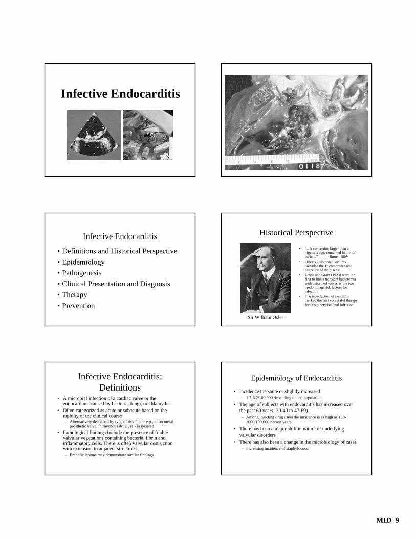
Infective Endocarditis
Infective Endocarditis
• Definitions and Historical Perspective • Epidemiology• Pathogenesis• Clinical Presentation and Diagnosis• Therapy• Prevention
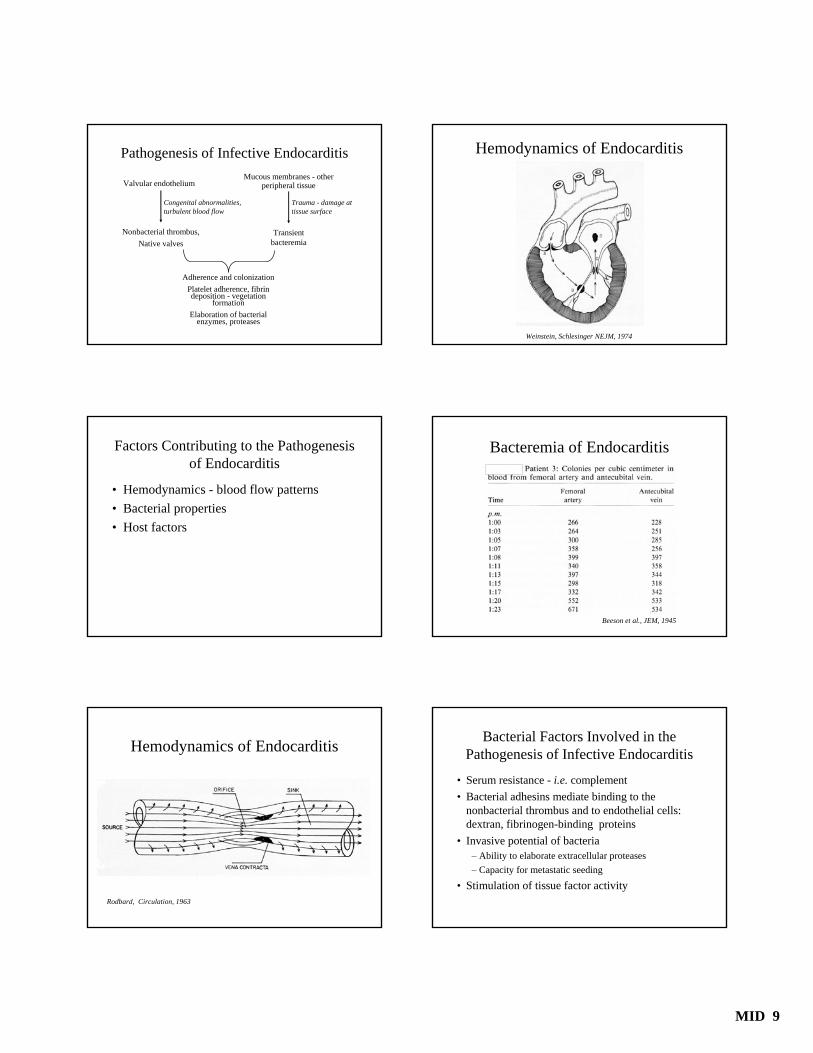
Infective Endocarditis: Definitions
• A microbial infection of a cardiac valve or the endocardium caused by bacteria, fungi, or chlamydia
• Often categorized as acute or subacute based on the rapidity of the clinical course– Alternatively described by type of risk factor e.g., nosocomial,
prosthetic valve, intravenous drug use - associated• Pathological findings include the presence of friable
valvular vegetations containing bacteria, fibrin and inflammatory cells. There is often valvular destruction with extension to adjacent structures. – Embolic lesions may demonstrate similar findings
Historical Perspective
• “.. A concretion larger than a pigeon’s egg; contained in the left auricle.” Burns, 1809
• Osler’s Gulstonian lectures provided the 1st comprehensive overview of the disease
• Lewis and Grant (1923) were the first to link a transient bacteremia with deformed valves as the two predominant risk factors for infection
• The introduction of penicillin marked the first successful therapy for this otherwise fatal infection
Sir William Osler
Epidemiology of Endocarditis• Incidence the same or slightly increased
– 1.7-6.2/100,000 depending on the population
• The age of subjects with endocarditis has increased over the past 60 years (30-40 to 47-69)– Among injecting drug users the incidence is as high as 150-
2000/100,000 person years
• There has been a major shift in nature of underlying valvular disorders
• There has also been a change in the microbiology of cases– Increasing incidence of staphylococci

MID 9
Epidemiology of Endocarditis
• There has been an increasing incidence of nosocomial endocarditis - both native and prosthetic valve
• There is an increased risk of IE among injecting drug users, patients on long-term hemodialysis, patients with intravenous catheters, diabetics and HIV-infected patients
Incidence of Underlying Heart Disease in Infective Endocarditis*
Pre-Antibiotic Era Present
Rheumatic ++++^ +Congenital + +Degenerative + ++Mitral valve prolapse + +++Normal valves + ++_______________________________________________________________________
*Incidence varies with population studied^ Relative incidence
Microbiology of Infective Endocarditis Prospective International Cohort Study
OrganismsStaphylococcus aureusCoagulase-negative staphylococciViridans group streptococciStreptococcus bovisOther streptococciEnterococcus spp.Gram negative bacilliFungiPolymicrobialCulture negative
Fowler, V. JAMA, 2006
Percent Cases31.610.5186.55.1
10.63.81.81.38.1
Risk Factors for Infective Endocarditis• Dental procedures, poor dental hygiene - viridans
streptococci, nutritionally variant streptococci, HACEK • Prosthetic valves
– Early: coagulase negative staphylococci, S. aureus– Late: coagulase negative staphylococci, viridans streptococci
• Gastrointestinal or genitourinary procedures - enterococci or S. bovis (colon carcinoma)
• Nosocomial - S. aureus (including MRSA), Gram negatives, Candida species
Brouqui and Raoult, Clin Microbiol Rev, 2001
Risk Factors for Infective Endocarditis• Nosocomial - S. aureus (including MRSA), Gram
negatives, Candida species • HIV - S. aureus• Animal or farm exposure: Coxiella, Chlamydia,
Brucella• History of homelessness, alcoholism (body lice):
Bartonella
Pathogenesis of Endocarditis
• Inoculation of bacteria colonizing a mucosal (e.g., oral mucosa) or peripheral tissue site into the bloodstream
• Transient bacteremia of a serum-resistant pathogen capable of adhering to a cardiac valvular surface
• Turbulent blood flow across the valve• Bacterial adherence to cardiac valvular surface • Pathogen - host tissue interaction resulting in vegetation
formation and local tissue damage– Bacterial persistence
• Dissemination of infection to other tissue sites and elicitation of systemic findings
MID 9
Valvular endotheliumMucous membranes - other
peripheral tissue
Trauma - damage at tissue surface
Congenital abnormalities, turbulent blood flow
Nonbacterial thrombus,Native valves
Transient bacteremia
Adherence and colonizationPlatelet adherence, fibrin deposition - vegetation
formation Elaboration of bacterial
enzymes, proteases
Pathogenesis of Infective Endocarditis
Factors Contributing to the Pathogenesis of Endocarditis
• Hemodynamics - blood flow patterns• Bacterial properties• Host factors
Hemodynamics of Endocarditis
Rodbard, Circulation, 1963
Hemodynamics of Endocarditis
Weinstein, Schlesinger NEJM, 1974
Bacteremia of Endocarditis
Beeson et al., JEM, 1945
Bacterial Factors Involved in the Pathogenesis of Infective Endocarditis
• Serum resistance - i.e. complement• Bacterial adhesins mediate binding to the
nonbacterial thrombus and to endothelial cells: dextran, fibrinogen-binding proteins
• Invasive potential of bacteria– Ability to elaborate extracellular proteases– Capacity for metastatic seeding
• Stimulation of tissue factor activity

MID 9
Vegetation Formation
Mills et al., Infect Immun, 1984
Dextran + Dextran –
Host Factors Involved in the Pathogenesis of Infective Endocarditis
• Valvular surfaces– Nonbacterial thrombus forms on damaged valves– Direct adherence to the endovascular surface of normal valves – Suture line, valve surface of prosthetic valves
• Platelets dual role – Platelet microbicidal proteins (α-granules)– Bacteria induce platelet aggregation– Part of nonbacterial thrombus surface
• Leukocytes, complement, cytokines
Immunologic Manifestations of Infective Endocarditis
• Hypergammaglobulinemia; both antigen specific and polyclonal B cell activation (e.g., rheumatoid factor)
– May block IgG opsonic response, accelerate microvascular damage or stimulate phagocytosis
• Vasculitis– Circulating immune complexes – Hypocomplementemia
• Clinical syndromes: “Lumpy-Bumpy” glomerulonephritiswith deposition of complexes plus complement, Osler’s nodes
The Pathogenetic Basis for the Clinical Manifestations of Infective Endocarditis
• Valvular destruction and local intracardiaccomplications
• Bland or septic embolization of vegetations• Sustained bacteremia• Immunologic phenomena
Osler, Gulstonian Lectures, Lancet, 1885Weinstein and Schlesinger NEJM, 1974
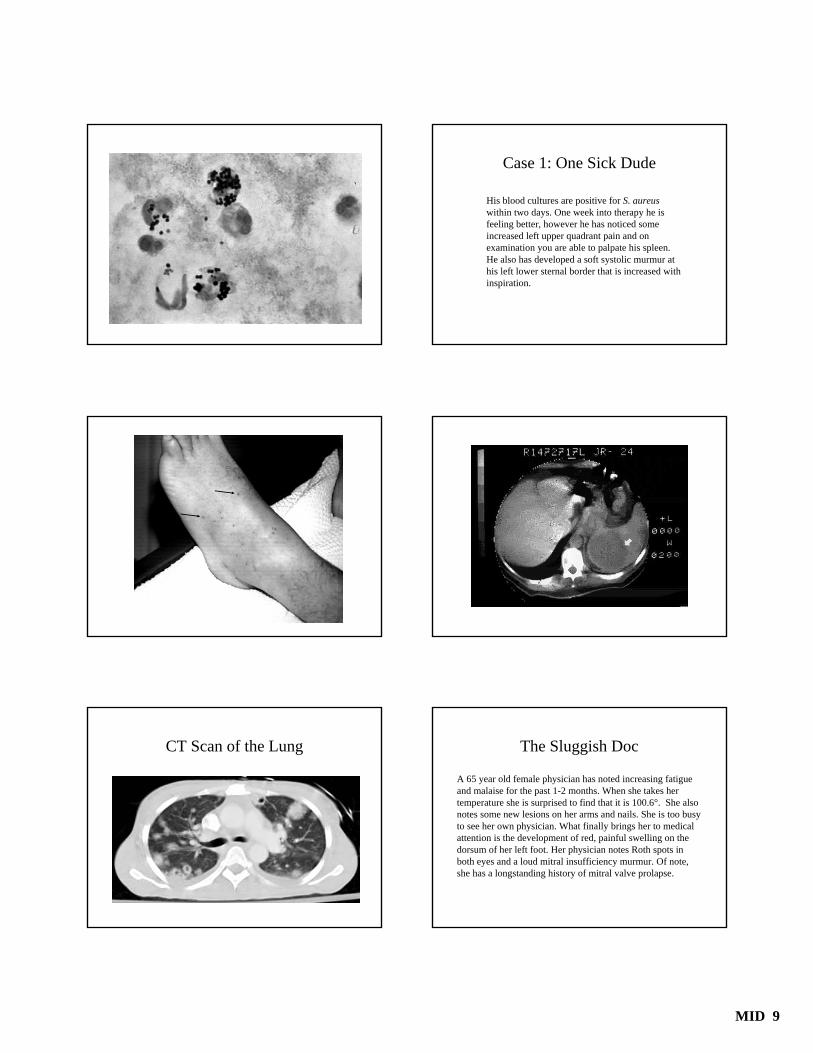
Case 1: One Sick Dude
A 33 year-old recreational drug user develops fever, chills and pleuritic chest pain 48h after injecting heroin. He appears acutely toxic with a temperature of 104°, rapid respirations and agitation. His sputum is purulent and bloody. His cardiac exam is unremarkable except for his sinus tachycardia. Examination of his skin reveals a petechial rash and a small soft tissue abscess at his injection site.
MID 9
CT Scan of the Lung
Case 1: One Sick Dude
His blood cultures are positive for S. aureus within two days. One week into therapy he is feeling better, however he has noticed some increased left upper quadrant pain and on examination you are able to palpate his spleen. He also has developed a soft systolic murmur at his left lower sternal border that is increased with inspiration.
The Sluggish Doc
A 65 year old female physician has noted increasing fatigue and malaise for the past 1-2 months. When she takes her temperature she is surprised to find that it is 100.6°. She also notes some new lesions on her arms and nails. She is too busy to see her own physician. What finally brings her to medical attention is the development of red, painful swelling on the dorsum of her left foot. Her physician notes Roth spots in both eyes and a loud mitral insufficiency murmur. Of note, she has a longstanding history of mitral valve prolapse.

MID 9
The Sluggish DocMultiple blood cultures grow viridans streptococcus. Her laboratory studies reveal a positive rheumatoid factor test. Despite antibiotic therapy, she develops refractory heart failure and requires mitral valve replacement
Diagnosis of Endocarditis
• History and physical - the "old fashioned way"• Laboratory studies -
– Microbiology - sustained positive blood cultures– Other studies - hematologic, rheumatologic, renal
• Echocardiography– Transesophageal echocardiography
AHA Diagnosis and Management of IE and Its Complications, Circulation 98:2936,'98
Cutaneous Manifestations of Endocarditis
Splinter Hemorrhages Osler’s Node Janeway Lesion

MID 9
Transesophageal Echocardiography
Daniel, Mugge NEJM,1995
What’s Wrong with this Picture?
Duke Criteria for the Diagnosis of Endocarditis
• Definite:– Pathologic criteria are histologic or culture confirmation
of vegetation or emboli.– Clinical criteria are 2 major, 1 major plus 3 minor or 5
minor criteria (e.g. positive blood cultures or echocardiogram, new regurgitant murmur)
• Possible:– 1 major, 1 minor; or 3 minor criteria
• Rejected:– Alternate diagnosis, resolution of infection with brief
therapy, no pathologic evidence of endocarditis Durack et al. AJM, 1994; Li et al. Clin Infect Dis, 2000
Mimics of Infective Endocarditis
• Atrial myxoma• Marantic endocarditis• Left atrial thrombus• Acute rheumatic fever with
carditis• Collagen vascular disease (SLE)• Neoplasms (carcinoid)

MID 9
Principles of Therapy• Bactericidal antibiotics must be used• Prolonged therapy is necessary (weeks)• Treatment is best started after multiple sets of
blood cultures have been taken. • Urgency in the initiation of therapy is required for
acute but not subacute endocarditis.• Synergistic combinations of antibiotics are used
when available.
Antimicrobial Prophylaxis of Endocarditis – Potential Mechanisms
• Bactericidal activity• Reduce bacterial adherence• Reduce bacterial density in the wound at the
time of surgery (for prosthetic valves)
Problems with Prophylaxis for Infective Endocarditis (IE)
• Most cases of IE not preventable with prophylaxis (absent history of a lesion)
• No controlled studies of efficacy• Most cases not associated with procedure
warranting prophylaxis• Dental treatment not a risk factor in population -
based, case-control study (Strom et al., AIM '98)– Prosthetic valves, antecedent endocarditis major risk
factors
Prevention of Infective Endocarditis• High risk
– Prosthetic valve – Complex congenital heart disease– Previous endocarditis– Cardiac transplantation with valvulopathy
• Moderate risk– Acquired valvular dysfunction (e.g. rheumatic valve)– Mitral valve prolapse with regurgitation
• Negligible risk– Mitral valve prolapse without regurgitation– Rheumatic fever without valvular dysfunction
AHA Recommendations, Circulation, 2007
Prophylaxis Recommended (or Not)!
• Dental procedures that involve manipulation of gingival tissue
• Respiratory tract, infected skin or skin structures• Prophylaxis not recommended for gastrointestinal
or genitourinary procedures
AHA Recommendations, Circulation, 2007
What Do I Need to Know?• What are the major epidemiologic features of
endocarditis and how have they changed?• Which bacteria are associated with which types of
exposure in the development of endocarditis?• What is the pathogenesis of endocarditis and what
is its correlation with the different clinical presentations?
• How is endocarditis diagnosed?• What are the basic principles of therapy and
prophylaxis?