ah 381 documents for student review. rsd (1) reflex sympathetic dystrophy reflex sympathetic...
TRANSCRIPT

AH 381
Documents for Student Review
RSD (1)
Reflex Sympathetic Dystrophy
• Reflex sympathetic dystrophy (RSD) is a complex, poorly understood disorder that is characterized by chronic, severe pain and progressive changes in skin, muscle, and bone. Although the precise causes of RSD are unknown, it often occurs following an injury, often minor in nature.
• Some experts believe that RSD represents an exaggerated response of the sympathetic nervous system to some form of injury or insult (eg. surgery) to the area resulting in chronic, severe, sometimes debilitating pain. Although the signs and symptoms of RSD vary depending upon the clinical stage of the disorder, the one common feature shared by all 3 clinical stages (early, established, or late RSD) is pain.

(1)
• Usually occurs in distal extremity when CNS produces continuous sympathetic stimulation of the limb; in the athletic setting, RDS most commonly follows a severe injury followed by immobilization or non weight bearing

RDS S&S (1)
Symptoms• “confounding symptom” =
severe localized pain• Pain disproportional to
injury• Skin hypersensitivity
( even to clothes and bed sheets)
• Extreme reluctance to move joint or bear weight
Signs• Swelling• Decreased ROM• Increase skin temp.• Atrophic skin, hair & nails
change sin affected limb
Over a period of time, you will see…(1)
• Atrophy & poor peripheral vascular control (cyanosis, intolerance to cold, pallor)
• After several months, sympathetic activity decreases and entire limb becomes atrophic, cool, pale, and so hypersensitive it can no longer function
Treatment of RSD (1)
• Recognition and treatment are CHALLENGING• Can be prevented by encouraging movement and
progressively weight bearing• Rehab includes : rhythmic weight bearing, gentle jt
distraction, AROM, desensitization techniques, and joint mobs
• Pain relief through transcutaneous nerve stimulation• In resistance cases, analgesics and anesthetic pain
blockage have be utilized• Persistent and aggressive TX increases probability of
successful outcome

Meningitis (2)• Meningitis is an infection of the fluid in the spinal cord and the fluid
that surrounds the brain. Meningitis is usually caused by an infection with a virus or a bacterium. Knowing whether meningitis is caused by a virus or a bacterium is important because of differences in the seriousness of the illness and the treatment needed.
• VIRAL MENINGITIS is usually relatively mild. It clears up within a week or two without specific treatment. Viral meningitis is also called aseptic meningitis.
• BACTERIAL MENINGITIS is much more serious. It can cause severe disease that can result in brain damage and even death.– Antibiotics will be prescribed for bacterial meningitis; the type will vary
depending on the infecting organism. Antibiotics are ineffective in viral meningitis. Treatment of secondary symptoms including brain swelling, shock, and convulsions will require other medications and intravenous fluids. Hospitalization may be required depending on the severity of the illness and the needed treatment.

(2)
• Bacterial meningitis caused by streptococcus or meningococcus bacteria can be fatal within hours of infection
• Other causes include: viruses, drugs, lead poisoning, and parasites

(2)
• Symptoms – fever and chills – severe headache – nausea and vomiting (may be violent) – stiff neck ("meningismus") or rigid neck– sensitivity to light (photophobia) – Rapid coma– Altered cognition, syncope, seizures, & coma may occur over
time
• Additional symptoms that may be associated with this disease: – decreased consciousness – rapid breathing – severe neck stiffness, ultimately resulting in a characteristic
arched posture-seen in infants or small children– "bulging fontanelles" may be seen in infants – poor feeding or irritability in children

Signs of Meningitis (2)
• Rash on head
• Inability to flex the neck passively without hip an knee flexion ( known as Brudzinski’s sign)
• EMERGENCY TRANSPORT for antibiotics

Abdominal Quadrants (4, 12)
• Upper right
• Upper left
• Lower right
• Lower left
• Palpation of organ will feel …

Using a Stethoscope (5, 6, 13)

Using and Otoscope (14)

Clinical S&S Associated with ENT conditions (180-187)
• Common cold• Conjunctivitis• Laryngitis• Phayngitis• Rhinitis• Sinusitis• Tetanus• Tonsillitis

Common Cold
• Viral
• Head and throat mainly affected
• Stuffy, sore throat, post nasal drip, fever possible but not required
• AKA URI

Conjunctivitis
• Inflammation of the lining of the posterior eyelid and eyeball margins (the conjunctiva)
• Can result from allergens or infection by bacteria or viruses
• Burning (infection) or itching (allergy) with purulent (infection) or mucoid (allergy) drainage from the eye occurs
• Commonly called “pink eye”

Laryngitis
• Infection or inflammation of the vocal cords
• Causes changes in the quality of the voice, such as hoarseness or inability to speak
• Itching of the throat, fever, difficulty or pain with swallowing, or even dyspnea may occur with severe laryngitis

Pharyngitis
• Infection or inflammation of the throat
• Causes throat pain, painful or difficult swallowing, and pain in the ears when swallowing
• Reveals a red (erythematous) throat with purulent or mucoid exudate covering the pharynx

Rhinitis
• Viral -causes infection of mucous membranes causing mucous secretion
• Allergic – AKA hay fever; experiences watery eyes
• Sore throat, runny nose, congestion, nasal discharge (clear or light colored); fever

Tetanus
• Lockjaw• Bacterial infection enters through puncture
/ wound and moves to CNS• S/S: pain around wound, local and
regional hyper tonicity and spasm• Eventually difficulty opening mouth within
48 hours• Severe – fever• Must have vaccination!!!!

Tonsillitis
• Infection or inflammation of the tonsils
• Produces throat pain, painful or difficult swallowing, and pain in the ears when swallowing
• Reveals a red (erythematous) throat with purulent or mucoid exudate covering the tonsils

Sinusitis
• Inflammation of the paranasal sinuses
• Caused by: URI from bacteria
• Nasal mucous membranes swell and block paranasal sinus; resulting pressure causes pain

Clinical S&S Associated with GI conditions(188-197)
• Appendicitis• Colitis• Constipation• Diarrhea• Esophageal reflux• Gastritis• Gastroenteritis• Indigestion• Ulcer• Irritable bowel syndrome

Appendicitis
• Inflammation of the appendix• Caused by physical irritants or infection• Causes abdominal pain in the lower right
quadrant, passive extension or active flexion of the right hip may be painful if psoas muscle is irritated
• Loss of appetite and nausea are usually present, although vomiting is rare
• Treatment is usually surgical removal

Colitis

Constipation
The abnormal retention of feces as a result of hardened (dehydrated) stool or decreased bowel motility
Poor diet (high sugar, low fiber), dehydration, medications, stress, inactivity, or GI disease can contribute
Appropriate lifestyle changes relieve constipation due to diet or inactivity, although laxatives may be needed in more severe cases

Diarrhea
• Frequent or loose bowel movements from increased motility, malabsorption syndromes, infection, or a combination of these factors
• Some medications and drugs cause temporary diarrhea; for instance, antibiotics allow overproduction of intestinal bacteria, which increases intestinal motility

Esophageal Reflux

Gastritis
• Stomach inflammation that results from erosion of the entire mucosa, chronic use of medications, H. pylori infection, or autoimmune disease
• It can be acute or chronic, erosive or nonerosive• Causes nausea, vomiting, and vague upper
abdominal pain • Treatments include dietary restrictions and and
symptomatic treatment with antacids

Irritable Bowel Syndrome
• Is thought to be a reaction to psychophysical stress and poor diet
• Produces abdominal pain and cramping, and is most prevalent among young adult females
• This disorder affects motility of the intestines, causing diarrhea, constipation, or alternating episodes of both
• Bloating or abdominal distension may appear• Relief of abdominal pain usually occurs after
defecation

Gastroenteritis
• The inflammation of the mucosal lining of the stomach and intestines, usually a result of infection
• Food poisoning, traveler’s diarrhea, and viral “stomach flu” are common manifestations of gastroenteritis
Gastritis
• Stomach inflammation that results from erosion of the entire mucosa, chronic use of medications, H. pylori infection, or autoimmune diseases
• Can be acute or chronic, erosive or nonerosive• Causes nausea, vomiting, and vague upper
abdominal pain• Treatment includes dietary restrictions and
symptomatic treatment with antacids

Indigestion

Ulcer

General Terms and Concepts
This information is assigned “float” due dates. The student may complete the proficiency through the accepted method at any time
between Jan 19 and March 8, 2005Due March 8, 2005

Observe and ID the Clinical S&S associated with elbow… (26, 27, 28, 29, 30,31,
32, 33, 34)
• Dislocation or subluxation• Fracture• efficiency of movement• bursitis• epicondylitis• Tenosynovitis and tendonitis• Osteochondritis dissecans• Sprain• strain

ID S&S of overuse injuries – ankle (58)
• Bursitis
• Exostosis
• Fascitis
• Stress fracture
• Tarsal syndrome
• Tendonitis/tenosynovitis
• Tibial stress syndrome

ID clinical S&S associated with..-ankle (59-63)
• Dislocation or subluxation
• Fracture
• Sprain
• Strain
• atrophy

ID clinical S&S associated with- knee (64-68)
• Atrophy
• Bursitis
• Fracture
• Sprain
• Strain
• tendonitis

ID clinical S&S associated with (69-78)
• Atrophy• Bursitis• Dislocation and subluxation• Efficiency of movement• Fracture• Sprain• Nerve injury• Strain• Symmetry• Tendonitis / tenosynovitis

ID Clinical S&S associated with…(102-112)
• Leg length discrepancy• Apophysitis• Dislocation & subluxation• Fracture• Stress fracture• Bursitis• Contusion• Sprain• Strain• Tendonitis

Unit 2

Functional &Activity specific test for the cervical spine (3)
• Goniometry not always used to assess functional ability here b/c of difficulty measuring
• Gross movement patterns should be completed; if any limitations are noted there, conduct a more specific, focused assessment of that motion and related anatomy
• Active, passive, then resistive ROM

Postural assessment of Cervical Head and spine (18)
• Normal position: chin in line with throat, throat centered, should be able to draw an imaginary line through the ears through the shoulder, hip, knee, and ankle
• Abnormal positions– Forward head: ears in front of acromium– Accentuated cervicothoracic hump – head in
constant flexed position over the spine

Torticollis (20)
• AKA wryneck or stiffneck
• S&S– Pain on side of neck upon awakening
• Acquired spasm of sternocleomastoid

Commonly used test for Nerve Root Compression ( 21)• Distraction test
– Pt seated, examiner has 1 hand on chin and other around occiput; examiner distracts head from trunk directly away from trunk
– Positive test if pain decreases of disappears & inidctaes nerve root; increase in pain may indicate muscle / ligament damage
– Note: vertebral artery test should be done prior to this test and do not use this test if possibility of cervical instability exists
• Compression test– Same position as above; examiner has hands on top of patient’s head
and compresses– Positive if pain is produced
• Spurling’s test ( Compression)– Pt seated; examiner’s palms on pt’s head, examiner applies downward
pressure while pt laterally flexes to each side (AROM & PROM used)– Positive finding is pain to flexed side
• Shoulder abduction tests– Pt seated / stand– Patient abducts shoulder until palm is resting n top of head– Decrease in symptoms may indicate nerve root compression; possibly
resting from herniated disk

Commonly used tests for neurovascular dysfunction (24)
• Vertebral Artery Test– Subject lies supine, examiner supports head– Examiner slowly extends, rotates and laterally
flexes subject head– Observe subject for dizziness, blurred vision,
slurred speech, LOC– This position pathologically occludes artery if
those present

Commonly used testes for Brachial Plexus Neuropathy (22)
• Brachial tension test– Must rule out bony trauma before performing– Examine stands behind athlete and passively flexes head to one
side while applying downward pressure through the patient's opposite shoulder
– Positive test if pain increases or radiates down arm
• Tinel’s sign– Tap skin over superficial nerve– Patient sits or lies supine– Tap Erb’s point(2cm superior to clavicle and anterior to
transverse process of C6– Positive if sensation on that side changes; indicates brachial
plexus pathology

Commonly used tests for Cervical Disk Herniation (23)
• Valslva Maneuver– Deep breath, bear down– Used to determine the presence of space
occupying lesion (herniateddisc, tumor, tec)– May decrease pulse, increase intracranial
pressure, decreases venous return, cause fainting
– Positive test if pain increases

Commonly used tests for neurovascular dysfunction (24)
• See previous slide

Functional and Activity Specific test for head and face injuries (46)
• Must rule out instability involving head and spine• Assess facial motions• Use cranial nerve assessment
– I Olfactory ID smells– II Optic read– III Oculomotor P(ea)RL, up and down gaze– IV Trochlear down & lat gaze– V Trigeminal face sensation, clench teeth– V I Abducens medial and lat gaze– VII Facial close eyes, smile\– VIII Vestibulocochlear sound, balance– IX Glossopharyngeal swallow, gag– X Vagus gag; swallow, say “Ahh”– XI Accessory resist shoulder shrug– XII Hypoglossal stick out tongue

ID clinical S&S of posturing (47)
• Sprengel’s deformity– Undescended scapula– Congenital– Normal scapula
• Base of scapular spine should be at T4 and inferior pole should be at T7

ID Clinical S&S associated with trunk pathology (90-93)
• Sprain– injury to a ligament of similar connective tissue
• Stenosis– Narrowing of foramen of vertebral bodies
• Step deformity– the spinous process of the vertebral segment involved
protrudes ventrally– may exist as a consequence of spondylolisthesis
• Strain– Muscular injury

Remember all those that were listed in Unit 1 we called float can
be done at any time but will be due on March 8, 2005

Unit 3

Kyphosis vs Normal Spine (15)
Results from trauma, developmental problems or degenerative diseases

Normal Spine (

Lordosis vs Normal Spine (16)

Scoliosis (17)
ID Clinical S&S associated with (79-88)
• Café au lait macules (spots)• Dislocation and subluxation• Spina bifida occulta• Facet syndrome• Intervertebral disc pathology• Spinal posture (kyphosis/lordosis/scoliosis) –
see #15-17)• Leg length discrepancy• Nerve root compression• sacroiliac dysfunction

Café au lait macules (spots)(79)
• Macule is a spot• Café au lait macules
may be solitary benign findings or may indicate the presence of neurofibromatosis with its associated complications
Dislocation and subluxation
• In terms of vertebrae, very serious
• Instability can compromise other structures
• How do you assess spinal instability– Motor an sensory deficits– Mechanisms– Signs and symptoms

Spina bifida occulta
• Congential condition in which lamina of verebrae (usually lumbar) do not unite which exposes the spinal cord
• Most people will not even be aware that they have spina bifida occulta unless it shows up on an X-ray which they have for some unrelated reason. It is usually just a small part of one vertebra low in the back which is missing
• Depending on the amount of neural involvement, symptoms can be absent, minimal, or severe. Symptoms can include:
– Weakness or sensory loss in the legs, feet – Leg length difference – Foot deformity – Problems with gait (walking) – Bowel or bladder infection or incontinence – Constipation – Scoliosis (sideways curvature of the spine) – Back pain – Continence problems

Facet syndrome
• The pain comes from the cervical facet joints, which are joints at the back of the vertebrae in your neck. When these joints are subluxated or irritated, local inflammation develops that can lead to subsequent irritation of surrounding nerves. When this happens, the irritated facet joints can refer (move) pain through these nerves to other areas of the body
• Neck pain, neck stiffness, headaches, shoulder pain and upper back pain are common symptoms of facet syndrome

ID Clinical S&S associated with vertebral pathology (89)
• Spondylitis: inflammation of one or more vertebrae
• Spondylolyisis: degeneration or deficient development in articulating part of vertebrae ( pars defects); fracture in region or pars (region between inferior and superior facets)
• Spondylolisthesis: forward slippage of vertebrae
• For more information, research pars interarticularis fractures

ID Clinical S&S associated with cervical (96-101)
• Dislocation & subluxation
• Vertebral fracture
• Head & neck posture
• Intervertebral disc herniation
• Nerve root compression
• ischemia

Sacroiliac dysfunction
• Relatively immobile
• Accessory movement, rotation, and .or translation of the ilium on the sacrum occurs
• When extreme, the ilium rotates to the point it subluxes on sacrum
• Resulting pain and dysfunction resemble nerve root lesion symptoms

Sacroiliac dysfunction• Special tests
– FABER• Overpressure in FABER position causes pain contra laterally
– Approximation/ Transverse stress• Pt side-lying and downward pressure is directed through hips• Pain or pressure is positive indicator as sprain
– SI rock• Pt hips and knees fully flexed toward opposite shoulder and his adducted; SI
is rocked by flexion and adduction of the hip; pain in posterior lateral thigh indicates SI involvement
– SI compression • Supine with ATC hands over opposte ASIS• Pressure applied spreads ASIS and compresses SI joint• Pian is positive sign
– SI distraction• Pt supine• Palms on anterior ilium, apply downward pressure; pain in gluts may indicate
SI sprain

Leg length discrepancy
• Have pt lie supine with his knees flexed to 90 degrees, feet flat on the table; if one knee appears higher, the tibia of that extremity is longer; if the knee projects further anteriorly, the femur is longer
• True -ASIS to medial malleolus • Functional / no bony inequality – umbillicus to
medial malleolus– Pelvic obliquity– Hip deformity ( adduction or flexion)

Administer appropriate sensory, circulatory and neurological test for the head and face
(57)
• Circulatory – assess carotid pulse or skin color
• Neurological– Review cranial nerves from previous
• Sensory– Assess different areas of the face for
sensation

Administer functional & activity specific test for thoracic & lumbar
spine (112)• Function
– Goniometry not normally used as difficult for individual reasons; assess gross movement then get specific
– AROM: willingness to perform; can complete range be attained; is range blocked by spasm, stiffness or pain, ALWAYS compare bilaterally
– PROM: no performed when sensory or motor deficient exist (may indicate spinal injury); Thoracic and lumbar passive motions are seldom performed; end ranges should feel “ the tissue stretch”
– RROM: done through full ROM and make sure you stabilize
• Activity– Have pt perform usual or limited activities and assess gross motor
ability; find activity that is limited or is limiting other activities and focus your assessment there

Commonly used special tests for thoracic and lumbar spine (113)
• Intervertebral disk herniation– Valsalva maneuver
• Deep breath, bear down• Used to determine the presence of space
occupying lesion (herniateddisc, tumor, tec)• May decrease pulse, increase intracranial
pressure, decreases venous return, cause fainting• Positive test if pain increases
Commonly used special tests for thoracic and lumbar spine (114)
• Neuropathy– SLR
• AKA Lasegue• Used for SI pain, sciatic irritation or tight hamstrings• Relaxed supine position; passively flex hip while knee extended until
pain r tension is in hams; lower leg until pain disappears then DF ankle and have athlete flex neck
• If pain does not increase with DF or neck flexion, you have tight hams
– Well SLR– Babinski’s reflex test
• Pt lying down w/ eyes closed, leg slightly elevated and flexed• Stroke pointed object along the plantar aspect of pt’s foot• Normal: toes curl down in flexion and adduction• Positive sign: suggests upper motor lesion and is demonstrated by
extension of big tow and abduction (slaying) of other toes

– Oppenheim’s gait test– Kerni’s sign /Brudzinski sign test
• Like SLR but active movements by pt• Brudzinki’s: pt supine with hands behind head; positive test if
c/o neck or low back pain and tries to relieve by involuntarily flexing knees and hips;
• Kerni’s: pt supine with hips flexed, knees extended; pain in head, neck and back indicate meningeal irritation and will be relived with flexion of knee
– Bowstring test• In SLR tets position, ATC flexes knee slightly (20 degrees)
reducing symptoms; with finger/thumb ATC applies pressure to the tibial portion fthe siactic nerve in popliteal space and radiating pain returns
– Hoover sign test• Under normal conditions, when pt tries to elevate one leg,
downward pressure is applied with opposite leg• ATC places heel of pt in palms

Commonly used special tests for thoracic and lumbar spine (115)
• Vertebral defects– Stork standing
• Pt stands on one leg and extends spine while balancing on the single leg
• Positive sign is pain in back; associated with pars interarticular stress fracture (spondylolisthesis)
• If stress fx if unilateral, standing on ipsilateral leg causes more pain
• If rotation is combines with extension and pain results, indicates possible facet pathology
– Spondylolisthesis test

Commonly used special tests for thoracic and lumbar spine (116)
• Joint instability– Spring Test
• Pressure applied through spinous process; process will rebound after pressure removed
• Indicates stability if rebounds• If does not return to normal position, indicates
potential problems

Postural Assessment of the Lumbo-thoracic Region(125)
• Know what is normal– Normal curvatures (recall earlier slides)– assess sagital and frontal planes– Inspect torso for muscle spasm of atrophy– Inspect each spinal segment for Malalignment
• Thoracic– Look at skin folds; should be equal bilaterally– Observe respiration for irregularity from normal and bilaterally
with that pt
• Lumbar– General motion and posture (improper sitting or lifting)– Observe curve of extenuation (tight hip flexors or weak abs) or
reduction ( acute pain, muscle spasm, hamstring tightness) is pain present
– Standing: look for lateral shift – may shift position to remove nerve root pressure

ID Clinical S&S associated with… (137-138)
• Weight bearing verses non weight bearing alignment– Shift based on status
• Gait– Hoppenfeld devotes entire chapter to nuances
of gait analysis – review with your peers and ACIs
– You are looking for the abnormal

Unit 4

Perform a postural assessment of the shoulder (19)
Look for faulty posture or congenital abnormalitiesLook from anterior, posterior and lateral aspectsNote position of the head (if tilted or rotated could
suggest spasm, pressure on cervical nerve root, or stretching of cervical nerves)
Check scapular position (base t4, inferior pole t7, with medial border equal distance from spinous process
Note position of arm (splinted or hanging limp)Movement by patient

ID S&S of scapulohumeral rhythm (48)
• As the arm abducts, the scapula must rotate to accommodate and allow necessary motion
• First 30 degrees abduction, no rotation, 30-90 1scapula rotation:2 humeral elevation, rest it’s 1:1

ID Clinical S&S associated with scapular winging (49)
• Medial border elevated
• If serratus anterior weak, affected side will be depressed (lower on chest wall), protracted (further from vertebral processes, Elevated (winged) off chest wall
• Note position of arm

ID Clinical S&S associated with step deformity (50)
• OF GH– When sulcus sign is positive
• Of AC– Elevated distal clavicle– Indicates AC sprain

Administer functional tests and activities for the shoulder (51)
• Activity specific– Have the athlete perform the motion
necessary to accomplish their given tasks– Look for pain, discomfort, weakness, or
compensation
• Function– A, P, ROM – assess the shoulder’s ability to
work

Commonly used special test for Glenohumeral Instability (52)
• Anterior drawer test (load & shift)– Pt seated, ATC behind with one hand stabilizing the shoulder
over clavicle and scapula– Grasp head of humerus and push into glenoid “seat”– Now, push head of humerus anteriorly and posteriror and note
amount of translation
• Posterior drawer tests– Posterior portion of above– 50% is considered normal
• Relocation test– Athlete supine, arm in position to be ER by ATC– Apply posteriorly directed pressure ands symptoms relieved

• Apprehension test– Same position and motion as relocation, no posterior pressure
used– Read athlete’s response (facial expression, muscular guarding,
pain, etc)• Clunk test
– Athlete supine, one hand on posterior as part of the shoulder under humeral head
– Grasp humerus above elbow and abduct arm fully over athlete’s head
– Humeral head hand applies anterior push while other hand moves humerus into lateral rotation
– Positive test results in a clunk or grinding sound , indicating labral tear
– May be painful if instability is present• Sulcus test
– With athlete sitting or standing, arm to side– Apply downward traction by grasping hand– Positive if space widens between acromion process and humeral
head

Commonly used special test for Acromioclavicular instability (53)
• Shear test – Mobilize clavicle be grasping and producing
shear translation
• Compression test– horizontal abduction of humerus across chest
compresses the AC joint and will also lead to increased pain in injured joint

Commonly used special test for Rotator cuff inflammation / Impingement (54)
• speed’s test– Bicipital tendonitis– Supinate arm with elbow fully extended– With one hand over groove, resist athlete flexing elbow– Positive test is tenderness over the groove
• Drop arm test– Integrity of supraspinatus muscle– Have athlete abduct shoulder 90 degrees with no rotation then
ask them to slowly lower– Positive if arm is not lower smoothly– Can apply pressure in abducted position, if drops, it’s positive
• Empty can test– Both arms abducted 90 degrees, hor. Abd. 330-60 degrees with
thumbs down– Apply downward pressure– Weakness or pain should be assessed

• Impingement test– Hawkins-Kennedy impingement test
• Athlete sitting, ATC hor. Abd and IR arm with elbow flexed 90 degrees
• Must maintain anterior pressure through scapula with posterior hand
– Neer impingement test• Athlete sitting, arm IR and forcible flexed (raised above head)
to jam greater tuberosity against anterior surface of acromion• ATC posterior hand over scapula, fingers grasp clavicle
• Pectoralis major contracture test

Commonly used special test for biceps & biceps tendon pathology
(55)
• Yergason’s test– Stabilized flexed arm against the body with forearm
pronated– Ask athlete to supinate forearm, flex elbow and ER
humerus while ATC resists– At the same time, apply downward traction of
humerus through your grasp on the elbow• Ludington test
– Athlete seated, hands clasped behind head– Biceps alt. Contracted and relaxed– As ATC palpates, if no contraction is felt, as rupture
exists

Commonly used special test for thoracic outlet syndrome (56)
• Adson’s maneuver– Extend and ER humerus while the athlete extends
head– Monitoring pulse
• Allen test– Abduct shoulder and flex elbow while athlete looks to
opposite shoulder– Monitoring pulse
• Military brace position– Athlete seated or standing– Have athlete force shoulder posteriorly

Unit 5

Identify the following (7, 8, 9)
• Symmetry– Review section on posture– Should look same unless upper body athlete
• Atrophy– Is there a decreases muscular definition on one side
visually; may test muscular strength to verify a weakness
• Nerve injury– Looking for weakness, lack of function, lack of
sensation, atrophy

Elbow: Soft Tissue (10)
• Muscles– Bicpes brachii– Brachioradialis– Brachialis– Pronator teres– Pronator quadratur– Triceps brachaii– Anconeus– supinator

• Ligaments

Functional &Activity specific test for the forearm, wrist & hand (11)
• Activity specific– Have the athlete perform the motion
necessary to accomplish their given tasks– Look for pain, discomfort, weakness, or
compensation
• Function– A, P, ROM – assess the shoulder’s ability to
work
Clinical S&S for carrying angle – Elbow ( 25)
• Angle between the longitudinal axis of the humerus and the ulna when the arm is in anatomical position
• Forearm angles away from body when carrying something in the hand
• 10-15degrees normally, greater in females than males (due to shape of trochlea)

Cubital Valgus ( 25)
• excessive angulations of the forearm with respect to the humerus is known as cubitus valgus
• the result being an increased carrying angle at the elbows - the lower arms stick out more.

Cubital Varus ( 25)
• a decreased carrying angle, also known as a "Gunstock Deformity", usually due to an improperly reduced supracondylar fracture or epiphyseal abnormality during growth.

Repeated SlideObserve and ID the Clinical S&S associated with elbow… (26, 27, 28,
29, 30,31, 32, 33, 34)
• Dislocation or subluxation• Fracture• efficiency of movement• bursitis• epicondylitis• Tenosynovitis and tendonisti• Osteochondritis dissecans• Sprain• strain

Administer Functional & Activity Specific test for the elbow (35)
• Activity specific– Have the athlete perform the motion
necessary to accomplish their given tasks– Look for pain, discomfort, weakness, or
compensation
• Function– A, P, ROM – assess the shoulder’s ability to
work

Commonly used tests for joint stability - elbow (36)
• Valgus
• Varus

Commonly used Special Tests – elbow (37)
• Inflammatory Conditions– Medial Epicondylitis
• Resisted wrist flexion and pronation painful
– Lateral Epicondyltis• Resisted wrist extension • Picking up cup is painful• Long finger test

Commonly used tests for neuropathy – elbow (38)
• Tinel’s sign– Tapping over cubital tunnel to produce tingling down
ulnar nerve into forearm and hand
• Pronator teres syndrome– Athlete sits with elbow flexed 90 degrees– AT resists forearm pronation while elbow is extended– Positive test is tingling or paresthesia
• Pinch grip test– Ask athlete to make an “O” with thumb and finger– Normally tip to tip; if pulp to pulp, entrapment of
interosseous nerve

Clinical S&S associated with- forearm (39,40)
• Dislocation or subluxation– Care for vascular structures and distal extremities
• Disease states– Club nails: domed, broader, and larger than normal
• Often due to hypertrophy of underlying soft tissues, but may indicate respiratory or congenital heart problems
– Spoon nails: weakly structured, appearing concave or dug out, result of a severe fungus infection

ID clinical S&S associated with soft tissue pathology- forearm, wrist and hand (41)
• Sprain• Flexion tendon avulsion
– FDP- jersey finger, DIP cannot flex; surgical repair– FDS- PIP cannot flex
• Extensor tendon avulsion– Mallet finger, DIP unable to extend and in partial
flexed position• Extensor tendon rupture
– Boutonniere Deformity – DIP extension, PIP flexion– Splint 5-8 wk w/ PIP in extension
• Volar plate rupture

• Dupuytren’s contracture– Unknown cause; development of nodules that limit finger extension and
cause flexion deformity– Ring finger flexes into palm and cannot be extended
• Ganglion– Synovial cyst; herniation of joint capsule or sheath– Feels rubbery, soft, or very hard– Apply pressure pad or drain
• Swan neck deformity– Volar plate injury from severe hyperextension force– Distal tear (swan neck deformity) proximal plate injury
(pseudoboutonniere)• Volkman’s contracture
– Pressure on the brachial artery inhibits circulation to forearm, wrist, etc– Pain in forearm with interruption of pulses; increase in pain when fingers
passively extended

ID clinical S&S associated with neurovascular involvement (42)
• Carpal tunnel syndrome• Bishop’s deformity / benediction deformity
– Ulnar nerve damage affecting the hypothenar and intrinsic muscle of ring and little fingers
– Ring and little finger flex toward palm• Ape hand
– Palsy of the median nerve– Thumb is pulled backward in line with fingers
• Claw fingers– Compression of median and ulnar nerve– All digits flexed toward palm
• Drop wrist deformity– Palsy of radial nerve– Paralysis of extensor muscles so wrist and fingers cannot be extended
• Volkmann’s contracture

Commonly used special test for inflammatory conditions –forearm.. (43)
• Finkelstein’s test– De Quervains syndrome– Make a fist with thumb tucked inside and
ulnarly deviate or wrist flex

Special Tests for joint instability (44)
• Valgus stress
• Varus stress
• Glide test– Anterior and posterior gliding of carpal bones,
metacarpals, and phalange

Common special test for neurovascular pathology (45)
• Tinel’s sign– Tap over palmer surface, mid wrist– Transverse carpal ligament– Will cause tingling and parasthesia over thumb, index
finger, middle finger, and lateral half of ring finger– Median nerve distribution
• Phalen’s test– Flex both wrists as far as possible and press them
together; hold for 1 minute– Pain in carpal tunnel is positive for carpal tunnel
syndrome

Unit 6
Repeated SlideID Clinical S&S associated with…(102-112)
• Leg length discrepancy – see previous week’s slide• Apophysitis• Dislocation & subluxation• Fracture• Stress fracture• Bursitis• Contusion• Sprain• Strain• Tendonitis

Measurement of hip and pelvis (94)
• Goniometer
• Tape measure– Hip abduction can be measured by measuring
the speration of the malleolar

Recognize the following postural deviations & predisposing conditions
(117-124)• Pelvic obliquity
– Relationship of the inominate bones; looking at proper allignment; iliac crests should be level; if not, entire pelvis may have sifted
• Tibial torsion– Tibial torsion is an inward twisting of the shin bones
(the bones that are located between the knee and the ankle). Tibial torsion causes the child's feet to turn inward, or have what is also known as a "pigeon-toed" appearance.
• Hip anteversion & retroversion• Genu valgum, varum, and recurvatum

• Rearfoot values & varus– See next slides
• Forefoot valgus and varus– See next slides
• Pes cavus and planus– Cavus (high) & PLanus (low) arhc
• Foot and toe posture– Abnormalities (claw toes, halicus valugus)

• Rearfoot varus

Rearfoot Valgus

Forefoot varus
• a foot deformity characterized by an inward bending of the front half of the foot.

Forefoot valgus

Postural assessment of the Hip and Pelvis (126)

Function & activity specific test for the hip and pelvis (167)

Common tests for… (168-170)
• Sacroiliac dysfunction– FABER / Patrick’s test– Gaenslen’s test: supine position, with affected side
on table edge, unaffected side flexed toward abdomen; pressure applied to knee hanging off the table moving SI into extension; positive if pain increases on affected side (hyperextension side)
– Pelvic compression/ distraction test
• Neuopathy– Femoral nerve traction test: hip exteneded and
knee flexed 90 degrees; as hip extended, pain occurs in anteriro thigh

• Neuromucular pathology– Trendelenburg test: glut medius weakness; standing,
unaffected foot lifted (hip flexes) hips should not drop– Thomas Test; for hip contractures; supine with legs hanging off
table; examiner places hand under lumbar spine; one leg is brought to chest; if tightness exists, the extended thigh will raise of of table
– Rectus femoris contracture test– Ober test: lying on unaffected side, knee flexed 90 degrees,
thigh abducted as far as possible; pelvis stabilized, abducted thigh relaxes and allowed to drop into adduction; if IT or TFL contracted, thigh will stay abducted
– Noble Test: supine, knee flexed 90 degrees and pressure applied to lateral epicondyle while knee gradually extended; if pain severe at 30 degrees, positive test
– Piriformis test: flexing both hip sto 90 degrees and lifting the top leg places it on stretch; increase in pain indicates tightness

Clinical S&S Associated with… (171-178)
• Hip retroversion: toe out, femoral neck directed posteriorly
• Hip anteversion: toe in interiorly directed femoral neck
• Legg-Calve-Perthes disease– AV of femoral head 4-10yo (boys more)- disruption of
circulation/idiopathic; c/o pain in groin; limping, limited hip movement
• Slipped capital femoral epiphysis– 10-17 yp (boys more); idiopathic- maybe related to
growth hormone; femoral head slips; pain in groin, pain with PROM&AROM of hip & knee when advanced

• Osteitis pubis– Chronic inflamm. of symphysis pubis; pain in groin
and over sym. Pubis; pain while running, sit ups, squats; point tender over pubic tubercle
• Athletic pubalgia– Chronic pubic / inguinal pain; pain during exertion for
several months, sharp burning pain local to abdomen which will radiate to adductors and testicles; pain increases with hip flexion, IR, and ab contraction; pain with resisted hip Adduction although adductors not tender
• Piriformis syndrome• IT band syndrome

Unit 7

Repeated SlideID clinical S&S associated with- knee (64-68)
• Atrophy
• Bursitis
• Fracture
• Sprain
• Strain
• tendonitis
Repeated SlideID clinical S&S associated with (69-78)
• Atrophy• Bursitis• Dislocation and subluxation• Efficiency of movement• Fracture• Sprain• Nerve injury• Strain• Symmetry• Tendonitis / tenosynovitis

Postural assessment of the knee (127)
• Athlete attitude about using the knee• Observe gait• Have athlete stand with both feet together
and observe alignment (valgus /varus /recurvatum)
• Patella high (alta) or low (baja) or facing each other (squinting)
• Tibial torsion (medially –feet point towards each other or laterally feet pointed away)

ID Clinical S&S associated with…(knee) (148-154)
• Chondromalacia patella– Degenerative process, softening pf articular surface– Irritation in patellar groove, slow but constant onset and gradual
progression; pain w/ knee flexion activities (squating, stairs, etc; c/o grinding or buckling during activities
• Dislocation & subluxation– Symptoms similar to internal derangement– Athlete describes giving away, popping or catching– Tender on lateral patella aspect and will resist any movement of
patella
• Fat pad contusion– Pain in region (generally around the patellar tendon)– Knee flexion activities increase pain

• Leg length – see previous• Meniscal tear
– Pop of tear felt at injury then pain– Pain and swelling local to knee joint– c/o giving out or buckling– Clicking and locking classic
• Osgood- Schlatter disease– Anterior knee pain– Traction on tibial tubercle– Aggravated by running and jumping and when use quads
• Osteochondritis dissecans– Avascular necrosis (usually at m. femoral condyle)– Pain, stiffness, and swelling worse with activity– refer

Administer functional and activity specific test for the Knee (161)
• A, P, RROM
• Have them perform activities related to sport or activity
• Prior to beginning these, ensure no structural deficits exist that may cause further injury with testing

ID Clinical S&S associated with patella alignment (155)
• Patella alta (high)• Patella baja (low)• Squinting patella (facing each other)• Q angle
– Intersection of a line from the ASIS to midpatella and another line from the mid patella to the tibial tubercle
– 15 degrees or less is considered normal– Greater than 15 degrees may be associated with
unstable mechanism and patellar instability

ID Clinical S&S associated with … (knee) (156-160)
• Patellar tendon rupture– Evaluate the extensor mechanism as fucntion will be
lost– Pain in anterior knee
• Peroneal nerve contusion / palsy– Nerve wraps around fibula head– Pain down anterior lateral aspect and dorsum of foot/
numbness and tingling– Weakness of peroneals and dorsiflexors; may
develop drop foot

• Popliteal cyst– Baker’s cyst– Large, soft, painless mass in popliteal space
• Tibial torsion– medially –feet pt towards each other or
laterally -feet pointed away
• Tibiofemoral alignment– Q angle

Common Knee tests for… (162)
• Uniplanar stress– Valgus stress test– Varus stress tests– Lachman Test– Anterior Drawer test– Posterior drawer test– Posterior sag sign

Common Knee tests for…(163)• Rotational stress (pg 357)
– Slocum test• Modifies pivot shift- sidlying on uninjured with uninjured knee and hip flexed
so injured knee can rest on exam table• With hands on each side of joint line, and push knee into flexion w/ slight
pressure• Positive if reduction felt as you pass 30 degrees of flexion
– Hughston’s Test• Jerk test- second portion of Lateral pivot shift• Beginning at hip flexion 45, knee 90, valgus force applied to knee and tibia
internally rotated• As leg extends, test is positive if jerk occurs
– Lateral pivot shift maneuver• Mcintosh: supine position; examiner lifts injured leg, IR tibia and flex knee w/
hand at fibular head while applying valgus force• Positive test subluxes tibial condyle which reduces past 30 degrees where it
reduces & produces “jump” – reverse procedures

Common Knee tests for…(164-166)
• Meniscal tears– McMurray’s
• While palpating joint lines with knee and hip in full flexion, IR and ER rotate the tibia
• Tibia outward rotation, extend leg – medial meniscus
• Tibia inward rotation and extend – lateral meniscus
– Apley’s• Prone compression w/ knee flexed 90 degrees and
IR/ER
• Patellofemoral dysfunction– Grind test– apprehension test
• Intra-extracapsular swelling– Sweep test– Ballottable patella

Unit 8

Repeated SlideID S&S of overuse injuries – ankle (58)
• Bursitis
• Exostosis
• Fascitis
• Stress fracture
• Tarsal syndrome
• Tendonitis/tenosynovitis
• Tibial stress syndrome

Repeated SlideID clinical S&S associated with..-ankle (59-63)
• Dislocation or subluxation
• Fracture
• Sprain
• Strain
• atrophy

Postural assessment of the ankle, foot and toes (128)
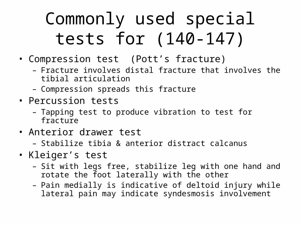
Commonly used special tests for (140-147)
• Compression test (Pott’s fracture)– Fracture involves distal fracture that involves the tibial
articulation– Compression spreads this fracture
• Percussion tests– Tapping test to produce vibration to test for fracture
• Anterior drawer test– Stabilize tibia & anterior distract calcanus
• Kleiger’s test– Sit with legs free, stabilize leg with one hand and rotate the foot
laterally with the other– Pain medially is indicative of deltoid injury while lateral pain may
indicate syndesmosis involvement

• Talar tilt test– Grasp talus and invert / evert
• Thompson Test– Achilles tendon rupture
• Tinel’s sign– Same as in other body regions
• Homan’s Sign– DVT; passive dorsiflexion of foot produces
pain in calf

Performing and interpreting flexibility tests (95)
• Passive ROM and measuring
ID Clinical S&S associated with.. (129-131)
• Achilles tendon rupture– Pain in distal calf– Positive Thompson test
• Compartment syndrome• Burning and tingling in foot• Weakness of lower extremity• Shinny appearance to skin• Cessation of distal pulses
• Apophysitis– Sever’s disease– Inflammation at tendon insertion; traction injury

ID Clinical S&S associated with foot / toe structure (132)
• Forefoot varus / valgus– See previous
• Equinus deformity
• Pes cavus/planus: high/low arch)
• Plantar flexed 1st ray
• Rearfoot (hindfoot) valgus/ varus

ID Clinical S&S associated with… (133-135)
• DVT– Homan’s Sign: pain in calf worsens with dorsiflexion– Look for predisposition – recent surgery, CV
problems, etc
• Neuroma– Mass on a nerve usually caused by pinching and
swelling; pain and radiating will occur along nerve’s path
• Osteochondritis dissecans

ID Clinical S&S associated with toe structure/alignment(136)
• Bunion (hallux valgus)• Claw toe
– Flexion of the distal interphalengeal joint• Hallux rigdus• Hallux valgus (bunion)
– Enlargement of bursa and lateral displacement of toe• Hammer toes
– Extension deformity of metatarsophalangeal join & flexion deformity of proximal interphalangeal joint of lesser toes
• Mallet toe• Morton’s Foot syndrome
– nerve becomes entrapped and swelling occurs b/t metatarsal heads (usually 3-4th)
– Sharp burning pain is presnet in 3rd web sapce and is worsen with activity, pain radiates into 3-4th toes and wering shoes aggrevates

Functional and activity specific test for the foot, ankle and lower leg (139)
• A, P, RROM
• Have the perform functional and activity / sport specific tests
• Ensure the athlete will no insure themselves further if they perform these tests

Instruct UBE use for upper body CR endurance (179)
• Make sure the seat/stool is appropriate height
• Have athlete assume correct posture• The chest pad should be comfortable and
level• Adjust arm length to appropriate for
person• Ensure the arm length is appropriate for
the person

Interrupt Isokinetic data to determine follow up care (198)
• Reading charts and graphs
• Usually need an equitable comparison (such as torque by body weight)
• Consult your isokinetic devices manufacturer for specific set ups

Perform isokinetic test for knee and shoulder (199)
• We will have to take a field trip – I am attempting to schedule … stay tuned