advances in laboratory testing for rheumatic diseases 2010 · advances in laboratory testing for...
TRANSCRIPT

1
Advances in Laboratory Testing for Rheumatic Diseases 2010
Jonathan Graf, M.D.Assistant Clinical Professor of MedicineUniversity of California, San Francisco
Division of RheumatologySan Francisco General Hospital
The black hole of medical knowledge: An internist’s view of rheumatologic lab tests
The ABIM’s view of rheumatologic lab testing
No Idea
Rh. Factor
ANA
ANCA
Typical ABIM Board Examination QuestionOn Rheumatology Lab Testing
Updates in Testing for Rheumatic Diseases
Chapter 1: Rheumatoid Arthritis: Recent advances in diagnostic and prognostic testing
Chapter 2: Systemic Lupus Erythematosis: Two steps forward and perhaps a step backwards

2
Rheumatoid Arthritis
• Chronic, inflammatory, predominantly small joint arthritis
• Affects up to 1% of the US population• Female:Male predominance of 3:1• Disability costs are high, both in terms of direct
and indirect medical costs– 35% of patients with 10 years disease duration are
work-disabled– Decline from 50% rate reported in 1987
Arthritis Rheum. 2008 Mar 27;59(4):474-480
RA: Chronic Joint Destruction and Disability – What We Try to Prevent
Improving Outcomes in RA: Three Pillars
• Improvement in timely and accurate diagnosis
• Development of better tools to predict disease severity
• Improvement in therapy
Improving Outcomes in RA: Three Pillars
• Improvement in timely and accurate diagnosis
• Development of better tools to predict disease severity
• Improvement in therapy

3
Early RA: The Window of Opportunity to Intervene
The Window of Opportunity Eventually Closes for Many….
• Chronic disease progression leads to permanent joint deformity, destruction, and disability
• Empirically, RA is a different disease the longer disease activity progresses without effective control– More difficult to suppress
activity and treat– More extra-articular
disease?
ACR Criteria for the Classification of Rheumatoid Arthritis
(>4 criteria required; 1-4 must be present > 6 wks)
• Morning stiffness > 1 hr• Arthritis of 3 or more joint areas• Arthritis of wrists, MCPs, and/or PIPs• Symmetric arthritis• Rheumatoid nodules• Serum rheumatoid factor• Radiographic changes
Limitations of ACR Classification Criteria for the diagnosis of early RA
• Developed for the classification of patients with longstanding disease (for clinical studies, not diagnosis)
• For early RA:– Specificity: 90%– Limited sensitivity: 40-65%
• Relying on criteria to make a diagnosis of RA can lead to delayed or inappropriate diagnosis

4
Diagnosis of early RA by ACR criteria van Gaalen et al Arth Rheum 50: 709, 2004
936 patients with early inflammatory arthritisInitial evaluation After 3 years
205 RA by ACR criteria
936 318 “undifferentiated 127 RAarthritis”
413 other diagnoses
Factors predictive of progression from undifferentiated arthritis to RA
van Gaalen et al Arth Rheum 50: 709, 2004
At initial evaluation OR (95% CI)
Positive rheumatoid factor 1.7 (0.5-5.6)
Positive anti-CCP antibody 38.6 (9.9-151.0)
Posttranslational modification of proteins:PADI converts arginine to citrulline RA-associated autoantibodies that
recognize peptides containing citrullineGirbal-Neuhauser et al J Immunol 162: 585, 1999
Peptide sequence Antibody recognition
ESSRDGSRHPRSHD No
PADI
ESSRDGScitHPRSHD Yes
Actual citrullinated antigen is not known

5
Antibodies to citrullinated peptides in RA
• Detected by ELISAs using synthetic cyclic citrullinated peptides (CCP)
• Sensitivity for very early RA: 50%
• Sensitivity for early-later RA: 70-80%• Specificity for RA: 95-98%
RF and anti-CCP testing in a cohort of 182 early RA patients
Quinn et al Rheumatology (Oxford) 45:478, 2006
RF-CCP+
RF+CCP+
RF+CCP-
RF-CCP-
Preclinical autoimmunity in RA:appearance of anti-CCP abs and
RF prior to onset of arthritis
Nielen et al Arth Rheum 50: 380, 2004
Improving Outcomes in RA: Three Pillars
• Improvement in timely and accurate diagnosis
• Development of better tools to predict disease severity– Possible to predict which patients require
more aggressive therapy up front?
• Improvement in therapy

6
Classic Predictors of Disease Severity
• More difficult to treat, destructive, extra-articular disease:– Rheumatoid factor positive– Erosive disease– Genetic factors: HLA DR4 withRR of 6:1
• Turns out that some DR4 alleles do not confer risk• And some non-DR4 alleles do!• There is something that most DR risk alleles share
in common!
HLA DR alleles: Shared Epitope Hypothesis for RA
amino acid position on the DRβ chainDRB1 allele 70 71 72 73 740101 Q R R A A
0401 Q K R A A0404 Q R R A A0405 Q R R A A0408 Q R R A A1402 Q R R A A
1001 R R R A ACONSENSUS Q/R R/K R A A
The shared epitope (DRB1*0401)
A74 Q70
A73 R72
HLA DR and rheumatoid arthritis: 25 years without clinical utility
• It’s actually this shared epitope that confers the risk of developing RA
• Homozygotes for SE (can mix and match) have more severe disease than do heterozygotes or those without DR4
• Still not practical to genotype all patients
HLA DR (MHC class II): Antigen presentation by APC’s to T-cells

7
Relationship between shared epitope and CCP antibodies in RA
Van Gaalen et al. Arthritis and Rheumatism 2004:50;7:2113-2121
Suggests that homozygotes for SE get CCP+ RA, those without SE get CCP- RA
Is anti-CCP+ RA a different genetic disease than anti-CCP neg RA??Huizinga, Criswell et al. Arthritis and Rheumatism 2005:52;11:3433-3438
42% 80% 49%At least onecopy of SE
In terms of SE genetics, patients with anti-CCP neg RA look like controls
Van Gaalen et al. Arthritis and Rheumatism 2004:50;7:2113-2121
Huizinga, Criswell et al. Arthritis and Rheumatism 2005:52;11:3433-3438
Presence of CCP antibodies, not SE, confers risk for more aggressive RA
Progression of joint damage in subgroups of early RA
Huizinga et al Arthritis Research& Therapy 7: 949, 2005
radiographicjoint damage
score
anti-CCP+
anti-CCP-
8
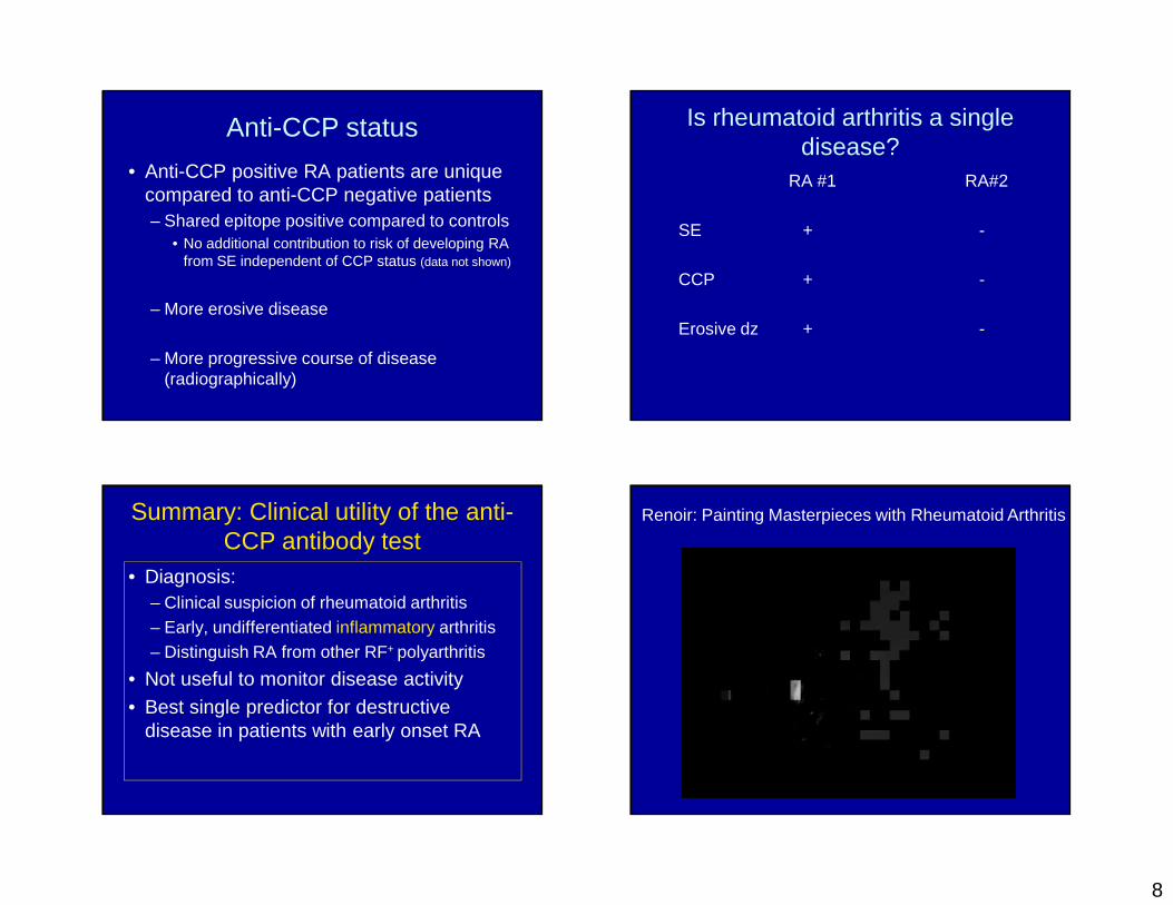
Anti-CCP status
• Anti-CCP positive RA patients are unique compared to anti-CCP negative patients– Shared epitope positive compared to controls
• No additional contribution to risk of developing RA from SE independent of CCP status (data not shown)
– More erosive disease
– More progressive course of disease (radiographically)
Is rheumatoid arthritis a single disease?
RA #1 RA#2
SE + -
CCP + -
Erosive dz + -
Summary: Clinical utility of the anti-CCP antibody test
• Diagnosis:– Clinical suspicion of rheumatoid arthritis– Early, undifferentiated inflammatory arthritis– Distinguish RA from other RF+ polyarthritis
• Not useful to monitor disease activity• Best single predictor for destructive
disease in patients with early onset RA
Renoir: Painting Masterpieces with Rheumatoid Arthritis

9
Sorting out the ANA Puzzle So that ANAs make sense
Advances in Laboratory Diagnoses of Rheumatic Diseases
• Anti Citrullinated Peptide Antibodies in the diagnosis, prognosis, and management of rheumatoid arthritis
• Automated autoantibody screening
Automation: Coming to a lab near you
• Allows for high throughput testing– Many patient samples– Many tests per sample
• Cheaper• Less prone to human
“interpretation”; standardized• Actually been around for a
while– Automated Chem panels– CBC’s
• Increasing use of lab automation for more “complex tests”– Serologies– Anti-nuclear antiboies

10
N
M
G
RER
What’s in Ourselves??What’s in a Cell?
PM
Cell membranePhospholipids
CytoplasmMitochondria
NucleusNucleic AcidsHistones
What is an Anti-Nuclear Antibody?
• Autoantibodies directed specifically against intra-nuclear antigens
• Most commonly (not always) detected by the technique of immunofluorecence on whole cells
• Specific antigen may or may not be known (most ANA’s aren’t known – only seen by staining)
• Smaller handful of specific antibodies target known nuclear antigens associated with rheumatic and other diseases
What is Indirect Immonoflorescence??
• Hep-2 cells fixed to slide & permeabolized
• Incubated in patient serum
• Washed vigorously to remove serum
• Fluorescently labeled Anti-hum Ig secondary Ab
• Wash again• Detect florescence of
bound secondary Ab
ANA Patterns
Depends upon what molecule(s) are recognized by patient antibodies– DNA is
homogeneously distributed
– Centromere’s seen in dividing cells
– Extractable nuclear antigens are speckled throughout cell

11
What does an indirect immunofluoresence look like??
ANA+ with Homogeneous StainingLabor intensive and subject to interpretation
ANA Patterns: Speckled
ANA Patterns: Nucleolar ANAs by immunoflurescence• Co$$$tly
• Low throughput– One test on one sample per patient– Time consuming
• Labor intensive– Someone has to prepare the test, titer out serum samples, etc…– Someone has to read the slide and make a judgment– Subject to human interpretation (what’s positive/what’s background?)
• Hep-2 cell lines contain antigens of unknown significance that lower specificity of test
• When ANA IIF is positive, actual autoantibody target is not known– Pattern may be suggestive, but not diagnostic– Is it something important like anti-Smith or is it a non-specific antigen?

12
The ANA IIF Test CharacteristicsSensitive for SLE, Not Specific
• Modern Hep-2 ANA IIF is nearly 99% sensitive for SLE (90% sensitive for scleroderma, etc.)
• ANA IIF is not nearly as specific for SLE as it is sensitive– Autoimmune thyroid disease– Other Collagen-Vascular diseases (>90% of SSc)– Medications– Malignancies– Infections (viral)– Normal people (especially low titers)
www.PeaceHealthLabs.org January 2010
True
True
TrueA Reference Lab’s Rationale forAutomated testing
Examples of Automated ANA’sCommercially Available Automated Solid-Phase ANA Testing Platforms
• Varelisa ANA CTD Screen• Quanta Lite ANA Screen (Inova Diagnostics,
USA)• Bioplex 2200 ANA Screen (Emeryville, CA)• AtheNA Multi-Lyte ANA Test System (Zeus
Scientific)• COBAS® Core HEp2 ANA EIA (Roche)• And the list goes on and on…. Startup anyone?

13
Multiplex Testing: Technique
• Very tiny color-coded beads are coated with a specific antigen, such that the bead’s color and antigen are linked together
• Several thousand of each specific antigen-coated bead are combined into a single reagent, which can be used for detecting multiple antibodies from one patient sample
• A second color-labeled Anti-human IgG is used to detect autoantibodies that bind to the beads
• The beads flow through a laser detector that detects 2 colors: that of the specific bead and that of any bound antibodies
A Crude Look at Automated ANAs
Smith dsDNA
Anti-SM +
Anti dsDNA+
Automated MultiPlex ANA Assays
• Can tell you with good specificity if ANA+– Laser beam and detector vs. human eye
• Can tell the specific antigen target of the anti-nuclear antibody(ies)
• Can tell you multiple antibody specificities with one sample (based on numbers of different antigen coated beads)
• Can run multiple different patient samples rapidly
14
Automated ANA Testing
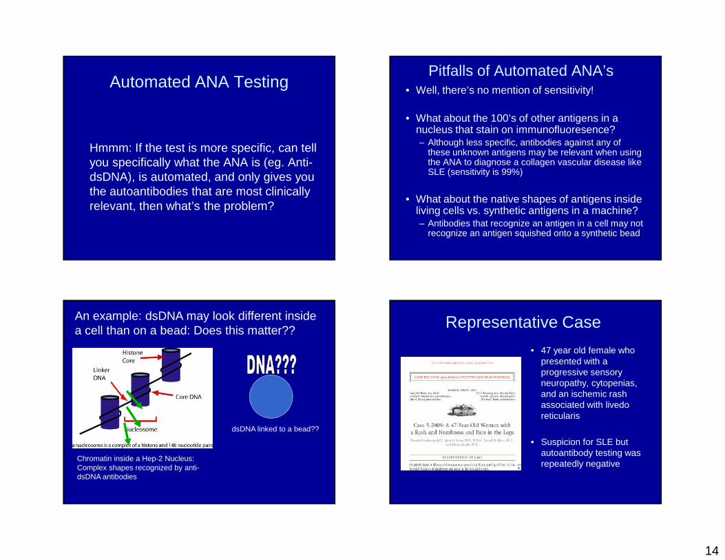
Hmmm: If the test is more specific, can tell you specifically what the ANA is (eg. Anti-dsDNA), is automated, and only gives you the autoantibodies that are most clinically relevant, then what’s the problem?
Pitfalls of Automated ANA’s• Well, there’s no mention of sensitivity!
• What about the 100’s of other antigens in a nucleus that stain on immunofluoresence?– Although less specific, antibodies against any of
these unknown antigens may be relevant when using the ANA to diagnose a collagen vascular disease like SLE (sensitivity is 99%)
• What about the native shapes of antigens inside living cells vs. synthetic antigens in a machine? – Antibodies that recognize an antigen in a cell may not
recognize an antigen squished onto a synthetic bead
An example: dsDNA may look different inside a cell than on a bead: Does this matter??
Chromatin inside a Hep-2 Nucleus: Complex shapes recognized by anti-dsDNA antibodies
dsDNA linked to a bead??
Representative Case
• 47 year old female who presented with a progressive sensory neuropathy, cytopenias, and an ischemic rash associated with livedo reticularis
• Suspicion for SLE but autoantibody testing was repeatedly negative

15
Clinical Case from Massachusetts General HospitalFrom Krashinsky, Stone, et al. New Engl J Med 2009; 360:711-720 NEJM Case 2009
Key points:1. ANA’s detected by repeat with IIF
– HIGH TITER– Likely not new seroconversion– Likely speckled antigen not represented in automated ANA
2. Andti-dsDNA (which had been negative in the automated assay) was detected using a different method (antigen preparation matters!!!)
Automated ANAs lack Sensitivity

16
“The main problem (among many others) was that multiple autoantibodies can be present in the sera of SLE patients that are not necessarily detected by the antigens that coat the beads. So many patients with documented SLE tested as ANA negative, and the diagnosis of SLE was missed among so many hospital admissions, that all 32 rheumatologists in our division signed a petition asking the Department of Clinical Pathology to reconsider the use of this new test.”
ACR: Position Statement 2/2009
• The ANA by immunofluorescence is the gold standard
• Newer automated ANA’s that test for handfulls of antigens may miss 100’s of potential autoantigens screened by IIF
• Laboratories should indicate which method they use
What should you do??
• Be aware of the method your lab uses– This can be very tricky as different insurance
companies use specific labs– Most of the major reference labs use automated
ANA testing now unless you ask!
• Know the patient whom you’re testing– What is the pre-test likelihood they have a
rheumatic disease like SLE??– If there remains a high pre-test likelihood of
disease, and automated ANA testing is negative, then re-test the patient with an IIF ANA