advanced applications of fibtem- in geosciences - fib … · fib/tem applications fib systems...
TRANSCRIPT

Advanced applications of FIB-TEM in Geosciences
Richard WirthHelmholtz Centre Potsdam
GFZ German Research Centre for Geosciences

FIB/TEM ApplicationsFIB systems operated at GFZ Potsdam
FEI FIB 200 TEM
FEI Quanta 3Dimage courtesy Anja Schreiber, GFZ, Potsdam

FIB/TEM Applications
• Applications in Geosciences– Nano-Inclusions in minerals (diamond)– Microstructure, Porosity– 3D imaging
• Applications in Geobiology– Monorhaphis Chuni– Precambrian acritarchs
• DAC experiments– Machining diamonds – Recovering samples from DAC experiments and TEM investigation
• Sample preparation– Synchrotron IR– Synchrotron XRF STXM (Scanning transmission X-ray microscopy) – NanoSIMS– FEG-microprobe

Application of FIB/TEM
Nano-diamonds in melt inclusions in OPX and CPX in Hawaiian Lava
SE image TEM foil, TEM bright field image
Opx
Wirth, R.; Rocholl, A. (2003): Nanocrystalline diamond from the Earth's mantle underneath Hawaii. Earth and Planetary Science Letters, 211, 3-4, 357-369

Application of FIB/TEM
Nano-diamonds in melt inclusions in OPX and CPX in Hawaiian Lava
Aggregates of nanocrystalline diamond (d)Dark field image of an aggregateof nanocrystalline diamond
Dark field STEM image
Wirth, R.; Rocholl, A. (2003): Nanocrystalline diamond from the Earth's mantle underneath Hawaii.
Earth and Planetary Science Letters, 211, 3-4, 357-369
Diamond
Olivine
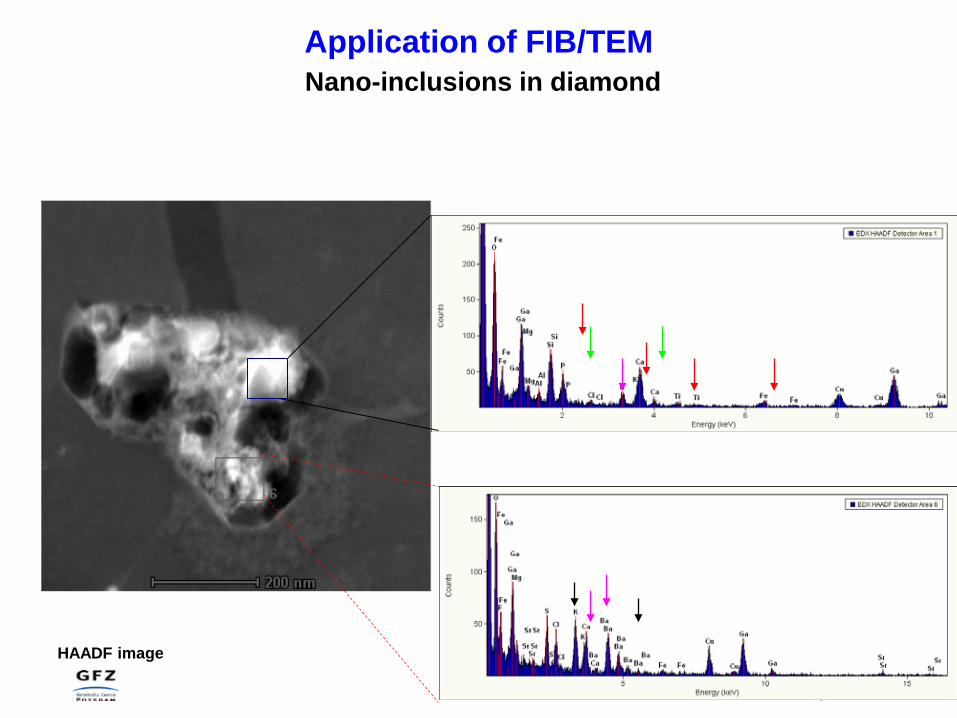
Application of FIB/TEMNano-inclusions in diamond
HAADF image

Diamond
Olivine
Nano-inclusions in diamond
A.M., Logvinova, R., Wirth, E.N., Federova, N.V., Sobolev., 2008, EUR. J. Mineral., 20, 317-331
Nano-inclusions in diamond are composed of:
•Carbonates (Ca, Mg, Fe, Ba, Sr) •Silicates (phlogopite, high silica mica)•Halides (NaCl, KCl)•Phosphate (F-Apatite)•Ti-phase (Ilmenite, rutile)•Sulphide (Fe-Cu-Ni)•Quench phase (Si, Ba, Sr, F)•Fluid
Inclusions represent High density fluid (HDF) enriched in Cl, K, P, Ba, Si, Sr + water + carbonate,the medium diamond has grown from.Klein BenDavid, Wirth, Navon, 2006, Amer. Min., 91, 353-365
New insights in diamond genesis
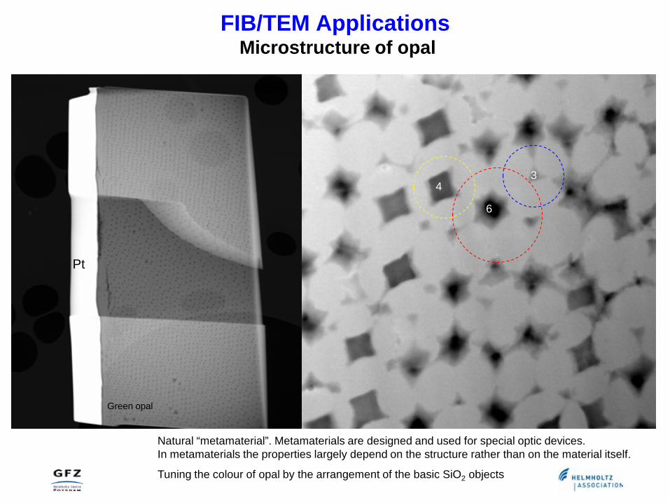
FIB/TEM ApplicationsMicrostructure of opal
Apatite
Thorium Silicate
Apatit
Thorium Silicateat
Apatite
Tuning the colour of opal by the arrangement of the basic SiO2 objects
Green opal
4
6
3
Pt
Natural “metamaterial”. Metamaterials are designed and used for special optic devices.In metamaterials the properties largely depend on the structure rather than on the material itself.

FIB/TEM ApplicationsPorosity
Apatite
Thorium Silicate
Apatit
Apatite
4
6
3
15 µm
Pt
500 nm
Original porosity is not changed by FIB sputtering!!
Ultracataclastic core samples recovered from 3194 m and 3294 m depth of the main bore hole of the
San Andreas Fault Observatory at Depth.
Janssen, C.; Wirth, R.; Rybacki, E.; Naumann, R.; Kemnitz, H.; Wenk, H.-R.; Dresen, G. (2010): Amorphous material in SAFOD core samples (San Andreas Fault): Evidence for crush-origin pseudotachylytes?. Geophysical Research Letters, 37, L01303
pores

FIB/SEM Dual Beam Cross-Sectioning

Symplectite Microstructure 3D imagingRainer Abart, University Vienna, Austria
3D reconstruction from successive slices, 100 slices at 100 nm intervals, each slize is imaged using BSE signal on an FEI Quanta 3D FEG-SEM-FIB
montmerw fo
Ca3MgSi2O8 Mg2SiO4at 900°C, 1.2 GPa

FIB/TEM ApplicationsGeobiology
Deep sea sponge monorhaphis chuni
Cross section through the fibre Sponge at the sea floor
M. Roux Univ. Reims

FIB/TEM ApplicationsGeobiology
Deep sea sponge monorhaphis chuni
In situ etching of a cross section of the fibre with FIB using XeF2
Not etched etched

FIB/TEM ApplicationsGeobiology
Deep sea sponge monorhaphis chuni
SE image of fibre cross section
TEM bright field image of FIB cut foil
C - element map (EELS) of
organic material“Natural fibre composite”
Amorphous silica

(Blue algae)
FIB/TEM ApplicationsGeobiology
Kempe, A.; Wirth, R.; Altermann, W.; Stark, R. W.; Schopf, J. W.; Heckl, W. M. (2005): Focussed ion beam preparation and in situ nanoscopic study of Precambrian acritarchs. Precambrian Research, 140, 1-2, 36-54.
Fossil Acritarch cell walls from 650 my old Chichkan Formation, Kazakhstan
Ancient cell wall
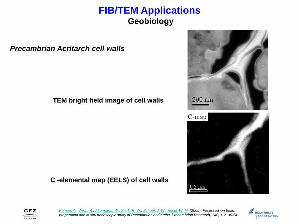
Precambrian Acritarch cell walls
TEM bright field image of cell walls
C -elemental map (EELS) of cell walls
FIB/TEM ApplicationsGeobiology
Kempe, A.; Wirth, R.; Altermann, W.; Stark, R. W.; Schopf, J. W.; Heckl, W. M. (2005): Focussed ion beam preparation and in situ nanoscopic study of Precambrian acritarchs. Precambrian Research, 140, 1-2, 36-54.

FIB/TEM ApplicationsMicromachining of diamond for DAC application
diamond
Wirth, R. (2004): A novel technology for advanced application of micro- and nanoanalysis in geosciences and applied mineralogy. European Journal of Mineralogy, 16, 6, 863-876

FIB/TEM ApplicationsMicromachining of diamond for DAC application
Synchrotron radiation
diamond
X-ray fluorescence
FIB milled section

FIB/TEM ApplicationsSample recovery from DAC experiments
sample
Rhenium gasket
0.2 mmsample
gasket

FIB/TEM ApplicationsSample recovery from DAC experiments
sample
Rhenium gasket
0.2 mmsample
gasketsample

FIB/TEM ApplicationsSample recovery from DAC experiments
sample
Rhenium gasket
0.2 mm

FIB/TEM ApplicationsTEM investigation of the foil
sample
Rhenium gasket
0.2 mm
Amorphous carbonate
diamond
Pt
Experiment: Sergio Speziale, GFZ Potsdam
Reacting Fe - MgO - CaCO3 at Lower Mantle conditions

FIB/TEM ApplicationsSample preparation : synchrotron IR spectroscopy
Zinco-Staurolite from Samos
Synchrotron IR-Measurement
FIB cut foil
30 x 20 x 5 micron

FIB/TEM ApplicationsSample preparation: Nano SIMS
diamond
carbonate
TEM foil on grid, optical micrograph
TEM foil TEM bright field image

FIB/TEM ApplicationsSample preparation: Nano SIMS
diamond
Y. Sano, D. Pinti, Center for Advanced Marine Res., Ocean Research Institute, Tokyo, Japan

FIB/TEM ApplicationsTEM and Nano SIMS
diamond
L. F. Dobrzhinetskayaa,, Ri. Wirth, J.Yang, I. D. Hutcheon, P. K. Weber, and H.W. Green II (2009)High-pressure highly reduced nitrides and oxides from chromitite of a Tibetan ophiolite. Proceedings of the National Academy of Sciences of the United States of America (PNAS), 106, 46, 19233-19238.
Coesite bearing chromitite from Luobasa, Tibet
TiN
High-pressure highly reduced nitrides and oxides from chromitite of a Tibetan ophiolite
TEM HAADF image

FIB/TEM ApplicationsSample preparation: STXM, EXAFS
diamond
Scanning transmission X-Ray microscopy STXM of the foil imaged at 500 eV
Daniel Fliegel, Richard Wirth, Antonio Simonetti, Anja Schreiber3, Harald Furnes and Karlis Muehlenbachs (2010)
Tubular textures in pillow lavas from a Caledonian West-Norwegian Ophiolite a: combined TEM, LA-ICP-MS and STXM study . G3 in press.

STXM image at the C-K edge
XANES spectra at the C-K edge
Bernard, S.; Horsfield, B.; Schulz, H.-M.; Schreiber, A.; Wirth, R.; Vu, T. T.; Perssen, F.; Kitzer, S.; Volk, H.; Sherwood, N.; Fuentes, D. (2010): Multi-scale detection of organic and inorganic signatures provides insights into gas shale properties and evolution.. Chemie der Erde - Geochemistry,70, Suppl. 3, 119-133.
FIB/TEM ApplicationsSample preparation: STXM, XANES
XANES images

FIB/TEM ApplicationsTEM element mapping after using the foil for STXM and XANES
Bernard, S.; Horsfield, B.; Schulz, H.-M.; Schreiber, A.; Wirth, R.; Vu, T. T.; Perssen, F.; Kitzer, S.; Volk, H.; Sherwood, N.; Fuentes, D. (2010): Multi-scale detection of organic and inorganic signatures provides insights into gas shale properties and evolution.. Chemie der Erde - Geochemistry,70, Suppl. 3, 119-133.

FIB/TEM Applicationssample preparation: Brillouin Spectroscopy
Flat and parallel surfaces required
S. Speziale1, H. Marquardt1, R. Wirth1, A. Schreiber1, K. Marquardt1, G. Neusser2, H.J. Reichmann1
(2010), AGU Fall Meeting, San Francisco

FIB/TEM Applicationssample preparation: FEG microprobe analysis
Interaction sample - electron beam (thin sample)
Incident electron beam
X-ray photonsX-ray photons
Excited volume
Sample surface
TEM foil (150 nm thick)
FEG microprobe analysis with thin TEM foils at reduced acceleration voltage (< 10 keV) improves the spatial resolution of the analysis << 100 nm!
