adjuvant liposomes containing lipid a: enhancementof ... · negative liposomes were identified as...
TRANSCRIPT
INFEcTION AND IMMUNITY, June 1992, p. 2438-2444 Vol. 60, No. 60019-9567/92/062438-07$02.00/0Copyright © 1992, American Society for Microbiology
Adjuvant Effects of Liposomes Containing Lipid A: Enhancement ofLiposomal Antigen Presentation and Recruitment of MacrophagesJITENDRA N. VERMA,' MANGALA RAO,1,2 SHIMON AMSELEM,' URSZULA KRZYCH,2 CARL R. ALVING,'
SHAWN J. GREEN,3 AND NABILA M. WASSEFl*Departments ofMembrane Biochemistry, 1 Immunology, 2 and Cellular Immunology, 3 Walter Reed Army
Institute of Research, Washington, D. C. 20307-5100
Received 12 December 1991/Accepted 27 March 1992
Liposomes containing lipid A induced potent humoral immune responses in mice against an encapsulatedmalaria antigen (R32NS1) containing NANP epitopes. The immune response was not enhanced by lipid A aloneor by empty liposomes containing lipid A. Experiments to investigate the adjuvant mechanisms of liposomesand lipid A revealed that liposome-encapsulated R32NS1 was actively presented by bone marrow-derivedmacrophages to NANP-specific cloned T cells. The degree of presentation was related to the amount ofliposomal antigen added per macrophage in the culture medium. At high cell densities, poor presentationoccurred when liposomes lacked lipid A but excellent presentation occurred when the liposomes contained lipidA. Liposomes containing lipid A and encapsulated antigen also activated gamma interferon-treated macro-phages to produce nitric oxide. Macrophage activation and antigen presentation occurred with liposomes thatcould not be detected by the Limulus amebocyte lysis assay. Intraperitoneal injection of liposomal lipid Acaused a marked increase in the recruitment of immature (peroxidase-positive) macrophages to the perito-neum. On the basis of these experiments, we propose that the mechanism of the adjuvant action of liposomallipid A is partly due to increased antigen presentation by macrophages and partly due to recruitment of anincreased number of macrophages serving as antigen-presenting cells.
In recent years, there has been a growing interest in theuse of liposomes as carriers of antigens (Ag) for immuniza-tion (1, 2, 17, 28). Liposomes are delivered avidly andefficiently to macrophages, and because of this, it has beenpresumed that macrophages could serve as Ag-presentingcells for liposomal Ag (1, 28). Support for this hypothesis hasbeen provided by in vitro model systems that have demon-strated that liposomal Ag are immunologically presented toT lymphocytes by cultured macrophages (9, 18).One of the theoretical advantages of liposomes is that they
can carry adjuvants in addition to Ag, and one adjuvant thathas been successfully employed is lipid A (LA), the endo-toxic moiety of gram-negative bacterial lipopolysaccharide(LPS) (2, 3, 4, 22, 23). Although liposomal LA has beenextensively utilized as an adjuvant, the mechanism of itsadjuvant activity is not yet fully understood. Liposomal LAcan be a potent mitogen for stimulation of murine B lympho-cytes (5), and this might contribute to its adjuvant activity inmice. It has also been reported elsewhere that liposomalLPS activates macrophages to develop tumoricidal activity(13, 14). However, the macrophage-activating properties ofliposomal LPS are controversial, and it has also been re-ported that when compared with LPS or LA alone, liposo-mal LPS or liposomal LA has a markedly diminished abilityto activate macrophages to induce tumoricidal activity (8,11) and also has a diminished capacity to cause secretion ofinterleukin-1 by macrophages (12).
In the present study, we investigated possible cellularmechanisms underlying the ability of liposomal LA to stim-ulate enhanced humoral immunity to a poorly immunogenicAg in mice. In vitro experiments demonstrated that liposo-mal LA caused marked enhancement of immunologicalpresentation of liposomal R32NS1 by cultured bone marrow-
* Corresponding author.
derived macrophages. We also found that intraperitonealinjection of liposomal LA resulted in vigorous recruitment ofperitoneal inflammatory macrophages.
MATERUILS AND METHODS
Lipids. Lipids were purchased from the following sources:dimyristoyl phosphatidylcholine and dimyristoyl phosphati-dylglycerol, Avanti Polar Lipids, Inc., Alabaster, Ala.;cholesterol, Calbiochem-Behring, La Jolla, Calif.; and LA(isolated from Salmonella minnesota R595), List BiologicalLaboratories, Campbell, Calif.Ag and antibodies. The Ag used in this study, R32NS1 and
R32LR, were kindly supplied by SmithKline Beecham Phar-maceuticals, Swedeland, Pa. R32 consists of 30 repeats ofthe tetrapeptide NANP interspersed with two tetrapeptideNVDP repeats from the immunodominant repeat region ofthe circumsporozoite (CS) protein of Plasmodium falci-parum. In the case of R32NS1, R32 is linked to 81 aminoacids from the nonstructural protein of influenza virus (NS1)(25). In the case of R32LR, R32 is linked to the first twoamino acids, leucine and arginine, from a nonsense readingof the tetracycline resistance gene of the vector (19). Amonoclonal antibody against CS protein, Pf 1B2.2 (31), wasthe primary antibody used in the enzyme-linked immunosor-bent assay (ELISA) described below.
Preparation of liposomes. Detailed procedures for prepa-ration of liposomes were described previously (6, 23, 24, 29).Liposomes were composed of dimyristoyl phosphatidylcho-line, dimyristoyl phosphatidylglycerol, cholesterol, and LAin molar ratios of 1.8:0.2:1.5:0.04 (Limulus positive) or1.8:0.2:1.5:0.005 (Limulus negative), in which the molarityof LA is expressed as LA phosphate. Limulus-positive andLimulus-negative liposomes either caused coagulation orfailed to cause coagulation, respectively, in the Limulusamebocyte lysate (LAL) assay, as described previously (24).
2438
on May 21, 2020 by guest
http://iai.asm.org/
Dow
nloaded from
EFFECTS OF LIPOSOMAL LIPID A ON ANTIGEN PRESENTATION 2439
LA contains approximately 1.1 ,g of LA per nmol of LAphosphate. Except in the experiments in which Limulus-negative liposomes were identified as having been used, allexperiments utilized Limulus-positive liposomes. For thepurpose of following the uptake of liposomes by macro-phages, a trace amount of cholesterol ['4C]oleate (58 mCi/mmol; E. I. du Pont de Nemours, NEN Research Products,Boston, Mass.) was included in the lipid bilayer as a nonex-changeable lipid marker.
Multilamellar liposomes were prepared by dispersion oflyophilized mixtures of lipids in Dulbecco's phosphate-buffered saline (PBS; GIBCO, Grand Island, N.Y.) contain-ing or lacking R32NS1, such that the liposomal phospholipidconcentration was 10 mM with respect to the buffer. Theliposomes were washed twice with 0.15 M NaCl at 27,000 xg for 10 min at 20°C to remove unencapsulated Ag. Theresulting liposomes were suspended in 0.15 M NaCl to givea final phospholipid concentration of 10 mM and stored at4°C until used. Liposome-encapsulated malaria Ag wasestimated to be approximately 10 ng/nmol of liposomalphospholipid.
Phagocytosis of liposomes by bone marrow-derived macro-phages. Macrophages derived from the bone marrow ofC3H/HeN mice (Frederick Cancer Research Facility, Fred-erick, Md.) were cultured in 24-well culture plates (Costar,Cambridge, Mass.) for 6 days, as described previously (7,29). Phagocytosis of liposomes by macrophages was deter-mined as previously described (30). Briefly, macrophagecultures were washed three times with RPMI 1640 mediumand incubated with liposome-encapsulated Ag (0.6 ,ug ofR32NS1 and/or 50 nmol of liposomal phospholipid per ml ofRPMI 1640 per 106 cells) at 37°C with 5% CO2 and 95%relative humidity for the indicated time periods. At the endof the incubation, the cultures were washed three times with1 ml of PBS to remove noningested Ag, covered with 1 ml ofPBS, and placed on ice. Cells were harvested by repeatedwashing and scraping of culture wells with ice-chilled PBS.The amounts of liposomal Ag phagocytosed by macrophageswere estimated by measuring uptake of cholesterol [1-14C]oleate-labelled liposomes. When metabolic inhibitors wereused to distinguish between phagocytosis and nonspecificbinding, the cultures were preincubated for 10 min withantimycin A and NaF, inhibitors of mitochondrial respira-tion and glycolysis, respectively. During subsequent incuba-tion of the macrophage cultures with liposomes, the inhibi-tors were present in the medium.
Immunizations. C3H/HeN mice were used for the immu-nization protocols. Five mice per group were injected witheach immunogen. Animals were injected intraperitoneallywith 7 ,g per dose of free Ag, free LA, liposomal Ag[L(Ag)], liposomes containing Ag and LA [L(Ag + LA)], orvarious combinations of these. Free LA was dissolved in0.5% triethylamine, diluted in 0.154 M NaCl, and injected atdoses similar to those of liposomal LA. Control groupsconsisted of animals immunized by direct injection of Aginto the peritoneal cavity. Blood was collected by tail veinbleeding on days 5, 10, and 15 following immunization. Serawere stored at -70°C until used.ELISA. Solid-phase ELISAs were performed to measure
the levels of immunoglobulin G (IgG) antibody against acapture Ag (R32LR) containing the same repeating units asthe Ag that was used for immunization (29). Assays wereperformed at room temperature (27°C) in 96-well, U-bottomImmulon-2 polystyrene plates (Dynatech Laboratories, Al-exandria, Va.). Ag (R32LR) in a solution of PBS containing
0.0004% boiled casein was added to the wells to give aconcentration of 10 ng of Ag per well in a volume of 50 ,ul.
Blocking buffer was prepared by dissolving 5 g of bovinetechnical grade milk casein in 100 ml of 0.1 M NaOH byboiling. The volume was brought to 1 liter with PBS after thecasein was dissolved, the pH was adjusted to 7.4 by usingHCI, and then 0.1 g of thimerosal and 0.2 g of phenol redwere added. All constituents of the blocking buffer werepurchased from Sigma Chemical Co., St. Louis, Mo.
Control wells used for creating a standard curve containedR32LR Ag in concentrations ranging from 0.15 to 10 ng, andan anti-CS protein monoclonal antibody was used as theprimary antibody. The plates were covered and storedovernight. The wells were emptied by aspiration, filled withblocking buffer, and allowed to stand for 2 h. Blocking bufferwas removed and, except for control wells which receivedblocking buffer alone, 50 .1l of mouse sera diluted 1:50 inblocking buffer was added to each well. The primary anti-body for standard curve was anti-CS protein Pf 1B2.2 (31),which was added in a concentration of 100 ng/50 ,ul ofblocking buffer per well. The plates were covered andincubated for 2 h. The well contents were aspirated, and thewells were washed three times with PBS containing 0.05%Tween 20 (J. T. Baker, Phillipsburg, N.J.). Fifty p,l ofperoxidase-conjugated goat anti-mouse IgG (heavy plus lightchains) (Bio-Rad, Richmond, Calif.) diluted 1:500 in block-ing buffer was added to each well and incubated for 1 h. Thewells were washed three times with PBS containing 0.05%Tween 20. ABTS peroxidase substrate (Kirkegaard & PerryLaboratories, Inc., Gaithersburg, Md.) was added, and A405was determined after 1 h with a Vmax Kinetic MicroplateReader (Molecular Devices, Palo Alto, Calif.).Ag presentation assay. Bone marrow-derived macrophages
were used as Ag-presenting cells in T-cell proliferativeassays. Marrows from femurs of 8- to 10-week-old C57BL/10(H-2") mice (Jackson Laboratory, Bar Harbor, Maine) wereisolated and cultured for 10 days in 96-well Costar 3590plates in macrophage growth medium (RPMI 1640 containing10% fetal bovine serum, L-glutamine [8 mM], penicillin [100U/ml], streptomycin [100 p.g/ml], and 10% L-929 cell-condi-tioned medium [7, 29]). Macrophages were seeded at densi-ties ranging from 5 x 10⁴ to 1 x 10 cells per well. On day 9,macrophage cultures were supplemented with 5 U of murinegamma interferon (IFN-,y) per ml (a gift from Carol Nacy,Walter Reed Army Institute of Research). Macrophagegrowth medium supplemented with fetal bovine serum wasreplaced with RPMI 1640 medium before Ag addition. Dif-ferent concentrations of liposomal Ag in a volume of 200 p,lof RPMI 1640 were added to the macrophage cultures asindicated. The cultures were incubated for 90 min, whichwas found to be the optimum length of time in otherexperiments (data not shown), at 37°C, with 5% CO2 and95% relative humidity. At the end of the incubation period,noningested Ag were removed by aspirating the medium,cells were washed three times with 200 ,ul of RPMI 1640maintained at 37°C, and 200 p,l of the same medium per wellwas added to the cultures. Macrophage cultures were ex-posed to 3,000 R irradiation from a cobalt source (Gamma-cell 220, cobalt 60 irradiator; Atomic Energy of CanadaLtd.). The media were aspirated from the wells and replacedwith 100 ,ul of T-cell growth medium (RPMI 1640 containingEHAA [1:1 ratio; Whittaker M. A. Bioproducts, Walkers-ville, Md.], 100 U of penicillin per ml, 100 pg of streptomy-cin per ml, 8 mM glutamine, 0.1 mM nonessential aminoacids, 1 mM sodium pyruvate, 100 p.M 2-mercaptoethanol,and 5% fetal bovine serum [HyClone, Logan, Utah]).
VOL. 60, 1992
on May 21, 2020 by guest
http://iai.asm.org/
Dow
nloaded from
2440 VERMA ET AL.
An (NANP)40-specific T-cell clone (a gift from G. Corra-din, University of Lausanne, Switzerland) was maintained inour laboratory as described previously (27). At 14 to 21 daysafter restimulation with the Ag in the presence of syngeneicsplenic cells, viable T cells were collected after removal ofdead cells by centrifugation over Ficoll-Metrizoate and wereadjusted to 105 cells per ml in T-cell growth medium. A100-,ul volume of the T-cell clone (104 cells) in completeRPMI 1640 was added to each well containing Ag-pulsedmacrophages. The cultures were maintained at 37°C in ahumidified 5% CO2 atmosphere for 72 h. During the last 16 hof incubation, 1 ,uCi of [3H]thymidine (specific activity, 6.7Ci/mM [NEN]) was added to each well. At the end of theculture period, cells were harvested and processed forscintillation spectroscopy (LKB betaplate).
Activation of macrophages to produce nitric oxide. Nitricoxide produced by macrophages treated with murine INF--yand liposomal LA was measured by a modification of previ-ously described methods (16). Cultured peritoneal macro-phages from C57BL/6 mice (1.2 x 10⁵ cells per well) wereincubated for 2 h with Dulbecco's modified Eagle medium(DMEM) containing 5% fetal calf serum (FCS) (16). Thecells were washed and incubated for 90 min with 100 [lI ofDMEM medium containing 5% FCS, 10 units of IFN-y perml, and either R32NS1 Limulus-negative or R32NS1 Limu-lus-positive liposomes. The cells were washed twice andincubated with DMEM containing 5% FCS. After 72 h, 50 ,ulof the medium was removed and assayed for nitric oxide bythe Griess reaction (15). Briefly, 50 RI of the medium wasincubated with 50 pAl of 1% sulfanilamide and 50 ,ul of 1%N-1-naphthylethylenediamine dihydrochloride in 2.5%H3PO4 (Sigma Chemical Co.) at room temperature for 5 min.Optical density was measured at 543 nm, and nitrite wasquantified by comparison with Na(NO)2 as standard.
Peritoneal cell collection. Seven-week-old C3H/HeN malemice were injected intraperitoneally with 0.1-ml volumes of(i) sterile PBS, (ii) 100 p,g of liposome-encapsulated R32NS1malaria Ag [L(Ag)], or (iii) 100 p,g of liposome-encapsulatedR32NS1 in which the liposomes also contained 20 jig of LAas an adjuvant [L(Ag + LA)]. Five mice for each group wereinjected. Cells were collected after 6 and 24 h by peritoneallavage with 10 ml of PBS. Peritoneal fluid was withdrawnthrough the anterior abdominal wall with an 18-gauge needleand collected in 50-ml tubes.
Peritoneal cells were counted in a hemocytometer. Differ-ential counts were made on Diff-Quick-stained cell smearsprepared by centrifugation of 0.2 ml of the cell suspension ina Cytospin centrifuge (Shandon Southern Instruments, Ltd.,Camberley, United Kingdom) at 10,000 rpm for 7 min. Theperitoneal cell suspensions were centrifuged at 400 x g for10 min at 4°C and adjusted to 106 cells per ml in PBS. Thespleens were removed from the animals and weighed.
Identification and quantitation of mature and immaturemacrophages. Cell smears were prepared on glass slides aftercytocentrifugation as described above. The slides were fixedin an ethanol-formaldehyde (9:1) solution for 1 min, washedin tap water, and air dried. Peroxidase staining was per-formed by Kaplow's method (20). Briefly, 20 mg of diami-nobenzidine hydrochloride (Sigma) was dissolved in 40 ml of0.1 M Tris-HCl buffer (pH 7.4), and 0.2 ml of 3% H202 wasadded to the solution. The cell preparations were incubatedin this mixture for 2 h in a water bath at 37°C, withintermittent shaking. The slides were then counterstained ineither acid hematoxylin solution or Giemsa stain for 10 min,washed in tap water, and mounted with Permount. Peroxi-
2.4 - L(Ag)LOo 0 L+Ag
2.0-
---LA+Agz 1.6-I Ag
1.2-0UI)
.40.8
0.4-
0 5 1 0 1 5
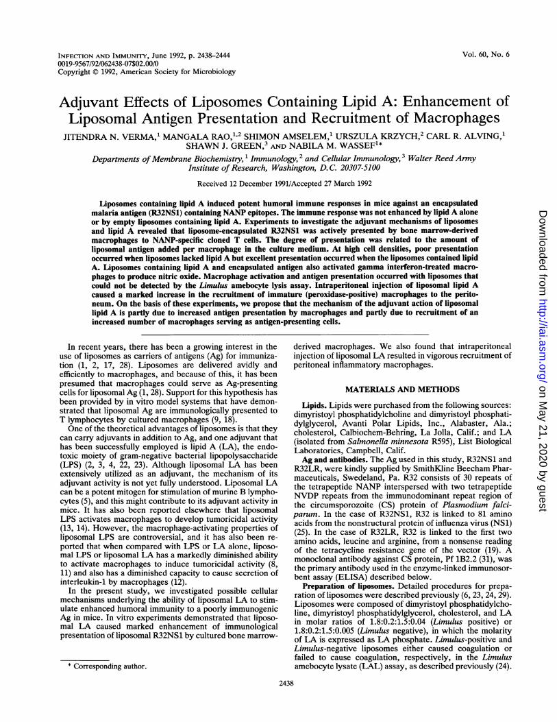
DAYS AFTER IMMUNIZATIONFIG. 1. Immune response of mice to different combinations of
liposomal malaria Ag and LA. The IgG antibody response ofC3H/HeN mice injected intraperitoneally with R32NS1 alone or invarious liposomal formulations was measured by binding to R32LRin a solid-phase ELISA. When free Ag or free LA was used, it wasinjected in amounts comparable to those of liposomal Ag [L(Ag)] orliposomal LA [L(LA)]. The sera obtained were tested by ELISA forIgG activity against R32NS1 antigen. Each absorbance value wascorrected by subtracting the absorbance value obtained for thecorresponding preimmune serum. The data shown represent themeans ± standard deviations of triplicate observations. At 10 and 15days, all L(Ag + LA) values were very significantly different fromthose of L(Ag) and L(Ag) + LA (P < 0.01), and the values of L(Ag)+ LA compared with those of L(Ag) were also significantly different(P < 0.03; two-tailed Student's t test).
dase-positive cells characterized by pink-to-light-purplegranules in the cytoplasm were counted.
RESULTS
Immune response to liposomal Ag. Liposomes, malaria Ag(R32NS1), and LA, either alone or in different combinations,were injected into the mice in order to induce serum anti-bodies (Fig. 1). A potent immune response was obtainedafter injection of liposomes containing R32NS1 Ag and LA[i.e., L(Ag + LA)]. In contrast, a relatively low level ofantibody response was obtained after injection of each of theother formulations, including liposomes containing Ag butlacking LA [L(Ag)], Ag alone, Ag mixed with LA (LA +Ag), empty (Ag-free) liposomes containing LA [L(LA) +Ag], or empty liposomes lacking LA (L + Ag), and lipo-somes containing Ag followed by injection ofLA [i.e., L(Ag)+ LA] (Fig. 1). The observations presented in Fig. 1 areconsistent both with the well-known poor immunogenicity ofnonencapsulated R32 Ag in rabbits and monkeys (22) andwith previous observations demonstrating excellent immu-nogenicity of R32 repeat epitopes encapsulated in liposomescontaining LA (2, 22). The data suggest that LA was the keyelement responsible for the enhanced immune response in
INFECT. IMMUN.
on May 21, 2020 by guest
http://iai.asm.org/
Dow
nloaded from
EFFECTS OF LIPOSOMAL LIPID A ON ANTIGEN PRESENTATION 2441
801 LIMULUS-NEGATIVELIMULUS-POSITIVE
0
E
=LU
%.o
I-
w
60
40
20
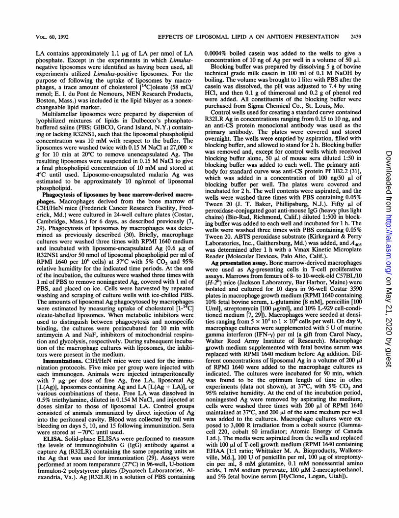
LIPID A (pmol P)FIG. 2. Effect of liposomal LA on macrophage activation. Lip-
osomes containing encapsulated antigen R32NS1 and different con-
centrations of LA (Limulus-negative and Limulus-positive lipo-somes) were incubated for 90 min with macrophage cultures (1.2 x10⁵ cells per well) that had been pretreated with murine IFN--y. Thecultures were incubated for 72 h at 37°C with 5% CO2 and 95%humidity. Culture supernatants were then collected for nitric oxidemeasurements as described in Materials and Methods. The data arethe means + standard deviations of triplicate observations.
vivo and that the simultaneous presence of Ag and LA in theliposomes was required for expression of adjuvant activity.
Effects ofLA on phagocytosis of liposomes by macrophages.A series of experiments was devised to try to elucidate thecellular mechanisms by which LA exerts its effects. Choles-terol ["4C]oleate-labelled liposomes (50 nmol of liposomalphospholipid in [L(Ag)] or [L(Ag + LA)]) were incubatedwith macrophage cultures (106 cells per well) for 6 h at 37°Cwith 5% CO2 and 95% humidity. Separate macrophagecultures were preincubated for 10 min with the metabolicinhibitors NaF and antimycin A in a final concentration of 10mM and 1 ,ug/ml, respectively, before addition of liposomes.At the end of 6 h, cells were harvested by washing with coldPBS and liposome uptake was determined by measurementof radioactivity. Ingestion of liposome-encapsulated Ag bycultured macrophages resulted in continuously increasinglevels of liposome uptake over a period of 24 h (data notshown). There was no evidence of increased phagocytosisinduced by LA. A slight but not significant reduction in therate of phagocytosis of [L(Ag + LA)] compared with that ofL(Ag) was observed. Phagocytosis was distinguished fromnonspecific binding of liposomes to macrophage surfaces byseparate control cultures incubated with metabolic inhibitors(antimycin A and NaF). The liposome uptake of L(Ag) (innanomoles of phospholipid per 106 macrophages + standarddeviations of triplicate observations) was 16.94 + 0.98without inhibitors and 0.83 + 0.17 with inhibitors. For L(Ag+ LA), the uptake was 12.87 + 0.80 without inhibitors and0.72 0.16 with inhibitors.
Effects of LA on activation of macrophages. Induction ofnitric oxide production by cultured macrophages with lipo-somes containing Ag and LA in the presence of IFN--y wasexamined (Fig. 2). Two different types of liposomes witheither a very low epitope density of liposomal LA (Limulusnegative) or a very high epitope density of liposomal LA(Limulus positive) were employed. The total amount of LA
w 50 - Cell Density: 1 x 10 cells AI-
IP AI40
30
z 30--ES- 20
10
0.0 0.4 0.8 1.2LIPOSOMAL ANTIGEN (jig/ml)
30 6Cell Density: 1 x 10 cells B
T-l20p- 0b0
zp E cn tao L(Ag+LA)c2e10Iw D L(Ag)cellswrprcsefoscniA L(LA) (control)
0d.0 0.4 0.8 1 .2LIPOSOMAL ANTIGEN (jig/mI)
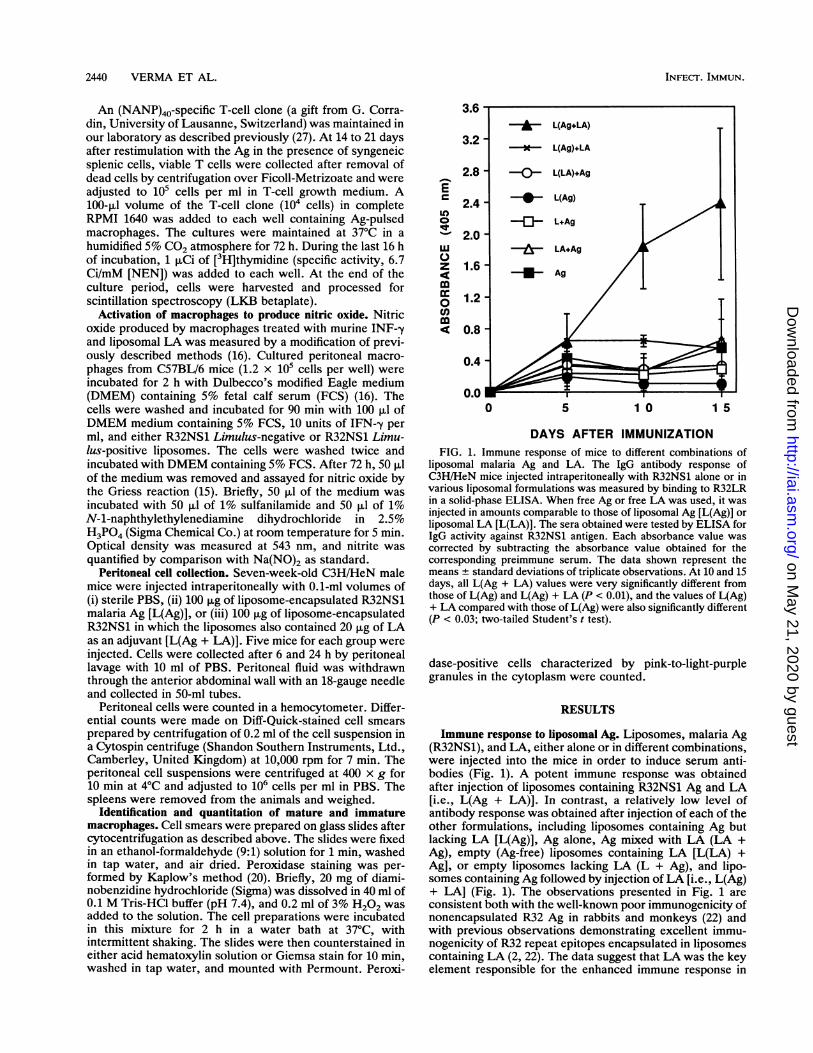
FIG. 3. Effects of celldensityand liposomal AgconcentrationdonT-cell proliferation. C57BL/10 bone marrow-derived macrophageswere seeded at10o(A) or 106 (B) cells per well. Macrophages werepulsed with different concentrations of L(Ag) or L(Ag + LA) for 90mm prior to the addition of an (NANP)4-specific T-cell clone (10icells per well). During the last 16 h of the 72-h culture period, 1 poCiof [3aHthymidine was added. At the end of the incubation period, thecells were processed for scintillation spectroscopy. Data are ex-pressed as mean counts per minute of [LHJthymidine uptake tstandard deviations of triplicate cultures.
added to the cells (as shown on the abscissa of Fig. 2) wasadjusted by adding different dilutions of either Limulus-negative or Limulus-positive liposomes. The data indicatethat nitrite production by stimulated macrophages was de-pendent on the total amount of LA added to the cells but wasnot dependent on -the epitope density of LA in the liposomes(Fig. 2).
It should be pointed out that our phagocytosis model islimited in sensitivity at low levels of liposomes. Because ofthis and because of the great disparity in the total amount ofL-A that is present in equal numbers of Limulus-negative andLimulus-positive liposomes, it was not technically possibleto compare extremely small amounts of Limulus-positiveand large amounts of Limulus-negative liposomes in order tomaintain identical total amounts of LA (Fig. 2). However,Fig. 2 suggests that this limitation in the assay system maynot be important, because despite differences in total LAthere was considerable overlap in the total amount of nitritegenerated by Limulus-negative and Limulus-positive lipo-somes.
Effects ofLA on Ag presentation by macrophages. Prolifer-ative activities of an (NANP)40-reactive T-cell clone wereassayed in cultures seeded with iO5 or 106 bone marrow (BM)precursor cells per well (Fig. 3). To prevent the inherent
VOL. 60, 1992
on May 21, 2020 by guest
http://iai.asm.org/
Dow
nloaded from
2442 VERMA ET AL.
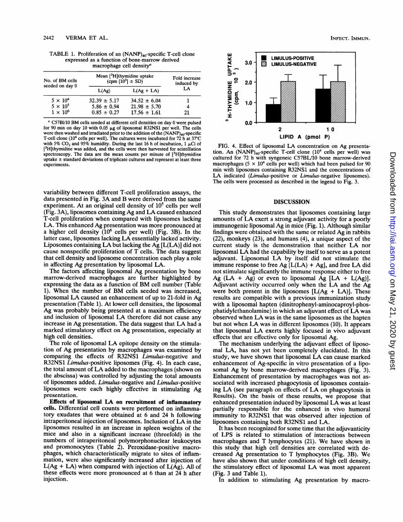
TABLE 1. Proliferation of an (NANP)40-specific T-cell cloneexpressed as a function of bone-marrow derived
macrophage cell density'
Mean [3H]thymidine uptake Fold increaseNo. of BM cells (cpm [103] ± SD) induced byseeded on day 0
L(Ag) L(Ag + LFA) LA
5 x 10⁴ 32.39 ± 5.17 34.52 ± 6.04 15 x 105 5.86 ± 0.94 21.98 ± 5.70 41 x 106 0.85 ± 0.27 17.56 ± 1.61 21
a C57BI/10 BM cells seeded at different cell densities on day 0 were pulsedfor 90 min on day 10 with 0.05 Lg of liposomal R32NS1 per well. The cellswere then washed and irradiated prior to the addition of the (NANP)40-specificT-cell clone (104 cells per well). The cultures were incubated for 72 h at 37'Cwith 5% CO2 and 95% humidity. During the last 16 h of incubation, 1 pCi of[3H]thymidine was added, and the cells were then harvested for scintillationspectroscopy. The data are the mean counts per minute of [3H]thymidineuptake ± standard deviations of triplicate cultures and represent at least threeexperiments.
variability between different T-cell proliferation assays, thedata presented in Fig. 3A and B were derived from the sameexperiment. At an original cell density of 105 cells per well(Fig. 3A), liposomes containing Ag and LA caused enhancedT-cell proliferation when compared with liposomes lackingLA. This enhanced Ag presentation was more pronounced ata higher cell density (106 cells per well) (Fig. 3B). In thelatter case, liposomes lacking LA essentially lacked activity.Liposomes containing LA but lacking the Ag [L(LA)] did notcause nonspecific proliferation of T cells. The data suggestthat cell density and liposome concentration each play a rolein affecting Ag presentation by liposomal LA.The factors affecting liposomal Ag presentation by bone
marrow-derived macrophages are further highlighted byexpressing the data as a function of BM cell number (Table1). When the number of BM cells seeded was increased,liposomal LA caused an enhancement of up to 21-fold in Agpresentation (Table 1). At lower cell densities, the liposomalAg was probably being presented at a maximum efficiencyand inclusion of liposomal LA therefore did not cause anyincrease in Ag presentation. The data suggest that LA had amarked stimulatory effect on Ag presentation, especially athigh cell densities.The role of liposomal LA epitope density on the stimula-
tion of Ag presentation by macrophages was examined bycomparing the effects of R32NS1 Limulus-negative andR32NS1 Limulus-positive liposomes (Fig. 4). In each case,the total amount of LA added to the macrophages (shown onthe abscissa) was controlled by adjusting the total amountsof liposomes added. Limulus-negative and Limulus-positiveliposomes were each highly effective in stimulating Agpresentation.
Effects of liposomal LA on recruitment of inflammatorycells. Differential cell counts were performed on inflamma-tory exudates that were obtained at 6 and 24 h followingintraperitoneal injection of liposomes. Inclusion ofLA in theliposomes resulted in an increase in spleen weights of themice and also in a significant increase (threefold) in thenumbers of intraperitoneal polymorphonuclear leukocytesand promonocytes (Table 2). Peroxidase-positive macro-phages, which characteristically migrate to sites of inflam-mation, were also significantly increased after injection ofL(Ag + LA) when compared with injection of L(Ag). All ofthese effects were more pronounced at 6 than at 24 h afterinjection.
uJw
I-0
za E>o.I-
3.0
2.0
1.0 -
0.0
* LIMULUS'POSITIVELIMULUS-NEGATIVE
T17 T T
2 1 0LIPID A (pmol P)
FIG. 4. Effect of liposomal LA concentration on Ag presenta-tion. An (NANP)40-specific T-cell clone (104 cells per well) wascultured for 72 h with syngeneic C57BL/10 bone marrow-derivedmacrophages (5 x 10⁴ cells per well) which had been pulsed for 90min with liposomes containing R32NS1 and the concentrations ofLA indicated (Limulus-positive or Limulus-negative liposomes).The cells were processed as described in the legend to Fig. 3.
DISCUSSION
This study demonstrates that liposomes containing largeamounts of LA exert a strong adjuvant activity for a poorlyimmunogenic liposomal Ag in mice (Fig. 1). Although similarfindings were obtained with the same or related Ag in rabbits(22), monkeys (23), and humans (4), a unique aspect of thecurrent study is the demonstration that neither LA norliposomal LA had the capability by itself to serve as a potentadjuvant. Liposomal LA by itself did not stimulate theimmune response to free Ag [L(LA) + Ag], and free LA didnot stimulate significantly the immune response either to freeAg (LA + Ag) or even to liposomal Ag [LA + L(Ag)].Adjuvant activity occurred only when the LA and the Agwere both present in the liposomes [L(Ag + LA)]. Theseresults are compatible with a previous immunization studywith a liposomal hapten (dinitrophenyl-aminocaproyl-phos-phatidylethanolamine) in which an adjuvant effect of LA wasobserved when LA was in the same liposomes as the haptenbut not when LA was in different liposomes (10). It appearsthat liposomal LA exerts highly focused in vivo adjuvanteffects that are effective only for liposomal Ag.The mechanism underlying the adjuvant effect of liposo-
mal LA, has not yet been completely elucidated. In thisstudy, we have shown that liposomal LA can cause markedenhancement of Ag-specific in vitro presentation of a lipo-somal Ag by bone marrow-derived macrophages (Fig. 3).Enhancement of presentation by macrophages was not as-sociated with increased phagocytosis of liposomes contain-ing LA (see paragraph on effects of LA on phagocytosis inResults). On the basis of these results, we propose thatenhanced presentation induced by liposomal LA was at leastpartially responsible for the enhanced in vivo humoralimmunity to R32NS1 that was observed after injection ofliposomes containing both R32NS1 and LA.
It has been recognized for some time that the adjuvanticityof LPS is related to stimulation of interactions betweenmacrophages and T lymphocytes (21). We have shown inthis study that high cell densities are correlated with de-creased Ag presentation to T lymphocytes (Fig. 3B). Wehave also shown that under conditions of high cell density,the stimulatory effect of liposomal LA was most apparent(Fig. 3 and Table 1).
In addition to stimulating Ag presentation by macro-
INFECT. IMMUN.
on May 21, 2020 by guest
http://iai.asm.org/
Dow
nloaded from
EFFECTS OF LIPOSOMAL LIPID A ON ANTIGEN PRESENTATION 2443
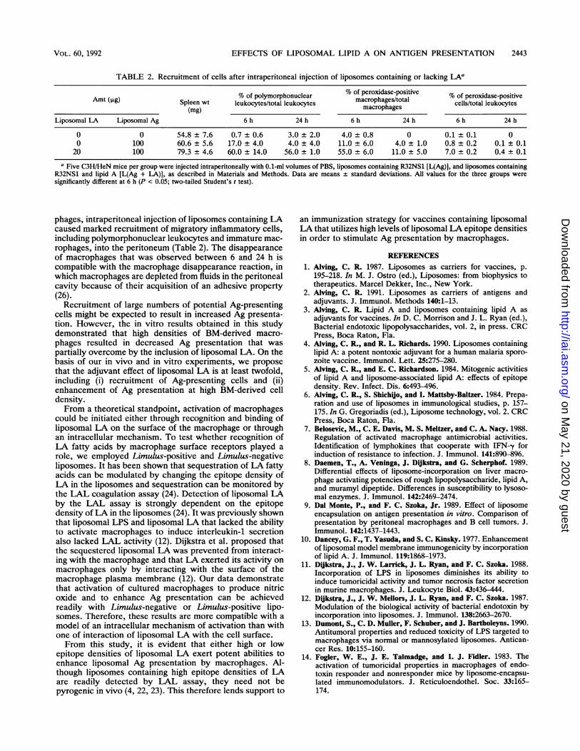
TABLE 2. Recruitment of cells after intraperitoneal injection of liposomes containing or lacking LAa
% of peroxidase-positive % of peroxidase-positiveNoof polymorphonuclear marohaestoa cell/otaldleukoytvesAmt (kLg) Spleen wt leukocytes/total leukocytes macrophages/total cells/total leukocytes(mg) macrophages
Liposomal LA Liposomal Ag 6 h 24 h 6 h 24 h 6 h 24 h
0 0 54.8 + 7.6 0.7 ±0.6 3.0 + 2.0 4.0 ± 0.8 0 0.1 t 0.1 00 100 60.6 t 5.6 17.0 + 4.0 4.0 t 4.0 11.0 + 6.0 4.0 t 1.0 0.8 t 0.2 0.1 t 0.120 100 79.3 t 4.6 60.0 t 14.0 56.0 t 1.0 55.0 t 6.0 11.0 t 5.0 7.0 t 0.2 0.4 t 0.1
a Five C3H/HeN mice per group were injected intraperitoneally with 0.1-ml volumes of PBS, liposomes containing R32NS1 [L(Ag)], and liposomes containingR32NS1 and lipid A [L(Ag + LA)], as described in Materials and Methods. Data are means ± standard deviations. All values for the three groups weresignificantly different at 6 h (P < 0.05; two-tailed Student's t test).
phages, intraperitoneal injection of liposomes containing LAcaused marked recruitment of migratory inflammatory cells,including polymorphonuclear leukocytes and immature mac-rophages, into the peritoneum (Table 2). The disappearanceof macrophages that was observed between 6 and 24 h iscompatible with the macrophage disappearance reaction, inwhich macrophages are depleted from fluids in the peritonealcavity because of their acquisition of an adhesive property(26).Recruitment of large numbers of potential Ag-presenting
cells might be expected to result in increased Ag presenta-tion. However, the in vitro results obtained in this studydemonstrated that high densities of BM-derived macro-phages resulted in decreased Ag presentation that waspartially overcome by the inclusion of liposomal LA. On thebasis of our in vivo and in vitro experiments, we proposethat the adjuvant effect of liposomal LA is at least twofold,including (i) recruitment of Ag-presenting cells and (ii)enhancement of Ag presentation at high BM-derived celldensity.From a theoretical standpoint, activation of macrophages
could be initiated either through recognition and binding ofliposomal LA on the surface of the macrophage or throughan intracellular mechanism. To test whether recognition ofLA fatty acids by macrophage surface receptors played arole, we employed Limulus-positive and Limulus-negativeliposomes. It has been shown that sequestration of LA fattyacids can be modulated by changing the epitope density ofLA in the liposomes and sequestration can be monitored bythe LAL coagulation assay (24). Detection of liposomal LAby the LAL assay is strongly dependent on the epitopedensity of LA in the liposomes (24). It was previously shownthat liposomal LPS and liposomal LA that lacked the abilityto activate macrophages to induce interleukin-1 secretionalso lacked LAL activity (12). Dijkstra et al. proposed thatthe sequestered liposomal LA was prevented from interact-ing with the macrophage and that LA exerted its activity onmacrophages only by interacting with the surface of themacrophage plasma membrane (12). Our data demonstratethat activation of cultured macrophages to produce nitricoxide and to enhance Ag presentation can be achievedreadily with Limulus-negative or Limulus-positive lipo-somes. Therefore, these results are more compatible with amodel of an intracellular mechanism of activation than withone of interaction of liposomal LA with the cell surface.From this study, it is evident that either high or low
epitope densities of liposomal LA exert potent abilities toenhance liposomal Ag presentation by macrophages. Al-though liposomes containing high epitope densities of LAare readily detected by LAL assay, they need not bepyrogenic in vivo (4, 22, 23). This therefore lends support to
an immunization strategy for vaccines containing liposomalLA that utilizes high levels of liposomal LA epitope densitiesin order to stimulate Ag presentation by macrophages.
REFERENCES1. Alving, C. R. 1987. Liposomes as carriers for vaccines, p.
195-218. In M. J. Ostro (ed.), Liposomes: from biophysics totherapeutics. Marcel Dekker, Inc., New York.
2. Alving, C. R. 1991. Liposomes as carriers of antigens andadjuvants. J. Immunol. Methods 140:1-13.
3. Alving, C. R. Lipid A and liposomes containing lipid A asadjuvants for vaccines. In D. C. Morrison and J. L. Ryan (ed.),Bacterial endotoxic lipopolysaccharides, vol. 2, in press. CRCPress, Boca Raton, Fla.
4. Alving, C. R., and R. L. Richards. 1990. Liposomes containinglipid A: a potent nontoxic adjuvant for a human malaria sporo-zoite vaccine. Immunol. Lett. 25:275-280.
5. Alving, C. R., and E. C. Richardson. 1984. Mitogenic activitiesof lipid A and liposome-associated lipid A: effects of epitopedensity. Rev. Infect. Dis. 6:493-496.
6. Alving, C. R., S. Shichjo, and I. Mattsby-Baltzer. 1984. Prepa-ration and use of liposomes in immunological studies, p. 157-175. In G. Gregoriadis (ed.), Liposome technology, vol. 2. CRCPress, Boca Raton, Fla.
7. Belosevic, M., C. E. Davis, M. S. Meltzer, and C. A. Nacy. 1988.Regulation of activated macrophage antimicrobial activities.Identification of lymphokines that cooperate with IFN--y forinduction of resistance to infection. J. Immunol. 141:890-896.
8. Daemen, T., A. Veninga, J. Dijkstra, and G. Scherphof. 1989.Differential effects of liposome-incorporation on liver macro-phage activating potencies of rough lipopolysaccharide, lipid A,and muramyl dipeptide. Differences in susceptibility to lysoso-mal enzymes. J. Immunol. 142:2469-2474.
9. Dal Monte, P., and F. C. Szoka, Jr. 1989. Effect of liposomeencapsulation on antigen presentation in vitro. Comparison ofpresentation by peritoneal macrophages and B cell tumors. J.Immunol. 142:1437-1443.
10. Dancey, G. F., T. Yasuda, and S. C. Kinsky. 1977. Enhancementof liposomal model membrane immunogenicity by incorporationof lipid A. J. Immunol. 119:1868-1973.
11. Dikstra, J., J. W. Larrick, J. L. Ryan, and F. C. Szoka. 1988.Incorporation of LPS in liposomes diminishes its ability toinduce tumoricidal activity and tumor necrosis factor secretionin murine macrophages. J. Leukocyte Biol. 43:436-444.
12. Di,kstra, J., J. W. Mellors, J. L. Ryan, and F. C. Szoka. 1987.Modulation of the biological activity of bacterial endotoxin byincorporation into liposomes. J. Immunol. 138:2663-2670.
13. Dumont, S., C. D. Muller, F. Schuber, and J. Bartholeyns. 1990.Antitumoral properties and reduced toxicity of LPS targeted tomacrophages via normal or mannosylated liposomes. Antican-cer Res. 10:155-160.
14. Fogler, W. E., J. E. Talmadge, and I. J. Fidler. 1983. Theactivation of tumoricidal properties in macrophages of endo-toxin responder and nonresponder mice by liposome-encapsu-lated immunomodulators. J. Reticuloendothel. Soc. 33:165-174.
VOL. 60, 1992
on May 21, 2020 by guest
http://iai.asm.org/
Dow
nloaded from
2444 VERMA ET AL.
15. Green, L. C., D. A. Wagner, J. Glogowski, P. L. Skipper, J. S.Wishnok, and S. R. Tannenbaum. 1982. Analysis of nitrate,nitrate and ['5N] nitrate in biological fluids. Anal. Biochem.126:131-138.
16. Green, S. J., R. M. Crawford, J. T. Hockmeyer, M. S. Meltzer,and C. A. Nacy. 1990. Leishmania major amastigotes initiate theL-arginine-dependent killing mechanism in IFN--y-stimulatedmacrophages by induction of tumor necrosis factor-a. J. Immu-nol. 145:4290-4297.
17. Gregoriadis, G. 1990. Immunological adjuvants: a role forliposomes. Immunol. Today 11:89-97.
18. Harding, C. V., D. S. Collins, J. W. Slot, H. J. Geuze, and E. R.Unanue. 1991. Liposome-encapsulated antigens are processedin lysosomes, recycled, and presented to T cells. Cell 64:393-401.
19. Hoffman, S. L., L. T. Cannon, Sr., J. A. Berzofsky, W. R.Majarian, J. F. Young, W. L. Maloy, and W. T. Hockmeyer.1987. Plasmodium falciparum: sporozoite boosting of immunitydue to a T-cell epitope on a sporozoite vaccine. Exp. Parasitol.64:64-70.
20. Kaplow, L. S. 1965. Simplified myeloperoxidase stain usingbenzidine dihydrochloride. Blood 26:215-218.
21. McGhee, J. R., J. J. Farrar, S. M. Michalek, S. E. Mergenhagen,and D. L. Rosenstreich. 1979. Cellular requirements for lipopoly-saccharide adjuvanticity. A role for both T lymphocytes andmacrophages for in vitro responses to particulate antigens. J.Exp. Med. 149:793-807.
22. Richards, R. L., M. D. Hayre, W. T. Hockmeyer, and C. R.Alving. 1988. Liposomes, lipid A, and aluminum hydroxideenhance the immune response to synthetic malaria sporozoiteantigen. Infect. Immun. 56:682-686.
23. Richards, R. L., G. M. Swartz, Jr., C. Schultz, M. D. Hayre,G. S. Ward, W. R. Ballou, J. D. Chulay, W. T. Hockmeyer, S. L.Berman, and C. R. Alving. 1989. Immunogenicity of liposomalmalaria sporozoite antigen in monkeys: adjuvant effects of
aluminium hydroxide and non-pyrogenic liposomal lipid A.Vaccine 7:506-512.
24. Richardson, E. C., B. Banerji, R. C. Seid, Jr., J. Levin, andC. R. Alving. 1983. Interactions of lipid A and liposome-associ-ated lipid A with Limulus polyphemus amoebocytes. Infect.Immun. 39:1385-1391.
25. Rickman, L. S., D. M. Gordon, R. Wistar, Jr., U. Krzych, M.Gross, M. R. Hollingdale, J. E. Egan, J. D. Chulay, and S. L.Hoffman. 1991. Use of adjuvant containing mycobacterial cell-wall skeleton, monophosphoryl lipid A and squalene in malariacircumsporozoite protein vaccine. Lancet 337:998-1001.
26. Shannon, B. T., S. H. Love, and Q. N. Myrik. 1980. Participationof hyaluronic acid in the macrophage disappearance reaction.Immunol. Commun. 9:357-370.
27. Smith, L. F., G. H. Lowell, W. R. Ballou, and U. Krzych. 1988.The role of lauroyl and cys-tyr-gly-gly in T cell activation bysynthetic peptides from Plasmodium falciparum circumsporo-zoite protein, p. 651-659. In L. Lasky (ed.), Technologicaladvances in vaccine development. Alan R. Liss, New York.
28. van RooiJen, N., and D. Su. 1989. Immunoadjuvant action ofliposomes: mechanisms, p. 95-106. In G. Gregoriadis, A. C.Allison, and C. Poste (ed.), Immunological adjuvants and vac-cines. Plenum Press, New York.
29. Verma, J. N., N. M. Wassef, R. A. Wirtz, C. T. Atkinson, M.Aikawa, L. D. Loomis, and C. R. Alving. 1991. Phagocytosis ofliposomes by macrophages: intracellular fate of liposomal ma-laria antigen. Biochim. Biophys. Acta 1066:229-238.
30. Wassef, N. M., and C. R. Alving. 1987. Complement-dependentphagocytosis of liposomes by macrophages. Methods Enzymol.149:124-134.
31. Wirtz, R. A., F. Zavala, Y. Charoenvit, G. H. Campbell, T. R.Burkot, I. Schneider, K. M. Esser, R. L. Beaudoin, and R. G.Andre. 1987. Comparative testing of Plasmodium falciparumsporozoite monoclonal antibodies for ELISA development.Bull. W.H.O. 65:39-45.
INFECT. IMMUN.
on May 21, 2020 by guest
http://iai.asm.org/
Dow
nloaded from