addendum arterial blood supply of cns
DESCRIPTION
artery of the brain and central nervous system blood supply for neuroscienceTRANSCRIPT

ARTERIAL SUPPLY OF THE CENTAL
NERVOUS SYSTEM

Arterial Supply of the Brain
• internal carotid arteries
• vertebral arteries

Internal Carotid Artery• begins at bifurcation of common carotid artery (upper border of thyroid cartilage)
• parts:
• cervical (from origin to base of the skull)
• petrous (within carotid canal in petrous part of temporal bone)
• cavernous (within cavernous sinus)
• cerebral (from the point it pierces the dura mater to its termination, where it divides into anterior and middle cerebral arteries)
• cavernous and cerebral parts together are referred to as “carotid siphon” (S-shaped configuration)

Netter’s Plate 130

Netter’s Plate 98

Branches of the Internal Carotid Artery
• cervical part: no branches
• petrous part: small branches to tympanic cavity
• cavernous part: inferior hypophysial branches
small branches supplying trigeminal ganglion, walls of cavernous sinus (including contained nerves) and adjacent dura mater
• cerebral part: ophthalmic artery
superior hypophysial branches
anterior choroidal artery
posterior communicating artery
anterior cerebral artery
middle cerebral artery


Netter’s Plate 132

Posterior Communicating Artery• extends from internal carotid artery to proximal part of posterior cerebral artery
• connects anterior (carotid) and posterior (vertebro-basilar) circulations
Anterior Choroidal Artery• runs posteriorly, inferior to optic tract
• supplies choroid plexus in inferior (temporal) horn of lateral ventricle, structures along medial aspect of temporal lobe (uncus, hippocampus, amygdala) , tail of caudate, globus pallidus, inferior part of internal capsule, optic tract, lateral geniculate nucleus and cerebral peduncle

Anterior Cerebral Artery (ACA)• it is joined to homologous artery by anterior communicating artery
• anterior communicating artery divides ACA into two segments: A1 (proximal/precommunicating) and A2 (distal/postcommunicating)
• A1 segment passes anteriorly and medially, above optic nerve
• A2 segment is distributed on medial surface of cerebral hemisphere

Anterior Cerebral Artery (ACA)• supplies (cortical branches):
• medial surface of cerebral hemisphere from frontal pole to parieto-occipital sulcus
• corpus callosum
• straight gyrus and olfactory bulb and tract
• distal branches extend over superior border of hemisphere and supply strip of cortex on superolateral surface

Anterior Cerebral Artery (ACA)• medial striate artery (recurrent artery of Heubner):
• originates from ACA proximal or distal (more common) to anterior communicating artery
• passes posteriorly and laterally and its branches enter brain via anterior perforated substance
• supplies inferior part of head of caudate nucleus and adjacent parts of anterior limb of internal capsule and putamen

Netter’s Plate 132


Fig. 8-9 A Haines Textbook


Middle Cerebral Artery (MCA)• runs laterally in lateral sulcus toward ventral part of insula, where it divides into superior and inferior trunks
• M1 segment: from origin to bifurcation into superior and inferior trunks
• M2 segment (insular part): trunks and their branches as they lie on insula
• M3 segment (opercular part): branches of trunks that curve around opercula
• M4 segment (cortical part): distal branches of trunks that exit lateral fissure and serve cortical areas above and below it
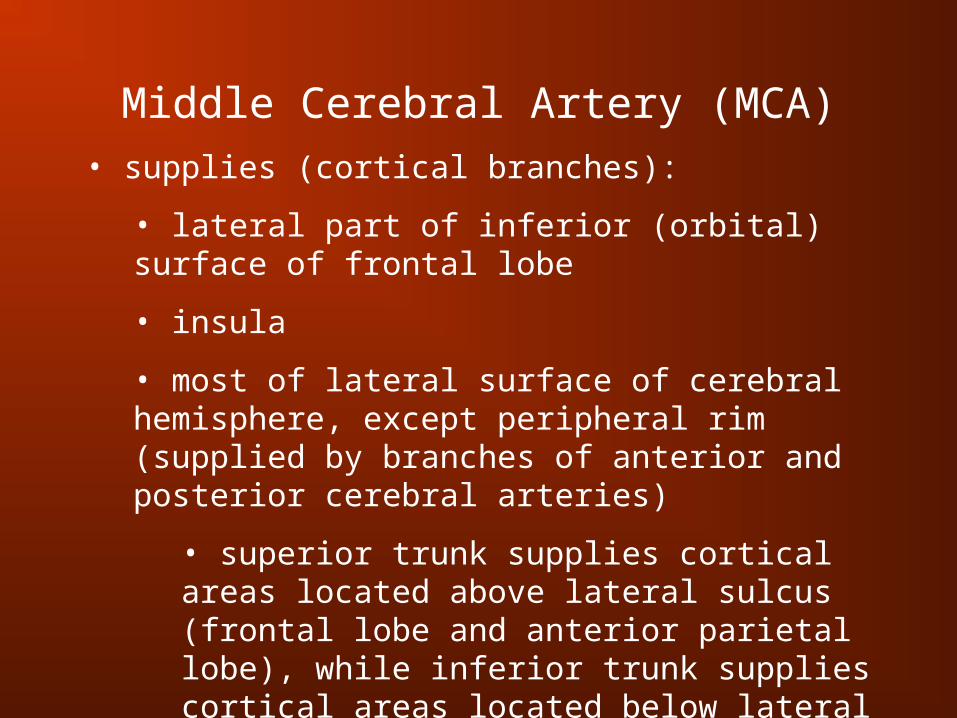
Middle Cerebral Artery (MCA)• supplies (cortical branches):
• lateral part of inferior (orbital) surface of frontal lobe
• insula
• most of lateral surface of cerebral hemisphere, except peripheral rim (supplied by branches of anterior and posterior cerebral arteries)
• superior trunk supplies cortical areas located above lateral sulcus (frontal lobe and anterior parietal lobe), while inferior trunk supplies cortical areas located below lateral sulcus (upper part of temporal lobe) and posterior part of parietal lobe

Netter’s Plate 132


Fig. 8-9 A Haines Textbook

Vertebral Artery
• enters cranial cavity through foramen magnum
• runs superiorly and medially and at pontomedullary junction joins homologous artery to form basilar artery
• branches:
• posterior spinal artery
• posterior inferior cerebellar artery
• anterior spinal artery

Posterior Spinal Artery
• it may be a branch of the vertebral artery (25%) or the PICA (75%)
• descends along posterolateral sulcus of spinal cord
Posterior Inferior Cerebellar Artery (PICA)
• curves around medulla to reach inferior cerebellar surface
• supplies posterolateral part of medulla, choroid plexus of 4th ventricle and medial parts of inferior surface of cerebellum
Anterior Spinal Artery
• the two arteries join to form a single anterior spinal artery, which runs along anterior median fissure of medulla and spinal cord
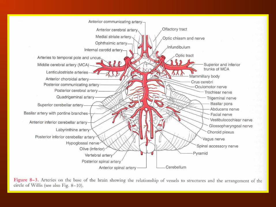

Fig. 11-16 B Haines Textbook

Basilar Artery• runs in basilar sulcus of pons from pontomedullary junction to pons-midbrain junction where it terminates by dividing into 2 posterior cerebral arteries
• branches:
• pontine branches (paramedian, short circumferential and long circumferential branches)
• anterior inferior cerebellar artery
• labyrinthine (internal auditory) artery
• superior cerebellar artery
• posterior cerebral artery

Anterior Inferior Cerebellar Artery (AICA)
• passes lateral along inferior border of middle cerebellar peduncle
• supplies lateral parts of inferior surface of cerebellum, lower pons, upper medulla and choroid plexus of 4th ventricle
Labyrinthine (Internal Auditory) Artery
• it may originate from basilar artery or AICA (more commonly)
• enters internal acoustic meatus with CNs VII and VIII to supply internal ear
Superior Cerebellar Artery
• passes laterally, just inferior to oculomotor nerve
• wraps around brainstem to supply upper pons, lower midbrain and superior surface of cerebellum


Posterior Cerebral Artery (PCA)• passes laterally, just above oculomotor nerve
• passes around midbrain and joins inferomedial aspect of temporal lobe
• parts:
• P1 segment: from origin to posterior communicating artery
• P2 segment: from posterior communicating artery to 1st temporal branch
• P3 segment: part of PCA that gives rise to temporal branches
• P4 segment: distal part of artery that gives rise to branches that supply occipital lobe (parieto-occipital and calcarine arteries)

Posterior Cerebral Artery (PCA)• supplies:
• inferior and medial surfaces of temporal and occipital lobes
• distal branches extend over superior and inferior borders of hemisphere to supply a strip of cortex on superolateral surface
• midbrain, thalamus and choroid plexuses of 3rd and lateral ventricles


Fig. 8-9 B Haines Textbook

Fig. 8-9 A Haines Textbook

Watershed Infarcts• border zones: areas where terminal branches of anterior, middle and posterior cerebral arteries overlap
• brain tissue in these border zones is susceptible to damage in case of sudden systemic hypotension or when there is hypoperfusion of distal vascular bed of a major cerebral artery
• inadequate perfusion of border zones may result in watershed infarcts

Fig. 8-9 C Haines Textbook

Cerebral Arterial Circle (of Willis)
• consists of the larger cerebral vessels and their interconnections on the ventral surface of the brain
• components: anterior communicating a.
anterior cerebral a.
internal carotid a.
posterior communicating a.
posterior cerebral a.
• serves as potential vascular shunt, assisting in development of collateral circulation if one of the proximal vessels is occluded

Netter’s Plate 133

Perforating (Central) Branches• small branches that originate from the arteries of the cerebral arterial circle and penetrate the ventral surface of the brain
• groups:
• anteromedial
• anterolateral
• posteromedial
• posterolateral

Anteromedial Arteries• originate from A1 segment of ACA and anterior communicating artery
• supply supraoptic part of hypothalamus

Anterolateral Arteries
• originate from M1 segment of MCA (lenticulostriate arteries) and a few may come from A1 segment of ACA
• enter brain via anterior perforated substance
• supply most of the caudate, putamen, globus pallidus and internal capsule
• most common site of intra-cerebral hemorrhage in hypertensive individuals

Posteromedial Arteries
• originate from posterior communicating artery and P1 segment of posterior cerebral artery
• enter brain via posterior perforated substance
• supply tuberal and mammillary regions of hypothalamus, subthalamus, anterior part of thalamus and medial parts of midbrain tegmentum and cerebral crus

Posterolateral Arteries
• originate from P2 segment of posterior cerebral artery
• supply posterior part of thalamus, including geniculate nuclei, choroid plexus of lateral and 3rd ventricles (posterior choroidal arteries) and midbrain


Arterial Supply of the Spinal Cord• there are 1 anterior and 2 posterior spinal arteries
• blood received by spinal arteries from vertebral arteries is sufficient only for upper cervical segments
• spinal arteries are reinforced at intervals by anterior and posterior radicular branches that originate from segmental arteries (vertebral, deep cervical, ascending cervical, posterior intercostal, lumbar, sacral arteries)
• largest anterior radicular artery artery of Adamkiewicz travels with ventral root of a lower thoracic or upper lumbar spinal nerve, most frequently on the left

Netter’s Plate 164

Arterial Supply of the Spinal Cord• branches of anterior spinal artery supply anterior 2/3 of spinal cord anterior horn, intermediate zone, basal part of posterior horn, anterior and lateral funiculi
• branches of posterior spinal artery supply posterior 1/3 of spinal cord most of posterior horn and posterior funiculus
• arterial vasocorona: fine arterial plexus connecting anterior and posterior spinal arteries supplies narrow zone of white matter beneath pia mater

Fig. 5-6 Haines Atlas