acute rheumatic fever and rheumatic heart disease: sudan’s...
TRANSCRIPT

1
Acute Rheumatic Fever and Rheumatic Heart Disease: Sudan’s Guidelines for Diagnosis, Management and Prevention

2

3
Sudan’s Federal Ministry of Health
Sudan Heart Society-Working Group on Pediatric Cardiology
Sudanese Association of Pediatricians
Sudanese Children’s Heart Society
Writing Committee:
Sulafa Khalid M Ali, FRCPCH, FACC, Consultant Pediatric Cardiologist
Professor-University of Khartoum
Mohamed Saeed Al Khaleefa, FRCP, Consultant Cardiologist
Professor-University of Al Zaem Al Azhari
Siragedeen Mohamed Khair, MD, Consultant Pediatrician
Professor- University of Al Zaem Al Azhari
Second Edition
Jan/2017
4
Contents
Chapter Title Page
Preface 5
Chapter 1 Rheumatic Heart Disease : General Considerations 6
Chapter 2 Diagnosis and Management of Streptococcal Pharyngitis
11
Chapter 3 Acute Rheumatic Fever 15
Chapter 4 Rheumatic Heart Disease 25
Chapter 5 Rheumatic Heart Disease in Pregnancy 49
Chapter 6 Acute Rheumatic Fever & Rheumatic Heart Disease Control
57
Appendices Rheumatic Heart Disease Protocols, Manuals, Brochures and Educational Websites
63

5
Preface to the Second Edition:
This is the second edition of Sudan’s Guidelines for acute rheumatic fever (ARF) and rheumatic heart disease (RHD) diagnosis, management and prevention.
RHD is a devastating sequel of ARF, initiated by a simple throat infection with group A streptococcus in susceptible population. Eradication of RHD can be achieved by improvement of health care system as has been witnessed in developed countries. In many developing countries like Sudan, RHD is still prevalent causing significant mortality and premature cardiovascular death as well as an undesired burden on the health system.
An RHD control program has been established in Sudan in 2012 aiming to increase the awareness of both the public and medical personnel, to introduce primary and consolidate secondary prevention and to strengthen the surveillance system.
This booklet serves as a reference for physicians and other health care providers containing relevant background knowledge and clinical guidelines for diagnosis, management and prevention of ARF and RHD. In addition, it contains brochures, awareness material as well as educational websites.
This second edition has been revised to include:
1.New data about RHD epidemiology in Sudan.
2.Updated diagnostic criteria for bacterial phyryngitis.
3.Posters for RHD in pregnancy.
4. Penicillin administration guidelines are updated with a section on management of anaphylaxis.
We hope that this book helps the medical community to have a better understanding for and management of ARF and RHD.
Acknowledgment:
We would like to acknowledge the contribution of Professor Khalid Yassin, Consultant Obstetrics and Gynecology, Bahri Hospital and Professor Aiden Long, Associate Professor of Immunology, Harvard Medical School for their valuable input.
Khartoum in Jan 2017

6
Chapter 1
Rheumatic Heart Disease:
General Considerations

7
Introduction
Acute Rheumatic Fever (ARF) is an immune reaction to the group A streptococcus (GAS) pharyngitis. ARF mainly affects the joint, the heart , the skin and the central nervous system, however, all the manifestations resolve spontaneously except the cardiac affection which leads to chronic valve damage; rheumatic heart disease (RHD). RHD is the leading cause of acquired heart disease in young people all around the world but is highly prevalent in countries with poor socioeconomic state and health systems.
The global burden was found to be 471,000 annual cases of ARF, with the incidence of ARF in children ages 5 to 15 years ranging from 10 cases per 100,000 in industrialized countries to 374 cases per 100,000 in the Pacific region. The overall burden of RHD was estimated to be 15.6 million prevalent cases with 282,000 new cases and over 233,000 deaths per year (1).
Pathogenesis:
Although not fully understood, there is strong evidence that molecular mimicry between GAS antigen and human proteins lead to auto immune (humoral and cellular) mediated damage to heart valves. Cytokines are the likely effectors of valve damage, they can recognize M protein and then produce inflammatory mediators including tumor necrosis factor alpha, interferon and interleukins which cross react with cardiac myosin. Further evidence of inflammation is supported by high levels of advanced oxidation protein products and high sensitive C-reactive protein in plasma detected in patients with RHD. (2). This was well shown in Sudanese patients who were found to have a high level of cytokines and C- reactive protein even during the chronic phase of RHD) indicating an ongoing inflammatory process (3, 4).
Rheumatic Heart Disease: natural history:
Once ARF occurs, there is a very high rate of recurrence and the valve damage becomes evident. As early as 1965, it was found that the prognosis of patients who have established RHD was poor. While mild RHD may improve if recurrence of ARF is prevented by secondary prophylaxis, only 11% of those with established RHD will be free of heart failure at 10 years of follow up (5). Recurrence of ARF is the most important factor that determines the severity and prognosis of RHD.
Rheumatic Heart Disease in Sudan:
The incidence of clinical RHD (detected by auscultation) in Sudan was found to be 100 per 100000/year and the prevalence had been reported as 10.2 per 1000 compared with 2.3 per 1000 in Saudi Arabia and 5.1 per 1000 in Egypt. (6) These numbers are likely to be underestimated as in many developing countries echocardiography (echo) screening of school children revealed much higher prevalence rates. In India, it was shown that the prevalence of RHD by echo screening is 20 per1000,

8
several folds higher than clinical prevalence (7) Similar results found in Uganda, and Fiji had unmasked an extremely high prevalence of subclinical carditis(8,9). In Sudan’s main Children’s hospital s, RHD represent the most common cause of admission to cardiology wards and the most common cause of death due to cardiac disease. Ninety five percent of patients with RHD seen have severe forms of valve disease needing surgical intervention. Ninety percent of those with RHD do not have any history of ARF indicating that the first episode passed unnoticed and 50% of them were not compliant with secondary prophylaxis. Only 30% of patients came back for follow up indicating poor access to tertiary care and the need to decentralize services and raise public awareness (10). The Ministry of Health (MOH) reported that in the year 2011 the total number of patients seen with ARF/RHD in outpatient clinics in the whole Sudan was 36877 (representing 13% of the global annual burden), out of them (11976) were seen in Khartoum state followed by Western States (Darfur and Kordofan) (9170) while the least number was seen in River Nile State (195) patients. The same report showed that 509 cases of RHD were admitted to hospitals, including 176 children between (5-14 years) . RHD caused 44 deaths in 2011(9%). (9). These findings are similar to those derived from review of over 300 patients referred to Khartoum. The majority were found to be coming from Darfur area followed by Kordofan. In addition most patients residing in Khartoum are originally coming from those areas (10) A RHD register has been established; so far about 900 cases are included. There is a well defined RHD belt that mainly affects Kordofan, Darfur , White Nile and Al Gazira areas (Figure)
Figure showing distribution of RHD cases in Sudan’s States/Regions
9
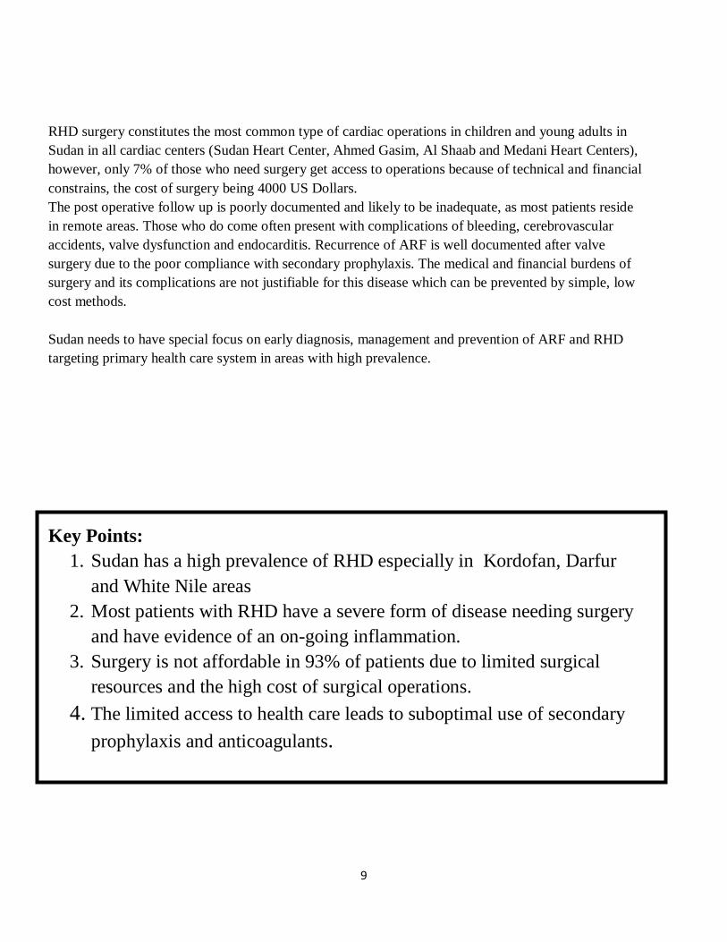
RHD surgery constitutes the most common type of cardiac operations in children and young adults in Sudan in all cardiac centers (Sudan Heart Center, Ahmed Gasim, Al Shaab and Medani Heart Centers), however, only 7% of those who need surgery get access to operations because of technical and financial constrains, the cost of surgery being 4000 US Dollars. The post operative follow up is poorly documented and likely to be inadequate, as most patients reside in remote areas. Those who do come often present with complications of bleeding, cerebrovascular accidents, valve dysfunction and endocarditis. Recurrence of ARF is well documented after valve surgery due to the poor compliance with secondary prophylaxis. The medical and financial burdens of surgery and its complications are not justifiable for this disease which can be prevented by simple, low cost methods. Sudan needs to have special focus on early diagnosis, management and prevention of ARF and RHD targeting primary health care system in areas with high prevalence.
Key Points: 1. Sudan has a high prevalence of RHD especially in Kordofan, Darfur
and White Nile areas 2. Most patients with RHD have a severe form of disease needing surgery
and have evidence of an on-going inflammation. 3. Surgery is not affordable in 93% of patients due to limited surgical
resources and the high cost of surgical operations. 4. The limited access to health care leads to suboptimal use of secondary
prophylaxis and anticoagulants.
10
References: 1.Carapetis JR, Steer AC, Mulholland EK, Weber M. The global burden of group A streptococcal diseases. Lancet Infect Dis 2005;5:685–94. 2.Chopra P, Gulwani H. Pathology and pathogenesis of rheumatic heart disease.Indian J Pathol Microbiol. 2007 ;50(4):685-97. 3. Khalifa Mutaz, and Ali KM Sulafa. Clinical and echocardiographic features of children with rheumatic carditis: correlation with high sensitivity C-reactive protein. Sudan JMS2013; 8: 131-134. 4. Sulafa Khalid Mohamed Ali, Inaam Noor Eldaim, Samia Hassan Osman, Sahar Mohamed Bakhite. Clinical and echocardiographic features of children with rheumatic heart disease and their serum cytokine profile. The Pan African Medical Journal. 2012;13:36 5. The Natural History of Rheumatic Fever and Rheumatic Heart Disease :Ten-Year Report of a Cooperative Clinical Trial of ACTH, Cortisone, and Aspirin. A Joint report by the Rheumatic Fever Working Party of the Medical Research Council of Great Britain and the Subcommittee of Principal Investigators of the American Council on Rheumatic Fever and Congenital Heart Disease, American Heart Association. Circulation1965; 32: 457-476 6.Rheumatic Fever and Rheumatic Heart Disease, Report of a WHO Expert Consultation, Geneva, World Health Organization (Technical Report Series, No.923) 2001. 7. Saxena A, Ramakrishnan S, Roy A, Seth S, Krishnan A, Misra P, Kalaivani M, Bhargava B, Flather MD, Poole-Wilson PP. Prevalence and outcome of subclinical rheumatic heart disease in India: the RHEUMATIC (Rheumatic Heart Echo Utilisation and Monitoring Actuarial Trends in Indian Children) study. Heart 2011;97:2018-22. 8.Steer AC, Kado J, Wilson N, Tuiketei T, Batzloff M, Waqatakirewa L, Mulholland EK, Carapetis JR.High prevalence of rheumatic heart disease by clinical and echocardiographic screening among children in Fiji. J Heart Valve Dis. 2009;18:327-35 9.Beaton A, Okello E, Lwabi P, Mondo C, McCarter R, Sable C.Echocardiography screening for rheumatic heart disease in Ugandan schoolchildren. Circulation 2012;125:3127-32. 10. Eynas Khalid, Hamid El Banna, Rehab Mahmoud, Hanadi Hassan, Laila El Mahdi, Sulafa Ali.Clinical and echocardiographic features of 370 children with rheumatic heart disease seen in Khartoum. Sudan Med J 2014;50:151-54 11. Ministry of Health Annual Report, 2011.

11
Chapter 2 Diagnosis and Management of Streptococcal Pharyngitis Pharyngitis is a clinical syndrome associated with infection/irritation of the pharynx and/or tonsils. It is mostly viral, however, Group A beta hemolytic streptococcus (GAS) is the most common cause for bacterial phayngitis all around the world, including Sudan, accounting for 25% of pediatric patients with sore throat (1). The diagnosis of streptococcal pharyngitis can either be clinical only or using clinical criteria supported by laboratory investigations. The gold standard diagnostic method is by using a Clinical Prediction rule (CPR) supported by rapid antigen test (RAT) and/or throat culture (2)
Importantly, antistreptolysin O titre (ASO) has no role in diagnosis of GAS pharyngitis as titers increase only 7 to 14 days after the onset of infection and remain high for weeks.
Limitations of Rapid Antigen Test and Throat Culture: 1. The cost of both tests is high and RAT is not available in Sudan. 2. Many people are asymptomatic carriers (10% of school age children). 3. Sensitivity and specificity of both tests are not optimal. 4. The need to wait for days for the culture result prohibits its routine use.
As countries with RHD usually have low resources, a CPR can be used without doing RAT or throat culture in order to diagnose GAS pharyngitis. Which CPR to choose?
Table1shows an example of CPR (3)
Sore Throat and: Points
Fever (temperature >38°C) 1
Absence of cough 1
Cervical LN enlargement 1
Tonsillar swelling or exudates 1
Age 3 to <15 yr 1
Antistreptolysin O (ASO) titre has NO ROLE in diagnosis of acute pharyngitis

12
Score Risk of GAS pharyngitis (%) 1 5 -10 2 11 - 17 3 28 -35 4 51 - 53 Another example is the one used in the original Integrated Management of Childhood Illness (IMCI) which states that the presence of fever or sore throat and 2 of the following: congested throat, tonsillar exudates or cervical lymph node enlargement, so it considers 3 points. However, when IMCI score was tested it was shown to be only 12% sensitive and 94% specific. It missed 88% of children with positive cultures (4). A similar CPR was tested in Egypt and used only 2 points and found a sensitivity of 80% and specificity of 40%. Similar results were reported from Turkey (5, 6).So in Sudan we decided to use a more sensitive score (2 or 3 points according to the risk of RHD) Sudan’s Protocol for Sore Throat Management: In patients 3-18 years of age: Bacterial pharyngitis is diagnosed as follows:
High risk area 1. Sore throat 2. No runny nose/cough
Sore throat and No runny nose/cough and one of the following:
1. Cervical lymph node 2. Enlarged congested tonsils 3. A membrane on the
throat/tonsills
Low risk area
High risk Areas: Kordofan, Darfur, White Nile and Gezira
13
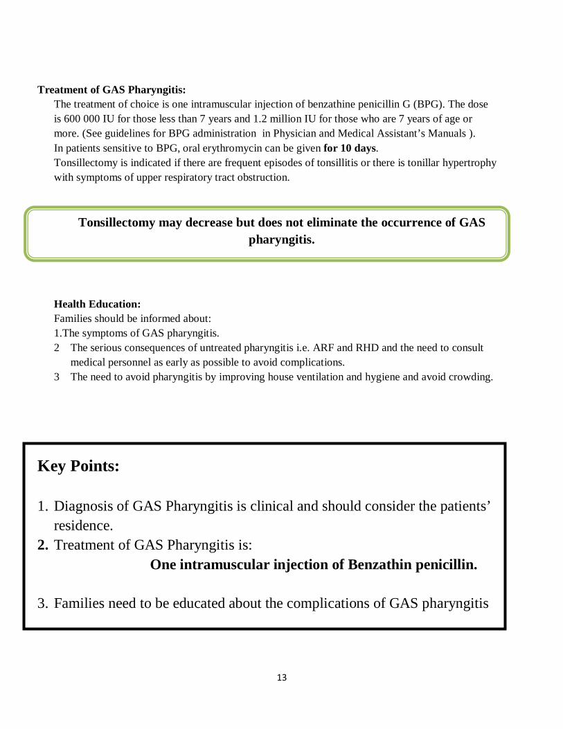
Treatment of GAS Pharyngitis:
The treatment of choice is one intramuscular injection of benzathine penicillin G (BPG). The dose is 600 000 IU for those less than 7 years and 1.2 million IU for those who are 7 years of age or more. (See guidelines for BPG administration in Physician and Medical Assistant’s Manuals ). In patients sensitive to BPG, oral erythromycin can be given for 10 days. Tonsillectomy is indicated if there are frequent episodes of tonsillitis or there is tonillar hypertrophy with symptoms of upper respiratory tract obstruction.
Tonsillectomy may decrease but does not eliminate the occurrence of GAS pharyngitis.
Health Education: Families should be informed about: 1.The symptoms of GAS pharyngitis. 2 The serious consequences of untreated pharyngitis i.e. ARF and RHD and the need to consult
medical personnel as early as possible to avoid complications. 3 The need to avoid pharyngitis by improving house ventilation and hygiene and avoid crowding.
Key Points: 1. Diagnosis of GAS Pharyngitis is clinical and should consider the patients’
residence. 2. Treatment of GAS Pharyngitis is:
One intramuscular injection of Benzathin penicillin.
3. Families need to be educated about the complications of GAS pharyngitis

14
References:
1. Ethar M Malik, Sulafa KM Ali. Prediction of bacterial pharyngitis in children using clinical features. Khartoum Medical Journal 2014,7: 967 – 971.
2. Micharl R. Wessels.Streptococcal Pharyngits. N Engl J Med 2011;364:648-55
3. McIsaac WJ, Kellner JD, Aufricht P, Vanjaka A, Low DE. Empirical validation of guidelines for the management of in children and adults. JAMA 2004;291:1587-95.
4. Rimoin AW, Hamza HS, Vince A, Kumar R, Walker CF, Chitale RA, daCunha ALA, Qazi S, Steinhoff MC.Evaluation of the WHO clinical decision rule for streptococcal pharyngitis. Arch Dis Child. 2005;90:1066–1070.
5. Steinhoff MC, Abd el Khalek MK, Khallaf N, Hamza HS, el Ayadi A, Orabi A, Fouad H, Kamel M Effectiveness of clinical guidelines for the presumptive treatment of streptococcal pharyngitis in Egyptian children. Lancet. 1997;27:918-21.
6. Sahin F, Ulukol B, Aysev D, Suskan E. The validity of diagnostic criteria for streptococcal pharyngitis in Integrated Management of Childhood Illness (IMCI) Guidelines. J Trop Pediatr. 2003;49:377–379.

15
Chapter Three
Acute Rheumatic Fever
Introduction:
Acute rheumatic fever (ARF) is a multisystemic auto immune disease that involves mainly the joints, the heart and the brain and rarely the skin. It takes place 2-3 weeks after an infection of upper respiratory tract with Lancefield group Aβ haemolytic streptococci (GAS). Streptococcal skin infection rarely been implicated in the disease process (1, 2).
ARF, like streptococcal infection occurs commonly between the ages of 5-15 years. It is rare in children under three years of age and adults, the peak incidence of first attack is between 6-8 years (3).In Sudan , ARF has been reported in patients 5 years of age or younger in 10% of patients (4).
Male to female ratio is similar regarding the overall incidence of ARF. While chorea and mitral valve disease are common in females, aortic valve disease is more common in males.ARF and RHD have been found in nearly every ethnic group. It is a worldwide phenomenon, and in every country its frequency is a function potentiated by crowding and poverty In cold climates in the past, there were two peaks of ARF in late winter and in autumn, but the difference is not marked in warm countries, other authors reported highest peak in winter and spring months in cold climate countries and this may be related to increased opportunity to spread streptococcal infection in cold and damp months. Prevention provided the most direct evidence showing that the relationship between GAS and ARF is casual rather than coincidental (2).
Nearly half a million people worldwide suffer an episode of ARF each year and at least 15 million people live with subsequent valve damage of RHD. Robust epidemiologic data for ARF and RHD is insufficient; the true burden of disease is likely to be several times higher than current estimates. Approximately half a million people die of RHD annually around the world. These deaths are premature; on average, people dying from RHD are below the age of 40 years (5).
Clinical Features of Acute Rheumatic Fever:
In one third of patients the streptococcal infection passes unnoticed and 54 to 70% of recurrences of ARF were caused by asymptomatic streptococcal infection. A latent period, the interval between the onset of symptoms of streptococcal infection and symptom of RF, duration may be 1-5 weeks, and in chorea may be 2-6 months (1).

16
Jones Criteria: 2015 Modification:
The American Heart Association published the revised Jone’s Criteria endorsed by the World Heart Federation. (table1)(ref 6)
Table 1: 2015 Modification of Jones Criteria
Important Modifications of Jones Criteria 2015
1. Inclusion of subclinical carditis (echo diagnosed) as a major criteria. ECHO SHOULD BE DONE TO ALL PATIENTS WITH SUSPECTED ARF
2. Consideration of the patients origin: in patients from high risk areas (defined by a prevalence of RHD more than 1 per 1000 or incidence > 2 per 100 000 per year) ARF is diagnosed using less strict criteria.
3. In high risk settings, monoarthritis and polyarthralgia are considered as Major Criteria and monoarthralgia a minor criteria.
4. Degree of fever and ESR in high risk areas is less than in low risk areas. (Ref 6)

17
Major Manifestations:
1. Carditis:
Occurs in 40% of patients during the first attack and almost 100% if ARF recurs. It may be the only major manifestations and usually appears in the first week of the illness.
Patients typically present with heart murmurs. Heart failure ensues if the valve regurgitation is severe, or can be asymptomatic if the valve affection is mild. Tachycardia presenting during sleep, disproportionate to fever or persisting after the control of fever is highly suggestive of carditis. In patients suspected with ARF ,without heart murmurs, echo should be done to exclude subclinical carditis.
Murmurs Heard In ARF: (see also chapter 4)
An organic apical pan-systolic murmur due to mitral regurgitation (MR) is the commonest murmur in ARF. This is sometimes accompanied by apical mid-diastolic(Cary Coombs) murmur denotes mitral valvitis and should be differentiated from that of mitral stenosis which does not occur in the acute phase.
Basal diastolic murmur of aortic regurgitation (in the second aortic area) can be present, usually associated with MR and occasionally isolated.
Mitral stenosis is a late manifestation of ARF, which are not present in the acute phase. Arrhythmias such as delayed AV conduction (see below) is common but does not necessarily indicate carditis. Nodal rhythm and atrial fibrillation are rarely caused by ARF.
Pericarditits manifested by a pericardial rub can rarely be present.
Diagnosis of Carditis:
Rheumatic carditis is diagnosed when there is clinical or echo evidence of valvitis with or without pericarditis.
The old theory that ARF causes histopathological pancarditis has been refuted by the finding that the rheumatic process does not involve the myocardium but rather affects the endocardium. This fact is supported by the absence of clinical myocarditi s. (7)
Subclinical carditis (SCC):
SCC is pathological valvular regurgitation/stenosis detected on echocardiography that is not evident clinically. With better definition of echocardiographic criteria for SCC that was recently published by World Heart Federation group, American Heart Association has now accepted SCC as a major manifestation (see below and see chapter 4) (8,6).

18
Jones Criteria (2015 Modification)
2. Arthritis:
Characteristically involves multiple big joints and is migrating. It is unusual to involve the central joints as spines, hips and the peripheral ones as the fingers and toes. Infrequently it involves the tempromandibular joint. Severe pain with inability to move the joint is characteristic. The pain continues for 1-2 days then flits to the other join. Examination of the joint reveals tenderness, swelling, heat and redness.
Monoarthritis or nonflitting polyarthritis can rarely occur and has been recently included as major criteria in high risk population and monoarthralgia included as a minor criterion. (See modified Jones Criteria 2015 above)
Arthritis typically improves dramatically with aspirin and is a self limiting condition without permanent sequelae.
3. Subcutaneous nodules:
Subcutaneous nodules are not pathognomonic of ARF as they occur in rheumatoid arthritis and systemic lupus. They rarely occur as isolated manifestation but most often with severe carditis appearing several weeks after its onset. They are round, firm, painless, 0.1 – 1cm in diameter sited over the bone prominences and over tendons, with overlying skin being mobile and not inflamed. They occur particularly over the extensor tendons of the fingers and toes and flexors of the wrists and ankles. They occur in crops and vary in numbers from one to usually three or four dozen. When numerous, they tend to be symmetrical. Their duration last for a week or two and rarely more than a month, and sometimes disappear within several days.
Minor Criteria
1. Fever
2. Polyrthralgia, monoarthralgia in high risk areas
3. Increased acute phase reactants
4. Prolonged PR interval
Major Criteria
1. Carditis (clinical or echo diagnosed)
2. Arthritis : polyarthritis ; monoarthritis & polyarthralgia in high risk areas
3. Chorea
4. Erythyma marginatum
5. Subcutaneous nodules
19
4. Erythema Marginatum:
Occurs in10% of patients with ARF commonly with other manifestations. The lesions are pink often slightly raised macules that fade centrally and coalesce to form serpiginous patterns. It usually occurs in the covered parts and may be manifested by local application of heat. They disappear within hours and may appear intermittently within weeks to months (9).
5. Chorea (Sydenham, St Vitus Dance):
It is involuntary purposeless movements, usually bilateral but sometimes unilateral that develops gradually over weeks. It is variable from being slight, brought about by excitement or conscious effort to being so violent that may result in self injury. It occurs most often in pre-pubertal girl and rarely among adults in either sex.
Chorea may occur as the only clinical signs or may precede, follow or exist concomitantly with other manifestations of ARF. Speech and handwriting abnormalities are common. Serial handwritings can be used to detect and follow up progress of the disease.
There is a difficulty in counting rapidly and maintaining the protrusion of the tongue. There is also hyperextension of the fingers and wrist when the arms are held extended over the head. The hand grip is weak and there may be intermittent muscular contractions or twitches. Patellar response shows a hung up type of response. Emotional liability is marked as is shown by inappropriate bouts of crying or laughter.
Minor manifestations:
1. Fever:
It is almost invariably present in the early stage, except in patients whose only manifestation is chorea or those receiving salicylates or steroids. It often becomes low grade after the first week and may persist at this level for 2-4 weeks.
2. Arthralgia:
Pain without objective changes of swelling, heat and redness may occur in some joints. Myalgia is rare. Arthralgia should not be counted as minor when arthritis is counted as major criteria.In high risk population , polyarthralgia is a major criterion and monoarthralgia a minor one.
3. Acute phase reactants:
Include elevated C-reactive protein (CRP) and erythrocyte sedimentation rate (ESR) are commonly raised in ARF.In high risk areas ESR>30 mm/hr is considered as a minor criterion
4. Prolonged PR interval:
Prolonged PR interval (first degree heart block) alone does not constitute evidence of carditis or predict long term cardiac sequelae and is transient. It should not be considered when carditis is counted as a major manifestation. Rarely, complete (3rd degree) heart block may occur.

20
Other Findings:
Include history of recent sore throat, family history of ARF, abdominal pain, epistaxis, tachycardia, rheumatic pneumonia, anemia, chest pain, weight loss and malaise.
Post Streptococcal Psychiatric Disorder:
Clinical & research findings in both immunology & neuropathology have established the existence of post streptococcal neuropsychiatric disorders. Pediatric autoimmune neuropsychiatric disorders associated with streptococcal infections (PANDAS) is acronym applied to subgroup of children with obsessive-compulsive or tics disorders occurring in association of streptococcal infections. In addition, there are recent reports of dystonia, chorea, encephalopathy and dystonic chereoasthetosis occurring as sequalae of streptococcal infections (10).
Recent Evidence of Group A Streptococcal infection:
For diagnosis of ARF there should be an evidence of recent GAS infection. It includes raised tites of antistreptolysin O (ASO), anti DNase B and antihyalurindase. About 80% of patients with ARF have elevated ASO; however, 95-100% have elevation if the three different antibodies are measured. (11). Evidence also includes a positive throat culture for (GAS), recent scarlet fever, and rapid antigen test for GAS.
Diagnosis of ARF:
For diagnosis of ARF in new patients, the revised Jones criteria should be adhered to which states that one major and two minor criteria or two major criteria plus recent evidence of GAS infection.
For diagnosis of ARF in patients known to have RHD: one major or two minor criteria are needed plus recent evidence of GAS infection.
New Episode
2 Major Criteria
OR
1 Major + 2 Minor Criteria
+ High ASO
Recurrent Episode
1 Major Criterion
OR
2 Minor Criteria
+ High ASO
Probable (atypical) ARF
Fewer criteria
Atypical (e.g. mono or poly) arthritis
+ High ASO
21
Special Situations:
1. Probable (Atypical) ARF:
Patients may present with non-flitting polyarthritis, polyarthralgia or monoarthritis and with several (3 or more) other minor manifestations, together with evidence of recent GAS infection. Some of these cases may later turn out to ARF. It is prudent to consider them as case of probable ARF (once other diagnoses are excluded) and advice regular secondary prophylaxis; such patients require close follow up and regular examination of the heart. This cautious approach is particularly suitable for patients in vulnerable age groups in high incidence settings (6)
2. Chorea:
Often presents without other manifestations and patients should be started on secondary prophylaxis.
3. Insidious (indolent) Carditis:
Slowly progressive valve damage without history of ARF is common in endemic countries and these patients are high risk as the valve damage has already started. Strict secondary prophylaxis is needed.
Treatment of ARF:
1. Antibiotics:
A single intramuscular injection of benzathine penicillin G (BPG) to eradicate GAS from upper respiratory tract. The dose is 600 000 IU for those less than 7 years and 1.2 million IU for those who are 7 years of age or more. (See guidelines for BPG administration in Physician and Medical Assistant’s Manuals). After this initial course of antibiotic therapy the patient should be started on long term secondary prophylaxis. Duration of secondary prophylaxis is for life in case of RHD and up to 25 years of age if there is no carditis.
2. Anti inflammatory therapy:
Patients should be treated with oral salicylates. The usual dose of aspirin is 75 mg/kg/24 hrs divided q.i.d. PO for 4 wk then tapering according to the acute phase reactants. Aspirin should be given after meals.
Indications for Steroids:
There is little evidence that steroids are superior to salicylates. (12). Steroids can be considered in the following situations:
1. Patients who do not tolerate aspirin.
2. Patients who fail to show improvement (by clinical or acute phase reactants) with aspirin
22
The usual dose is 2 mg/kg/day divided 6 to 8 hourly for 2weeks followed by tapering of the dose by 5 mg/day every 2-3 days. At the beginning of tapering the dose aspirin should be started at dose of 60 mg/kg/day divided qid for 6 wk keeping a period of overlap.
3. Supportive Treatments:
Supportive therapies for patients with moderate to severe carditis include fluid & salt restriction, diuretics and angiotensin converting enzyme inhibitors. Physicians need to consult cardiologists.
Treatment of Sydenham’s chorea:
Anti-inflammatory agents are usually not indicated. Sedatives may be helpful early in the course of chorea. Haloperidol (0.01-0.03 mg/kg/day divided bid PO) or choropromazine (0.5 mg/kg 4-6 hr PO) should be initiated and continues for 3-4 months.
Prevention of ARF: (also see chapter 6)
Depends on eradication of group A streptococci from upper respiratory tract. It is divided into:
Primordial Prevention:
Improving socioeconomic conditions, nutrition, housing conditions (decreasing crowding) and improving access to health care can all decrease the incidence of ARF.
Primary prevention:
Prompt treatment of GAS pharyngitis with one injection of IM BPG is highly effective in preventing first attacks of ARF. However, about 1/3 of patients with ARF do not recall preceding episode of pharyngitis
A vaccine for GAS is being developed but has not yet been used in clinical practice (see chapter 6)
Secondary prevention:
Secondary prevention requires continuous BPG administration, which should begin as soon as the diagnosis of ARF is made. For those with no evidence of carditis in their initial attack should receive BPG every three weeks up to 25 years of age; however those with evidence of RHD should receive it for life. The interval of 3 weeks has been questioned and some countries are administering BPG every 2 weeks. To modify the protocol, BPG brands should be tested to confirm their bioavailability.
BPG Quality:
In developed countries, good brands of BPG do not require skin testing as the purity of the drug is assured while in developing countries bearing the burden of ARF/RHD, BPG purity has been questioned. (13)

23
Therefore, countries need to work on importing high quality BPG and test its bioavailability in order to determine the secondary prophylaxis interval and allergenic properties.
The doses are the same as for primary prevention. In cases of documented sensitivity to penicillin, erythromycin can be given as 250 mg (below 7years of age) and 500 mg (above 7 years of age) PO BD for the same period.
Improving Medical Personnel and Public Awareness:
ARF is considered to be a neglected disease even in endemic countries, therefore continuous training of physicians and public awareness campaigns need to be conducted in order to improve its recognition and management. (14)
Key points: • Modified Jones Criteria for 2015 included subclinical carditis
polyarthralgia and monoarthritis as major criteria and monoarthralgia as a minor criteria in high risk settings.
• Diagnosis of recurrent episode of ARF utilizes less criteria than the first episode
• In highly endemic areas if criteria are short of ARF, probable ARF needs is a new category.
• Benzathine penicillin, salicylates and supportive treatment should be started in patients with ARF
• Secondary prophylaxis should continue for life in case of RHD and up to 25 years of age if no carditis.
• Strict follow up is needed to assure adherence of patients to secondary prophylaxis

24
References
1.Abdel Rahman SMK. Rheumatic fever with special reference to prevention and recommendations to Sudan, written document presented within the collaborative program between Uppsala University Hospital and Ministry of Health, Sudan, International Child Health Unit, Uppsala, 1985.
2.McDonald, M.I. et al. Low rates of streptococcal pharyngitis and high rates of pyoderma in Australian aboriginal communities where acute rheumatic fever is hyperendemic. Clin. Infect. Dis. 2006; 43: 683-843.
3.Chan LT, Reddy V, Rhoads GG: Occurrence and rheumatic fever among ethnic group of Hawii. AJDC 1984, 138: 476-78. 4. Eynas Khalid, Hamid El Banna, Rehab Mahmoud, Hanadi Hassan, Laila El Mahdi, Sulafa Ali.Clinical and echocardiographic features of 370 children with rheumatic heart disease seen in Khartoum. Sudan Med J 2014;50:151-54 5.Wyber R, Grainger-Grasser A, Thompson D et al. Tools for implementation rheumatic fever disease control programmes, Quick Tips summary. World Heart Federation and RhEACH, Perth, Australia 2014:2. 6. Michael H Gewitz et al.Revision of the Jones Crtieria for diagnosis of acute rheumatic fever in the era of echo/Doppler.Circulation 2015;131:1806-18. 7. Tandon R. Rheumatic fever pathogenisis: Approach in research needs change. Ann Pediatr Cardiol 2012 Jul; 5(2): 169-78. 8.Reményi B, Wilson N, Steer A, Ferreira B, Kado J, Kumar K, Lawrenson J, Maguire G, Marijon E, Mirabel M, Mocumbi AO, Mota C, Paar J, Saxena A, Scheel J, Stirling J, Viali S, Balekundri VI, Wheaton G, Zühlke L, Carapetis J.World Heart Federation criteria for echocardiographic diagnosis of rheumatic heart disease--an evidence-based guideline. Nat Rev Cardiol. 2012;9:297-309. 9. Michael A Gerber Rheumatic fever. In: Nelson Text Book of Pediatrics 19th ed. Saunders, Philadelphia 2011: 874-879. 10. Lisa A. Snider and Susan E. Swedo post-streptococcal auto immune disorders of central nervous system.Current option inNeurology2003,16:359-365. 11.Egyptian Ministry of Health, Rheumatic fever control & prevention program. Rheumatic Fever, Rheumatic Heart Disease Guidelines. 12.Cilliers A, Manyemba J, Adler AJ, Saloojee HAnti-inflammatory treatment for carditis in acute rheumatic fever.Cochrane Database Syst Rev. 2012;6. 13. Rosemary Wyber, Kathryn Tauberty, Stephen Markoz, Edward L. Kaplan. Benzathine Penicillin G for the Management of RHD,Concerns About Quality and Access, and Opportunities for Intervention and Improvement. Global Heart 2013; 8: 227-234. 14.Abdel Rahman SMK, Ali SKM. The control of rheumatic fever and rheumatic heart disease: a call to raise the awareness. Sudan J Pediatr 2014; 14(1): 21-24.

25
Chapter 4 Rheumatic Heart Disease
ARF is associated with carditis in 50-75% of patients with the first episode, the rate of carditis increases significantly with recurrence of ARF hence the importance of secondary prophylaxis. Rheumatic carditis affects mainly the heart valves. Although by histopathology the myocardium and pericardium may show the Aschoff nodules, the characteristic lesions of RHD , however, clinical symptoms and signs are mainly due to valvular involvement. The most common valves affected are the mitral and aortic valves. In Sudanese patients, isolated mitral regurgitation (MR) is reported in about 40% , followed by combined MR and aortic regurgitation (AR) in 35%, combined MR and mitral stenosis (MS) in 7%, isolated AR in 4% and isolated MS in 3%. In about 11% of patients, combination of more than 2 valves was detected. (1). In older children and adults, stenotic lesions become more dominant, aortic stenosis is rare in all ages. In another cross sectional study MS was found to be 9% and all the patients were found to be older children with severe disease, the postulation was that mild MS may be missed by clinical examination as it is asymptomatic and the signs are subtle. This is supported by the finding that MS is the commonest lesion in young adults which indicates low rate of detection in children (2) Types of RHD: Rheumatic carditis can be categorized into subclinical and clinical types. Recently, echo screening of asymptomatic school children revealed a very high prevalence of subclinical carditis , up to 10 times the clinical RHD (3)

26
Subclinical Carditis
Definition: “The echocardiographic diagnosis of RHD in asymptomatic patients without audible murmurs” Echo diagnosis of RHD without heart murmur can be made in 2 situations: A: RHD discovered during screening programs at community level. B: RHD discovered on screening in patients with ARF (e.g. with arthritis or chorea): in this situation we have a lower threshold to diagnose subclinical carditis. Subclinical carditis detected on screening of school children utilized strict guidelines published by the World Heart Federation (4). Although long term follow up of such patients had so far shown a favorable outcome, guideline are still not well developed and more follow up time is needed . (5). Sub clinical echo-diagnosed RHD in the setting of screening programs is divided into 2 types:
1. Definite RHD
2. Borderline RHD
( See tables 1-3 -Ref 3)
Echocardiographic types of Subclinical Carditis
Definite RHD
1. Pathological MR and 2 morphological features of RHD
2. MS mean gradient 4 mmHg or more
3. Pathological AR and 2 morphological features of RHD
4. Borderline disease of both AV and MV
Borderline RHD
1. Two Morphological features of MV
2. Pathological MR
3. Pathological AR

27
Morphological Features of Mitral and Aortic Valves
Doppler Features of Pathological MR and AR
Management of Subclinical Carditis:
In the setting of screening programs, individuals with definite RHD need to be started on BPG prophylaxis in the same protocol of clinical RHD.
In borderline RHD, patients should be closely monitored by echo every 6 months to determine the progression of echo abnormalities and the need for prophylaxis.
In Sudan, echo screening is used:
1. As a research tool to measure the prevalence of RHD
2. For early detection in highly endemic areas order to start secondary prophylaxis for definite cases.
Pathological MR
1. Seen in 2 views
2. Jet length 2 cm or more
3. Velocity 3m/s or more
4. Pan systolic jet
Pathological AR
1. Seen in 2 views
2. Jet length 1cm or more
3. Velocity 3 m/sec or more
4. Pan diastolic jet
Morphological Features of Mitral Valve
1. AML >3 mm
2. Chordal thickening
3. Restricted leaflet motion
4. Excessive leaflet tip motion in systole
Morphological Features of Aortic Valve
1. Irregular/focal thickening
2. Coaptation defect
3. Restricted leaflet motion
4. Prolapse
28
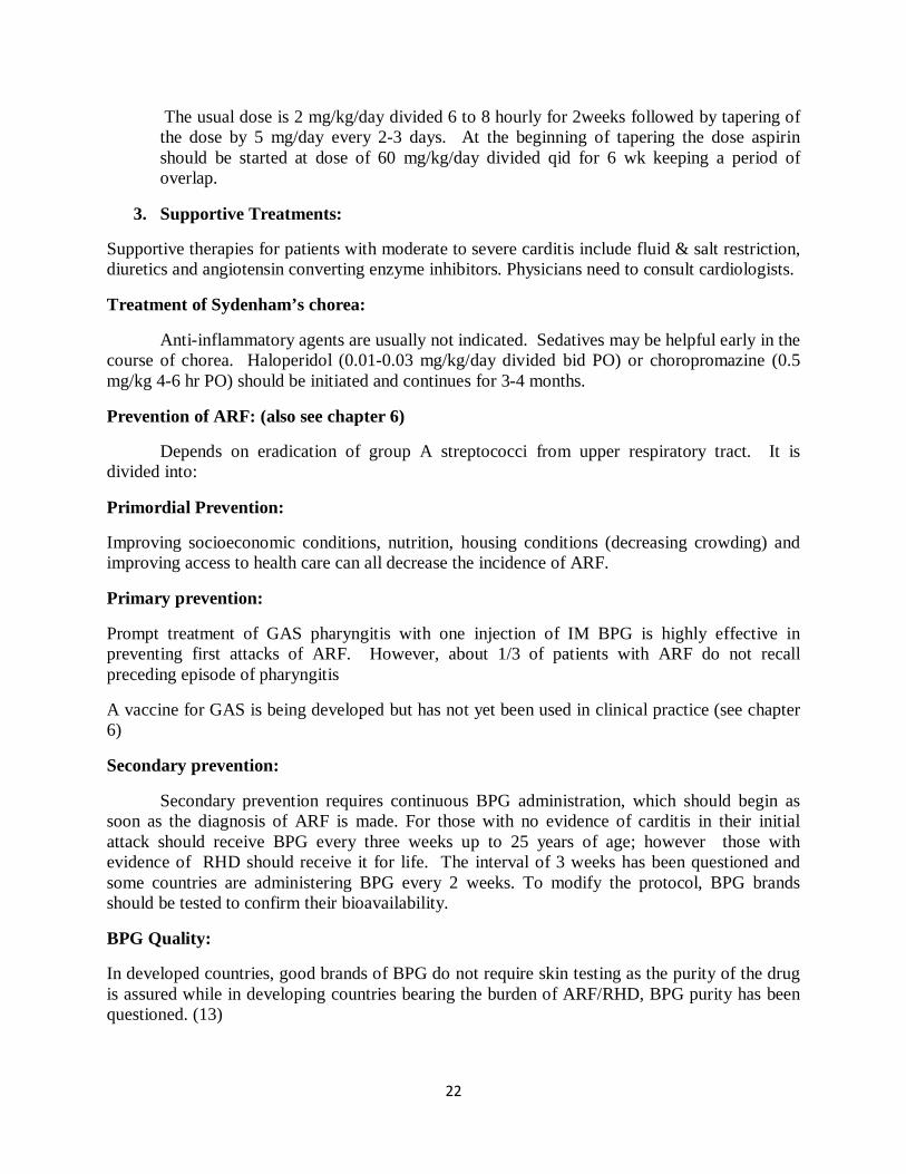
Clinical Rheumatic Carditis
Patients with clinical carditis almost always present with heart murmurs, depending on the severity of valve dysfunction, variable degrees of heart failure ensue. Initial presentation can be with other features of ARF (arthritis, chorea) but many patients deny any history of preceding ARF.
Pathophysiology of RHD:
Involvement of the valves is the fundamental issue of RHD which affects mainly the mitral and aortic valves. Clinically, it presents with features of pulmonary congestion or left heart failure (LHF) initially, then it may progress with time to develop pulmonary hypertension (PHT) and right sided heart failure (RHF) with features of both LHF and RHF i.e. congestive heart failure (CHF) then with the progression of PHT the features of pulmonary congestion may diminish and the picture becomes that of severe RHF and end stage cardiac disease.
Symptoms start with shortness of breathing associated with cough, frothy sputum (LHF) that may be accompanied by hemoptysis. However with the ongoing pulmonary congestion the pulmonary vascular bed reacts by hypertrophy to protect the lungs from the pulmonary edema, this will result in increased pulmonary vascular resistance so the symptoms of cough, frothy sputum and hemoptysis may became less with the progression of the PHT while the symptoms of RHF ( swelling of the lower limbs due to systemic venous congestion and its associated symptoms of abdominal pain and dyspepsia due to the congestion of the portal circulation) may ensue leading to features of CHF.
Rheumatic Heart DiseaseMurmurs+/- Heart failure
With ARF: Commonly arthritis, chorea
Without ARF: ARF not present(Indolent carditis) or missed

29
Clinical Features:
Symptoms:
1.Congestion:
Pulmonary: cough, shortness of breathing. Systemic: abdominal discomfort, lower limb edema
2.Low Cardiac Output & compensatory mechanisms:
Palpitations (could be atrial fibrillation), sweating, fatigue.
Systemic embolization: hemiplegia
Signs (see diagrams below)
-Isolated MR, MR with AR
-Isolated AR , Isolated MS
-MR/MS
-TR/TS and AS are rare
-Systemic embolization: hemiplegia
30
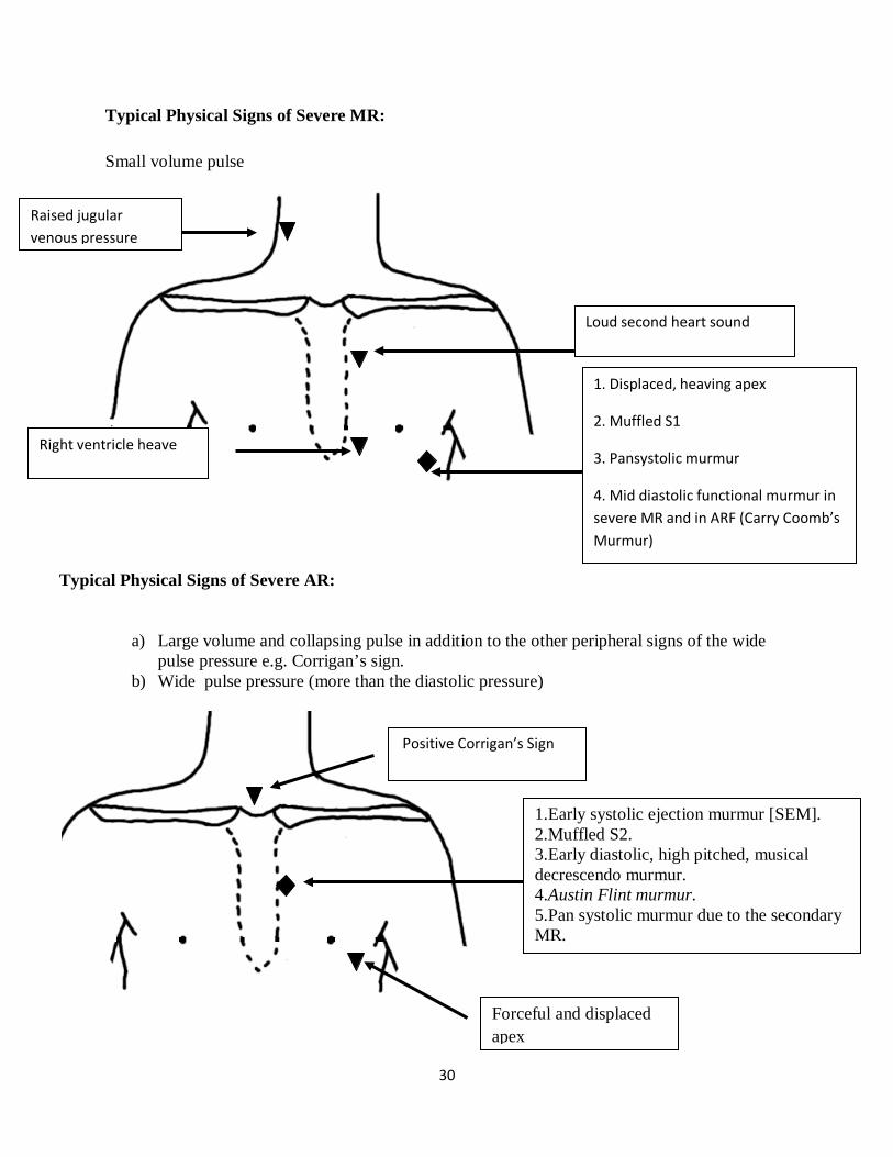
Typical Physical Signs of Severe MR: Small volume pulse
Typical Physical Signs of Severe AR:
a) Large volume and collapsing pulse in addition to the other peripheral signs of the wide pulse pressure e.g. Corrigan’s sign.
b) Wide pulse pressure (more than the diastolic pressure)
1. Displaced, heaving apex
2. Muffled S1
3. Pansystolic murmur
4. Mid diastolic functional murmur in severe MR and in ARF (Carry Coomb’s Murmur)
Loud second heart sound
Right ventricle heave
Forceful and displaced apex
1.Early systolic ejection murmur [SEM]. 2.Muffled S2. 3.Early diastolic, high pitched, musical decrescendo murmur. 4.Austin Flint murmur. 5.Pan systolic murmur due to the secondary MR.
Positive Corrigan’s Sign
Raised jugular venous pressure
31
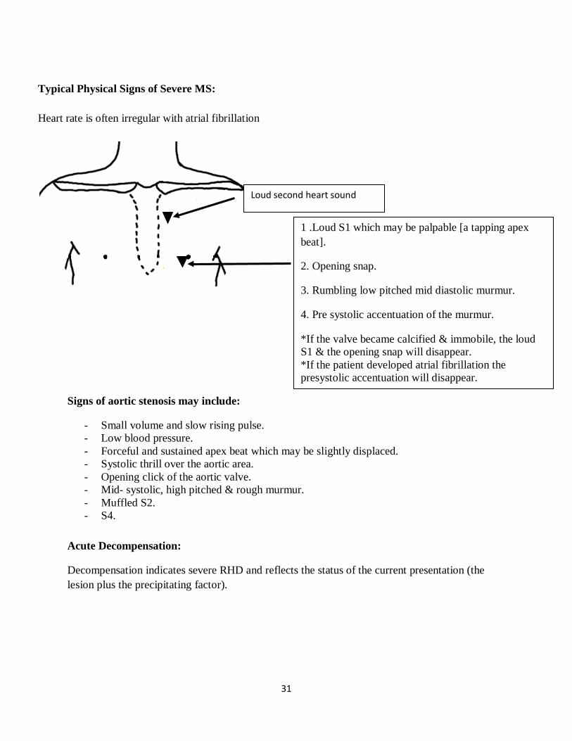
Typical Physical Signs of Severe MS: Heart rate is often irregular with atrial fibrillation
Signs of aortic stenosis may include:
- Small volume and slow rising pulse. - Low blood pressure. - Forceful and sustained apex beat which may be slightly displaced. - Systolic thrill over the aortic area. - Opening click of the aortic valve. - Mid- systolic, high pitched & rough murmur. - Muffled S2. - S4.
Acute Decompensation:
Decompensation indicates severe RHD and reflects the status of the current presentation (the lesion plus the precipitating factor).
1 .Loud S1 which may be palpable [a tapping apex beat].
2. Opening snap.
3. Rumbling low pitched mid diastolic murmur.
4. Pre systolic accentuation of the murmur.
*If the valve became calcified & immobile, the loud S1 & the opening snap will disappear. *If the patient developed atrial fibrillation the presystolic accentuation will disappear.
Loud second heart sound

32
Causes of Decompensation:
1. Recurrence of ARF: related to discontinuation of secondary prophylaxis is the most common cause of decompensation in young patients.
2. Volume overload
- Loss of volume control by discontinuation of medications “diuretics and vasodilators”
- Causes of hyper dynamic circulation e.g. fever, anemia, pregnancy.
3. Pressure overload
- Sudden rise in blood pressure.
- Pulmonary embolism.
4. Intrinsic pump problem
- Systolic dysfunction: related to long standing valve regurgitation
- Diastolic dysfunction: e.g. rapidly accumulating pericardial effusion.
5. Arrhythmias:
a. Tachyarrhythmia: Fast atrial fibrillation [AF]. b. Brady arrhythmia: Could be related to drugs that slow the heart e.g. B Blockers and Digoxin

33
The following precipitating factors are more relevant to RHD:
1- Discontinuation or non compliance with anti failure medications , the problem is even more serious with warfarin treatment (thrombotic or bleeding complications) or secondary prophylaxis with penicillin( progression of disease with recurrent ARF)
2- As these patients are of low socioeconomic status they are prone to recurrent infections and febrile illnesses. Fever is serious and correct diagnosis is essential for the cause of fever which may addressed in three categories:
a- Recurrence of ARF (see above) it may be the initial presentation or as a precipitating factor and it indicates lack of secondary prophylaxis and may aggravates the lesion.
b- Endocarditis (may be acute or sub acute) serious complication which may lead to rapid deterioration and destruction of the valve and may need urgent intervention
Endocarditis is common with regurgitant lesions and rare in mitral stenosis
Assessment of Severity:
Group 1: Severe lesion but the patient is asymptomatic:
- This may be encountered more with Stenotic lesions (MS and AS), symptoms may be late and the patient may show rapid downhill course when they appear.
- Careful history may reveal subtle symptoms - They may need intervention to decrease morbidity and mortality.
Group 2: Severe lesion and symptomatic:
- Needs intervention at the right time before developing resistant heart failure and adverse surgical outcome measures.
Objective Assessment of Severity:
This may be assessed with great accuracy by physical signs (see above) and basic investigations (Echo, CXR and ECG)
CXR
- The primary role is to assess the heart size and the pulmonary vascular bed . - Cardiomegaly is defined as the greatest transverse cardiac diameter > ½ the chest
diameter

34
The pulmonary vascular bed:
This can be affected in four ways:
1- Pulmonary congestion. 2- Pulmonary plethora. 3- Pulmonary hypertension. 4- Pulmonary oligemia. In regard to RHD pulmonary congestion and hypertension are important as mitral and aortic valves lesions lead to increased LA pressure which leads early to pulmonary congestion and eventually to pulmonary hypertension.
PULMONARY CONGESION
Grade 1:PV WP=15-20 Grade 2: PVWP=20-25 Grade 3: PVWP=25-30
Pulmonary congestion is defined as increased pulmonary venous pressure due to high left atrial (LA) pressure (Passive congestion).
There are three grades of pulmonary congestion
Grade 1: Upper lobe diversion:
The veins of the upper lobes will become prominent due to increased vascularity of the upper lobes (which are well aerated) this correlates with pulmonary wedge pressure PWP of 15—20 cm water .
Grade 2:Interstitial edema:
Evident as Kerly B lines, this correlates with PWP of 20 – 25 cm water .
Grade 3:Frank alveolar edema:
Upper Lobe
Diversion
Interstitial
Edema
Alveolar
Edema

35
Fluffy white patches and butterfly shadow radiating from the hila, this correlates with PWP > 25 cm water.
Pulmonary Hypertension: This is evident on CXR as prominent pulmonary hila due to enlarged pulmonary arteries plus peripheral pruning (cutting) of the pulmonary circulation.
So the main indication of taking a chest x ray in a patient with RHD for the follow up patient is to assess the pulmonary vascular bed and to detect the signs of transition form of pulmonary congestion to pulmonary hypertension so as to interfere at the right time as severe pulmonary hypertension increases the operative risk and makes the long term prognosis less promising.
ECG : - To assess chamber enlargement or hypertrophy
- To assess the rhythm e.g. AF - effects of drugs e.g. Digitalis
Echo: Echo is the main tool for assessing valvular lesions. In RHD Echo is important to:
- Confirm the diagnosis of RHD - To assess the severity of the lesion (see blew) - To assess prosthetic valves’ function - Detect pericardial effusion ( due to AFR or severe heart failure) - Detect vegetations - Left atrial echo contrast/ clots
Assessment of Valve Dysfunction: (Simplified Approach)
Mitral Regurgitation severity
Mild Moderate Severe
• Small central jet <4 cm2 or <20% of LA area
• Vena contracta width <0.3 cm
• Signs of MR>mild present, but no criteria for severe MR
• Vena contracta width ≥ 0.7cm with large central MR jet (area < 40% of LA) or with a wall-impinging jet of any size, swirling in LA
• Systolic reversal in pulmonary veins
• Prominent flail MV leaflet or ruptured papillary muscle.
36
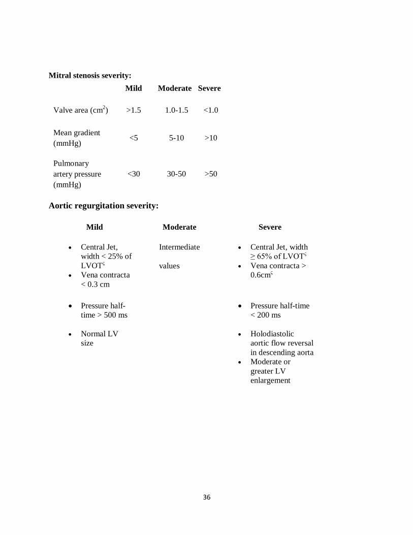
Mitral stenosis severity: Mild Moderate Severe
Valve area (cm2) >1.5 1.0-1.5 <1.0
Mean gradient (mmHg)
<5 5-10 >10
Pulmonary artery pressure (mmHg)
<30 30-50 >50
Aortic regurgitation severity:
Mild Moderate Severe
• Central Jet, width < 25% of LVOTς
• Vena contracta < 0.3 cm
Intermediate
values
• Central Jet, width ≥ 65% of LVOTς
• Vena contracta > 0.6cmς
• Pressure half-time > 500 ms
• Normal LV size
• Pressure half-time < 200 ms
• Holodiastolic aortic flow reversal in descending aorta
• Moderate or greater LV enlargement

37
Assessment of AS severity:
Mild Moderate Severe
Aortic jet velocity (m/s)
≤2.5 m/s
2.6-2.9 3.0-4.0 >4.0
Mean gradient (mmHg)
- <20 (<30a) 20-40 (30-50a)
>40b (>50)
AVArea (cm2)
- >1.5 1.0-1.5 <1
(Modified from: Classification of valve stenosis and regurgitation http://www.echopedia.org/wiki/Classification_of_valve_stenosis_and_regurgitation)
Assessment of Left Ventricle Function and Pulmonary Pressure:
Ejection Fraction (EF) needs to be measured and carefully monitored by echo. EF is usually high normal in patients with valve regurgitation. Decreasing EF is an indication for surgery and a low/normal EF is a surgical risk factor that may not improve after the operation.
Measurement of pulmonary artery systolic pressure from the tricuspid valve regurgitation velocity helps to assess the severity of the lesion and the timing as well as risk assessment of surgery.
Management of RHD:
At the time of first presentation: First step is to make sure that there is no active disease: Do ESR, CRP, ASO titre and consider recurrence of ARF is there is a major or 2 minor criteria and positive ASO. If there is active disease, anti inflammatory therapy should be started till the acute phase reactants return to normal (4-6 weeks) and the patients should be re evaluated at that time.

38
It should be emphasized to the patients that they should continue Benzathine penicillin prophylaxis for life

39
Management of Patients with Severe valve regurgitation
The most common problems faced in treating patients with RHD are: 1. It is a disease of childhood and young adults 2. It is a disease of low socioeconomic communities. 3. Recurrence of rheumatic activity, on-going inflammation and failure to control factors of
volume overload and hyper dynamic circulation may lead to severe disease that mandates early intervention in childhood as well as it may lead to failure of valve repair.
4. Endocarditis in patients with prosthetic as well as native regurgitant valves. 5. Treatment (both surgical and catheter-based) is expensive and at best is still palliative, as all
of them have limited longevity. 6. Problems of monitoring anticoagulation treatment in patients with prosthetic valves or AF,
leading to cerebro-vascular accidents (CVA) due to thrombosis/bleeding. 7. Grown up children and adults may need re do surgery due to valve mismatch.
Medical treatment:
- CHF needs to be managed with diuretics, angiotensin converting enzyme inhibitors (ACEI) and spironolactone.
- Digoxin can be added to patients with severe hemodynamic compromise or atrial fibrillation.
- Patients with atrial fibrillation should be started on beta blockers/digoxin and anticoagulation.

40
Management of patients with severe heart failure: Patients may present with features of cardiogenic shock including tachypnea, , dizziness or loss of consciousness, low blood pressure and multisystem failure.
Signs of edema and raised jugular venous pressure in the presence of hypotension should alert the physician to cardiogenic shock.
-Intensive care unit admission if possible, for cardio respiratory, blood gas, urine output and renal function as well as other system function monitoring. -ECG, pulse oximetery and when feasible central venous pressure and arterial (invasive) blood pressure monitoring, if not available non-invasive blood pressure can be used. -Low systolic pressure needs to be managed by inotropic support e.g dopamine 5-10 microgram /kg/minute. This helps to increase blood pressure but will increase systemic vascular resistance therefore it needs to be combined with dobutamine at similar dose, the latter causes peripheral vasodilatation and inotropy with minimal chronotropy. -Intravenous furosemide needs to be started, patients with massive edema benefit from infusion of frusemide, they need to be monitored for hypokalemia.Potassium sparing diuretics needs to be added. -Urgent surgical referral is needed.
Atrail Fibraillation
Betablockers (propranolol, metoprolol)
Digoxin (dose 250 micrograms BD)
Cardioversion if still symptomatic on
medications

41
Palliative and prognostic treatment
-Interventional treatment for RHD is regarded as ‘good Palliation’ as most procedure are associated with a high rate of reintervention.
-As the disease starts in childhood every effort should be done to delay the intervention (importance of secondary prophylaxis and control of factors leading to decompensation). -Patients should be followed carefully by Echo to determine the right time for intervention (late intervention carries high operative risk and less favorable prognosis.
Indications for intervention
1. Asymptomatic Patients: Asymptomatic patients with severe valvular lesion (see Echo criteria for severity) should be closely monitored to decide the appropriate time for intervention.
The following are indications for intervention: - Severe Pulmonary hypertension. - For regurgitant lesions (MR and AR), decrease in LV ejection fraction or Increasing in
LV dimensions especially LV end systolic dimension. Importantly, when the ejection fraction approaches 50% progression of heart failure and death occur in up to 25% of these patients per year if surgery is not performed (cardiomyopathy secondary to MR). When surgery is performed in patients with ejection fraction less than 50% the 10 year survival is only 30% compared to 100% for those whose ejection fraction is 60%.(6)
- Severe MS. 2. Symptomatic patients:
Symptomatic patients with severe valve dysfunction should be referred for intervention.
Treatment options
1- Valve repair - This is done mainly for MR but needs careful assessment by Echo to select the suitable
candidates. - To guide MV surgical repair techniques, the Carpentier’s classification for rheumatic
MR may be used (7): Type I: Annular dilatation
In pure type I dysfunction, the main mechanism is rapid progressive dilatation of the left ventricle and mitral annulus caused by inflammatory pancarditis, which also frequently alters left ventricular contractility.
42
Type IIa: Anterior Leaflet Prolapse
On echocardiography, in true prolapse, the anterior leaflet overrides the mitral annulus plane during systole. Causes of true anterior leaflet prolapse are elongation or rupture of anterior primary chordae tendinae or elongation of the papillary muscles.
Type II Anterior Leaflet Pseudoprolapse /III Posterior
Mixed-type: pseudo IIa (anterior leaflet Pseudoprolapse) and IIIp (restricted posterior leaflet motion).
On echocardiography, contrary to pure type IIa dysfunction, in the mixed type pseudo IIa/IIIp, the anterior leaflet does not override the mitral annulus plane during systole but there is lack of coaptation, resulting mainly from significant restricted motion of the posterior leaflet, allowing for the anterior leaflet to move up to the annular plane during systole without overriding it.
Type III: Restricted Leaflet Motion
(Restricted anterior and/or posterior leaflet) dysfunction.
The restricted motion mainly affected the closure of the leaflets during systole, resulting in lack of leaflet coaptation generating mixed stenosis and regurgitation, either predominantly regurgitant (mitral orifice area >1.5 cm2 ), or stenotic (mitral orifice area < 1.5 cm2) on echocardiography.
In the majority of such cases, associated annular dilatation contributes to the degree of mitral regurgitation by aggravating the lack of coaptation between both mitral leaflets, as well as in the mixed type pseudo IIa/IIIp dysfunction.
There may be variable degrees of calcification and multi valvular involvement making valve repair difficult.(8)
Prognosis after valve repair:
- Even after successful valve repair, recurrence of RHD can occur despite strict adherence to penicillin prophylaxis. Patients with chronic RHD have persistently elevated inflammatory markers (cytokines and C-reactive protein) which indicate an ongoing inflammation that leads to failure of their valve repair.(9)
- Valve repair is superior to replacement, however, even after successful valve repair, long term follow up of patients showed that late mortality was 15%, and the reoperation rate was 27% , mainly related to the ongoing inflammation (10)
- Patients are usually living in distant areas and commonly fail to follow up and may have a wrong impression that their disease has been cured with the operation.
43
2.Percutaneous transvalvular mitral commissurotomy (PTMC):
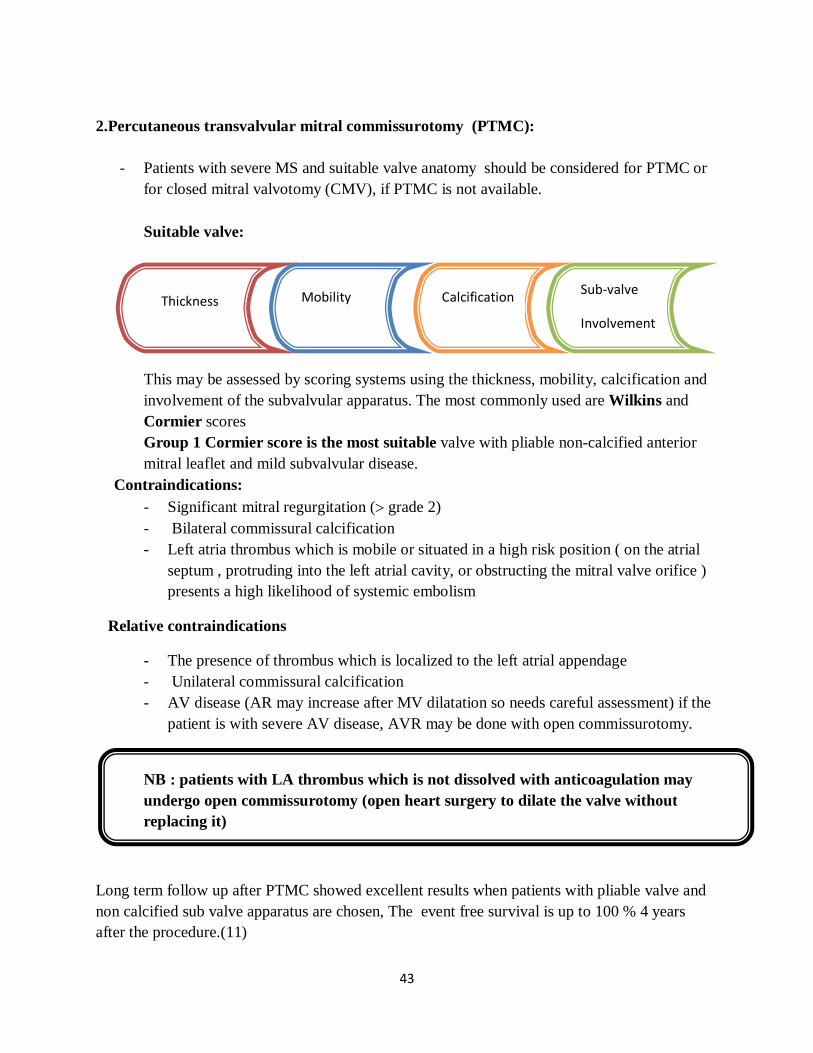
- Patients with severe MS and suitable valve anatomy should be considered for PTMC or for closed mitral valvotomy (CMV), if PTMC is not available. Suitable valve:
This may be assessed by scoring systems using the thickness, mobility, calcification and involvement of the subvalvular apparatus. The most commonly used are Wilkins and Cormier scores Group 1 Cormier score is the most suitable valve with pliable non-calcified anterior mitral leaflet and mild subvalvular disease.
Contraindications: - Significant mitral regurgitation (> grade 2) - Bilateral commissural calcification - Left atria thrombus which is mobile or situated in a high risk position ( on the atrial
septum , protruding into the left atrial cavity, or obstructing the mitral valve orifice ) presents a high likelihood of systemic embolism
Relative contraindications
- The presence of thrombus which is localized to the left atrial appendage - Unilateral commissural calcification - AV disease (AR may increase after MV dilatation so needs careful assessment) if the
patient is with severe AV disease, AVR may be done with open commissurotomy.
NB : patients with LA thrombus which is not dissolved with anticoagulation may undergo open commissurotomy (open heart surgery to dilate the valve without replacing it)
Long term follow up after PTMC showed excellent results when patients with pliable valve and non calcified sub valve apparatus are chosen, The event free survival is up to 100 % 4 years after the procedure.(11)
Thickness Mobility Calcification Sub-valve
Involvement

44
3. Valve replacement: - Prosthetic valve replacement is the option when valve repair or commissurotomy is not
feasible for MV disease and it is the main procedure for AV disease. - The main problems related to valve replacement are the need for life-long anticoagulation
and the risk of endocarditis. Both problems impose a huge burden on patients and the health system.
- It has been shown that prosthetic valve replacement in the presence of poor socio economic conditions, and the presence of chronic inflammation together with preceding poor echocardiographic indices like low ejection fraction , all lead to a poor outcome.(12)
- Late mortality related to prosthetic valve like valve dysfunction, endocarditis, bleeding and cerebrovascular events are not uncommon especially in countries with low resources and poor health care system.
RHD in Adolescents: RHD typically affects older children, adolescents and young adults who bear most of the burden of chronic disease . The patients are faced with different challenges including the symptoms of heart failure with restriction of physical activity in this critical age, the need to take regular painful penicillin injections and the need for cardiac surgery and frequent hospital visits. All these factors may affect the adolescent’s physical and emotional attitudes leading to refusal and non compliance. As with other chronic diseases, this category of patients need special attention and it is highly recommended to be managed in special clinics which addresses the psychological needs of adolescents. Potential solutions include: 1. A team of young experts including psychologist
45
2. Peer groups with interactions through group discussions and youth activities e.g. in social media, games and songs.
3. Involving the patients in their management and encouraging them to give suggestions and solutions to their own problems.
Infective Endocarditis: Infective endocarditis (IE) is an important complication of heart disease in general and valvular heart disease in particular. As RHD constitutes the major cause of valve disease in Africa, it is the most commonly encountered heart disease predisposing to IE.(13).In Sudan , RHD was found to be the underlying cardiac lesion in 40% of patients seen in a cohort of children with IE, and the mortality in this cohort was 20%. (14).IE needs to be considered whenever patients with RHD present with fever. Diagnosis of IE: Diagnosis should be guided by the Duke’s Criteria (15): Major criteria: 1. Positive Echocardiogram:
a. Oscillating intracardiac mass on valve or supporting structures in the path of regurgitant jets or on implanted material in the absence of an alternative anatomic explanation. b. Intramural abscess.
c. New partial dehiscence of a prosthetic valve. 2.Histopathological evidence of IE from excised heart valve. 3. Positive blood culture with an organism consistent with IE. Minor criteria:
1. Predisposing heart condition or IV drug use. 2. Fever. 3. Vascular phenomena: major arterial emboli, septic pulmonary infarcts, mycotic aneurysm, intracranial hemorrhage, conjunctival hemorrhages and Janeway lesions. 4. Immunologic phenomena: gluomerulonephritis, Osler nodes, Roth’s spots and positive rheumatoid factor. 5. Microbiological evidence: positive blood culture, but does not meet a major criterion. 6.Echocardiographic abnormalities that fell short of typical lesions described above.
In patients with RHD and fever: IE and Recurrence of ARF need to be ruled out

46
Criteria for diagnosis:
Management:
- Two blood samples should be taken for culture and sensitivity - General investigations: Complete blood count, renal function, ESR, CRP. - After taking blood culture , the patient should be started on antibiotics: the standard is
benzyle penicillin (4-6 weeks) plus gentamicin (2 weeks) - In case of prosthetic valve endocarditis , the drugs should include vancomycine - Monitoring for progress and complications (systemic embolization) - Surgical intervention may be needed if there is no improvement by medical treatment or
there is a high risk of embolization (size, site and mobility of vegitaion)
Endocarditis Prophylaxis:
Patients with RHD should be instructed to keep a good oral hygine and avoid dental cares. If they are going for procedures associated with bactremia (e.g dental, renal or other), antibiotic prophylaxis should be instituted one hour before the procedure.
Definite IE:
2 major criteria; or
1 major criterion and 2 minor criteria; or
5 minor criteria
Possible IE
1 major criterion and 1 minor criterion; or
3 minor criteria
Rejected (one of the following)
-Firm alternative diagnosis explaining the features of IE.
-Resolution of IE syndrome with antibiotic therapy for 4 days or less.
-No pathological evidence of IE at surgery or autopsy, with antibiotic therapy for 4 days or less; or Does not meet criteria for possible IE as above.
47
References: 1. Eynas Khalid, Hamid El Banna, Rehab Mahmoud, Hanadi Hassan, Laila El Mahdi, Sulafa Ali.Clinical and echocardiographic features of 370 children with rheumatic heart disease seen in Khartoum. Sudan Med J 2014;50:151-54 2. M S Alkhaleefa A, S A Ibrahim, Samia H Osman, Patterns & severity of rheumatic valvular lesions in Sudanese children. Eastern Mediterranean Health Journal 2008;(14): 29-35 3.Carapetis JR, Hardy M, Fakakovikaetau T, Taib R, Wilkinson L, Penny DJ, Steer AC.Evaluation of a screening protocol using auscultation and portable echocardiography to detect asymptomatic rheumatic heart disease in Tongan schoolchildren. Nat Clin Pract Cardiovasc Med. 2008;5:411-7 4.Reményi B, Wilson N, Steer A, Ferreira B, Kado J, Kumar K, Lawrenson J, Maguire G, Marijon E, Mirabel M, Mocumbi AO, Mota C, Paar J, Saxena A, Scheel J, Stirling J, Viali S, Balekundri VI, Wheaton G, Zühlke L, Carapetis J.World Heart Federation criteria for echocardiographic diagnosis of rheumatic heart disease--an evidence-based guideline. Nat Rev Cardiol. 2012;9:297-309. 5.Saxena A, Ramakrishnan S, Roy A, Seth S, Krishnan A, Misra P, Kalaivani M, Bhargava B, Flather MD, Poole-Wilson PP. Prevalence and outcome of subclinical rheumatic heart disease in India: the RHEUMATIC (Rheumatic Heart Echo Utilisation and Monitoring Actuarial Trends in Indian Children) study. Heart 2011;97:2018-22.
Key Points: 1. Intervention in patients with RHD is a palliative but with good effect on
morbidity and mortality 2. Asymptomatic patients with severe valve dysfunction should be assessed
carefully by echocardiography in order to time intervention. 3. Conservation of the native valve when possible (Valve repair or
commissurotomy) is preferred to valve replacement 4. Patients with prosthetic valve need close follow up to monitor anticoagulation
and valve function 5. Strict BPG prophylaxis should be continued for life in patients with RHD even
after surgical/cath interventions. 6. Fever in patients with RHD should alert physician to rule out endocarditis and
recurrence of ARF
48
6.Bonow, R., Carabello, BA, Chatterjee, K, et al,ACC/AHA 2006 guidelines for the management of patients with valvular heart disease: a report of the American College of Cardiology/AmericanHeart Association Task Force on Practice Guidelines (writing committee to revise the1998 guidelines for the management of patients with valvular heart disease) developed in collaboration with the Society of Cardiovascular Anesthesiologists endorsed by the Society for Cardiovascular Angiography and Interventions and the Society of Thoracic Surgeons. J Am CollCardiol, 2006. 48(3): p. e1-148. 7. Carpentier A. Cardiac valve surgery - “The French correction”. J Thorac Cardiovasc Surg 1983;86:323-337 8.Skoularigis J, Sinovich V,Joubert G, Sareli P. Evaluation of the long-term results of mitral valve repair in 254 young patients with rheumatic mitral regurgitation. Circulation 1994, 90:II167-74 9. . Khalifa Mutaz, and Ali KM Sulafa. Clinical and echocardiographic features of children with rheumatic carditis: correlation with high sensitivity C-reactive protein. Sudan JMS2013; 8: 131-134. 10. Wisenbaugh T, Skudicky D, Sareli P. Prediction of outcome after valve replacement for rheumatic mitral regurgitation in the era of chordal preservation. Circulation. 1994;89:191–197. 11.Short- and long-term results of catheter balloon percutaneous transvenous mitral commissurotomy Jui-Sung Hung, MD, Ming-Shyan Chern, MD, Jong-Jen Wu, MD, Morgan Fu, MD, Kou-Ho Yen, MD, Yahn-Chyurn Wu, MD, Wen-Jin Cherng, MD, Sarah Chua, MD, Ching-Bin Lee, MD The American Journal of Cardiology 1991; 67: 854–862. 12.M Enriquez-Sarano; A J Tajik; H V Schaff; T A Orszulak; K R Bailey; R L Frye. Echocardiographic prediction of survival after surgical correction of organic mitral regurgitation Circulation. 1994; 90: 830-837 13. Baskerville CA1, Hanrahan BB, Burke AJ, Holwell AJ, Rémond MG, Maguire GP. Infective endocarditis and rheumatic heart disease in the north of Australia. Heart Lung Circ. 2012;21:36-41. 14.Samia H.Osman, Ghada Sh.Mohamed.Infective endocarditis:4 years experience in a tertiary care hospital in Sudan. Cong Cardio Today 2011;9:5-8. 15. Durack D, Lukes A, Bright D. New criteria for diagnosis of infective endocarditis: utilization of specific echocardiographic findings. Duke Endocarditis Service". Am J Med 96: 200–9.

49
Chapter 6
RHD in Pregnancy
RHD in Pregnancy:
RHD in Sudanese Pregnant Ladies:
In a review of 65 pregnant ladies with heart disease, RHD constituted 69% of causes of heart
disease. Preconception counseling was available for only 10% of the patients. Maternal
complications included arrhythmias (21%), heart failure (9%) and mortality (3%). Thirty percent
had adverse fetal outcome including five cases of fetal loss.(1) It is important to include RHD in maternal health program at MOH especially in areas with high
prevalence.
Pre conception Counseling: The risks of anticoagulation during pregnancy should be clearly discussed Ladies with the severe valve dysfunction are high risk and should be advised not to get pregnant. (see below)
RHD
- Exacerbation of heart Failure in regurgitating lesions - Increase mitral stenosis gradient due to tachycardia and
increased blood volume
Normal Pregnancy
• 30–50% increase in blood volume • Reduction in systemic vascular
resistance • Tachycardia • Increase in cardiac output.

50
Mild –Moderate MR or AR:
• Regurgitant lesions are better tolerated in pregnancy than obstructive lesions • If regurgitation is more severe can present with CHF in 3rd trimester
• Angiotensin receptor antagonists and ACE inhibitors are contraindicated during pregnancy.
• Hydralazine and nitrates,or dihydropyridine calcium channel blockers (e.g.nifedipine) should be used if vasodilator therapy is needed, tachycardia is a common side effect.(2) • Vaginal delivery spontaneously or assisted can be achieved in most patients • Cardiac surgery should be avoided during pregnancy as fetal loss can occur in 30% (3)
Mitral Stenosis: The most common type of RHD in pregnancy (4). Mild- moderate MS: diuretics, betablockers and digoxin to control the pregnancy induced tachycardia.
Indication for PTMC: Symptomatic patients with severe MS and suitable mitral valve should go for PTMC at the end of the second or beginning of the third trimesters.(5) Classification of valvular heart disease risk in pregnancy
Low maternal and fetal risk • Asymptomatic AS with mean gradient <50 mmHg and normal LV function • AR, NYHA Class I/II and normal LV function • MR, NYHA Class I/II and normal LV function • Mild–moderate MS, no severe pulmonary hypertension
High maternal and fetal risk • Severe AS with or without symptoms • AR and NYHA Class III or IV symptoms • MS with NYHA Class II or higher • MR with NYHA Class III or IV symptoms • AV disease, MV disease, or both, resulting in severe pulmonary hypertension (PA pressure >75% systemic pressure) • AV disease, MV disease, or both, with LV dysfunction (LVEF <40%) • Reduced functional status (NYHA Class III or IV)
High maternal risk • Impaired LV systolic function (LVEF <40%) • Previous heart failure • Previous CVA or TIA
High neonatal risk • Maternal age <20 or >35 years • Use of anticoagulant therapy throughout pregnancy • Smoking during pregnancy • Multiple gestations
51
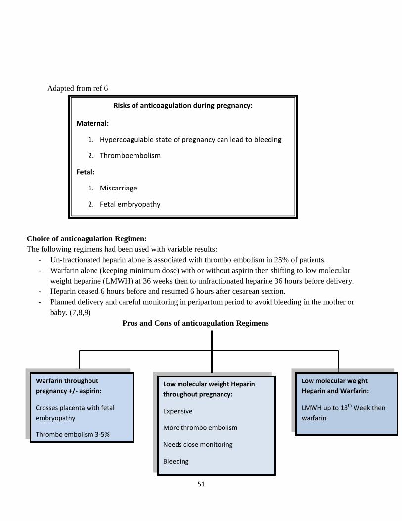
Adapted from ref 6
Choice of anticoagulation Regimen: The following regimens had been used with variable results:
- Un-fractionated heparin alone is associated with thrombo embolism in 25% of patients. - Warfarin alone (keeping minimum dose) with or without aspirin then shifting to low molecular
weight heparine (LMWH) at 36 weeks then to unfractionated heparine 36 hours before delivery. - Heparin ceased 6 hours before and resumed 6 hours after cesarean section. - Planned delivery and careful monitoring in peripartum period to avoid bleeding in the mother or
baby. (7,8,9) Pros and Cons of anticoagulation Regimens
Warfarin throughout pregnancy +/- aspirin:
Crosses placenta with fetal embryopathy
Thrombo embolism 3-5%
Low molecular weight Heparin throughout pregnancy:
Expensive
More thrombo embolism
Needs close monitoring
Bleeding
Low molecular weight Heparin and Warfarin:
LMWH up to 13th Week then warfarin
Risks of anticoagulation during pregnancy:
Maternal:
1. Hypercoagulable state of pregnancy can lead to bleeding
2. Thromboembolism
Fetal:
1. Miscarriage
2. Fetal embryopathy

52
Key Points: 1. Pre-conception counseling of ladies with RHD is needed, patients
in the high risk group should be advised not to get pregnant 2. Patients with moderate to severe regurgitation need to be on
maximum anti-CHF drugs , avoid angiotensin receptor blockers and ACE inhibitors
3. Patients with moderate to severe MS need to be referred for PTMC 4. Patients with prosthetic valves need to be followed closely to
determine the best anticoagulation and to monitor bleeding profile. 5. Delivery of patients with significant valve dysfunction should be
planned with the cardiologist, anesthetist and intensivist.
References:
1.Amira El Faeil , Khalid Yassein . Cardiac disease in Sudanese pregnant women. 2013 ). 2.Task Force on the Management of Cardiovascular Diseases During Pregnancy
of the European Society of Cardiology, ESC guidelines on the management of cardiovascular diseases during pregnancy. Eur Heart J, 2011. 32(24): p. 3147-3197. 3.De Souza, J., Martinez, Jr, EE, Ambrose, JA, et al, Percutaneous balloon mitral valvuloplasty in comparison with open mitral valve com-missurotomy for mitral stenosis during pregnancy. J Am Coll Cardiol, 2001. 37(3): p.900-903. 4.Elkayam, U., Bitar, F, Valvular heart disease and pregnancy part I: native valves. J Am Coll Cardiol, 2005. 46(2): p. 223-230. 5.Nobuyoshi, M., Arita, T, Shirai, S, et al,Percutaneus balloon mitral valvuloplasty. Circulation, 2009. 99(12): p. 1580-1586. 6.Reimold SC, Rutherford JD (2003). Valvular heart disease in pregnancy.N Engl J Med 349(1):52–59. 7. Yinon, Y., Siu, SC, Warshafsky, C et al. , Use of low molecular weight heparin in pregnant women with mechanical heart valves. Am J Cardiol, 2009. 104(9): p. 1259-1263. 8. Salazar, E., Izaguirre, R, Verdejo, J, et al. , Failure of adjusted doses of subcutaneous heparin to prevent thromboembolic phenomena in pregnant patients with mechanical cardiac valve prostheses.J Am Coll Cardiol, 1996. 27(7): p. 698-703. 8. Bates, S., Greer, IA, Pabinger, I, Sofaer, S, Hirsh,J, Venous Thromboembolism,
Thrombophilia,Antithrombotic Therapy, and Pregnancy:American College of Chest Physicians Evidence-Based Clinical Practice Guidelines (8th Edition) Chest, 2008. 133(6): p. 844S-886S.

53
Mild Disease
- Can Proceed to Pregnancy - Needs regular follow up by
Cardiologist
Moderate to severe Disease
Advice Contraceptives to all
Avoid Pregnancy if:
• Decreased LV systolic function • Significant aortic and mitral
stenosis • Moderate or severe
pulmonary hypertension • Heart failure symptoms
before pregnancy • Mechanical valve prostheses • Atrial fibrillation requiring
warfarin
Pre- Conception Planning - Cardiologist Assessment and Echo
- Advice Folic Acid to all
Severe lesions for Intervention before pregnancy:
- Trans catheter balloon dilatation for severe MS - Consider surgery in mlutivalvular lesions or severe
symptoms specially in the presence of As or MS (not suitable for PTMC)
- Counseling for prosthetic versus tissue valve
54
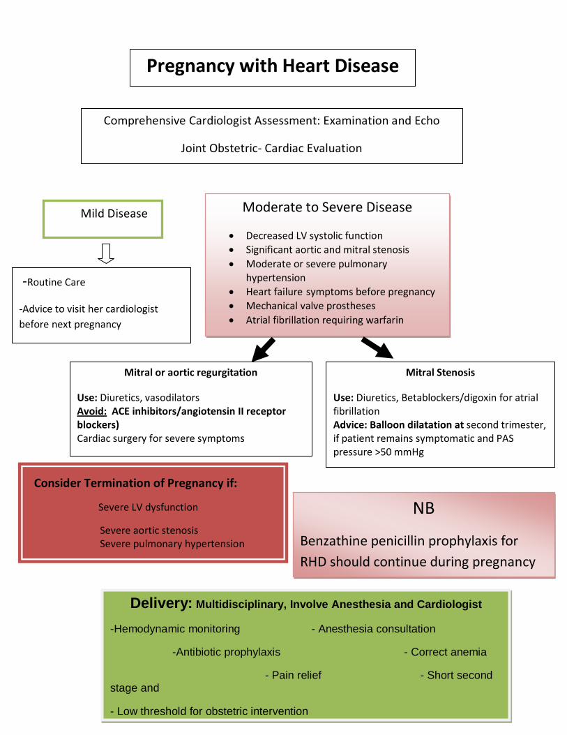
Pregnancy with Heart Disease
Comprehensive Cardiologist Assessment: Examination and Echo
Joint Obstetric- Cardiac Evaluation
Mild Disease
Mitral or aortic regurgitation
Use: Diuretics, vasodilators Avoid: ACE inhibitors/angiotensin II receptor blockers) Cardiac surgery for severe symptoms
-Routine Care
-Advice to visit her cardiologist before next pregnancy
Consider Termination of Pregnancy if:
Severe LV dysfunction
Severe aortic stenosis Severe pulmonary hypertension
Delivery: Multidisciplinary, Involve Anesthesia and Cardiologist -Hemodynamic monitoring - Anesthesia consultation
-Antibiotic prophylaxis - Correct anemia
- Pain relief - Short second stage and
- Low threshold for obstetric intervention
Moderate to Severe Disease
• Decreased LV systolic function • Significant aortic and mitral stenosis • Moderate or severe pulmonary
hypertension • Heart failure symptoms before pregnancy • Mechanical valve prostheses • Atrial fibrillation requiring warfarin
Mitral Stenosis
Use: Diuretics, Betablockers/digoxin for atrial fibrillation Advice: Balloon dilatation at second trimester, if patient remains symptomatic and PAS pressure >50 mmHg
NB
Benzathine penicillin prophylaxis for RHD should continue during pregnancy

55
Pregnant Ladies on Anti coagulation
(Prosthetic valves, atrial fibrillation)
One of 2 Regimens

56
نصائح للمرأة التي تعاني من أمراض القلب
هل لديك مرض بالقلب؟
مرة40الحمل يضاعف عمل القلب حوالي
البد من زيارة طبيب القلب للتأكد من نوع المرض و و شدته قبل الزواج و الحمل
أنواع أمراض القلب الشائعة في عمر اإلنجاب: روماتزم القلب وأمراض الصمامات - - امراض عضلة القلب - - ارتقاع الضغط الرئوي - العيوب الخلقية -
ماذا أفعل اذا فكرت بالزواج و الحمل؟
ازور طبيب القلب لمعرفة شدة المرض (بسيط- شديد) - اسأل طبيب القلب عن استعمال حبوب القلب و اثرها على األم و الطفل - اسأل طبيب النساء عن وسائل تنظيم األسرة -
تجنبي الحمل وإستعملي الموانع عند وجود أمراض القلب
االتية:
. ضعف شديد في عضلة القلب 1
.ضيق شديد في الصمام2
.إرتفاع الضغط الرئوي3
.وجود أعراض هبوط القلب :(ضيق النفس- تورم 4 الجسم)
.الحوجة ألستعمال أدوية مسيلة للدم5
هل أمراض القلب تكون عائقا للزواج أو الحمل؟
معظم أمراض القلب يمكن التحكم فيها قبل أو أثناء الحمل والوالدة-
الكشف المبكر والتواصل مع الطبيب المختص واستشارته قبل الزواج والحمل - يجنب كثير من المخاطر
المتابعة أثناء الحمل والوالدة ضرورية لصحة األم والطفل-
يجب اتباع ارشادات الفريق الطبى إذا نصح بعدم الحمل أو إسقاط الجنين - حفاضا على صحة األم
ماذا أفعل اذا حدث الحمل؟
-ازور طبيب القلب لمعرفة شدة المرض
-ازور طبيب النساء ومعي تقارير القلب
-قد يصف لك الطبيب أدوية جديدة يجب إتباع تعليماته بدقة.
-ال تتوقفي عن إستعمال البنسلين الوقائي لرومتزم القلب
-اسأل طبيب النساء عن وسائل تنظيم األسرة بعد الوالدة
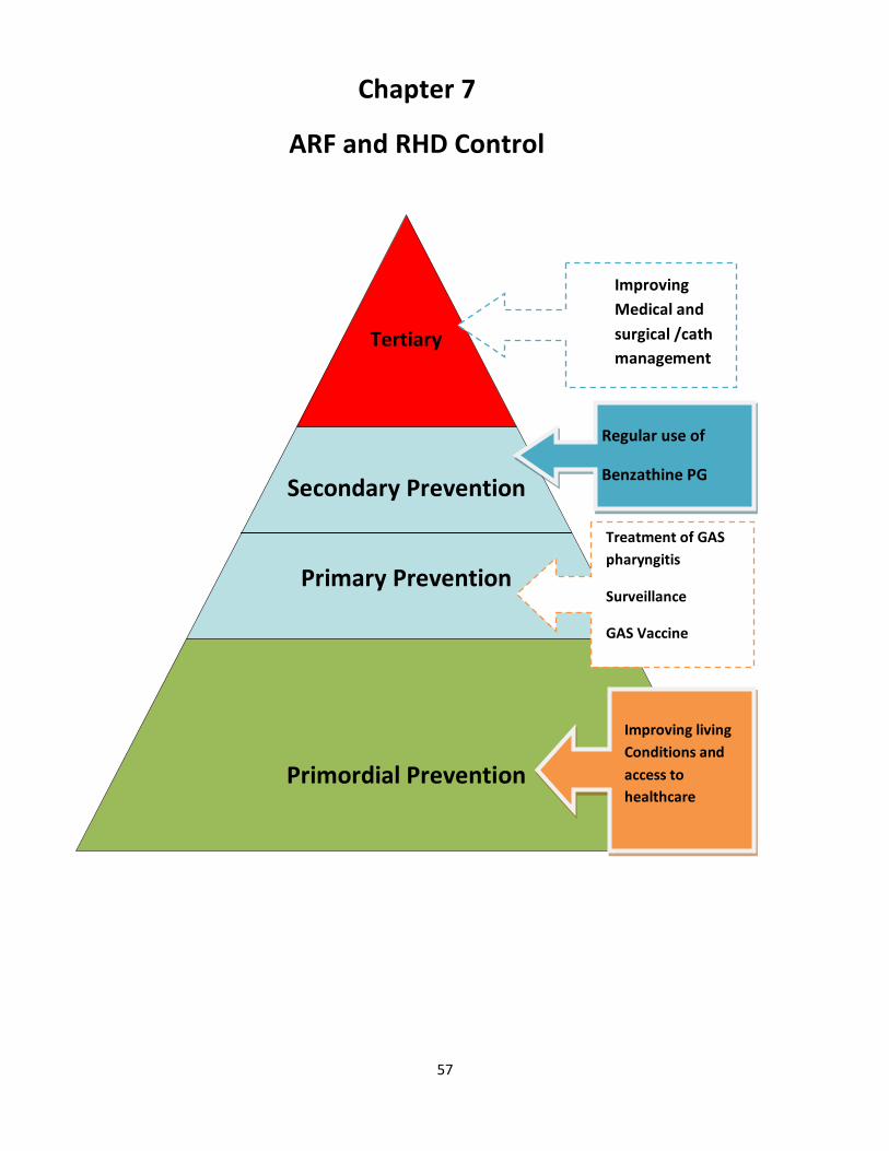
57
Tertiary
Secondary Prevention
Primary Prevention
Primordial Prevention
Regular use of
Benzathine PG
Treatment of GAS pharyngitis
Surveillance
GAS Vaccine
Improving Medical and surgical /cath management
Improving living Conditions and access to healthcare
Chapter 7
ARF and RHD Control

58
The previous Rheumatic Heart Disease Control Program: Lessons learned
In 1986-90, a World Health Organization (WHO) campaign involving 16 countries including Sudan was conducted in collaboration with the Sudanese MOH. The campaign aimed at screening and raising awareness with emphasis on secondary prophylaxis. Screening of 13322 children was done and 146 cases of RHD were reported in Khartoum Town (giving a prevalence of 10 per 1000). In this campaign Secondary prophylaxis coverage was found to be 72%. . Phase II was planned to extend the program to other states, however more financial and technical support was needed in terms of logistics, surveillance and basic research; therefore this program stopped in 2000. (Data from a report by Dr.Nawal Kurdufani, and reference (1).
Lessons Learned from the Previous Program: 1. RHD control programs need to be modified to involve primary as well as secondary prevention. 2. More advocacy is needed, namely by involving local and international nongovernmental organizations (NGOs), the public and the patients. 3. More internal will and cooperation with regional organizations from countries with similarly high RHD prevalence are needed in order to assure continuity.
The current RHD Control Program
Mission and Vision
Mission:
To minimize the burden of acute rheumatic fever and rheumatic heart disease in Sudan
Vision:
Elimination of acute rheumatic fever and rheumatic heart disease
RHD Control Program Objectives and Description:
General Objective:
“To decrease the incidence of ARF and RHD by 25% in those less than 25 years by the year 2025”

59
This objective is derived from the World Heart Federation (WHF)position statement which is aligned with the World Health Organization’s 65th World Health Assembly, that includes a reduction in premature deaths from non-communicable diseases (NCDs) by 25% by the year 2025.(2,3)
Specific Objectives:
A. To increase the awareness about RHD in through structured programs directed to physicians, nurses, health workers, teachers and the public.
B. Training of health personnel including physicians, nurses, medical assistants in target areas.
C. Establishing a national registry for RHD.
D. Establishing screening programs for RHD for school children in Khartoum and target States.
E. Establishing strong advocacy through social, political and media channels.
F. Consolidation of primary and secondary prevention through awareness and training.
G. Ensuring the continuous supply of good quality benzathine penicillin.
H. To plan and conduct research that can have an impact on RHD control
I. To strengthen partnership with national, regional and international organizations concerned with RHD control.
Description of the Current RHD Control Program:
In June 2012, the Working Group on Pediatric Cardiology and the Sudan Heart Society together with the Sudanese Association of Pediatricians initiated a program for RHD control that was conveyed to the MOH. The program was inspired by the 1st All Africa Workshop on Rheumatic Fever and Rheumatic Heart Disease held in South Africa in October 2005 which was convened by the Pan African Society of Cardiology (PASCAR), sponsored by the World Health Organization -Africa Office (WHO-AFRO), and the World Heart Federation (WHF).(3) Similar programs based on primary prevention were adopted in India and Pacific Islands and had led to eradication of RHD in Cuba and Costa Rica.(4,5) The Tools for Achieving the Objectives of RHD Control Program:
A. Robust awareness raising campaigns through utilizing different information, education
communication means including mass media, printing of health education and promotional
materials, workshops and seminars as well as home and school visits. The main target group will
be the population at most risk.
B. Training workshops for medical staff

60
C. Strengthening the early detection and surveillance system through establishing more clinics
and training physicians in Khartoum and each of the target regions (Darfur, Kordofan, Gezira
and White Nile) and school screening by auscultation and echocardiography.
D. Establishing regional and national registries.
E. Advocacy among the health professionals (through workshops and seminar) as well as among
social and political groups through media programs like films, radio and TV advertisement,
street signs, mobile messages etc. Also through attending international meetings and
conferences.
All the above mentioned activities will be carried in close collaboration and coordination with
national and state health authorities and related partners and associations.
Components of a Comprehensive RHD Control Program (Ref7)
Integration:
RHD program is planned to be integrated in the following programs:

61
Non-Communicable Disease program (NCD)
Integrated Management of Childhood Illness (IMCI)
Mother and Child Health (MCH)
Adolescent’s Health
Package of Essential Noncommunicable Disease (PEN)
Monitoring:
The following parameters will be used for monitoring;
• Prevalence of RHD in school children before and after the program.
• Number of cases seen in outpatient of main hospitals • Number of cases admitted to main hospitals • Awareness of physicians, school staff and the public monitored by questionnaire before
and after the start of the program.
Future Directions in RHD Control: GAS Vaccine:
There has been extensive research to develop a vaccine against GAS. Currently vaccines are divided into M-Protein and non M-Protein types. M protein is the major determinant of the organism’s virulence. The 2 vaccines that have entered or are nearing clinical investigation are the N-terminal M proteinbasemultivalent vaccines (26-valent and 30-valent vaccines) and conserved M protein vaccines (the J8 vaccine and the StreptInCor vaccine). There are a variety of other vaccine candidates that are at various stages of discovery and development (8). There are major obstacles facing vaccine development including safety concerns, an incomplete understanding of immune protection in humans, inadequate epidemiological data and minimal development of combination antigen vaccines. It is expected that with more intensive efforts these obstacles can be overcome and a vaccine can be made available.

62
Key Points 1. Primary prevention is an important tool in the current RHD
Control Program 2. Public awareness raising and training of health workers are
needed 3. All society sectors concerned with RHD need to be involved
(teachers, media, scientific and nongovernmental organizations) and should work in collaboration with health personnel to achieve the program targets.
References:
1.WHO and principal investigatorsWHO programme for the prevention of rheumatic fever/rheumatic heart disease in 16 developing countries: report from Phase I (1986-90) .Bulletin of the World Health Organization, 70(2): 213-218 (1992) 2.United Nations General Assembly. Political Declaration of the High-level Meeting of the General Assembly on the Prevention and Control of Non-communicable Diseases (A/RES/66/2) [online], http://daccess-ddsny.un.org/doc/ /UNDOC/GEN/N11/458/94/PDF/N1145894.pdf?OpenElement (2012). 3.Remenyi, B. et al. Position statement of the World Heart Federation on the prevention and control of rheumatic heart disease Nat. Rev. Cardiol 2013; 10:284–292. 4. Bongani Mayosi. A proposal for the eradication of rheumatic fever in our Lifetime. South AMJ 2006;96:229-230
5.Nordet P, Lopez R, Duenas A, Sarmiento L. Prevention and control of rheumatic fever and rheumatic heart disease: the Cuban experience (1986–1996–2002). Cardiovasc J Afr. 2008;19:135–140.
6. Arguedas A, Mohs E. Prevention of rheumatic fever in Costa Rica. J Pediatr. 1992;121:569 –572.
7. Wyber, R. Grainger Gasser, A. Thompson, D. Kennedy, D. Johnson, T. Taubert, K. Carapetis, J. Tools for Implementing RHD Control Programmes, Quick TIPS Summary. World Heart Federation and RhEACH. Perth, Australia 2014.Page 4.
8. Andrew C. Steer, James B. Dale,and Jonathan R. Carapetis Progress Toward a Global Group A Streptococcal Vaccine. The Pediatric Infectious Disease Journal 2013; 32:180-82

63
Appendices
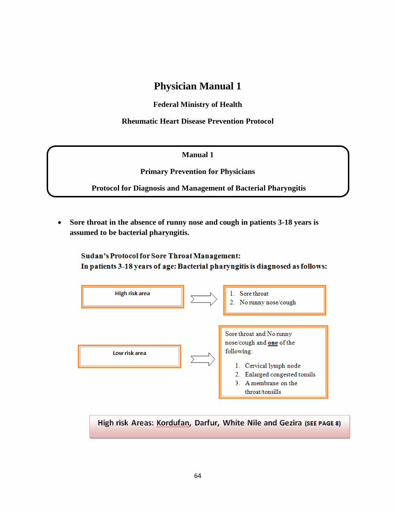
64
Physician Manual 1
Federal Ministry of Health
Rheumatic Heart Disease Prevention Protocol
Manual 1
Primary Prevention for Physicians
Protocol for Diagnosis and Management of Bacterial Pharyngitis
• Sore throat in the absence of runny nose and cough in patients 3-18 years is assumed to be bacterial pharyngitis.
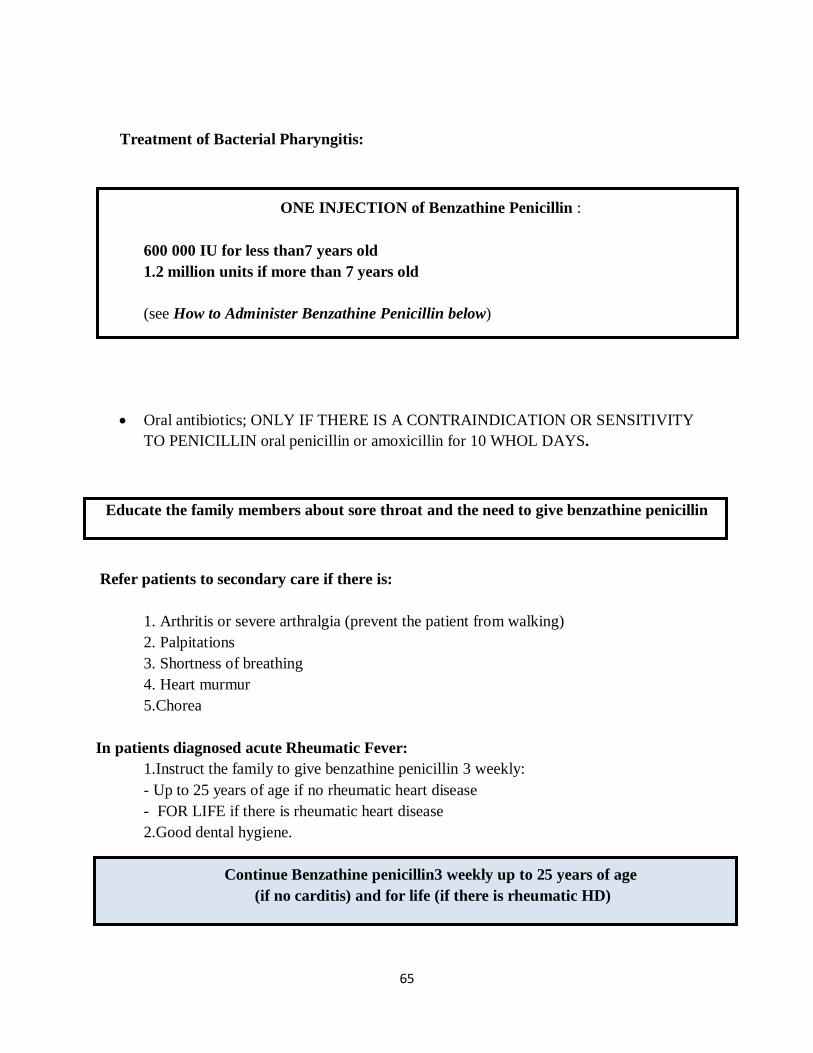
65
Treatment of Bacterial Pharyngitis:
ONE INJECTION of Benzathine Penicillin : 600 000 IU for less than7 years old 1.2 million units if more than 7 years old
(see How to Administer Benzathine Penicillin below)
• Oral antibiotics; ONLY IF THERE IS A CONTRAINDICATION OR SENSITIVITY TO PENICILLIN oral penicillin or amoxicillin for 10 WHOL DAYS.
Educate the family members about sore throat and the need to give benzathine penicillin
Refer patients to secondary care if there is:
1. Arthritis or severe arthralgia (prevent the patient from walking) 2. Palpitations 3. Shortness of breathing 4. Heart murmur 5.Chorea
In patients diagnosed acute Rheumatic Fever: 1.Instruct the family to give benzathine penicillin 3 weekly: - Up to 25 years of age if no rheumatic heart disease - FOR LIFE if there is rheumatic heart disease 2.Good dental hygiene.
Continue Benzathine penicillin3 weekly up to 25 years of age (if no carditis) and for life (if there is rheumatic HD)

66
Physician Manual 2
Federal Ministry of Health
Rheumatic Heart Disease Prevention Protocol
Manual 2
Secondary Prevention for Physicians
Management of Acute Rheumatic Fever
Acute Rheumatic Fever (ARF) is diagnosed by :
The Jones Criteria: Modified 2015
Major Criteria: Five
1.Arthritis/arthralgia : migratory, polyarthritis, monoarthritis or polyarthralgia
2. Carditis:
A: Clinical carditis: Typical murmurs:
1. Pansystolic murmur at apex radiating to the axilla (mitral regurgitation)
2. Mid diastolic apical murmur (Carry Coombs murmur) due to mitral valvulitis
3. Early diastolic murmur at aortic area 2 associated with large volume pulse.(aortic regurgitation)
Murmurs may or may not be associated with:
Congestive heart failure (CHF)
Pericardial rub
B: Subclinical Carditis: send patients with suspected ARF for echo as murmurs can be absent
3. Chorea (can be the only manifestation)
4. Subcutaneous nodules
5. Erythema marginatum

67
Minor Criteria: Four
1. Fever
2.Mono arthralgia
3.Prolonged PR interval on ECG
4. Raised acute phase reactants (ESR, CRP, Total WBC count)
New patients are different from those with known rheumatic heart disease (RHD)
Chorea alone is diagnostic of ARF
Treat with haloperidol or carbamazepine and start prophylaxis
Evidence of streptococcal infection (high or rising ASO)

68
Management
1. Anti inflammatory: - Aspirin 75 mg per kilogram per day divided 6 hourly after meals for 4 weeks. Do
ESR 2 weekly, taper aspirin by decreasing the dose by 2 tablet every week - Patients not responding (clinically and/or by acute phase reactants) or not tolerating
aspirin can be started on prednisolone 2mg per kilogram per day for 2 weeks - then aspirin is added (dose 60 mg per kilogram per day) for another 2 weeks - then prednisolone is tapered & discontinued. - Do ESR 2 weekly, taper aspirin by decreasing the dose by 2 tablets every week.
2. Antibiotic: One injection of benzathine penicillin for treatment then regular 3 weekly benzathine penicillin for secondary prophylaxis(dose and precautions below).
3. Duration of prophylaxis:
- Patients without carditis: till 25 years of age. - Patients with carditis: For life
4. Carditis: if there is heart failure start furosamide 1-2 mg per kilogram per day.
Consult cardiologist
5. Bed rest: 4 weeks
6. NOTIFY the Ministry of Health, National Program of RHD Control.
7. Ask about family members: those with sore throat are given one injection of benzathine or oral antibiotics for 10 days.
8. Bacterial endocarditis prophylaxis: for those with valve lesions going for procedures leading to bactremia, e.g. dental procedures give one dose of amoxicillin 50mg/kg one hour prior to procedure.
9. Family education:
Continue benzathine penicillin3 weekly up to 25 years of age (if no carditis) and for life (if there is rheumatic HD)
Good dental hygiene prevents complications of heart disease
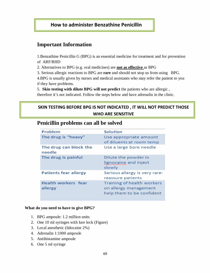
69
Important Information 1.Benzathine Penicillin G (BPG) is an essential medicine for treatment and for prevention of ARF/RHD 2. Alternatives to BPG (e.g. oral medicines) are not as effective as BPG 3. Serious allergic reactions to BPG are rare and should not stop us from using BPG. 4.BPG is usually given by nurses and medical assistants who may refer the patient to you if they have problems. 5. Skin testing with dilute BPG will not predict the patients who are allergic , therefore it’s not indicated. Follow the steps below and have adrenalin in the clinic.
Penicillin problems can all be solved
What do you need to have to give BPG?
1. BPG ampoule: 1.2 million units 2. One 10 ml syringes with lure lock (Figure) 3. Local anesthetic (lidocaine 2%) 4. Adrenalin 1:1000 ampoule 5. Antihistamine ampoule 6. One 5 ml syringe
How to administer Benzathine Penicillin
SKIN TESTING BEFORE BPG IS NOT INDICATED , IT WILL NOT PREDICT THOSE WHO ARE SENSITIVE

70
Step 2: Prepare the injection:
I. Draw appropriate amount of local anesthetic as diluents for the BPG powder (make sure it’s not cold)
II. Inject the diluents into the BPG vial III. Mix gently till dissolved IV. Draw in 5 ml syringe with Luer Lock V. Change the needle to a large bore (10 ml syringe) needle
Step 3: Prepare the patient and give the injection:
I. Ask the patient to lie on the abdomen
II. Mark the site of the injection on the gluteus muscle (Figure)
III. To minimize pain: press with your thumb over the site for 10 seconds
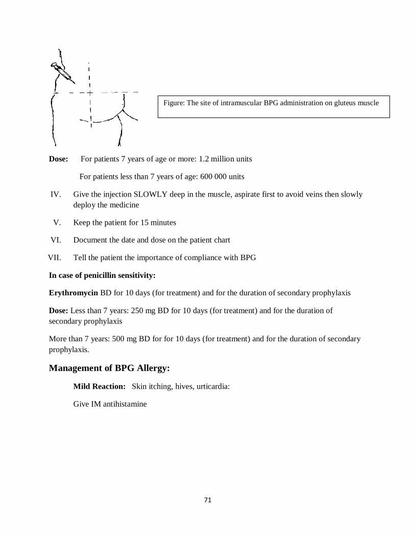
71
Dose: For patients 7 years of age or more: 1.2 million units
For patients less than 7 years of age: 600 000 units
IV. Give the injection SLOWLY deep in the muscle, aspirate first to avoid veins then slowly deploy the medicine
V. Keep the patient for 15 minutes
VI. Document the date and dose on the patient chart
VII. Tell the patient the importance of compliance with BPG
In case of penicillin sensitivity:
Erythromycin BD for 10 days (for treatment) and for the duration of secondary prophylaxis
Dose: Less than 7 years: 250 mg BD for 10 days (for treatment) and for the duration of secondary prophylaxis
More than 7 years: 500 mg BD for for 10 days (for treatment) and for the duration of secondary prophylaxis.
Management of BPG Allergy:
Mild Reaction: Skin itching, hives, urticardia:
Give IM antihistamine
Figure: The site of intramuscular BPG administration on gluteus muscle

72
Management of Anaphylaxis:
4.Lie the patient with legs up
5.If distressed, give O2
6.IV line: give IV normal saline10ml /kgboluses. Can give adrenaline infusion
3.Inject adrenaline: 0.3 ml (<7 y), 0.5 ml (>7 y) of 1:1000 solution. Can be repeated in 15 minutes
2.Call for help
1.Assess ABC: if needed perform CPRDO STEPS 2,3,4 QUICKLY AT SAME TIME
World Allergy Organization Guidelines for anaphylaxis management. WAO2011;4:13-37
1
2
4
3
5
6

73
برنامج مكافحة روماتزم القلب
وزارة الصحة االتحادية
إدارة الرعاية الصحية األساسية
للمساعد الطبيعالج التهاب الحلق البكتيري و الحمى الروماتزمية
الرسائل األساسيه:
. التهاب الحلق البكتيري يؤدي إلى الحمى الروماتزمية 1
سيؤدي الى روماتزم القلب الذي يدمر صمامات القلب و ال يمكن عالجه. الحمى الروماتزمية مرض خطير 2
التهاب الحلق البكتيري
تمنع الوقاية األوليه
الحمي الروماتزمية
الوقاية الثانويه تمنع
روماتزم القلب (اليمكن عالجه)
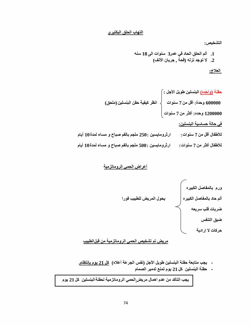
74
التهاب الحلق البكتيري
التشخيص:
سنه 18 سنوات الى 3ألم الحلق الحاد في عمر .1 ال توجد نزله (قحة , جريان األنف) .2
العالج:
البنسلين طويل األجل : (واحده)حقنة
(ملحق) انظر كيفية حقن البنسلين سنوات 7 وحدة: أقل من 600000
سنوات 7 وحده: أكثر من 1200000
في حالة حساسية البنسلين:
أيام 10 ملجم بالفم صباح و مساء لمدة 250 سنوات: ارثرومايسين : 7لألطفال أقل من
أيام 10 ملجم بالفم صباح و مساء لمدة 500 سنوات: ارثرومايسين : 7لألطفال أكثر من
أعراض الحمي الروماتزمية
ورم بالمفاصل الكبيره
ألم حاد بالمفاصل الكبيره يحول المريض للطبيب فورا
ضربات قلب سريعه
ضيق التنفس
حركات ال إرادية
الطبيب مريض تم تشخيص الحمي الروماتزمية من قبل
يوم بإنتظام.21كل يجب متابعة حقنة البنسلين طويل األجل (نفس الجرعة أعاله) - يوم تمنع تدمير الصمام 21البنسلين كل حقنة -
يوم21البنسلين كل الحمي الروماتزمية لحقنة يجب التأكد من عدم اهمال مريض
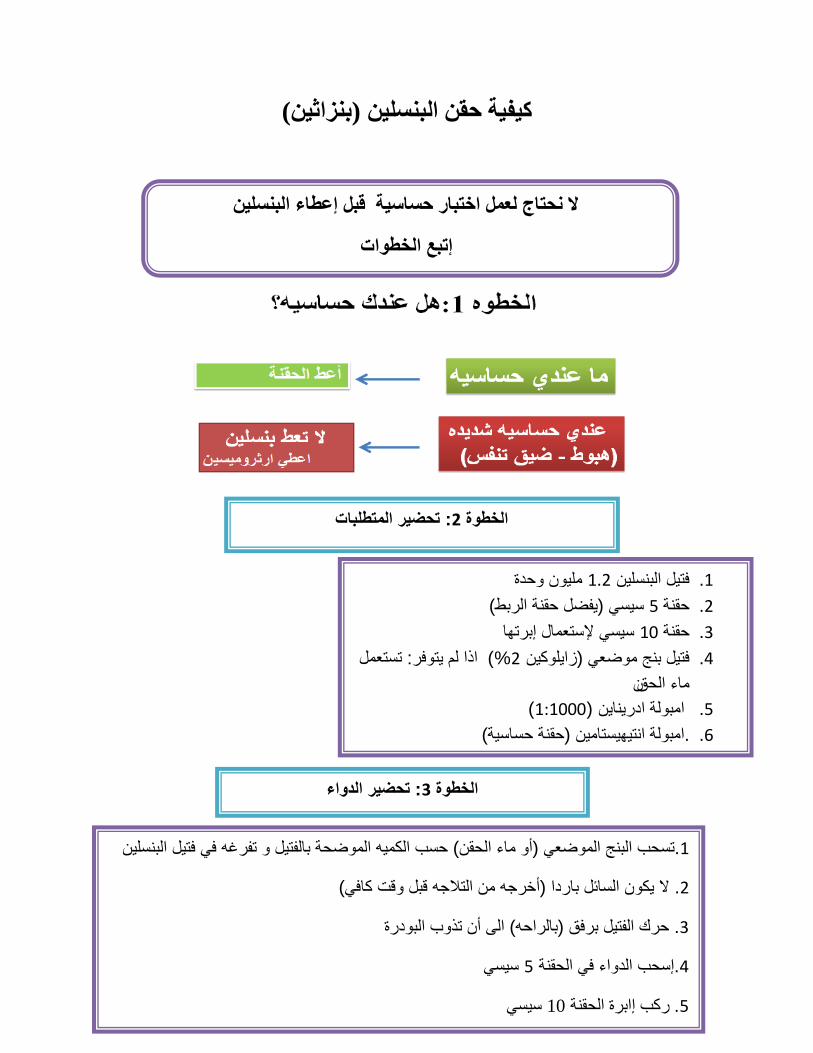
75
كيفية حقن البنسلين (بنزاثين)
ال نحتاج لعمل اختبار حساسية قبل إعطاء البنسلين
إتبع الخطوات
تتحضير المتطلبا: 2الخطوة
مليون وحدة1.2فتيل البنسلين .1 سيسي (يفضل حقنة الربط)5حقنة .2 سيسي إلستعمال إبرتها 10حقنة .3 اذا لم يتوفر: تستعمل )%2 فتيل بنج موضعي (زايلوكين .4
نماء الحق )1:1000 ( امبولة ادريناين .5 .امبولة انتيهيستامين (حقنة حساسية) .6
: تحضير الدواء3الخطوة
:تحضير المريض و إعطاء الحقنة4الخطوة
.تسحب البنج الموضعي (أو ماء الحقن) حسب الكميه الموضحة بالفتيل و تفرغه في فتيل البنسلين 1
. ال يكون السائل باردا (أخرجه من التالجه قبل وقت كافي)2
. حرك الفتيل برفق (بالراحه) الى أن تذوب البودرة3
سيسي5إسحب الدواء في الحقنة .4
سيسي10برة الحقنة ا ركب إ.5

76
جرعة االدرينالين
)1:1000( العمر
سنوات7اقل من خط 3
سنوات7أكثر من خط)5نصف سيسي (
: تحضير المريض و اعطاء الحقنة4 الخطوة
ينام المريض على بطنه .1
(الرسم)الحقنة حدد مكان .2
لتخدير المكان ثواني في مكان الحقنة10إضغط باالصبع الكبير لمدة .3
– اسحب لتجنب الوريد – فرغ الدواء ببطء بعمقحقنإ .4
دع المريض يرتاح ربع ساعه .5
؟إذا اشتكى المريض من هذه األعراضماذا أفعل
سنوات- 7): أمبوله لعمر اعطي حقنة (انتي هستامين: حكة بالجلد سنوات7نصف أمبوله ألقل من
استدع الطبيب •
هبوط بالجسم أو ضيق بالتنفس:
انزل المريض على االرض وضع رجليه لألعلى •
استدع الطبيب اعطي حقنة (ادرينالين ) في العضل بدون تأخير - •
هام.....هام....هام حقنة البنسلين في الوريد تؤدي إلى الوفاة

77
RHD Educational Websites: Sudanese Children’s Heart Society:
http://www.sudankidsheart.org/ World Heart Federation- RHD Net: http://www.world-heart-federation.org/what-we-do/rheumatic-heart-disease-network/ Australian RHD Guidelines http://www.rhdaustralia.org.au/sites/default/files/guideline_0.pdf TIPPS http://www.pascar.org/uploads/files/QUICK-TIPS_World-Heart-Federation_RhEACH.pdf
Wi Red
http://www.wiredhealthresources.net/EchoProject/
RHD Action: www.rhdaction.org
تم بحمد هللا
Last updated: Jan 2017