acute kidney injury (aki) - congresshome.com · 2018-06-15 · acute kidney injury (aki) represents...
TRANSCRIPT

Acute Kidney Injury (AKI)A Guide to Clinical Practice
All rights are reserved by the author and publisher, including the rights of reprinting, reproduction in any form and translation. No part of this book may be reproduced, stored in a retrievable system or transmitted, in any form or by any means, electronic, mechanical, photocopying, recording, or otherwise, without the prior written permission of the publisher.
First edition: September 2012
European Dialysis and Transplant Nurses Association/ European Renal Care Association (EDTNA/ERCA)Pilatusstrasse 35, Postfach 3052, 6002 Luzern, Switzerlandwww.edtnaerca.org
ISBN: 978-84-615-9445-0
D.L.: M-25234-2012
Layout, Binding and Printing: Imprenta Tomás Hermanos Río Manzanares, 42-44 · E28970 Humanes de MadridMadrid - Spainwww.tomashermanos.com

5
Acknowledgements

6
Acute Kidney Injury (AKI)
7
Acknowledgements
AcknowledgementsThis book is an initiative of EDTNA/ERCA (European Dialysis & Transplant Nurses Association/European Renal Care Association), EfCCNa (European federation of Critical Care Nursing associations) and HENNA (Hellenic Nephrology Nurses Association).Thanks to all of the authors for their considerable and valuable contributions which made this publication become a reality. Our deepest compliments to Anastasia Laskari, EDTNA/ERCA’s Immediate Past President: Rósa Thorsteinsdóttir, EfCCNa’s President and Panagiota Tsougia, HENNA’s President for their support , trust and commitment.A special mention to Maria Cruz Casal, EDTNA/ERCA’s Publications Coordinator for her additional work in coordinating the printing.
Editors and ReviewersJohn W. Albarran, RN, Dip.Nurs, BSc(Hons), MSc, PG Dip (HE), DPhil, Chair of R&D and ISC for EfCCNa, Associate Professor in Critical and Cardiovascular Nursing, Centre for Health and Clinical Research, University of the West of England, Bristol, UK
Maria Saraiva, RN, BSN, MSN, PhD, Coordinator Nursing Research, Coordinator for Master in Nephrology Nursing, Escola Superior de Enfermagem de Lisboa. Lisbon, PT
CollaboratorKaren Pugh-Clarke, MSc, BSc (Hons), RN PhD (c), Department of Nephrology, Keele University, Staffordshire, UK


9
Table of Contents

10
Acute Kidney Injury (AKI)
Preface .............................................................................................................. 15 John Sedgwick, MSc.N. MSc.HSR, BSc (Hons), RN, RMN, Dip.Nurs, Renal Cert, Cert.ED, PhD(c), EDTNA/ERCA Education Consultant, Director Multi-Professional Programmes & Principal Lecturer (Nephrology). Teesside University, UNITED KINGDOM
1. The concept of Acute Kidney Injury ............................... 21 Filipe Ramos,
RN, BSN, MSc, Hospital de São José- Unidade de Urgência Médica. Lisbon, PORTUGAL
Maria Saraiva, RN, BSN, MSN, PhD, Coordinator Nursing Research, Coordinator for Master in Nephrology Nursing, Escola Superior de Enfermagem de Lisboa. Lisbon, PORTUGAL
2. Epidemiology and Pathogenesis of AKI .................... 29 John W. Albarran,
RN, Dip.Nurs, BSc(Hons), MSc, PG Diploma in Education, DPhil, Chair of R&D and ISC for EfCCNa, Associate Professor in Critical and Cardiovascular Nursing. University of the West of England, Bristol, UNITED KINGDOM
3. Early Diagnosis and Prevention of AKI ...................... 39 Jona Palina Grimsdottir,
RN, MSN (stud), Landspitali, Universitary Hospital Reykjavik, ICELAND
Guðrún Jónsdóttir, RN, MSN, Landspitali, Universitary Hospital Reykjavik, ICELAND
4. Continuous Renal Replacement Therapy Program on ICU ............................................................. 55
Jackie Younker, RN, MSN, Senior Lecturer, University of the West of England, Faculty of Health & Life Sciences, Bristol, UNITED KINGDOM
11
Table of Contents
5. Vascular Access for HD and CRRT .................................. 67 Margaret McCann,
RGN, RNT, Certificate in Nephrology, Dialysis and Transplantation, BNS (Hons), MSc Nursing, FFNMRCSI, School of Nursing and Midwifery, Trinity College, Dublin, IRELAND
Glenda Taylor, RGN, RNT, BNS (Hons), Post Graduate Diploma in Renal Specialist Nursing, MSc in Nursing (Education), Adelaide and Meath hospital, incorporating the National Children’s Hospital, Dublin, IRELAND
6. Nutritional support during Renal Replacement Therapy ................................................... 81
Ada Azar, MSc, Clinical Nutrition, Nutrition Department, Assaf Hrofeh Medical Center. ISRAEL
7. Meeting the needs of the critically ill patient and family ................................................ 91
Cândida Durão, RN, BSN, MSc, Senior Lecturer, Coordinator for Master Degree in Critical Care Nursing, Escola Superior de Enfermagem de Lisboa. Lisbon, PORTUGAL
Maria Saraiva, RN, BSN, MSN, PhD, Coordinator Nursing Research, Coordinator for Master in nephrology Nursing, Escola Superior de Enfermagem de Lisboa. Lisbon, PORTUGAL
8. Acute Kidney Injury in children ........................................ 103 Chaski Diamanto,
RN, Pediatric Renal Nurse, Renal Unit of General Pediatric Hospital of Athens “Aglaia Kyriakou”, GREECE
Tsougia Panagiota, RN, Renal Nurse, President of HENNA, Director of Nursing Department in General Pediatric Hospital of Athens “Aglaia Kyriakou”, GREECE
12
Acute Kidney Injury (AKI)
9. Key principles of nursing care for the patient with Acute Kidney Injury ............................ 119
Karen Pugh-Clarke, MSc, BSc (Hons), RN PhD (c), Department of Nephrology, Keele University, Staffordshire, UNITED KINGDOM

13
Table of Contents

Notes

15
Preface
16
Acute Kidney Injury (AKI)
PrefaceAcute kidney injury (AKI) represents a significant challenge in clinical practice. AKI is avoidable but when it develops, astute management and intervention on the part of the whole multidisciplinary team ensures that further insult to kidney function is minimised. Understanding the challenges in managing AKI requires an awareness of the historical background to AKI. Chapter 1 in this handbook provides an overview of the evolution of Acute Renal Failure to the now internationally agreed definition of AKI. The difficulties which clinicians encountered in managing AKI were not helped by the multiple definitions of what actually constituted AKI and how it could be staged. The development of a consensus on a definition of AKI as well as the establishment of the AKI staging framework – RIFLE (Risk, Injury, Failure, Loss of kidney function, and End-stage renal failure) has improved the clinical management of patients.
Unfortunately, evidence continues to emerge of patients developing AKI where this should not have occurred1 . Failing to recognise the silent features of AKI or inadequate attention to the fundamentals of monitoring and assessment of renal function result in serious consequences for patients. Unrecognised, AKI has devastating consequences and significantly impairs and further damages both renal function as well as affecting other organs. The RIFLE framework provides an agreement on classifying AKI, ensuring management and interventions are appropriate to the particular stage of the AKI. Ensuring strategies are implemented which reduce the damaging effects upon renal function is critical. Astute and vigilant clinical staff play a part in identifying those susceptible to AKI; skilled assessment and the application of evidenced based care in dealing with AKI are vital.
The epidemiology and pathogenesis of AKI is varied. The RIFLE classification provides a clear indication as to the severity of AKI. An agreed staging / classification framework
17
Preface
for AKI facilitates comparisons between incidences of AKI along with a comparison of outcomes and effectiveness of interventions (Chapter 2). Evidence has already been published of the beneficial impact of RIFLE in highlighting how AKI severity relates to outcomes and mortality.
As highlighted in Chapter 3, early diagnosis of AKI is essential along with developing evidenced-based strategies focused upon preventing AKI developing. Various risk factors exist which predispose to the development of AKI; understanding such ‘ trigger factors ‘ enables clinicians to pre-empt the chances of AKI developing. Understanding the role of hydration status and volume loading in AKI is important.
When AKI develops, renal replacement therapy (RRT) is frequently needed to manage the altered fluid and biochemical status of the patient. Continuous Renal Replacement Therapy (CRRT) requires skilled expertise on the part of the clinical team to ensure the patient receives supportive therapy. An awareness of optimum approaches to delivering CRRT, coupled with skilled fluid management, vascular access care, anticoagulation management and preventing hypothermia due to CRRT is a goal of care (Chapter 4). The need to maintain optimum vascular access in providing renal replacement support is vital. Choosing the most appropriate means of vascular access has a significant impact upon patient outcomes. Patients with AKI are highly susceptible to secondary infections; therefore preventing vascular access infection is paramount and necessitates evidenced- based nursing care in the management of vascular access (Chapter 5).
Nutritional support for patients with AKI focuses upon preventing malnutrition, optimising recovery and patient outcomes. Protein and caloric requirements vary depending on the patient’s condition and catabolic state. Understanding the patients nutritional requirements in AKI is highly complex and a balance between understanding the impact of renal

18
Acute Kidney Injury (AKI)
replacement support on nutritional requirements, fluids, vitamins and minerals; the expertise of dietician involvement in the care of AKI is essential (Chapter 6).
In its most severe form, patients with AKI require the specialist care & support of an intensive care unit. Nursing care within this highly technological environment further highlights the importance of the art & science of nursing (Chapter 7). Within this environment, patients are usually at their most vulnerable. The importance of family care and support is vital to those who often experience high levels of uncertainty and anxiety.
Renal healthcare professionals are increasingly encountering children with AKI and whilst there are similarities in the principles of managing children to adults there are also important variations as outlined in Chapter 8. Whilst staging and definitions of AKI have greatly enhanced our understanding of AKI controversies remain in the appropriateness of the RIFLE criteria in children2 . This systematic review found wide variations in the application of RIFLE and conflicting associations between RIFLE and outcomes such as mortality, length of stay, illness severity. Concerns have been raised about the need to monitor more closely the long term follow up of paediatrics who have survived an episode of AKI and the potential development of CKD; in one study 10% of children developed CKD 1-3 years after AKI3.
AKI is a serious complication and is associated with high levels of morbidity and mortality. All members of the healthcare team must work in partnership to prevent AKI occurring through developing a heightened level of awareness of those at risk and where appropriate removing risk factors leading to AKI. When AKI occurs, patient outcomes and survival depends on the practices of the whole multidisciplinary team who must work together in delivering evidenced- based interventions (Chapter 9) known to enhance patient recovery and ultimately reduce the long term effects of AKI on kidney function following patient discharge from hospital.

19
Preface
I would like to congratulate the writers of this handbook in producing a most succinct and comprehensive overview of key issues related to the prevention, care and management of patients who develop AKI.
References1. Stewart J, Findlay G, Smith N, Kelly K, Mason M. Adding insult to
1 injury. A review of patients who died in hospital with a primary diagnosis of acute kidney injury. NCEPOD, 2009. www.ncepod.org.uk/2009aki.htm.
2. Slater MB, Anand V, Uleryk EM, Parshuram CS. (2012) A systematic review of RIFLE criteria in children, and its application and association with measures of mortality and morbidity. Kidney Int. Apr;81(8):791-8
3. Mammen C, Al Abbas A, Skippen P, Nadel H, Levine D, Collet J.P, Matsell DG.(2012) Long-term Risk of CKD in Children Surviving Episodes of Acute Kidney Injury in the Intensive Care Unit: A Prospective Cohort Study. Am J Kidney Dis. Apr; 59(4):523-30

The Concept of Acute Kidney Injury
21
Acute Kidney Injury (AKI)
2322
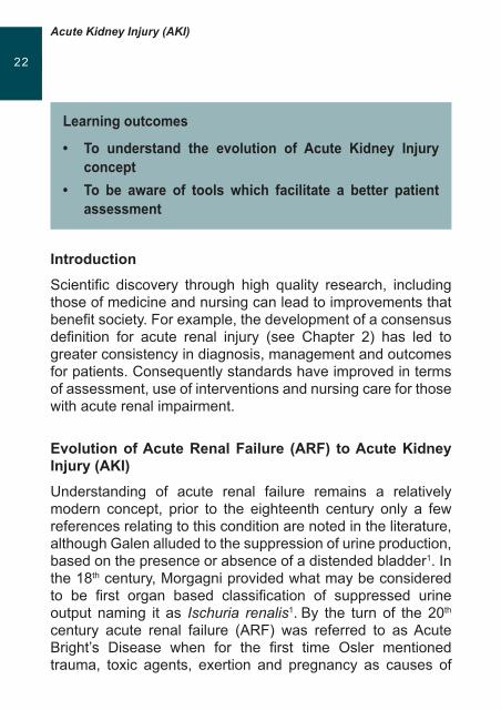
IntroductionScientific discovery through high quality research, including those of medicine and nursing can lead to improvements that benefit society. For example, the development of a consensus definition for acute renal injury (see Chapter 2) has led to greater consistency in diagnosis, management and outcomes for patients. Consequently standards have improved in terms of assessment, use of interventions and nursing care for those with acute renal impairment.
Evolution of Acute Renal Failure (ARF) to Acute Kidney Injury (AKI)Understanding of acute renal failure remains a relatively modern concept, prior to the eighteenth century only a few references relating to this condition are noted in the literature, although Galen alluded to the suppression of urine production, based on the presence or absence of a distended bladder1. In the 18th century, Morgagni provided what may be considered to be first organ based classification of suppressed urine output naming it as Ischuria renalis1. By the turn of the 20th century acute renal failure (ARF) was referred to as Acute Bright’s Disease when for the first time Osler mentioned trauma, toxic agents, exertion and pregnancy as causes of
Learning outcomes
• To understand the evolution of Acute Kidney Injuryconcept
• To be aware of tools which facilitate a better patientassessment

The Concept of Acute Kidney Injury
2322 acute Bright’s disease1. However, it is the contribution of the knowledge acquired in World War I, through military medicine, surgery and treatment of traumatic shock, that this entity took the name of War Nephritis2. This term remained throughout World War II until 1941, when Bywaters and Bell defined it as ‘Crush Syndrome’, in which they were able to describe the natural history of renal disease, examining the pathology of the kidney and widespread tubular damage and pigmented casts inside the tubular lumen2. These findings prompted several studies which subsequently increased knowledge in key areas that became central to the development of the term ARF. It is not until after 1950, that the term ‘Acute Renal Failure’, first appeared where the biomedical term is proposed and defined3
Figure 1, Schematic chronology of acute renal failure concepts1.
2nd Century 1760 1888 1917 1941 1951 2004
Empty Blader
Ischuria Renalis
Acute Bright’s Disease
War Nephritis
Crush Sindrome
Acute Renal Failure
Acute Kidney Injury
Galen Morgagni Bright Davies Bywaters H. Smith ADQI
ARF has traditionally been characterized by a rapid decline in renal function in hours or days with inability to regulate fluid, electrolyte and acid base balance4. However, within the literature there is noted to be over 35 definitions of ARF, leading to a lack of consensus on diagnostic criteria3 albeit a common point between the various definitions is the immediacy of the deterioration in renal function.
Due to operational problems of a common and shared definition, the term Acute Renal Failure (ARF), was recently replaced by the concept of Acute Kidney Injury (see chapter
Acute Kidney Injury (AKI)
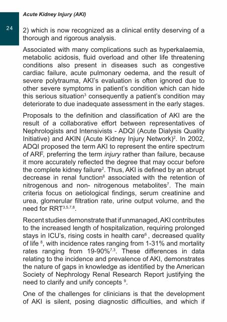
2524 2) which is now recognized as a clinical entity deserving of a thorough and rigorous analysis.
Associated with many complications such as hyperkalaemia, metabolic acidosis, fluid overload and other life threatening conditions also present in diseases such as congestive cardiac failure, acute pulmonary oedema, and the result of severe polytrauma, AKI’s evaluation is often ignored due to other severe symptoms in patient’s condition which can hide this serious situation3 consequently a patient’s condition may deteriorate to due inadequate assessment in the early stages.
Proposals to the definition and classification of AKI are the result of a collaborative effort between representatives of Nephrologists and Intensivists - ADQI (Acute Dialysis Quality Initiative) and AKIN (Acute Kidney Injury Network)2. In 2002, ADQI proposed the term AKI to represent the entire spectrum of ARF, preferring the term injury rather than failure, because it more accurately reflected the degree that may occur before the complete kidney failure2. Thus, AKI is defined by an abrupt decrease in renal function6 associated with the retention of nitrogenous and non- nitrogenous metabolites7. The main criteria focus on aetiological findings, serum creatinine and urea, glomerular filtration rate, urine output volume, and the need for RRT3,5,7,8.
Recent studies demonstrate that if unmanaged, AKI contributes to the increased length of hospitalization, requiring prolonged stays in ICU’s, rising costs in health care6 , decreased quality of life 8, with incidence rates ranging from 1-31% and mortality rates ranging from 19-90%7,3. These differences in data relating to the incidence and prevalence of AKI, demonstrates the nature of gaps in knowledge as identified by the American Society of Nephrology Renal Research Report justifying the need to clarify and unify concepts 9.
One of the challenges for clinicians is that the development of AKI is silent, posing diagnostic difficulties, and which if

The Concept of Acute Kidney Injury
2524 unrecognised can result in profound damage to renal structures and function, which in turn can precipitate cardiovascular, respiratory and neurological deleterious changes for patients.
With the recent guidance and recommendations provided by RIFLE (ADQI8, see Chapter 2), healthcare teams are able to confidently classify the severity of AKI and implement appropriate interventions to support the patient. Additionally, studies using the RIFLE criteria confirm that the identification of AKI incidence rates of 67%10, unlike previous studies which demonstrated that in ICUs these were lower and between 6-25%11.
Variations in mortality rates between populations with and without AKI have also been reported with mortality rates of 26.3% for patients referred to ICU, compared to figures of 5.5% for those who did not develop this condition. These findings reinforce the importance of adopting and implementing the RIFLE classification in ICU’s, with the aim of early diagnosis and the decreasing the burden of costs to the individual and healthcare providers12.
For health professionals caring for patients with AKI, the challenges not only include implementing a classification of prevention and early diagnosis, but also intervening with strategies and measures that reflect effective responses with the aim of minimizing the damage and decisively prevent its occurrence in those who may be at risk. Generally, nurses because of their proximity to patients are the first to observe and be aware of a deterioration of renal function. Their knowledge of renal function and pathophysiology, and skilled expertise in patient assessment, monitoring and interpreting data as well as understanding of current evidence based interventions play a pivotal role in preventing complications and improving the outcomes and well-being of individuals with AKI13-14. Nurses must be aware of the new classification systems of AKI and in this sense, the main priority in nursing management, apart from providing patient centred care, is to know and
Acute Kidney Injury (AKI)
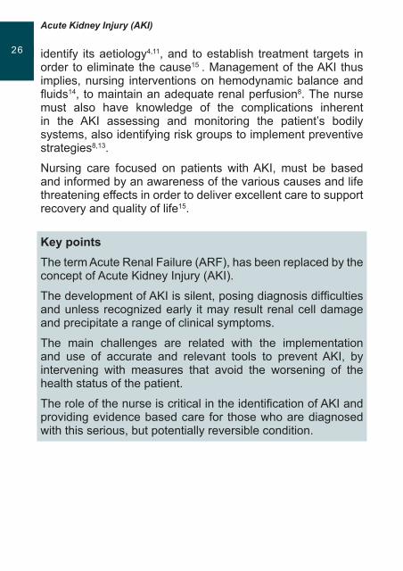
2726 identify its aetiology4,11, and to establish treatment targets in order to eliminate the cause15 . Management of the AKI thus implies, nursing interventions on hemodynamic balance and fluids14, to maintain an adequate renal perfusion8. The nurse must also have knowledge of the complications inherent in the AKI assessing and monitoring the patient’s bodily systems, also identifying risk groups to implement preventive strategies8,13.
Nursing care focused on patients with AKI, must be based and informed by an awareness of the various causes and life threatening effects in order to deliver excellent care to support recovery and quality of life15.
Key pointsThe term Acute Renal Failure (ARF), has been replaced by the concept of Acute Kidney Injury (AKI).
The development of AKI is silent, posing diagnosis difficulties and unless recognized early it may result renal cell damage and precipitate a range of clinical symptoms.
The main challenges are related with the implementation and use of accurate and relevant tools to prevent AKI, by intervening with measures that avoid the worsening of the health status of the patient.
The role of the nurse is critical in the identification of AKI and providing evidence based care for those who are diagnosed with this serious, but potentially reversible condition.
The Concept of Acute Kidney Injury
2726 References1. Eknoyan, G., Emergence of the concept of acute kidney injury. Adv.
ChronicKidneyDis, normal 2008, 15 (3) 308-13
2. Lewington, A. P., & Sayed, A. (2010). Acute kidney injury: how do we define it? AnnalsOfClinicalBiochemistry , 2010, 47 (1), 4-7.
3. Kellum, J., Bellomo, R., & Ronco, C. Definition and classification of acute kidney injury. Nephron.ClinicalPractice , 2008, 109 (4) 182-187.
4. Perkins, C., & Kisiel, M. Utilizing physiological knowledge to care for acute renal failure. BritishJournalOfNursing ,2005, 14 (14) 768-773.
5. Liãno, F., Álvarez, L., & Junco, E. Definiciones de insuficiencia renal aguda. Nefrología , 2007, 27 (3), 3-14.
6. Rosner, M. H. , Acute kidney injury: turning the tide. CurrentDrugTargets, 2009, 10 (12), 165-1668.
7. Lameire, N., Van Biesen, W., & Vanholder, R. , Acute renal failure. Lancet , 2005, 365, 417-430.
8. Dirkes, S. , Acute kidney injury: not just acute renal failure anymore? Critical Care Nurse , 2011, 31 (1), 37-49.
9. Hsu, C-y, Mc Culloch, C.E., Fan, D., Ordoñez, J.D., Cherton, G.M., Go, A.S. Community-based incidence of acute renal failure. Kidney International , 2007, 72 (2) 208-212.
10. Hoste, E., Clermont, G., Kersten, A., RIFLE criteria for acute kidney injury are associated with hospital mortality in critically ill patients: a cohort analysis. Critical Care, 2006, 10 (3) 1022–1030
11. Byrne, G., & Murphy, F., Acute kidney injury and its impact on the cardiac patient... first in a series. British Journal of CardiacNursing, 2008, 3 (9) 416-422
12. Ricci, Z., Cruz, D., & Ronco, C., Response to ‘The RIFLE criteria and renal prognosis in acute kidney injury’. Kidney International , 2008, 14 (11), 1492-1493.
13. Redmond, A., McDevitt, M., Barnes, S., Acute renal failure: recognition and treatment in ward patients. Nursing Standard Royal College Of Nursing , 2004, 18 (22), 46-53.
14. Yaklin, K. M. ,Acute Kidney Injury: An Overview Of Pathophysiology and Treatments. Nephrology Nursing Journal: Journal Of TheAmericanNephrologyNurses’Association , 2011, 38 (1) , 13-18.
15. Campbell, D., How acute renal failure puts the brakes on kidney function. Nursing, 2003, 33 (1), 59-64

Notes
Epidemiology and Pathogenesis of AKI(Incidence, classification and survival outcomes)
29
Acute Kidney Injury (AKI)
3130
IntroductionAcute Kidney Injury (AKI) is the currently adopted terminology which replaces the previous and often confusing definitions of acute renal failure. This revised concept aims to enable the early recognition and management of this condition, but more importantly it encourages healthcare professionals to view AKI as a spectrum of the syndrome that ranges from milder forms to more severe extreme cases that require Renal Replacement Therapy (RTT)1,2. The indices of AKI include abrupt decline, usually within 48 hours, in renal function that is associated with an inability to maintain fluid, electrolyte and acid-base balance. It is also vital to acknowledge that for in-hospitalised patients minor changes in renal function such as a rise in serum creatinine may be highly important as these may influence medium to long-term outcomes.
IncidenceFigures on the incidence of AKI, whether community or hospital acquired, remain unknown. It is suggested that AKI affects approximately 7% of in-hospital patients3. However, some of
Learning outcomes
• TounderstandtheincidenceofAcuteKidneyInjuryandassociatedmortalityratesforthiscondition,
• TohaveknowledgeandawarenessofcurrentdefinitionofAcuteKidneyInjuryandoftheRIFLEcriteriausedtodetermineseverityofrenaldysfunction,
• TogainconfidenceinbeingabletodiscussthevariousaetiologicalcausesforAcuteKidneyInjury
Epidemiology and Pathogenesis of AKI(Incidence, classification and survival outcomes)
3130
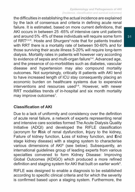
the difficulties in establishing the actual incidence are explained by the lack of consensus and criteria in defining acute renal failure. It is estimated, based on more current definitions, that AKI occurs in between 25 -65% of intensive care unit patients and around 5% -8% of these individuals will require some form of RRT2,4,6. Hoste and Shurgers4 note that for patients treated with RRT there is a mortality rate of between 50-60% and for those surviving their acute illness 5-20% will require long-term dialysis. Mortality rates in patients will however vary according to evidence of sepsis and multi-organ failure3-4. Advanced age, and the presence of co-morbidities such as diabetes, vascular disease and hypertension may also compound mortality outcomes. Not surprisingly, critically ill patients with AKI tend to have increased length of ICU stay consequently placing an economic burden on healthcare systems due to expensive interventions and resources used3-4. However, with newer RRT modalities trends of in-hospital and six month mortality may improve outcomes7.
ClassificationofAKIDue to a lack of uniformity and consistency over the definition of acute renal failure, a network of experts representing renal and intensive care societies formed The Acute Dialysis Quality Initiative (ADQI) and developed the RIFLE classification (acronym for Risk of renal dysfunction, Injury to the kidney, Failure of kidney function, Loss of kidney function, and End stage kidney disease) with a staging system to characterise various dimensions of AKI8 (see below). Subsequently, an international guidelines group of leading experts from various specialties convened to form Kidney Disease: Improving Global Outcomes (KDIGO) which produced a more refined definition and staging system for AKI that built on earlier work9.
RIFLE was designed to enable a diagnosis to be established according to specific clinical criteria and for which the severity is confirmed based upon a staging system. Furthermore, the
Acute Kidney Injury (AKI)
3332
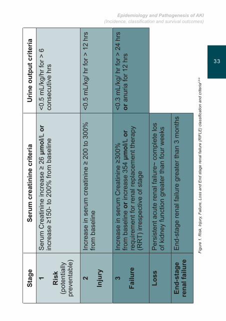
staging of a patient gives an indication of increasing severity of AKI with two outcomes possible, loss and end-stage renal failure (see Figure 1). Finally, by using RIFLE comparisons of incidence, outcomes and effectiveness of interventions can be made1,3. Indeed, a recent systematic review of the literature which included 13 studies reported that RIFLE was a good at predicting outcome and the relationship between increased mortality with worsening RIFLE classification10. Other work concludes that RIFLE is valuable in predicting recovery, need for RRT, length of stay and mortalities as well as reveal that there is a higher incidence of AKI within the general population than previously conceived11.
The RIFLE classification relies on rising serum creatinine values and or a fall in urine output with the three stages providing an index of increasing severity2 (see Figure 1). It also worth noting that an increase in ‘stage’ is associated with poorer survival outcomes1,3. Current modifications also recommend that the time span for diagnosis is reduced to 48 hours and a decreased threshold for the elevation of serum creatinine from baseline to peak value is applied2.
Epidemiology and Pathogenesis of AKI(Incidence, classification and survival outcomes)
3332
Stag
eSe
rum
cre
atin
ine
crite
riaU
rine
outp
ut c
riter
ia
1
Risk
(pot
entia
lly
prev
enta
ble)
Ser
um C
reat
inin
e in
crea
se ≥
26 μm
ol/L
or
incr
ease
≥15
0- to
200
% fr
om b
asel
ine
<0.5
mL/
kg/h
r for
> 6
co
nsec
utiv
e hr
s
2
Injury
Incr
ease
in s
erum
cre
atin
ine
≥ 20
0 to
300
%
from
bas
elin
e<0
.5 m
L/kg
/ hr f
or >
12
hrs
3
Failure
Incr
ease
in s
erum
Cre
atin
ine
≥300
%
from
bas
elin
e or
incr
ease
354
μmol
/L o
r re
quire
men
t for
rena
l rep
lace
men
t the
rapy
(R
RT)
irre
spec
tive
of s
tage
<0.3
mL/
kg/ h
r for
> 2
4 hr
s or
anu
ria fo
r 12
hrs
Loss
Per
sist
ent a
cute
rena
l fai
lure
~ co
mpl
ete
los
of k
idne
y fu
nctio
n gr
eate
r tha
n fo
ur w
eeks
End-stage
renalfailure
End
-sta
ge re
nal f
ailu
re g
reat
er th
an 3
mon
ths
Figu
re 1
. Ris
k, In
jury
, Fai
lure
, Los
s an
d E
nd s
tage
rena
l fai
lure
(RIF
LE) c
lass
ifica
tion
and
crite
ria2,
8-9
Acute Kidney Injury (AKI)
3534
AetiologyofAKIThe aetiology of AKI can be classified into three main areas3, 12
with each delineating specific causative factors leading to kidney failure and function.
• Pre-renal failure• Intrinsic renal failure• Post-renal failure
Pre-renalfailurePre-renal failure is typically induced in response to a sudden reduction in circulating volume (due to severe vomiting, haemorrhage, burns, dehydration, shock, anaphylaxis, severe vomiting and diarrhoea) and therefore can have a detrimental effect on renal perfusion and as a consequence decrease glomerular filtration rate (GFR). To maintain circulating volume, the kidneys will reabsorb sodium but as consequence the patient may become oliguric with a modest increase in urea and other waste products. However, in pre-renal failure, if the circulatory deficit is corrected promptly the condition can be immediately reversed and the nephrons remain structurally intact. If pre-renal failure is not managed appropriately, there is a possibility that this may deteriorate into intrinsic renal failure.
IntrinsicrenalfailureIntrinsic renal failure is a main reason for admission to a critical care setting and here structural damage occurs which can be sub-divided into tubule-interstitial, glomerular and or micro-vascular. Structural alternations will develop within the nephrons despite correcting the precipitating causative factors. Most forms of AKI have a tubular aetiology and the result in acute tubular necrosis (ATN) which can be either of an ischaemic or toxic form. The causes of ATN associated with an ischaemic event include prolonged hypo-perfusion and sepsis which compromise renal blood flow and GFR. When ATN is

Epidemiology and Pathogenesis of AKI(Incidence, classification and survival outcomes)
3534
the result of ischaemic insult this leads to changes at cellular level, which turn precipitates damage to cell wall membranes causing disruption to electrolytes, cell swelling and death. It is these and other structural changes within the tubules lead to renal dysfunction and explain why there is a delay in recovery of the kidneys despite intervention with RRT 12.
There are other factors which may lead to ATN for example nephrotoxins such as cephalosporins, aminoglycoside antibi-otics, non-specific anti-inflammatory drugs and radiographic contrast media. However inflammatory insults can also pre-cipitate ATN due to waste products of septicaemia and en-dogenous toxins3,12. With reference to tubulo-interstitial con-ditions, allergic interstitial nephritis and cast nephropathy are key causes. Glomerulonephritis can lead to AKI due to rapidly progressive glomerulonephritis which in turn can give rise to Nephritic syndrome although this is an unusual condition3,12. Other examples of glomerular causes are Goodpastures syn-drome, Lupus and Wegener granulomatosis. Finally, micro-vascular changes triggering intrinsic AKI can include malig-nant hypertension, haemolytic uraemic syndrome, scleroder-ma renal crisis and renal artery obstruction due to an emboli, dissection or a thrombus.
Post renal Finally, post renal failure is typically caused by mechanical obstructions to the passage of urine. Removing the obstruction through non-invasive or surgical manoeuvres can ameliorate the condition and improve renal functioning3,12.

Acute Kidney Injury (AKI)
3736
Keypoints
• Previous interpretations of incidence and outcomes associated with ARF have been unhelpful due to a lack of a standardised definition.
• The term Acute Renal Injury is more encompassing as it describes a spectrum of the syndrome that ranges from milder forms to more severe extreme cases that require Renal Replacement Therapy.
• The incidence of AKI ranges between a third and two thirds of intensive care patients, a variation which is accounted by the presence of co-morbidities, advancing age, sepsis and multi-organ dysfunction.
• Managing the care of critically ill AKI patients is expensive and around 5-20% of those who survive will require some form of RRT.
• The introduction of RIFLE criteria, which relies on measuring changes in serum creatinine, glomerular filtration rate and urine output, provides a uniform and standardised approach for assessing, diagnosing and managing patients with AKI.
• A number of clinical studies report that the RIFLE tool is a valid and reliable tool for predicting recovery, need for RRT, length of stay and mortalities.
• The aetiology of AKI is categorised into three discreet areas which in part explain the pathophysiology these include pre-renal failure, intrinsic AKI and post renal AKI.
Epidemiology and Pathogenesis of AKI(Incidence, classification and survival outcomes)
3736
References1. Lewington A and Kanagasundaram S. Clinical practice guidelines:
Acute Kidney Injury (5th edition) UK Renal Association 2010; [available at: www.renal.org/guidelines]
2. Kellum JA. Acute renal injury. Crit Care Med 2008; 36 (4 suppl): S141-S145
3. Griffiths L and Kanagasundaram S. Assessment and initial management of acute injury. Medicine 2011; 39 (7):390-397
4. Hoste EA, and Schurgers M Epidemiology of acute kidney injury: How big is the problem? Critical Care Medicine 2008; 36 (Suppl 4): S146-152
5. Hoste EA, Clermont G, Kersten A, Venkataraman R, Angus D, De Bacquer D and Kellum J RIFLE criteria for acute kidney injury are associated with hospital mortality in critically ill patients: a cohort analysis. Critical Care 2006, 10:R73 doi:10.1186/cc4915
6. Metnitz PGH, Krenn CG, Steltzer H, et al. Effect of acute renal failure requiring renal replacement therapy on outcome in critically ill patients. Crit Care Med 2002; 30: 2051–2058.
7. Vaara ST, Pettila V, Reinikainen M, Kaukonen KM, Population based incidence, mortality and quality of life in critically ill patients treated with renal replacement therapy- A nationwide retrospective cohort study in Finnish ICUs. Critical Care 2012; 16:R13 doi:10.1186/cc11158
8. Bellomo R,Ronco C, Kellum J A, Mehta R L, PalevskyP,andtheADQI .Acute renal failure – definition, outcome measures, animal models, fluid therapy and information technology needs: the Second International Consensus Conference of the Acute Dialysis Quality Initiative (ADQI) Group. Critical Care 2004;8:R204-212
9. Kidney Disease: Improving Global Outcomes. Clinical practice guideline on acute kidney injury 2010: www.kdigo.or
10. Ricci Z, Cruz D and Ronco C. The RIFLE criteria and mortality in acute renal injury: A systematic review. KidneyInternational 2008; 7: 538-546
11. Ali T, Khan I, Simpson W, Prescott G, Townsend J, Smith W, and Macleod A. Incidence and outcomes of Acute Kidney Injury: A comprehensive population based study. Journal of theAmericanSocietyofNephrology 2007; 18: 1292-1298
12. Workeneh B, Agraharkar M, Gupta R, Lederer E, Mulloy LL,TalaveraF.Acute Renal Failure, 2011 Medscape http://emedicine.medscape.com/article/243492-overview (accessed 1st of December 2011).

Notes
Early Diagnosis and Prevention of AKI
39

Acute Kidney Injury (AKI)
4140
Introduction
When acute kidney injury has occurred, timely individualized and evidence based supportive care may result in full recovery and function. Curative treatment of acute kidney injury is currently unavailable. The aim of modern treatment is directed at prompt and effective medical intervention in order to prevent complete and irreversible acute renal injury1. Kidney function as well as deterioration is diagnosed on the basis of serum creatinin e and urea levels. These parameters are, however, considered insensitive and non-selective in detecting changes of acute renal injury and function2.
Acute kidney injury is often a combination of many factors that are harmful and toxic to renal structures. Frequently, this is a situation triggering reduced renal blood flow and in addition, patients may receive drugs or contrast media which further increase the strain on the kidneys1. In order to prevent kidney failure, it is important to be aware of risk factors that may predispose patients to kidney injury or failure and take measures to reduce such a risk (see Chapter 2); moreover, it is imperative to eliminate the causes of reduced blood flow through the kidneys, if possible, and avoid administering
Learning outcomes
• To understand the need and importance of earlydiagnosisofAcuteKidneyInjury
• TorecognisetheriskfactorsforAcuteKidneyInjury• To have a comprehensive understanding of current
evidencebaseddataonhowtopreventAcuteKidneyInjury
Early Diagnosis and Prevention of AKI
4140
substances and drugs which are likely to have nephrotoxic effects1.
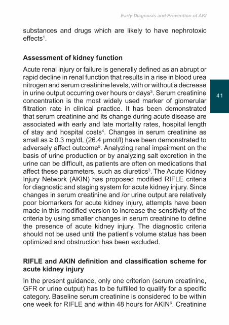
AssessmentofkidneyfunctionAcute renal injury or failure is generally defined as an abrupt or rapid decline in renal function that results in a rise in blood urea nitrogen and serum creatinine levels, with or without a decrease in urine output occurring over hours or days3. Serum creatinine concentration is the most widely used marker of glomerular filtration rate in clinical practice. It has been demonstrated that serum creatinine and its change during acute disease are associated with early and late mortality rates, hospital length of stay and hospital costs4. Changes in serum creatinine as small as ≥ 0.3 mg/dL (26.4 µmol/l) have been demonstrated to adversely affect outcome5. Analyzing renal impairment on the basis of urine production or by analyzing salt excretion in the urine can be difficult, as patients are often on medications that affect these parameters, such as diuretics3. The Acute Kidney Injury Network (AKIN) has proposed modified RIFLE criteria for diagnostic and staging system for acute kidney injury. Since changes in serum creatinine and /or urine output are relatively poor biomarkers for acute kidney injury, attempts have been made in this modified version to increase the sensitivity of the criteria by using smaller changes in serum creatinine to define the presence of acute kidney injury. The diagnostic criteria should not be used until the patient’s volume status has been optimized and obstruction has been excluded.
RIFLEandAKINdefinitionandclassificationschemeforacutekidneyinjuryIn the present guidance, only one criterion (serum creatinine, GFR or urine output) has to be fulfilled to qualify for a specific category. Baseline serum creatinine is considered to be within one week for RIFLE and within 48 hours for AKIN6. Creatinine
Acute Kidney Injury (AKI)
4342
is an aminoacid derivative that is easily filtrated through the glomerulus and its secretion in the tubule is usually rather scarce. Creatinine that is measured in the serum is a metabolite of creatine which is released from muscle cells. Therefore, individual muscle mass can affect the amount of creatinine in the serum. Among the elderly, who have a somewhat reduced muscle mass, serum creatinine levels might be within normal limits, even though renal function is considerably impaired. Meat consumption and various drugs as well as muscle degradation also affect serum creatinine levels. Rigorous rehydration might reduce serum creatinine levels. Serum creatinine levels are not sensitive to changes in renal function. Increase in serum creatinine will not be significant until renal function is reduced by 30 to 60%1,3,7.
Urea is a metabolite of protein metabolism and is excreted by the kidneys. Elevated urea levels in the serum can indicate kidney failure. Various external factors influence the amount of urea in serum apart from kidney function, such as liver metabolism and the amount of protein ingested. Catabolic state, gastrointestinal bleeding, and steroid therapy may increase serum urea levels. In case of dehydration, the kidneys increase the absorption of urea from the tubules, thereby increasing serum levels. Urea levels decrease in the event of liver failure, malnutrition and volume overload. Therefore, an increase in urea levels alone is not sufficiently indicative of renal failure. Elevated urea and creatinine levels in serum need to compound in order to establish possible kidney failure8. Several studies have been conducted with the purpose of pinpointing substances in the urine or the serum that might be more sensitive and specific than those currently used. These substances, however, need to meet certain requirements. They must be sensitive enough to detect early damage and also need to reflect deterioration or improvement in renal function. These substances have to be specific in such a way that one is able to ascertain the location of the damage within the kidney. Moreover, they must be simple and quick to perform,

Early Diagnosis and Prevention of AKI
4342
accurate, reliable and inexpensive. However, these efforts have not yet yielded adequate results9,10. Studies into Serum cystatin C turned out to be a promising marker for evaluation of renal function and a more sensitive indicator of impaired kidney function than creatinine; serum cystatin C is detected earlier than serum creatinine. Cystatin C is a small protein produced by all nucleated cells in the body; it readily filters through the glomerulus and is broken down in the renal tubule. Its accumulation suggests impaired glomerular filtration. The production of cystatin C is not dependent on age, gender, muscular mass or hydration level. It has, however, not been evaluated for acutely ill patients and its measurements have not become general practice7.
RiskfactorsforAKICertain populations are more vulnerable than others, for example patients with impaired renal function due to kidney diseases are at greatest risk for AKI. Age-related deterioration of renal function normally begins from the age of forty, and can be reduced by up to 50% by the age of eighty. Thus, the elderly (aged over 65) are also at greater risk3,11. The presence of heart failure and chronic hypertension can also be considered risk factors. In the case of heart failure, inadequate myocardial function may compromise renal perfusion and for those with chronic hypertension the blood vessels in the kidneys may constrict in order to control their blood flow. To complicate matters, these patients are often on antihypertensive drugs that might further reduce blood flow through the kidneys.
Diabetes is known to be a major risk factor for kidney failure. Major surgeries, including surgeries that involve a temporary clamping of the aorta, as well as use of a heart-lung machine, are also regarded as major risk factors12,13. Intra-abdominal hypertension and abdominal compartment syndrome are associated with acute kidney injury at relatively low levels of intra-abdominal pressure (IAP). IAP is defined

Acute Kidney Injury (AKI)
4544
as sustained or repeated pathological elevation of IAP ≥12 mmHg. Sustained elevation of IAP of >20 mmHg is associated with organ dysfunction. For most patients the critical IAP at which microcirculatory disturbance is observed is 10 – 15 mmHg. Abdominal perfusion pressure (APP) is defined as the difference between the mean arterial pressure (MAP) and the IAP and implies that as the IAP rises the perfusion of organs or vessels in or near the abdomen falls, even in the absence of a drop in MAP. In patients with IAP efforts should be made to maintain APP ≥ 60 mmHg14.
Evidence suggests a link between positive pressure ventilation and acute kidney failure. Several mechanisms have been proposed to explain the association15. Positive-pressure mechanical ventilation can markedly affect cardiac performance by acting on preload and cardiac output and thereby on renal perfusion. Hypercapnia is inversely correlated with renal blood flow (RBF) by direct and indirect mechanisms. The effects of moderate hypoxemia on renal hemodynamics are less understood but severe hypoxemia (PaO2 < 40 mmHg) causes renal vasoconstriction and vascular resistance leading to renal hypoperfusion. In addition to altering RBF mechanical ventilation can alter renal function through the release of pro inflammatory cytokines. Lung protective procedures can, on the other hand, reduce hemodynamic changes and inflammatory mediators 15,16.
Sepsis is a common cause of acute renal failure in the ICU. About 19 % of those suffering from moderate sepsis and about 51% of those diagnosed with septic shock are likely to suffer acute kidney failure. The causes of acute kidney failure in sepsis are often not only due to decreased arterial blood pressure and induction of vasoactive hormones, but may also be attributed to the release of inflammatory mediators, oxidizing substances, accumulation of white blood cells and bacterial toxins, all of which can contribute to cell damage17. It is estimated that 4-33% of patients with rhabdomyolysis suffer

Early Diagnosis and Prevention of AKI
4544
acute kidney injury or failure18. In patients with rhabdomyolysis large amounts of myoglobin and other intracellular proteins and electrolytes are released into the bloodstream18. Myoglobin causes toxicity in tubular cells and can as well block the tubules themselves. It is also thought to interfere with blood flow in the kidneys by having vasoconstrictive effects. Rapid dehydration that is often accompanied by rhabdomyolysis further increases the risk of renal failure19.
Contrast media has traditionally been known to cause acute renal failure in susceptible patients12. Although the toxicity of contrast media is not fully known it appears to be mainly associated with ischemia in the renal medulla12. Decreased renal blood flow coinciding with high pressure to eliminate a large amount of contrast media causes ischemia and multiple tubular damage20. Medical conditions such as hepatorenal syndrome and cardiogenic shock are also risk factors for acute kidney injury21. Various drugs and hyperosmolar therapeutic agents can have nephrotoxic effects by several mechanisms, especially if patients have underlying risk factors22.
Prevention
Hydrationandvolumeloading
Usually, initial damage to the kidney is caused by ischemia and hypoxia or by toxic effects of chemicals on tubular cells1. It may be difficult to assess the effects of fluid administration alone in preventing renal failure as fluid replacement is usually part of a comprehensive treatment of patients; it has been recognized, however, that intravascular volume depletion is an important risk factor for the development of acute kidney injury as well as other organ dysfunction23. In those with rhabdomyolysis and among predisposed patients undergoing cardiac catheterization with intravenous radio-contrast media, early and aggressive fluid resuscitation and
Acute Kidney Injury (AKI)
4746
pre-hydration have clearly proved beneficial to prevent acute kidney injury24,25.
Recently, early and aggressive fluid resuscitation and use of inotropic medication (early goal-directed therapy) has proven to be successful in septic patients to prevent multiple organ failure, including acute kidney failure26. In order to avert end-organ hypoperfusion and the consequent failure of end–organ function, important preventive therapy consisting of ensuring adequate hydration in the vascular volume expansion, adequate cardiac output and adequate blood flow, are recognized to be essential2. Where volume replacement is indicated this should be in a controlled fashion directed by hemodynamic monitoring as imprudent use of fluids carries its own inherent risk. Several observational studies have demonstrated a correlation between fluid overload and mortality in critically ill adults and children with acute kidney injury26.
Special attention should be paid to careful assessment of fluid balance in all patients. Bedside examination, including assessment of venous pressure, capillary refill time, blood pressure, pulse and postural blood pressure changes should be performed. Hourly urine-output and fluid-input and all fluid losses, including estimated insensible losses, drain/stoma output and nasogastric losses should be recorded. If possible, patients should be weighed daily. Where invasive hemodynamic measurements are in place changes in central venous pressure or pulse pressure can give clues to volume changes. In addition technological devices and functional monitoring can add further information about patients’ volume status and needs27. The role of colloid compared with crystalloids remains unclear. In the SAFE-study, a multi-centered study of 6997 critically ill patients, the investigators found no difference between albumin 4% and saline for fluid resuscitation in terms of risk of acute renal failure. Although the statistical significance was not attained, patients with severe sepsis who were given albumin did better than others28. All
Early Diagnosis and Prevention of AKI
4746
colloids, such as albumin, gelatins and hydroxyl starch may if administered in isolation cause “osmotic nephrosis” (osmotic tubular damage). Because isotonic saline, Ringer lactate or Ringer acetate is less expensive than albumin, the consensus has emerged that isotonic crystalloid solutions should be the preferred fluid in critically ill patients. In some cases albumin is, however, considered to be beneficial along with isotonic fluids26.
Hydroxyethyl starch (HES) is a less expensive colloid alternative for albumin; however HES has negative effects on coagulation and can cause “osmotic nephrosis” that may lead to renal impairment. A randomized trial compared HES to gelatins and found greater incidence of acute kidney injury with HES29. Another study compared HES (a low molecular-weight HES) with modified Ringers lactate for fluid resuscitation in patients with severe sepsis and found that the HES group exhibited a significantly higher rate of acute kidney injury. Thus, in septic and critically ill patients the use of HES is contraindicated30. Newer lower molecular weight hydroxyethyl starches with lower osmolality are considered less harmful but should be used cautiously in patients with pre-existing renal impairment (daily dose should not exceed 33 ml/kg/day)22. Volume therapy alone is not always sufficient to alleviate hypotension and maintain renal perfusion; these patients may therefore benefit from inotropic or/and vasopressor therapy31.
Drugs
Animal and human studies have shown that reversing hypotension with norepinephrine increases diuresis and creatinine clearance; whether this is due to an increase in renal blood flow and thus implies renal protection is unknown 32. Several studies and meta-analyses have concluded that even though dopamine also increases diuresis and possible creatinine clearance it does not protect against AKI1,23.
Acute Kidney Injury (AKI)
4948
Dobutamine and Dopexamine are used to increase cardiac output and can thereby increase MAP but controlled clinical trials have not shown protective effects on renal function33. Vasopressin increases blood pressure and can enhance diuresis but has not yet proven to prevent acute kidney injury33. Studies indicate that any MAP ≥ 60 mmHg may be considered adequate for patients with septic shock; additional benefits to renal function have not been observed when target MAP was raised from 65 mmHg to 85 mmHg. However, those studies have not involved ICU patients and those with preexisting risk factors and comorbidities. For those patients target MAP may have to be individually tailored according to premorbid blood pressure or to ensure adequate abdominal perfusion pressure1,34. Randomized trials and meta-analyses have shown that the use of loop diuretics in established renal failure does not improve renal function or mortality. They are useful in handling volume overload but have not been shown to protect or improve renal function nor do they decrease mortality34.
Radiocontrast medium can cause nephrotoxicity in susceptible patients. In animal experiments dehydration in conjunction with the infusion of radiocontrast medium has been shown to increase the incidence of acute kidney injury35. Studies have shown that using nonionic, low osmolal or iso-osmolal contrast medium in the lowest volume necessary in conjunction with adequate volume expansion prior to procedure reduces the risk of nephrotoxicity in high risk patients. The use of N-Acetylcysteine on a prophylactic basis remains unclear; however, with regard to its safety, low cost and possible advantages it has been considered beneficial in combination with adequate intravenous hydration in susceptible patients 36. In this context early and rigorous hydration has been considered vital in preventing or lessening the severity of acute renal injury. A minimum urine output goal of 2 ml/kg/h is recommended. In addition to hydration, sodium bicarbonate has been used to alkaline urine which serves to decrease cast formation and lessen the direct toxic effects of myoglobin. Mannitol has been
Early Diagnosis and Prevention of AKI
4948
used to increase urine output and thereby washing myoglobin out of the tubule. The effectiveness of combined crystalloid, Mannitol and bicarbonate therapy versus that of standard crystalloid resuscitation alone in prevention of acute kidney injury is debated19.
The complex nature of critical illness often necessitates the use of multiple therapeutic agents, many of which may individually or in combination have the potential to cause renal injury.
Aminoglycosides have a well–established nephrotoxicity. They are primarily excreted by glomerular filtration and are thought to accumulate in tubular cells where they interfere with normal cellular function eventually leading to cell death. Risk factors for aminoglycoside’s nephrotoxicity are the type of aminoglycosides used, high peak serum levels, cumulative dose, the duration and frequency of administration and patient related risk factors as well as the use of concomitant nephrotoxic drugs. Once-daily dosing and appropriate monitoring of drug levels are the best way to avoid kidney injury 22,23,37.
Vancomycin in high doses or in combination with other nephrotoxic drugs or known risk factors can cause kidney injury23. Angiotensin-Converting Enzyme inhibitors and angiotensin receptor blockers can in circumstances of already decreased renal blood flow cause an exacerbation of acute renal injury by modulating intra-renal blood flow; this may in turn cause a decline in glomerular filtration rate and a raise of serum creatinine, but the condition usually stabilizes within a few days, if not, drug administration must be halted38. Nonsteroidal anti-inflammatory drugs (NSAIDs) are in most circumstances not harmful. However, in cases of reduced renal perfusion which is common in critically ill patients the inhibitation of prostaglandin-induced vasodilation with the use of NSAIDs may further compromise renal blood flow and exacerbate ischemic injury12,13,38. Patients with pre-existing risk factors and concomitant use of other potential nephrotoxic drugs or procedures are vulnerable.
Acute Kidney Injury (AKI)
5150
Glucose control with intravenous insulin therapy in critically ill patients has been shown to improve outcome, including a decreased incidence of acute renal failure. This favorable result might be explained by modulation of inflammatory response 39.
KeypointsAcute Kidney Injury is often a combination of many factors that are harmful to the kidneys.
Early diagnosis of AKI that leads to supportive renal care may result in full recovery.
In order to prevent AKI a thorough assessment of blood pressure, fluid balance, urine output and management of known risk factors are important.
It has been demonstrated that serum creatinine and its change during acute disease are associated with early and late mortality rates, hospital length of stay and hospital costs.
Patients with pre-existing risk factors and concomitant use of other potential nephrotoxic drugs or procedures are vulnerable.
Recent evidence suggests that mechanical ventilation may contribute to the pathogenesis of acute kidney injury and several mechanisms have been proposed to explain the association.
The nature of critical illness often necessitates the use of multiple therapeutic agents, many of which may individually or in combination have the potential to cause renal injury.
Early Diagnosis and Prevention of AKI
5150
References1. Gill N, Nally JV, Fatica RA. Renal Failure Secondary to Acute Tubular
Necrosis*. Chest. 2005;128(4):2847-63.
2. Hoste EAJ, Kellum JA. Acute kidney dysfunction and the critically ill. Minervaanestesiologica. 2006;72(3):133.
3. Cheung CM, Ponnusamy A, Anderton JG. Management of Acute Renal Failure in the Elderly Patient: A Clinicians Guide. Drugs & Aging. 2008;25(6):455-476.
4. Coca SG, Yalavarthy R, Concato J, Parikh CR. Biomarkers for the diagnosis and risk stratification of acute kidney injury: a systematic review. Kidneyinternational. 2007;73(9):1008-16.
5. Chertow GM, Burdick E, Honour M, Bonventre JV, Bates DW. Acute kidney injury, mortality, length of stay, and costs in hospitalized patients. Journal of the American Society of Nephrology. 2005;16(11):3365-70.
6. Gopaluni S, Lines S, Lewington AJP. Acute kidney injury in the critically ill patient. CurrentAnaesthesia&CriticalCare. 2010;21(2):60-4.
7. Lisowska-Myjak B. Serum and urinary biomarkers of acute kidney injury. BloodPurif. 2010;29(4):357-65.
8. Perkins C, Kisiel M. Utilizing physiological knowledge to care for acute renal failure. Britishjournalofnursing. 2005;14(14):768-73.
9. Bagshaw SM, Langenberg C, Haase M, Wan L, May CN, Bellomo R. Urinary biomarkers in septic acute kidney injury. Intensivecaremedicine. 2007;33(7):1285-96.
10. Trof RJ, Di Maggio F, Leemreis J, Groeneveld AB. Biomarkers of acute renal injury and renal failure. Shock. 2006;26(3):245.
11. Zhou XJ, Rakheja D, Yu X, Saxena R, Vaziri ND, Silva FG. The aging kidney. Kidneyinternational. 2008;74(6):710-20.
12. Dishart MK, Kellum JA. An evaluation of pharmacological strategies for the prevention and treatment of acute renal failure. Drugs. 2000;59(1):79-91.
13. Huerta C, Castellsague J, Varas-Lorenzo C, García Rodríguez LA. Nonsteroidal anti-inflammatory drugs and risk of ARF in the general population. Americanjournalofkidneydiseases. 2005;45(3):531-9.
14. Mohmand H, Goldfarb S. Renal Dysfunction Associated with Intra-abdominal Hypertension and the Abdominal Compartment Syndrome. JournaloftheAmericanSocietyofNephrology. 2011;22(4):615-21.
Acute Kidney Injury (AKI)
5352
15. Broden CC. Acute Renal Failure and Mechanical Ventilation: Reality or Myth? CriticalCareNurse. 2009;29(2):62-75.
16. Ko GJ, Rabb H, Hassoun HT. Kidney-lung crosstalk in the critically ill patient. BloodPurif. 2009;28:75-83.
17. Devarajan P. Update on mechanisms of ischemic acute kidney injury. JournaloftheAmericanSocietyofNephrology. 2006;17(6):1503-20.
18. Bagley W, Yang H, Shah K. Rhabdomyolysis. Internal andEmergencyMedicine. 2007;2(3):210-8.
19. Malinoski DJ, Slater MS, Mullins RJ. Crush injury and rhabdomyolysis. Criticalcareclinics. 2004;20(1):171-92.
20. Weisbord SD, Palevsky PM. Radiocontrast-induced acute renal failure. Journalofintensivecaremedicine. 2005;20(2):63-75.
21. Ricci Z, Cruz D, Ronco C. The RIFLE criteria and mortality in acute kidney injury: a systematic review. Kidney international. 2007;73(5):538-46.
22. Pannu N, Nadim MK. An overview of drug-induced acute kidney injury. Criticalcaremedicine. 2008;36(4):S216.
23. Venkataraman R. Can we prevent acute kidney injury? Criticalcaremedicine. 2008;36(4):S166.
24. Huerta-Alardín AL, Varon J, Marik PE. Bench-to-bedside review: Rhabdomyolysis-an overview for clinicians. Crit Care. 2005;9(2):158-69.
25. Solomon R, Werner C, Mann D, D’Elia J, Silva P. Effects of saline, mannitol, and furosemide on acute decreases in renal function induced by radiocontrast agents. NewEnglandJournalofMedicine. 1994;331(21):1416-20.
26. Schrier RW. Fluid administration in critically ill patients with acute kidney injury. Clinical Journal of the American Society ofNephrology. 2010;5(4):733-9.
27. Bagshaw SM, Bellomo R. Early diagnosis of acute kidney injury. Currentopinionincriticalcare. 2007;13(6):638.
28. Finfer S, Bellomo R, Boyce N, French J, Myburgh J, Norton R. SAFE Study Investigators. A comparison of albumin and saline for fluid resuscitation in the intensive care unit. New England Journal ofMedicine. 2004;350(22):2247-56.
29. Schortgen F, Lacherade JC, Bruneel F, Cattaneo I, Hemery F, Lemaire F, et al. Effects of hydroxyethylstarch and gelatin on renal function
Early Diagnosis and Prevention of AKI
5352
in severe sepsis: a multicentre randomised study. The Lancet. 2001;357(9260):911-6.
30. Brunkhorst FM, Engel C, Bloos F, Meier-Hellmann A, Ragaller M, Weiler N, et al. Intensive insulin therapy and pentastarch resuscitation in severe sepsis. NewEnglandJournalofMedicine. 2008;358(2):125-39.
31. Ronco C, Bellomo R. Prevention of acute renal failure in the critically ill. NephronClinicalPractice. 2003;93(1):c13-c20.
32. Bellomo R, Wan L, May C. Vasoactive drugs and acute kidney injury. Criticalcaremedicine. 2008;36(4):S179.
33. Lameire NH, De Vriese AS, Vanholder R. Prevention and nondialytic treatment of acute renal failure. Current opinion in critical care. 2003;9(6):481.
34. Joannidis M, Druml W, Forni LG, Groeneveld ABJ, Honore P, Oudemans-van Straaten HM, et al. Prevention of acute kidney injury and protection of renal function in the intensive care unit. Intensivecare medicine. 2010;36(3):392-411.
35. Weisbord SD, Palevsky PM. Prevention of contrast-induced nephropathy with volume expansion. Clinical Journal of theAmericanSocietyofNephrology. 2008;3(1):273-80.
36. Goldenberg I, Shechter M, Matetzky S, Jonas M, Adam M, Pres H, et al. Oral acetylcysteine as an adjunct to saline hydration for the prevention of contrast-induced nephropathy following coronary angiography. Europeanheartjournal. 2004;25(3):212.
37. Rougier F, Claude D, Maurin M, Maire P. Aminoglycoside nephrotoxicity. CurrentDrugTargets-InfectiousDisorders. 2004;4(2):153-62.
38. Adhiyaman V, Asghar M, Oke A, White AD, Shah IU. Nephrotoxicity in the elderly due to co-prescription of angiotensin converting enzyme inhibitors and nonsteroidal anti-inflammatory drugs. JRSM. 2001;94(10):512-4.
39. Kellum JA, Leblanc M, Gibney RTN, Tumlin J, Lieberthal W, Ronco C. Primary prevention of acute renal failure in the critically ill. Current opinionincriticalcare. 2005;11(6):537.

Notes
Continuous Renal Replacement Therapy Programme in ICU
55
Acute Kidney Injury (AKI)
5756Introduction
Acute renal failure, also known as Acute Kidney Injury (AKI) is a common complication in critically ill adult patients in intensive care units. It is defined as an abrupt (within 48 hours) reduction in kidney function resulting in a failure to maintain fluid, electrolyte and acid-base homoeostasis. The AKI network has defined the reduction in kidney function as the presence of any one of the following1:
• An absolute increase in serum creatinine of ≥ 0.3 mg.dl-1(≥ 26.4 mcmol.l1)
• A percentage increase in serum creatinine of ≥ 50% (1.5-fold from baseline)
• A reduction in urine output (< 0.5 ml.kg1per hour for more than six hours)
Despite advances in treatment, an estimated one third of patients in the critical care setting develop an AKI2. Approximately 5% of patients with AKI will need renal replacement therapy3. Earlier studies suggest that the hospital mortality for patients with an AKI requiring RRT is up to 60%4.Factors that may influence the rates include the increasing
Learning outcomes
• ToreviewAcuteKidneyInjuryincludingdiagnosisandtreatment
• TogainknowledgeofCRRTandthemodesoftherapythatcanbeusedforcriticallyillpatients
• To consider key issues when caring for patientsundergoingCRRT
Continuous Renal Replacement Therapy Programme in ICU
5756
age of patients and the existence of comorbid conditions (e.g., diabetes, preexisting renal disease, vascular disease).
Management of AKI includes medication management and RRT. Renal replacement therapy may be done by intermittent hemodialysis (IHD), continuous renal replacement therapy (CRRT), or hybrid therapy (Slow Low Efficiency Dialysis – SLED), which aims to combine IHD and CRRT. This chapter will cover CRRT in the adult intensive care unit.
OverviewofCRRTCRRT is an extracorporeal process that uses a peristaltic blood pump to remove blood from the arterial lumen of a catheter. Blood is then pushed through a semipermeable membrane before being returned to the patient through the venous lumen of the catheter. Blood purification takes place by three key processes: diffusion, convection and ultrafiltration5. Vascular access is typically obtained through the internal jugular vein or subclavian vein.
IndicationsforCRRTCRRT mimics the function of the kidney by a continuous process of regulating water, electrolytes and wastes. The slow removal of fluid and solutes is an ideal therapy for critically ill patients who are hemodynamically unstable. The Acute Dialysis Quality Initiative (ADQI) has provided the following indications for RRT:6
• Oliguria (<200 ml/12 h)• Anuria (0-50 ml/12h)• Urea >35 millimoles per litre• Creatinine >400 micromoles per litre• Potassium > 6.5 millimoles per litre or rapidly rising
Acute Kidney Injury (AKI)
5958
• Pulmonary oedema resistant to diuretics• Uncompensated metabolic acidosis• Sodium <110 or >160 millimoles per litre• Temperature >40°C• Uraemic encephalopathy, myopathy, neuropathies,
pericarditis• Overdose with dialyzable toxin (e.g. Lithium)
There has been recent evidence to support the use of CRRT to treat severe sepsis/septic shock due to the ability of hemofiltration to remove inflammatory mediators. A higher treatment dose of 35 ml/kg1/hr-1 or greater has been shown to decrease vasopressor requirements in patients with sepsis7.
PrinciplesofCRRTThe aim of CRRT is water and solute removal.
• Membranes: High-efficiency membranes are used in CRRT to achieve optimum water and waste removal. The capability of the membrane is determined by surface area, membrane thickness, pore size and density and potential to absorb proteins.
• Water removal: Ultrafiltration is the process where plasma water and crystalloids are separated from whole blood across a semi-permeable membrane (filter). This is achieved by applying a transmembrane pressure gradient (pump)5.
• Solute removal: Convection is the movement of solutes under pressure through a membrane along with the movement of water. Diffusion is the creation of an electrochemical gradient across the membrane. This causes the movement of atoms or molecules from an area of higher concentration to an area of lower concentration5.
• Replacement fluids: The fluid (ultrafiltrate) that is produced by the CRRT machine from these processes (ultrafiltration,

Continuous Renal Replacement Therapy Programme in ICU
5958
convection and diffusion) is discarded and needs to be replaced by balanced electrolyte solutions also known as replacement fluid5. Replacement fluids include a lactate or bicarbonate buffer. Fluid with a lactate buffer is used on most patients, but may worsen metabolic acidosis. The decision regarding the use of replacement fluids will depend on the body’s ability to convert lactate into bicarbonate. In some critically ill patients (e.g. severe liver disease), this is not the case and a bicarbonate-based fluid is used5. Replacement fluids are infused into the arterial side of the circuit before the hemofilter, a method called “predilution/prefilter.” They may also be infused into the venous side of the circuit after the hemofilter, a method called “postdilution/postfilter.” Both methods of fluid replacement achieve the goal of replacing ultrafiltrate volume and electrolytes while removing wastes by convection8.
ModesofTherapy• Continuous Venovenous Hemofiltration (CVVH) – is a
venovenous technique whereby blood is driven through a highly permeable filter. The ultrafiltrate produced during membrane transit is replaced in part or completely to achieve blood purification and control fluid volume. Convection and ultrafiltration are used to remove waste products5.
• Continuous Venovenous Hemodialysis (CVVHD) – Waste products are removed by diffusion and ultrafiltration during CVVHD. Dialysate fluid is infused countercurrent to blood flow into an outside compartment of the hemofilter, rather than being directly infused into the blood. Small molecular weight wastes and electrolytes move from the high concentration in the blood to the dialyzing fluid and get removed in the ultrafiltrate. Dialysate solutions provide a range of electrolyte compositions and the choice of

Acute Kidney Injury (AKI)
6160
bicarbonate or lactate-based solutions to suit individual patient needs8.
• Continuous Venovenous Hemodiafiltration (CVVHDF) – Diffusion, convection and ultrafiltration are all used to remove wastes and water in this method. Dialysate and replacment fluids are both used. The goal of this therapy is to remove middle molecular weight molecules through convection and smaller molecular weight molecules through diffusion.
DoseofTherapyThe flow rate refers to the ultrafiltrate produced by the filtration process as well as any effluent dialysis flow. The flow rate is a marker of solute clearance and is referred to as the dose of RRT. Ultrafiltration is prescribed according to patient’s body weight. Current practice ranges from 20 – 45 ml/kg/hr. Recent trials suggest there is no significant benefit to increasing the flow rate to 35 ml/kg/hr or greater9. Clinicians need to consider that there may be a difference in prescribed dose and delivered dose due to therapy downtime (e.g. time in 24h period when system not running due to clotting, access problems or prescription errors). The current guidance suggests prescribing a dose with a safety margin that targets 30-35 ml/kg/hr to make sure the ‘adequate’ dose is delivered10.The ideal treatment for patients with septic shock and AKI is currently being reviewed by multi-centre trials.
AnticoagulationThe patient’s blood is outside the body and comes in contact with artificial filters and tubing during CRRT. This can result in activation of the clotting cascade. Anticoagulation may used during CRRT to reduce clotting in the hemofilter and to maximize the life of the CRRT circuit. Interruptions of the daily therapy due to clotting can significantly decrease the effectiveness of the therapy8. The clinician may choose to

Continuous Renal Replacement Therapy Programme in ICU
6160
provide CRRT without anticoagulation therapy in patients who have recently had surgery, have sepsis or immunosuppression or have hepatic failure or thrombocytopenia.
Routine monitoring is required for patients receiving anticoagulants. The most common test used is the activated partial thromboplastin time (aPTT) with a target range of 1.5-2.5 times normal11. Heparin is a widely used anticoagulant. Other options include low molecular weight heparin, direct thrombin inhibitors (Argatroban and Lepirudin), Prostaglandins (Epoprostenol), and Sodium citrate plus calcium.
CareIssues• Fluid Management – Patients receiving CRRT are usually
oliguric, anuric and potentially volume overloaded. The hourly ultrafiltrate volume removed will depend on the hourly fluid balance calculation and assessment of the patient’s volume status8. Fluid management includes hourly calculation of the patient’s intake (e.g. IV infusions, medications, feeds, oral intake) and non-CRRT system output (eg urine, blood loss, fluid loss from drains). CRRT fluid removal is calculated based on the patient’s hourly fluid balance. The goal of CRRT is usually to reduce fluid overload so clinicians should consider ways of reducing fluid intake and concentrate medications and infusions to minimize fluid intake8.
• Access and infection – The insertion site requires regular observation (at least daily). An intact, clean dressing should be maintained. Aseptic technique must be used with all procedures. Internal jugular catheters may be left in place for up to three weeks without a high risk of bacteraemia. Femoral catheters in bed-bound patients should be removed after one week12.
• Hypothermia – Patient cooling is a complication of CRRT due to blood being outside the body (approximately 110-200 mL) during treatment as well as high volume fluid

Acute Kidney Injury (AKI)
6362
replacement. Hypothermia can cause dysfunction of clotting factors and platelets, activation of fibrinolysis and cardiac arrhythmias. It may also mask signs of infection. The patient’s temperature is monitored throughout therapy and warming interventions are done when necessary. Some manufacturers offer a blood warmer in the circuit. Other interventions may include increasing room temperature and warming blankets13.
• Patient transport – Patients may need to leave the intensive care unit for a number of reasons (diagnostic procedures, tests, and transfer to other units). The connection with the CRRT system is discontinued before the patient leaves the unit and the patient’s blood is returned to the patient by flushing the blood back with an isotonic saline solution8. Most machines have a recirculation mode that is used while the patient is away and therapy can be resumed promptly when the patient returns.
Continuous Renal Replacement Therapy Programme in ICU
6362
KeyPoints• Acute kidney injury is a potentially life-threatening problem
in critically ill adult patients.• CRRT provides a continuous process for removing fluid
and solutes and is an ideal therapy for critically ill patients who have AKI and are hemodynamically unstable.
• The choice of therapy depends on the patient’s underlying disease process, degree of AKI, what needs to be removed from the plasma and cardiovascular stability.
• Current guidance suggests prescribing a dose with a safety margin that targets 30-35 ml/kg/hr to make sure the ‘adequate’ dose is delivered.
• Anticoagulation may used during CRRT to reduce clotting in the hemofilter and to maximize the life of the CRRT circuit.
• Fluid management is an important care issue that requires close monitoring and documentation.
• Vascular access sites should be monitored and kept clean and dry with an intact dressing.
• Hypothermia is a common effect of CRRT. Measures should be taken to keep the patient normothermic.
Acute Kidney Injury (AKI)
6564
References
1. Mehta RL, Kellum JA, Shah SV, Molitoris BA, Ronco C, Warnock DG, Levin A. Acute Kidney Injury Network: report of an initiative to improve outcomes in acute kidney injury. CriticalCare 2007; 11: R31.
2. Ostermann M, Chang RW: Acute kidney injury in the intensive care unit according to RIFLE. CriticalCareMedicine 2007; 35: 1837–43.
3. Uchino S, Kellum JA, Bellomo R, et al. Acute renal failure in critically ill patients: a multinational, multicenter study. JAMA 2005; 294: 813–8.
4. Bagshaw SM, Laupland KB, Doig CJ, Mortis G, Fick GH, Mucenski M, Godinez-Luna T, Svenson LW, Rosenal T. Prognosis for long-term survival and renal recovery in critically ill patients with severe acute renal failure: a population-based study.CriticalCare, 2005; 9: 700-9.
5. Bellomo R, Ronco C. An introduction to continuous renal replacement therapy. In: Bellomo R, Baldwin I, Ronco C, Golper T, eds. AltasofHemofiltration.London: WB Saunders; 2002: 1-10.
6. Bellomo R, Ronco C, Kellum JA et al. Acute renal failure—definition, outcome measures, animal models, fluid therapy and information technology needs: the Second International Consensus Conference of the Acute Dialysis Quality Initiative (ADQI) Group. CriticalCare2004; 8: R204–12.
7. Honore PM, Jamez J, Wauthier M, et al. Prospective evaluation of short-term, high-volume isovolemic hemofiltration on the hemodynamic course and outcome in patients with intractable circulatory failure resulting from septic shock.CriticalCareMedicine. 2000; 28:3581–3587.
8. Dirkes S, Hodge K. Continuous Renal Replacement Therapy in the Adult Intensive Care Unit: History and Current Trends. CriticalCareNurse 2007; 27:61-80.
9. Ricci A, Polito A, Polito A, Ronco C. The implications and management of septic acute kidney injury. Nat.Rev.Nephrol. 2011; 7:218-225.
10. Van Wert R, Friedrich JO, Scales DC, Wald R, Adhikari NKJ. High-dose renal replacement therapy for acute kidney injury: Systematic review and meta-analysis. CriticalCareMedicine. 2010: 38:1360-1369.
11. Oudemansvan Straaten HM, Wester JP, de Pont AC, Schetz MR. Anticoagulation strategies in continuous renal replacement therapy: can the choice be evidence based? IntensiveCareMed.2006;32:188–202.
Continuous Renal Replacement Therapy Programme in ICU
6564
12. Oliver, M et al. Risk of bacteraemia from temporary haemodialysis catheters by site of insertion and duration of use: a prospective study. KidneyInternational. 2000: 58: 2543-2545.
13. Baldwin I, Elderkin TD, Bridge NP. Nursing management concepts for CRRT in ICU. In: Bellomo R, Baldwin I, Ronco C, Golper T, eds. AltasofHemofiltration. London: WB Saunders; 2002:83-96.

Notes
Vascular Access for HD and CRRT
67

Acute Kidney Injury (AKI)
6968 Introduction
One of the management options for patients who develop Acute Kidney Injury is Renal Replacement Therapy, be it continuous renal replacement therapy (CRRT) or intermittent haemodialysis (IHD)1. Access to the vascular system is required prior to the initiation of RRT, and this is achieved through the insertion of a Central Venous Catheters (CVCs) into the femoral vein or one of the upper body’s central veins.
Central Venous Catheters
Non-tunnelled CVCs are known as short term, temporary, non cuffed or acute catheters. Short term femoral catheters should be replaced at least every 7 days while upper body catheters can be replaced every two to three weeks or as per local policy 1. In circumstances where patients require RRT for a period greater than two to three weeks, consideration should be given to using a tunnelled CVC1. Tunnelled catheters are
Learning outcomes
• To identify the various types and designs of centralvenouscathetersthatcanbeusedforrenalreplacementtherapy(RRT)inpatientswithAcuteKidneyInjury(AKI)
• To demonstrate an understanding of the differencebetweennon-tunnelledandtunnelledCVCs
• Todiscussthenursingcareofpatientspre,duringandafterCVCinsertionandremoval
• ToexplorethedifferentnursingstrategiesthatmaybeusedtopreventCVC-relatedinfections

Vascular Access for HD and CRRT
6968
often referred to as long term, chronic, permanent or cuffed catheters2. Associated problems with CVCs are increased mortality risk and high morbidity due to infection, thrombosis and stenosis2, 3.
Typesanddesign
There are various designs of CVCs such as:• Single or dual lumen catheters;• Single catheter with dual lumen and multiple side holes;• Catheter that is stepped whereby the artery and venous
tips are staggered;• Split catheter where the artery and venous tips are not
next to each other; • Catheter with two single lumen catheters inserted into the
same vein4.
A dual lumen CVC is the catheter of choice for patients with AKI. As CVCs are manufactured in various lengths, when selecting a catheter it is important to consider the length of the host vein. For example, a catheter length of 20cm is generally required for a right internal jugular vein, whilst a catheter length of 24cm is required for the left internal jugular vein1.
Whenafemoralveinisused,evidencefromresearchfindings5 suggests that catheters of at least 24cm in length produce more favourable flowswhen compared to catheters shorterthan 20cm. Non-tunnelled CVCs consist of polyurethane or semi rigid polyurethane while tunnelled CVCs are made from silicone6.
Insertion sites
The CVC insertion site is essentially patient dependent. However,inpatientswithAKIwhoarecriticallyill,confinedto
Acute Kidney Injury (AKI)
7170
bed, and who may have cardiac failure, respiratory failure, or bleeding tendencies, the femoral vein is commonly used.
The preferred insertion sites, where possible, are:• Right internal jugular vein• Left internal jugular vein• Right or left external jugular vein• Femoral vein
Due to the high risk of venous stenosis, the subclavian vein is not recommended in patients who may require, at a later date, the formation of permanent vascular access1, 2, 7.
CVC insertionA non-tunnelled CVC is inserted into the vein over a guide wire via a needle placed in either the femoral vein or one of the upper body’s central veins. The CVC is secured by suturing it to the skin just outside the exit site. For patients with AKI, in emergency cases, non-tunnelled catheters can be inserted at the bedside. In contrast, a tunnelled CVC is percutaneously tunnelled from the vein insertion site to a distant exit site and held inpositionbyacuff that isaffixed to thecatheter.Thepresence of the cuff acts as an anchor and a barrier to bacteria at the exit site, preventing bacteria migrating down the tunnel. It is also affixed to the skin by sutures,which are removed7-14 days post insertion8. CVCs inserted into the femoral vein can be used immediately while those inserted into upper body central veins can only be used post chest x-ray.
For both non-tunnelled and tunnelled CVC insertion a clean environment is required. Although a radiological intervention laboratory is preferable, given the critical nature of patients with AKI, this may not always be feasible. CVC insertion should be performed by trained senior personnel using strict asepsis. If the patient’s condition permits, the Trendelenberg position should be used for the insertion of catheters in upper
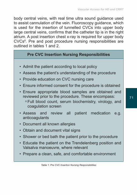
Vascular Access for HD and CRRT
7170
body central veins, with real time ultra sound guidance used to assist cannulation of the vein. Fluoroscopy guidance, which is used for the insertion of tunnelled CVCs into upper body largecentralveins,confirmsthatthecathetertipisintherightatrium. A post insertion chest x-ray is required for upper body CVCs8. Pre and post procedure nursing responsibilities are outlined in tables 1 and 2.
PreCVCInsertionNursingResponsibilities
• Admit the patient according to local policy• Assess the patient’s understanding of the procedure• Provide education on CVC nursing care• Ensure informed consent for the procedure is obtained• Ensure appropriate blood samples are obtained and
reviewed prior to the procedure. These encompass:ᵒ Full blood count, serum biochemistry, virology, and
coagulation screen
• Assess and review all patient medication e.g. anticoagulants
• Document all known allergies• Obtain and document vital signs• Shower or bed bath the patient prior to the procedure• Educate the patient on the Trendelenberg position and
Valsalva manoeuvre, where relevant• Prepare a clean, safe, and comfortable environment
Table 1: Pre CVC Insertion Nursing Responsibilities
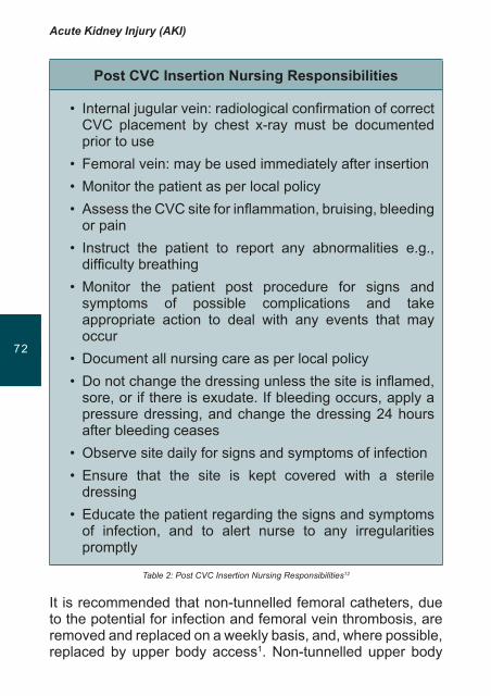
Acute Kidney Injury (AKI)
7372
PostCVCInsertionNursingResponsibilities
• Internaljugularvein:radiologicalconfirmationofcorrectCVC placement by chest x-ray must be documented prior to use
• Femoral vein: may be used immediately after insertion• Monitor the patient as per local policy• AssesstheCVCsiteforinflammation,bruising,bleeding
or pain• Instruct the patient to report any abnormalities e.g., difficultybreathing
• Monitor the patient post procedure for signs and symptoms of possible complications and take appropriate action to deal with any events that may occur
• Document all nursing care as per local policy• Donotchangethedressingunlessthesiteisinflamed,
sore, or if there is exudate. If bleeding occurs, apply a pressure dressing, and change the dressing 24 hours after bleeding ceases
• Observe site daily for signs and symptoms of infection • Ensure that the site is kept covered with a sterile
dressing• Educate the patient regarding the signs and symptoms
of infection, and to alert nurse to any irregularities promptly
Table 2: Post CVC Insertion Nursing Responsibilities12
It is recommended that non-tunnelled femoral catheters, due to the potential for infection and femoral vein thrombosis, are removed and replaced on a weekly basis, and, where possible, replaced by upper body access1. Non-tunnelled upper body

Vascular Access for HD and CRRT
7372
CVCs should be replaced every 2-3 weeks or as indicated by local policy1.
The CVC can be locked between dialysis treatment using either heparin9 or an antimicrobial lock solution such as trisodium citrate10. CVC manufacturing guidelines will indicate the lumen volumes to be used when locking the CVC at the end of each dialysis treatment.
CVC Care and Management Patients dialysed using a CVC have a 41% higher risk of infection related death when compared to patients using an arterio-venousfistula3. Given the high risk of infection, CVC manipulations should be conducted by trained nursing staff. Table 3 outlines nursing activities that reduce the risk of CVC-related infections.
PreventionofCVC-RelatedInfection
•Reserve the CVC for CRRT or IHD only• Implement strict hand hygiene measures in accordance
with local and national infection control policies•Undertake frequent assessment of the CVC and exit
site. To include:o Status of sutureso Observation of skin for signs of infection at exit siteo If a tunnelled CVC was inserted assess for signs of
tunnel infectiono Assess skin around the exit site for signs of skin
reaction to the antiseptic cleansing solution or dressing used to cover the CVC exit site
o Assess catheter clamps and lumens for signs of tape residue, breakage and malfunction
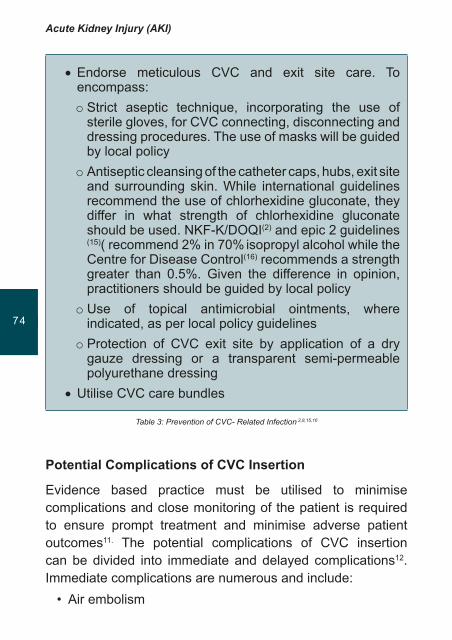
Acute Kidney Injury (AKI)
7574
•Endorse meticulous CVC and exit site care. To encompass:o Strict aseptic technique, incorporating the use of
sterile gloves, for CVC connecting, disconnecting and dressing procedures. The use of masks will be guided by local policy
o Antiseptic cleansing of the catheter caps, hubs, exit site and surrounding skin. While international guidelines recommend the use of chlorhexidine gluconate, they differ in what strength of chlorhexidine gluconate should be used. NKF-K/DOQI(2) and epic 2 guidelines
(15)( recommend 2% in 70% isopropyl alcohol while the Centre for Disease Control(16) recommends a strength greater than 0.5%. Given the difference in opinion, practitioners should be guided by local policy
o Use of topical antimicrobial ointments, where indicated, as per local policy guidelines
o Protection of CVC exit site by application of a dry gauze dressing or a transparent semi-permeable polyurethane dressing
•Utilise CVC care bundles
Table 3: Prevention of CVC- Related Infection 2,8,15,16
PotentialComplicationsofCVCInsertion
Evidence based practice must be utilised to minimise complications and close monitoring of the patient is required to ensure prompt treatment and minimise adverse patient outcomes11. The potential complications of CVC insertion can be divided into immediate and delayed complications12. Immediate complications are numerous and include:• Air embolism
Vascular Access for HD and CRRT
7574
• Nurses must closely observe for the development of an air embolism. If an air embolism is suspected place the patient in the Trendelenberg position as this reduces the risk of air embolism as venous pressure is higher than atmospheric pressure. Place the patient on left side and administer oxygen13.
• Pneumothorax/Haemothorax• After the insertion of the CVC into one of the upper body’s
large central veins a chest x-ray is preformed to detect for the development of a pneumothorax (a collection of air in the pleural cavity) or haemothorax (a collection of blood in the pleural cavity). If either complication is detected the patient may require the insertion of a chest drain2,12.
• Haemorrhage• The CVC exit site must be observed for post insertion
haemorrhage or the development of a haematoma (a collection of blood in the tissues from a leaking blood vessel). If an arterial puncture is observed, pressure must be placed on the CVC for 15 minutes in conjunction with medical review12,14.
• Pericardial tamponade• This is impairment of heart function due to effusion or
haemorrhage. • Cardiac arrhythmia • This may occur if the catheter or wire is inserted too far
into the right ventricle.
The delayed complications of CVC insertion include infection. Observe the CVC exit site for redness, ooze, tenderness or pain, swelling around the catheter or in the neck or arm. Close observation of the patient’s vital signs is required in order to observe for signs of infection e.g., pyrexia and tachycardia. Thrombosis (obstruction of blood flow by a blood clot) orstenosis (abnormal narrowing) of the host vein may present with oedema of the neck or arm, requiring thrombolytic therapy
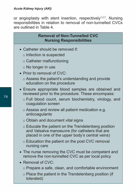
Acute Kidney Injury (AKI)
7776
or angioplasty with stent insertion, respectively1,2,7. Nursing responsibilities in relation to removal of non-tunnelled CVCs are outlined in Table 4.
RemovalofNon-TunnelledCVC NursingResponsibilities
•Catheter should be removed if:o Infection is suspectedo Catheter malfunctioningo No longer in use
•Prior to removal of CVC:o Assess the patient’s understanding and provide
education on the procedure•Ensure appropriate blood samples are obtained and
reviewed prior to the procedure. These encompass:o Full blood count, serum biochemistry, virology, and
coagulation screeno Assess and review all patient medication e.g.
anticoagulantso Obtain and document vital signso Educate the patient on the Trendelenberg position
and Valsalva manoeuvre (for catheters that are placed in one of the upper body’s central veins)
o Education the patient on the post CVC removal nursing care
•The nurse removing the CVC must be competent and remove the non-tunnelled CVC as per local policy
•Removal of CVC:o Prepare a safe, clean, and comfortable environmento Place the patient in the Trendelenberg position (if
tolerated)
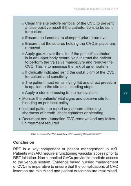
Vascular Access for HD and CRRT
7776
o Clean the site before removal of the CVC to prevent a false positive result if the catheter tip is to be sent for culture
o Ensure the lumens are clamped prior to removalo Ensure that the sutures holding the CVC in place are
removedo Apply gauze over the site. If the patient’s catheter
is in an upper body central vein instruct the patient to perform the Valsalva manoeuvre and remove the CVC. This is to minimise the risk of air embolism
o If clinically indicated send the distal 5 cm of the CVC for culture and sensitivity
o Thepatientmustremainlyingflatanddirectpressureis applied to the site until bleeding stops
o Apply a sterile dressing to the removal site•Monitor the patients’ vital signs and observe site for
bleeding as per local policy.• Instruct patient to report any abnormalities e.g.
shortness of breath, chest tightness or bleeding •Document non- tunnelled CVC removal and any follow
up treatment required
Table 4: Removal of Non-Tunnelled CVC - Nursing Responsibilities12
Conclusion RRT is a key component of patient management in AKI. Patients with AKI require a functioning vascular access prior to RRT initiation. Non-tunnelled CVCs provide immediate access to the venous system. Evidence based nursing management of CVCs is imperative to ensure that the complications of CVC insertion are minimised and patient outcomes are maximised.

Acute Kidney Injury (AKI)
7978
Keypoints
• Non-tunnelled CVCs (short term, temporary, non cuffed or acute catheters) are inserted to ensure vascular access is achieved to facilitate RRT.
• CVCs require evidence based nursing care to ensure patients do not develop immediate or delayed complications from CVC insertion or removal.
• Specificallytrainedcompetentrenalnursesareessentialin reducing the risk of patients developing CVC-associated infections.
Vascular Access for HD and CRRT
7978
References1. The UK Renal Association. Clinical Practice Guidelines, Acute
Kidney Injury, 5th Edition, Final Version. Available on line from http://www.renal.org/Libraries/Guidelines/Acute_Kidney_Injury_-_Final_Version_08_March_2011.sflb.ashx, accessed on 14th October 2011; 2011.
2. NKF K/DOQI. Clinical practice guidelines and clinical practice recommendations 2006 updates: Vascular access. New York. Retrieved from http://www.kidney.org/professionals/kdoqi/pdf/VA_guideline.pdf on 31st January 2011: National Kidney Foundation2006.
3. Astor B, Eustace J, Powe N, Klag M, Fink N, Coresh J. Type of vascular access and survival among incident hemodialysis patients: the Choices for Healthy Outcome in Caring for ESRD (CHOICE) Study. JournaloftheAmericanSocietyofNephrology. 2005;16(5):1449-55.
4. Levy J, Morgan J, Brown E. Oxford Handbook of Dialysis, 2nd Edition. Oxford Oxford University Press; 2004.
5. Uldall R. Vascular acccess for renal replacement therapy. SeminarinDialysis. 1996;9:93-7.
6. Vijayan A. Vascular access for continuous renal replacement therapy. SeminarinDialysis .2009;22(2):133-6.
7. Vanholder R, Canaud B, Fluck R, Jadoul M, Labriola L, Marti-Monros A, et al. Diagnosis, prevention and treatment of haemodialysis catheter-related blood stream infections (CRBSI), a position statement of European Renal Best Practice (ERBP). NDTplus. 2010;3:234-46.
8. McCann M, Einarsdottir H, Van Waeleghem J, Murphy F, Sedgwick J. Vascular access management III: Central Venous Catheters. Journal ofRenalCare. 2010;36(1):25-33.
9. Ashley C. The Renal Drug Handbook. 3rd Edition. Oxford: Radcliffe Publishing; 2009.
10. O’Horo J, Germana L, Nasia Safdar A. Anti-Infective Locks for Treatment of Central Line-Associated Bloodstream Infection: A systematic Review and Meta-Analysis. American Journal ofNephrology . 2011;31:415-22.
11. Daugirdas J, Ing T, Blake P, editors. Handbook of Dialysis. (4th Edition). Philadelphia: Lippincott Williams & Wilkins; 2007.
12. Dougherty L, Lister S. The Royal Marsden Hospital Manual of Clinical Nursing Procedures. 7th Edition. London: Wiley-Blackwell; 2008.
13. Adam S, Osborne S. Critical Care Nursing: Science and Practice. 2nd Edition 2nd Edition. ed. Oxford: Oxford University Press; 2009.
14. Molzahn A, Butera E, editors. Contemporary Nephrology Nursing: Principles and Practice. 2nd edition. New Jersey: AmericanNephrologyNursesAssociation(ANNA); 2006.
15. Pratt R, Pellowe C, Wilson J, Loveday H, Harper P, Jones S, et al. epic2: National evidence based guidelines for preventing healthcare associated infections in NHS hospitals in England. Journal ofHospitalInfection. 2007;65(Suppl 1):S1-S64.
16. Centers for Disease Control and Prevention. Guidelines for the prevention of intravascular catheter-related infections, 2011. Available on line from http://www.cdc.gov/hicpac/pdf/guidelines/bsi-guidelines-2011.pdf; 2011 [21st October 2011].
Nutritional support during Acute Kidney Injury
81
Acute Kidney Injury (AKI)
8382
Introduction
Nutritional support is, especially in Intensive Care Units (ICU’s), an important part of care of the critically ill patient. Together, with other therapies it helps prevent malnutrition during this serious condition and optimises recovery and survival outcomes.
Acute Kidney Injury (AKI) is a clinical syndrome characterized by a sudden deterioration of kidney function represented by electrolyte, acid-base and fluid imbalances. The causes for AKI are many and related to either ischaemic or toxic events such as prolonged hypo perfusion, medication misuse, accidents – crushing injuries in extensive parts of the body, cardiogenic shock, septic shock, glomerulonephritis, and contrast media.
Clinical and Laboratory Overview
The clinical and laboratory overview of the disease is characterized by:
• Sudden reduction in the amount and quality of the urine – the patient may secrete a large volume of urine, but the glomerular filtration rate (GFR) will be reduced as is the ability to excrete toxins of metabolism.
• Increase in the concentration of serum urea and creatinine.
Learning outcomes
• Tounderstand theeffectsof catabolic andnutritionalstatesintreatmentofpatientswithAcuteKidneyInjury
• TobeawareofthedietaryrecommendationsforpatientswithAKIaccordingtothepatient’scatabolicstate

Nutritional support during Acute Kidney Injury
8382
• When the patient exhibits oliguria or serious compromise of renal function the amount of potassium in blood increases5.
• Lack of appetite, nausea, vomiting, cramp and in extreme cases loss of consciousness.
• Failure to regulate acid-base balance will alter the pH of blood, to compensate for this, patients may hyperventilate.
• Due to the inability to excrete electrolytes such as potassium, there may be signs of cardiac irritability as evident through ectopic beats on the cardiac monitor.
DietaryTreatmentDietary treatment is affected by the following1,2,5,6.• Thecauseof thedisease is important as it affects the
patient’s catabolic rate.• The patient’s catabolic rate determines the
recommendations for protein and calories increase or decrease.
• Dialysis (renal replacement therapy) changes the recommendations for protein and calories.
There are a number of ways to estimate a patient’s catabolic rate. In patients without kidney damage, the most appropriate way is to measure the amount of nitrogen secreted in the urine. In patients suffering from kidney disease this measurement is problematic as some of the patients do not produce urine. Despite this, a number of studies relate to this measurement as long as the patient is urinating1.
Patients are divided into three groups according to the level of nitrogen secreted as urea:
• Less than 5 g/ day . – non catabolic state• 5-10 g/day. – moderate catabolic state• Above 10 g/ day. – severe catabolic state
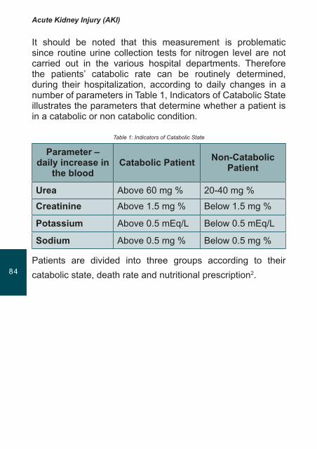
Acute Kidney Injury (AKI)
8584
It should be noted that this measurement is problematic since routine urine collection tests for nitrogen level are not carried out in the various hospital departments. Therefore the patients’ catabolic rate can be routinely determined, during their hospitalization, according to daily changes in a number of parameters in Table 1, Indicators of Catabolic State illustrates the parameters that determine whether a patient is in a catabolic or non catabolic condition.
Table 1: Indicators of Catabolic State
Parameter – daily increase in
thebloodCatabolic Patient Non-Catabolic
Patient
Urea Above 60 mg % 20-40 mg %
Creatinine Above 1.5 mg % Below 1.5 mg %
Potassium Above 0.5 mEq/L Below 0.5 mEq/L
Sodium Above 0.5 mg % Below 0.5 mg %
Patients are divided into three groups according to their catabolic state, death rate and nutritional prescription2.
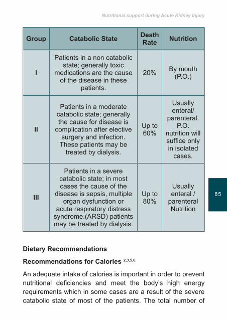
Nutritional support during Acute Kidney Injury
8584
Group Catabolic State DeathRate Nutrition
I
Patients in a non catabolic state; generally toxic
medications are the cause of the disease in these
patients.
20% By mouth (P.O.)
II
Patients in a moderate catabolic state; generally the cause for disease is
complication after elective surgery and infection. These patients may be
treated by dialysis.
Up to 60%
Usually enteral/
parenteral. P.O.
nutrition will suffice only in isolated
cases.
III
Patients in a severe catabolic state; in most cases the cause of the
disease is sepsis, multiple organ dysfunction or
acute respiratory distress syndrome.(ARSD) patients may be treated by dialysis.
Up to 80%
Usually enteral /
parenteral Nutrition
Dietary Recommendations
RecommendationsforCalories2,3,5,6.
An adequate intake of calories is important in order to prevent nutritional deficiencies and meet the body’s high energy requirements which in some cases are a result of the severe catabolic state of most of the patients. The total number of
Acute Kidney Injury (AKI)
8786
calories recommended for each patient should be based according to their corrected nutritional and catabolic state.
A corrected weight should be calculated, according to the following formula. For patients whose weight is equal to, or more than, 15% of the ideal weight
Ideal weight + 0.25 x (existing weight –ideal weight) = corrected weight
A corrected weight should be calculated, according to the following formula, for patients whose weight is less than 95% of the ideal weight for height:
Existing weight + 0.25 x |( ideal weight - existing weight) = corrected weight
The nutritional recommendation for calories is 25-35 calories per kilogram of ideal/corrected body weight. It is very important not to give more than 130% of the nutritional recommendations even in cases of severe catabolic states5.
The nutritional recommendation for calories is determined according to the patient’s catabolic state, as follows.
• Non catabolic state and normal state of nutrition - 25 calories/kilogram of body weight/corrected weight per day.
• Moderate catabolic state – 25-30 calories/kilogram of body weight/corrected weight per day.
• Severe catabolic state – 25-35 calories/kilogram of body weight/corrected weight per day.
Inallcasesitisrecommendedtostartfeedingatthelowercaloricrangeandgraduallyincreasetothehigherrange5
RecommendationsforProtein2, 4, 5.
The nutritional recommendations for protein are also given based on the patient’s catabolic state.
Nutritional support during Acute Kidney Injury
8786
GroupIpatients–normalornoncatabolicstateIt is recommended that patients are prescribed between 0.6-0.8 grams of protein per kilogram of given/corrected body weight. At the beginning of feeding 0.6 grams of protein per kilogram of body weight is given, and the quantity is gradually increased in the menu until 0.8 grams/kilogram of body weight. In parallel, the level of urea in the blood is monitored. The level should be less than 100 mg/dl (at all stages of protein intake)
GroupIIpatients–moderatecatabolicstateIt is recommended that patients are prescribed between 0.8-1.0 grams of protein per kilogram of given/corrected body weight. In these patients it is necessary to pay attention to the level of urea in the blood. If the patients are being treated by one of the types of dialysis, between 1.2 and 1.5 grams/kilogram of body weight can be given per day.
GroupIIIpatients–severcatabolicstateIt is recommended that patients are prescribed between 1.0 and 1.5 grams of protein per kilogram of given/corrected body weight. Patients treated with Dialysis (renal replacement therapy) should receive at least 1/5 g/kg/day/5.
It is important to note that it was customary to give parenteral nutrition to Group II and III patients, but the current accepted approach recognizes the importance of nutrition through the digestive system, since this preserves gastric functioning and integrity. In studies on animals it has been observed that enteral nutrition increases the rate of plasma flow and improves kidney function2, 5. For patients with AKI, there are a number of enteral nutrition options which can be prescribed based on the needs of the individual. Guidelines indicate that the enteral preparations should provide the recommended calorie and protein intake in a minimum amount of fluid 5.
Acute Kidney Injury (AKI)
8988
FluidsIn Acute Kidney Injury patients may either cease producing urine or the volume can be substantially reduced. For patients with a reduced urine output it is recommended that they should have a fluid intake based on the amount of urine secreted the previous day.
Vitamins and Minerals5, 6.
The levels of water soluble minerals are generally low in the patients who , treated by dialysis apart from vitamin C that represents an important precursor of oxalic acid, which settles in the soft tissue and may cause oxalosis.
It is recommended to add B complex daily, according to the following breakdown:
• Folic acid - 1 mg• B6 Pyridoxine - 5-10 mg• B2 Riboflavin - 1.2-6 mg• B12 - 6 mcg
It is not recommended to:• Give more than 200 mg vitamin C per day, including food• Add vitamins K, E and A
It is necessary to check the levels of selenium and zinc in the blood and, if necessary, to supplement these6. Appropriate and effective nutritional support improves the patient’s conditions reduces duration of ICU stay, decreases mortality, incidence of co morbidity and complications. These issues also have a positive influence in reducing the health costs and will contribute to a better quality of life and a better body endurance for the patients after the hospital discharge.
Nutritional support during Acute Kidney Injury
8988
KeyPointsIn determining the nutritional plan for patients in Acute Kidney Injury, the following considerations are important:
• The cause of the disease.• The patient’s catabolic measure and nutritional state.• Whether or not the patient is undergoing dialysis.• Recommendation for protein between 0.6-1.5 g /kg of body
weight and 25-35 calories per kilogram of body weight.• Method of nutrition –preferably this should be by the oral
route, where this is not possible either enteral or parenteral routes should be considered taking patient safety issues into account.

Acute Kidney Injury (AKI)
MT90
References1. Druml W. Mitch WE: Metabolic abnormalities in acute renal failure.
Global prevalence of diabetes. Seminars Dialysis 1996; 9:484-490.
2. Druml W: Nutritional Management of acute Renal Failure. AmericanJournalKidneyDisease. 2001; 37 (sup2) 589-594.
3. Schneeweiss B. Graninger W. , Soteckenhuber FD: Energy metabolism in acute and chronic renal failure 1990; Am.J.Clin.Nutr. 52:596-601.
4. Druml W: Protein metabolism in acute renal failure. Miner Electrolyte Metab 1998; 24:47-54.
5. Enrico Flaccadori ,Elena Cremaschi , and Gluseppe Regolisti Seminars in Dialysis, Vo 24 ,No 2 2011 pp 169-175 /.
6. A.S.P.E.N.( AmericanSocietyforParenteralandEnteralNutrition) Clinical Guidelines: J. Parenteral Enteral Nutrition, 2010 34:366.
Meeting the needs of critically ill patients and families
91

Acute Kidney Injury (AKI)
9392
Introduction
When assessing a critically ill patient with AKI it is essential to be aware of the advances made in medicine and technology which have taken place over centuries1,2. The second half of the 20th century was the cornerstone of this evolution, when the therapeutic processes, that remain central to the management of critical patients, became available. These ‘four pillars’ of critical care include artificial airways and mechanical ventilation, cardiopulmonary resuscitation and coronary care, the artificial kidney and total parenteral nutrition1.
It is only in Critical Care Units (CCU) or Intensive Care Unit (ICU) where a concentration of technology for diagnostics and therapeutics handled by members of the multidisciplinary teams is available. In addition, within these environments experts are able to respond effectively and quickly to deviations in clinical patients parameters and implement appropriate evidence based interventions1,2,3. These units are especially created to manage multiorgan failure situations, where the risk of death is high. Nurses perform clinical decision making and problem
Learning outcomes
• Tointegratetheconceptsoftechnologicalcompetenceandcriticalnursingcare.
• To recogniseandrespond to theneedsof thecriticalpatientandfamily,asvitalfortheexcellenceinnursingcare.
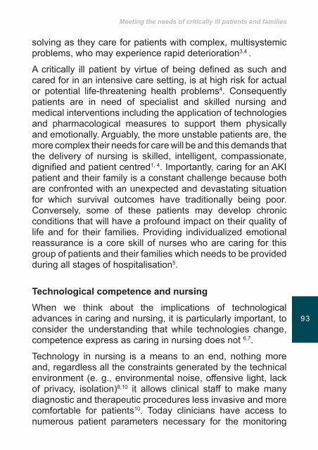
Meeting the needs of critically ill patients and families
9392
solving as they care for patients with complex, multisystemic problems, who may experience rapid deterioration3,4 .
A critically ill patient by virtue of being defined as such and cared for in an intensive care setting, is at high risk for actual or potential life-threatening health problems4. Consequently patients are in need of specialist and skilled nursing and medical interventions including the application of technologies and pharmacological measures to support them physically and emotionally. Arguably, the more unstable patients are, the more complex their needs for care will be and this demands that the delivery of nursing is skilled, intelligent, compassionate, dignified and patient centred1, 4. Importantly, caring for an AKI patient and their family is a constant challenge because both are confronted with an unexpected and devastating situation for which survival outcomes have traditionally being poor. Conversely, some of these patients may develop chronic conditions that will have a profound impact on their quality of life and for their families. Providing individualized emotional reassurance is a core skill of nurses who are caring for this group of patients and their families which needs to be provided during all stages of hospitalisation5.
TechnologicalcompetenceandnursingWhen we think about the implications of technological advances in caring and nursing, it is particularly important, to consider the understanding that while technologies change, competence express as caring in nursing does not 6,7.
Technology in nursing is a means to an end, nothing more and, regardless all the constraints generated by the technical environment (e. g., environmental noise, offensive light, lack of privacy, isolation)8,10 it allows clinical staff to make many diagnostic and therapeutic procedures less invasive and more comfortable for patients10. Today clinicians have access to numerous patient parameters necessary for the monitoring

Acute Kidney Injury (AKI)
9594
and control of their clinical status. To continuously care for critical patients without needing multiple manipulations, uncomfortable and sometimes painful, allows to comply with the periods of rest and sleep for each patient, usually so scarce in the ICUs10,13.
The technology and technological competence must be seen as essential to the production of more effective and efficient care, and that their use by nurses is congruent with the socio-political and economic health systems models and, used according to their relevance and purpose, may itself be a factor of humanization10-13.
Nursing care takes place in nursing situations, the shared lived experience in which the caring between nurse and the one nursed, enhances personhood” 14. Technological competence can be seen as an expression of caring in nursing.
Nursing care in ICU requires high-tech expertise but despite technological advances, particularly in modern medical and nursing practices, continue to challenge definitions of person, always keeping in mind that the focus of nursing care, is the person and its family15 .
CriticalcarenursingIn ICU's nurses provide care to individuals with a range of clinical conditions. This can be a challenge and involves more than patient’s appraisal. It is mandatory to include members of the patient’s family in all the process in order to provide it’s continuous involvement and to find better coping mechanisms to deal with the situation16,17.
Nursing practice occurs by building a relation of partnership and co-existence and although, non-familiar, nurses, patients and family can quickly connect when confronted, simultaneously, by the crisis triggered by a devastating disease as AKI in the complex, demanding and strange environment of ICU19.

Meeting the needs of critically ill patients and families
9594
Due to this crisis people are more vulnerable and, nurses in particular, need to be aware that cultural sensitivity is needed to to comfort families and ensure their well being19. Because individual responses and values, may vary depending on culture, the patient and his family must be supported with sensitivity and through acknowledging their individuality20. True communication it is more than words; it really comes from the heart. It is a flow of energy between the nurse and the patient or family2 .
Nurses can achieve this involvement when their practices rely on knowledge, experience, practical skills, ethics, intuition, caring, human spirit and wisdom; elements that Ashworth 19,20 considers constituting the “Nursing lens”, that reassures and gives security.
Boykin and Schoenhofer in their Theory of Nursing as Caring projected a model that serves as a guide for caring in situations where technology of high complexity and technological support is needed14. The aim of nursing is to nurture persons living caring and growing in caring. To truly nurse is to value this person in this moment, to form the committed intention to care and to communicate that caring effectively is the real meaning of being nursed14. Nursing care in high-technology settings [critical care nursing] is grounding in two fundamental principles: intentionality and knowing which means that all humans are caring persons, that to be human is to be called to live one’s innate caring nature. Developing and have a deep understanding of the full potential of expressing caring is an ideal and for practical purposes, is a lifelong process21.
CriticalpatientandfamilyneedsHistorically, hospitals had restrictive visiting policies limiting family visits but, over time, many hospital floors, and even ICUs have liberalized visiting policies22. Liberalizing family visitations is an emerging concept in providing a holistic

Acute Kidney Injury (AKI)
9796
approach to healing. Family members can provide the spiritual and emotional support to patients in an unfamiliar situation, and they can help give meaning and understanding of the experience of illness for the patient10,21.
The hospital is open to families, but the ICU’s are the places where these came last. Recognized as placeholders, the ICU represent something forbidden, sacred, where it plays symbolically the good and the evil, success and failure, life and death. Having someone admitted in “intensive care” is reason to silences, despair and unspoken words that accompany periods of great uncertainty. The fear of asking questions, touching on something that harms the patient, to get close to him, is associated with the anguish of loss and the silent need for responses. Since the 1970s when Hampe first and, after her Molter and Molter and Leske, in 1983 with their Critical Care Family Needs Inventory (CCFNI)23 first gave attention to the situation and problems of families in ICU, the “needs of visitors,” family and significant others have been the aim of several studies23,24, placing them in the core of the concerns of nurses, along with the patient.
“Molter’s early examination of family members’ needs led to the development of the Critical Care Family Needs Inventory which has been used in its original and adapted forms in much of the research conducted to date. Family needs are often categorized into 5 domains: (1) assurance, (2) proximity, (3) comfort, (4) support and, (5) information25.
Most research25, 26 in this area have used it and it is considered reliable and consistent of multiple family members. So, we can assume that these are the main domains that critical nurses must consider in their daily practice to provide excellent care
Despite advances in medical technology and the increased inclusion of families in ICU care, relatives continue to express the same needs: honest information, to visit at any time and to feel that the healthcare team care about the patient26.

Meeting the needs of critically ill patients and families
9796
Nursing has modified its practice, from a patient-centered model to an approach that recognizes the needs of the family as inseparable from those of the patient11. An exploratory study of nurses’ experiences of caring for family of ICU patients refers that caring for families and addressing both their emotional and physical needs, places heavy demands on nurses. There is great role of ambiguity and role conflict associated with caring for families with relatives in intensive care units11.
Examining the visiting preferences of patients in ICU Gonzalez et al 27 concluded that patients value the fact that visitors could interpret information for the patients while providing information to assist the nurse in understanding them.27 Patients in the intensive care unit were very satisfied with a visiting guideline that is flexible enough to meet their needs and those of their family members27.
Based in their theory of Nursing As Caring, Boykin and Schoenhofer29,conceived a model for relating: a dance, represented by an open circle: in this circle each person brings his or her way of doing in order to understand what really matters to those seeking care and, responding to that understanding. In the dance of caring persons, the dancers are all the individuals who were part of the multidisciplinary care team (family included), but now they are encouraged in a way of interact with each other that conveys respect for and honoring of person-as-person. In this dance, all dancers are valued, respected, and supported in their unique role (e.g., nursing, medicine, radiology, family) without issues of power and authority restricting their unique contribution to the dance29. It is assumed in this model that each dancer is a caring person and that each discipline and role is valued.
Adhering to this way of thinking and doing helps to capture the hearts and hands of nurses who really are the front line interface with patients and their significant others2.

Acute Kidney Injury (AKI)
9998
KeypointsThe more critically ill the patient is, the more likely he or she is to be highly vulnerable, unstable and complex, thereby requiring intense and vigilant nursing.
Regardless all the constraints generated by the technical environment, it also allows to make many diagnostic and therapeutic procedures less invasive and more comfortable for patients.
Nursing has modified its practice, from a patient-centered model to an approach that recognizes the needs of the family as inseparable from those of the patient.

Meeting the needs of critically ill patients and families
9998
References1. Wiles Vanda, Daffurn Kathy. There’a a bird in my hand and a bear by
the bed - I must be in the ICU: The pivotal years of Australian Critical Care Nursing. Marrickville: ACCCN, 2002. ISBN 0-9579703-0-7.
2. Zalumas J. Caring in crises: An oral history of critical care nursing. Philadelphia: University of Pennsylvania Press, 1995. ISBN 0-8122-1510-9.
3. ANCCN www.aacn.org/WD/PressRoom/Content/aboutcriticalcarenursing.pcms?pid=1&&menu= .[Online] [Quotation ]
4. Ponce, P. Cuidados intensivos. O paradigma da nova medicina tecnológica. AnáliseSocial,2002; XXXVIII:166 139-153 [141].
5. Paul F., & Rattray J., Short- and long-term impact of critical illness on relatives: literature review. JournalofAdvancedNursing, 2008, 62(3), 276–292.
6. Watson, Jean. Foreword. [autor do livro] Rozzano C Locsin. Advancing Techology, Caring, and Nursing. Westport: Auburn House, 2011.
7. Locsin R. Advancing Technology, Caring, and Nursing. Westport: Aubur House, 2001. 0-86569-302-1.[ xviii]
8. Petterson, M. , Reduced noise levels in ICU promote rest and healing. CriticalCareNurse, 2000, 20(5), 104.
9. Ulrich, R., & Zimring, C. (2004). The role of the physical environment in the hospital of the 21st century: Once-in-a-lifetime opportunity. Report to the Center of Health Design for designing the 21st Century Hospital Project. Retrieved February 22, 2005, from http://www.healthdesign.org/research/reports/pdfs/role_physical_env.pdf
10. Durão M. C. Ser enfermeiro numa UCI: um sentido para cuidar [Master’s dissertation]. Lisboa, Escola Superior de Enfermagem de Lisboa; 2004.
11. Ray A. Complex Culture and Technology: Toward a Global Caring Communitarian Ethics of Nursing. In: Advancing Technology, Caring, and Nursing. Westport. Locsin, 2001. ISBN 0-86569-302-1, 41-52.
12. Ozbolt, J. Nursing and technology: a dialectic. Holistic Nursing Practice. Philadelphia, ISSN 0887-9311. 11:1 (Oct 1996) 1-5.
13. Barnard A, Sandelowski M. Technology and humane nursing care: (ir)reconcilable or invented difference. JournalofAdvancedNursing. 2001, 34:3, 367-375.
14. Boykin A., & Schoenhofer S. Nursing as Caring: A Model for transforming practice. Sudbury: Jones and Bartlett , 2001.
Acute Kidney Injury (AKI)
101100
15. Locsin R, Purnell M. Rapture and suffering with technology in nursing. InternationalJournalofHumanCaring, 2007; 11:1, 38-43.
16. Basto M, Portilheiro C., Concepções de cuidados de enfermagem: o que dizem os enfermeiros. RevistaInvestigaçãoemEnfermagem, 2003; 34:3 42-51.
17. Purnell J. Who really makes the bed? Uncovering technologic dissonance in nursing. Holistic Nursing Practice. Philadelphia, ISSN 0887-9311. 12:4 (Jul 1998) 12-22.
18. Dean B. Reflections on technology: increasing the science but diminishing the art of nursing?. Accident and Emergency Nursing. 6(1998) 200-206.
19. Ashworth P. High technology and humanity for intensive care. IntensiveandCriticalCareNursing. 1990, September, 150-160.
20. Ashworth P. Long term intensive care – a challenge for nurses and patients. IntensiveandCriticalCareNursing. 1990 , September, 109-110.
21. Schoenhofer S. A Framework for Caring in a Technologically Dependent Nursing Practice Environment. In: Advancing Technology, Caring, and Nursing. Westport. Locsin, 2001. ISBN 0-86569-302-1, 3-11.
22. Berwick,D.,& Kotagal, M., Restricted visiting hours in ICUs: Time to change. Journalof theAmericanMedicalAssociation, 2004, 292(6), 736–737.
23. Linnarsson J, Bubini J, Perseius K. Review: a meta-synthesis of qualitative research into needs and experiences of significant others to critically ill or injured patients. JournalOfClinicalNursing. 2010, Nov,
24. Stayt L. Nurses’ experiencies of caring for families with relatives in intensive care units. JournalofAdvancedNursing. 2007, Mar 15 57(6): 623-630.
25. Baileya J., Sabbaghb M, Loiselle C, Boileaug J, McVeyh L. Supporting families in the ICU: A descriptive correlational study of informational support, anxiety, and satisfaction with care. IntensiveandCriticalCareNursing, 2010, 26, 114-122
26. Kinrade T, Jackson A, Tomnay J. The psychosocial needs of families during critical illness: comparison of nurses’ and family members’ perspectives. AustralianJournalOfAdvancedNursing [serial on the Internet].2009,, [cited March 20, 2012]; 27(1): 82-88. Available from: CINAHL Plus with Full Text.
27. Gonzalez C, Carroll D, Elliott J, Fitzgerald P, Vallent H. Visiting preferences of patients in the intensive care unit and in a complex

Meeting the needs of critically ill patients and families
101100
care medical unit. AmericanJournalOfCriticalCare: An Official Publication, American Association Of Critical-Care Nurses [serial on the Internet]. 2004,, [cited March 21, 2012]; 13(3): 194-198. Available from: MEDLINE with Full Text.
28. Boykin, A., & Schoenhofer, S.O. Nursing as Caring: A Model for transforming practice. Sudbury: Jones and Bartlett, 2001.
29. Boykin A, Schoenhofer S. The role of nursing leadership in creating caring environments in health care delivery systems. NursingAdministrationQuarterly 2001,; 25(3): 1-7.
30. Boykin A, Bulfin S, Schoenhofer S, Baldwin J, McCarthy D. Living caring in practice: the transformative power of the theory of nursing as caring. InternationalJournalForHumanCaring. 2005, 9(3): 15-19.

Notes
Acute Kidney Injury (AKI) in children
103
Acute Kidney Injury (AKI)
105104
IntroductionThe term acute kidney injury (AKI) is the inability of the kidney to maintain water balance, acid-base balance and electrolytes. Currently, AKI is characterized by decreased urine output (<1ml/kg/h) in parallel with increased urea and creatinine levels in blood and other useless metabolic products1. In some patients the AKI may be unaccompanied with oliguria but can coexist with polyuria.
Epidemiology of AKI in children It is difficult to estimate the true incidence and prevalence of the paediatric patients with AKI due to lack of clearly defined diagnostic criteria 2. It is suggested that 8 in 1.000.000 of children in the population per year 3 are diagnosed with AKI compared to 1 in 5 compared adults1. Mortality rates among children with AKI vary widely and depend on the nature of the underlying disease, with the highest mortality rates in children who develop multi organ failure 4,5,6,7 . The mortality in neonates and infants undergoing cardiac surgery for congenital heart disease and who subsequently develop AKI is 51%1, while this
Learning outcomes
• To understand the epidemiology of children whodevelop acute kidney injury.
• Todemonstratea thoroughgraspof themainclinicalmanifestations and complications of the disease for childrenpresentingwithacutekidneyinjury.
• Toappreciatekeytherapeutictreatmentmodalitiesandthe potential for complications.

Acute Kidney Injury (AKI) in children
105104
is greatly reduced to 3-6% among children with haemolytic uremic syndrome7. Use of specific biomarkers in early stages might reveal a much greater incidence of AKI8.
Causes for AKI in children
The causes of acute kidney injury in children, are distinguished into pre-renal, renal, post- renal 8 and based on the volume of urine output, into oliguric and non- oliguric phase 9. This is not unlike adults (see Chapter 2). The most common causes for AKI in children are mainly related with: renal parenchyma damage, inadequate renal perfusion, arterial or venous obstruction of the renal vessels and obstruction of the drainage portion of the urinary system /track7.
Pre-renal causes
Are caused due to a reduced renal blood perfusion either due to decreased fluid intake (circulating volume), cardiac output or as a result of vascular injury:
• hypovolaemia from any cause (dehydration, vomiting, haemorrhage)
• osmotic diuresis• fluid loss in the third space • decreased cardiac output • septic shock• burns9,10.
Renal causes
In children these may associated with glomerular or tubular injury (ischemic or nephrotoxic) or vascular10:
Acute Kidney Injury (AKI)
107106
1. Glomerulonephritis 2. Systemic disease with renal damage 3. Hemolytic-uremic syndrome 4. Vasculitides 5. Henoch-Schonlein purpura6. Thrombosis of renal artery or vein7. Metabolic cause 8. Infections9. Cancer 10. Rhabdomyolysis (myoglobinuria)11. Severe hemolysis (hemoglobinuria)12. Severe anaphylactic reaction13. Contrast substance14. Toxins and heavy metals15. Non steroid and inti-inflammatory drugs16. Antibiotics (Antimicrobials)17. Other medication
Post-renal causes Due to obstruction in the drainage parts of the urinary system11.
1. Bilateral ureteral obstruction or urethral obstruction. 2. Posterior valve urethra 3. Congenital obstructive ouropatheia
All these causes are summarized in the table below.
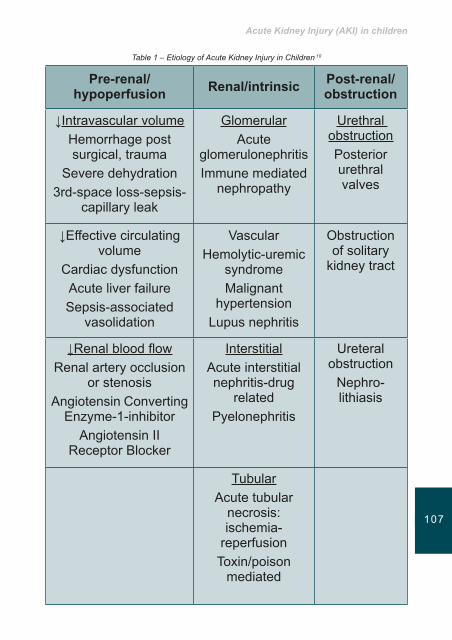
Acute Kidney Injury (AKI) in children
107106
Table 1 – Etiology of Acute Kidney Injury in Children 10
Pre-renal/hypoperfusion Renal/intrinsic Post-renal/
obstruction
↓Intravascular volumeHemorrhage post surgical, trauma
Severe dehydration3rd-space loss-sepsis-
capillary leak
GlomerularAcute
glomerulonephritisImmune mediated
nephropathy
Urethral obstructionPosterior urethral valves
↓Effective circulating volume
Cardiac dysfunctionAcute liver failureSepsis-associated
vasolidation
VascularHemolytic-uremic
syndromeMalignant
hypertensionLupus nephritis
Obstruction of solitary
kidney tract
↓Renal blood flowRenal artery occlusion
or stenosisAngiotensin Converting
Enzyme-1-inhibitorAngiotensin II
Receptor Blocker
InterstitialAcute interstitial nephritis-drug
relatedPyelonephritis
Ureteral obstruction
Nephro- lithiasis
TubularAcute tubular
necrosis: ischemia-
reperfusionToxin/poison
mediated

Acute Kidney Injury (AKI)
109108
Clinical manifestations of AKI in children • Oliguria or anuria
This is the main clinical manifestation of acute kidney disease in children20. The term oliguria refers to the urine output of <350ml/24h (<0,5ml/kg/h in children or <1ml/kg/h in infants), while anuria is characterized by a urine output of less 180ml/24h10,11. Morbidity and mortality rate is lower in children that are in non oliguric phase than in children who develop oliguria12,15.
Acute kidney injury should be suspected in any dehydrated child due to fluid depleting states like prolonged diarrhea or vomiting. In children who do not a have a history of gastroenteritis then the focus must be on other causes of AKI14,16. The history should also focus on the amount of urine recently excreted, quality of urinary stream, drug intake, evidence of fever, rash, arthropathy, urinary symptoms, recent weight and any history of weight loss and whether there is family history of renal problems15.
Another important issue is related with the electrolyte imbalance which commonly occurs in critically ill patients in combination with other conditions. Children are most predisposed to this condition because of their small physical complexion. It is very difficult to properly prescribe the exact fluids dose in critical situations 15. The most common imbalance, in the first stages of AKI in children, is related with potassium, a major intracellular ion. Chapter 3 gives an excellent overview about this issue.
• Metabolic disorders Increase of the urea and creatinine levels due to the functional impairment of the glomerular filtration.
Metabolic acidosis: when GFR is considerably decreased (<50ml/min/1,73 m2) , the ability of the kidney to produce

Acute Kidney Injury (AKI) in children
109108
ammonium is also reduced, resulting in retention of H+, sulphate and phosphate radicals and finally in the appearance of metabolic acidosis10,11.
• Electrolyte imbalance10,11,12
Hyponatremia (Na+serum <30mEq/L) usually due to fluid retention and rarely due to increased Na+ loss in urine output.
Hyperkalemia: mainly due to the failure of of the kidneys to excrete potassium (K+) in urine, but also due to the leakage of K+ from the cells as a consequence of metabolic acidosis. Disorders of K+ have direct and potentially fatal effects on cardiac function. For this reason, children with AKI should be under regular ECG monitoring.
Hypocalcaemia: is attributed to the hyperphosphatemia and the impaired production of 1,25-OH-VitD
Hyperfosfatemia: is the inability of the kidneys to eliminate phosphates.
Hypermagnesemia: Mg ++ movement from intracellular to extracellular space.
Hypertension: the treatment of hypertension is proportional to the level of blood pressure and the presence or absence of symptoms10. The child may present with hypertensive crisis, vague symptoms, heart failure or hypertensive encephalopathy (headache, irritability, convulsions). In most cases, hypertension in children with AKI is mild and responds to salt restriction and oral antihypertensive medication11.
Management of AKI in ChildrenInitially, an accurate assessment of the patient’s intravascular status (hypervolemia or hypovolaemia) is essential to

Acute Kidney Injury (AKI)
111110
help guide clinical decisions and treatments. Overall, the management of the patient will be dictated by the underlying condition of the patient. Key principles in the management of a child with AKI include:
• Maintain fluid and electrolyte balance• Treatment of underlying disease• Diet, which aims to meet the caloric needs, to reduce the
intake of potassium, sodium, phosphate, and protein level (0,5-1g/kg/d), to the limitation of the amount of fluids given according to the child’s daily needs11.
• Cardiovascular monitoring
Bladder catheterization and in more serious situations measurement of central venous pressure (CVP) through a central vascular catheter will be required.
ThehypovolemicpatientThe hypovolemic child shows signs of dehydration and shock. The patient needs immediate rehydration with iv fluids. The preferred solution is NaCl 0.9% (20ml/kg). This dose may be repeated two or three times to restore the patient diuresis. The kidneys may respond to this method and other methods of renal replacement may be avoided.
Fluid status of the patient in AKI is difficult to assess. In children, fluid administration so needed in the early stages to correct hypovolemia, can potentially lead to an overload so fluid balance monitoring is of utmost importance to prevent hypervolemia.
The hypervolemic patients usually presents oedema, increase in body weight possible increase in blood pressure, signs of heart failure such as tachycardia and presence of rale sounds on auscultation12,15. Oedema, due to fluid overload must be solved urgently to avoid further severe complications. It is used commonly, furosemide (up to 2mg/kg) or Mannitol
Acute Kidney Injury (AKI) in children
111110
(0.25-0.5 g/kg). Limitations in the daily fluids intake in 400ml/m2/day are also important as the administration of electrolytes according to the laboratory results. It is also important to check signs of dehydration and prevention of shock.
Hyperkalaemia Particular attention is still needed in the treatment of hyperkalemia: usually occurs in the oliguric phase: severe hyperkalemia (K+>7.5mEq/L), to moderate hyperkalemia (K+=6.5-7.5mEq/L) and mild hyperkalemia (K=,6.5 mEq/L) potassium disorders have immediate and potentially fatal effects on cardiac function and children with AKI should be under continuous EKG monitoring1.
The appearance of pointed T waves in the ECG is one of the first lesions that can be detected on ECG. These changes if not corrected, can be followed by the fall of the ST segment in the EKG and the prolongation of the PR segment and at last from the widening of the QRS (ventricular fibrillation and cardiac arrest)1.
TreatmentofHyperkalaemiaAdministration of Kayexalate (1g/kg per mouth or per rectum) (mild form).
In severe hyperkalemia calcium gluconate 10% is used or crystalline insuline with glucose. In severe cases the patient should be placed on dialysis (continuous or intermittent). In patients with severe metabolic acidosis (pH-7) sodium bicarbonate is administered but with great caution for hypernatremia and water retention.1
In children and in infants there is a wide choice of renal replacement therapies, which includes all methods that applies to adult patients. However, there are important technical issues regarding dialysis in children because of their body weight that
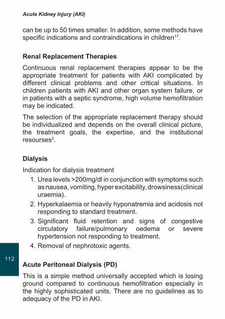
Acute Kidney Injury (AKI)
113112
can be up to 50 times smaller. In addition, some methods have specific indications and contraindications in children17.
RenalReplacementTherapiesContinuous renal replacement therapies appear to be the appropriate treatment for patients with AKI complicated by different clinical problems and other critical situations. In children patients with AKI and other organ system failure, or in patients with a septic syndrome, high volume hemofiltration may be indicated.
The selection of the appropriate replacement therapy should be individualized and depends on the overall clinical picture, the treatment goals, the expertise, and the institutional resourses2.
Dialysis Indication for dialysis treatment
1. Urea levels >200mg/dl in conjunction with symptoms such as nausea, vomiting, hyper excitability, drowsiness(clinical uraemia).
2. Hyperkalaemia or heavily hyponatremia and acidosis not responding to standard treatment.
3. Significant fluid retention and signs of congestive circulatory failure/pulmonary oedema or severe hypertension not responding to treatment.
4. Removal of nephrotoxic agents.
Acute Peritoneal Dialysis (PD)This is a simple method universally accepted which is losing ground compared to continuous hemofiltration especially in the highly sophisticated units. There are no guidelines as to adequacy of the PD in AKI.

Acute Kidney Injury (AKI) in children
113112
Advantages of PD1. It does not require specialized equipment or technical
expertise.2. It does not need special vascular intervention.3. There is no need the circuit to be filled with blood and
anticoagulation. 4. The hemodynamic instability is unusual.
The surgical insertion of catheter (type Tenckhoff) is preferred and in emergency situations an acute peritoneal catheter may be used. Initially a solution with the lowest glucose concentration is used (1.36%) 10-20ml/kg(300-600ml/m2) with cycles of one hour during the first 24h ( when the entire cycle should last ½ hour the method is facilitated by means of automatic change machine (cycler) which reduces the workload of the nursing staff and the repeated interventions in the catheter) and gradually increase the volume up to 800-1000ml/m2. Attention should be given to hyperglycaemia which can lead to hyperosmosis and to reduce filtration5,16. Prophylactic dose of cefuroxime 125mg/L for 48 h and heparin should be added to the bag solution in order to avoid complications of the method.
Complications of the method • Leakage of peritoneal fluid• Obstruction of the peritoneal catheter• Peritonitis
Acute Dialysis (AD)Acute dialysis applies when the PD is contraindicated due to intra abdominal pathology (recent surgery, abdominal hernia, omphalocele and gastroshisis) or respiratory limitations. Acute dialysis in infants and small children requires experience and technical expertise and adequate size of filters, bloodlines and vascular catheters15. In very small patients (<5kg) may

Acute Kidney Injury (AKI)
115114
need to fill the circuit with blood or human albumin 5% (5-8kg). The small size of the patient allows the efficient and rapid clearance of solutes (e.g. ammonia) where appropriate, but careful approach is needed because too rapid osmotic changes can precipitate the occurrence of seizures. There are filters available in a variety of sizes, suitable from newborns, infants to large adolescents. The choices at very small filters are limited16.
Continuous renal replacement (SCUF, CVVH, CVVHD, CVVHDF)These techniques do not differ from those in adults (SCUF, CVVH, CVVHD, CVVHDF) and the same vessels are used but smaller catheters and filters. The purpose of the continuous is that the patient is cleared 12-24 hours18 and the hemodynamic stability is not affected as in the intermittent treatments with the forcible removal of excess fluid. Prerequisite for the proper function of the operating system, is the choice of the suitable size of the filters, lines and vascular catheters. The vessel of choice is the femoral vein, followed by the internal jugular and the subsclavian vein with the use of catheter in adequate size in order to achieve adequate blood flow. In infants catheterization of the vessel is often difficult. Limited factor remains the maintenance of the adequate blood flow in the vascular access in the small vessels of children. The circuit must contain less than 10% of the total blood flow of the patient. The proposed blood flow in the system is 6-9ml/kg/min. If there are flow problems, two different catheters may be used in different positions. The low blood flow, the high hematocrit and the high protein in blood may pose problems in the proper functioning of the system17.

Acute Kidney Injury (AKI) in children
115114
Table 2- Age, type of catheter and choice of vessel for Hemodialysis 1
Age of the patient Type–sizeofcatheter Vessel
Newborn Single-lumen 5 Fr (COOK) Femoral artery or vein
Dual-Lumen 7.0 French
(cook/medcomp)
Internal-external jugularSubclavian or femoral vein
3-6 Kg Dual-Lumen 7.0 French
(cook/medcomp)
Internal-external jugularSubclavian or femoral vein
Triple-Lumen 7.0 Fr
(medcomp, arrow)
Internal-external jugularSubclavian or femoral vein
6-30 Kg Dual-Lumen 8.0 French
(kendall, arrow)
Internal-external jugularSubclavian or femoral vein
>15-Kg Dual-Lumen 9.0 French
(medcomp)
Internal-external jugularSubclavian or femoral vein
>30 Kg Dual-Lumen 10.0 French
(arrow, kendall)
Internal-external jugularSubclavian or femoral vein
Acute kidney injury is a serious life-threatening condition in children. The recognition in the early stage and the identification of risk factors, can contribute significantly to improved long-
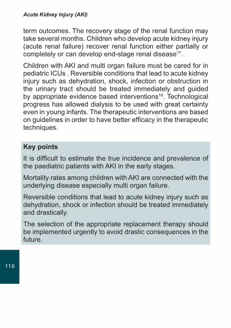
Acute Kidney Injury (AKI)
117116
term outcomes. The recovery stage of the renal function may take several months. Children who develop acute kidney injury (acute renal failure) recover renal function either partially or completely or can develop end-stage renal disease17.
Children with AKI and multi organ failure must be cared for in pediatric ICUs . Reversible conditions that lead to acute kidney injury such as dehydration, shock, infection or obstruction in the urinary tract should be treated immediately and guided by appropriate evidence based interventions18. Technological progress has allowed dialysis to be used with great certainty even in young infants. The therapeutic interventions are based on guidelines in order to have better efficacy in the therapeutic techniques.
Key points It is difficult to estimate the true incidence and prevalence of the paediatric patients with AKI in the early stages.
Mortality rates among children with AKI are connected with the underlying disease especially multi organ failure.
Reversible conditions that lead to acute kidney injury such as dehydration, shock or infection should be treated immediately and drastically.
The selection of the appropriate replacement therapy should be implemented urgently to avoid drastic consequences in the future.
Acute Kidney Injury (AKI) in children
117116
References 1. Iliadis A. Acute Renal Failure Acute Renal Failure-children. 13th
Congress of Intensive Care, Emergency Medicine. Guidelines, Athens, 2010 www.icutopics.gr
2. Brophy P. Acute Kidney Injury in Pediatric Patient (2010) www.tochnephrology.com
3. Moghal N, Brocklebank J, Meadow S. A review of acute renal failure in children: incidence, etiology and outcome. Clin Nephrol , 1998, 49 91-95
4. Gallego N, Perez-Caballero C, Gallego A, Estepa R, Liafo F, Ortuno J Prognosis of patients with acute renal failure without cardiopathy. Arch Dis Child 2001 84 (3) 258-260
5. Bunchman TE, McBryde K.D., Mottes TE, Gardner JJ, Maxvold NJ, Brophy PD. Pediatric acute renal failure: outcome by modality of disease. Pediatric Nephrol 2001 16 (12) 1067-1071
6. Flynn JT, Kershaw DB, Smoyer WE, Brophy PD, McBryde K.D., Bunchman TE. Peritoneal dialysis for management of pediatric acute renal failure. Perit Dial Int 2001 21(4) 390-394
7. Amal Abdel Ghani, Bassam Al Helal, Naser Hussain Acute renal failure in pediatric patiens: Etiology and predictors of outcome. Saudi JournalofKidneyDiseasesandTransplantation ,2009 20 (1) 69-768 Acute Renal Failure (ARF) and Chronic Kidney Disease (CKD) www.jimmoisiadis.com
8. Stamou SC et al. Quality improvement program decreases mortality after cardiac surgery, JThoracCardiovascSurg,2008, 136:494-499
9. Bajit K. Basu and Derek Wheeler Approaches to the Management of Acute Kidney Injury in Children. Recent Patents on Biomakers 2011, 1, 49-59
10. Kossyva L. Lagkona E., Kapogiannis A., Georgakis E. Acute renal failure. Emergency management protocols in pediatrics. Sheet A Pediatrics University of Athens (2006) 53 134-141
11. Andreoli S. Acute Kidney Injury in children. Pediatric Nephrology 2009 24 (2) 253-263 www.nil.gov
12. Andreoli S. Acute Renal Failure. Curr Opin Pediatr 2002 14 183-188
13. Martin -Ancel A, Garcia-Alix A, Gaya F, Cabanas F, Burguerow M, Quero J. Multiple organ involvement in perinatal asphyxia J Pediatr 1995, 127 786-793
14. Karlowivz M., Adelman R. nonoliguric and oliguric acute renal failure. Pediatr Nephrol 1995 9 718-722
15. Andreoli S. Acute Renal Failure in the newborn. Semin Perinatol 2004, 28 112-123
16. Mendley S. dialysis in neonates and children In: Daugiras J. Blake P. Ihg T. Manual 4th edition dialysis in neonates and children (2008) pp 650-670 Williams & Wilkins
17. Foland,J., et. al. Fluid overload before continuos hemofiltration and survival in critically ill children. Critical Care Medicine (in bold), 2004: 32, 1771-1776
18. Whyte D., Fine R. Acute Renal Failure in Children. Pediatrics in Review 2008, 29
Key principles of nursing care for the patient with acute kidney injury
119

Acute Kidney Injury (AKI)
121120
IntroductionAs described earlier in this handbook, AKI is a clinical syndrome denoted by an abrupt decline (over days to a few weeks) in glomerular filtration rate (GFR) sufficient to decrease the elimination of nitrogenous waste products (urea and creatinine) and other uraemic toxins1. The multifactorial aetiology of AKI, combined with the rapid onset, poses a unique challenge to the nurse in terms of managing both the root cause and the resultant kidney failure. The focus of this chapter is to outline the key principles of nursing care for the patient with AKI. The opening sections of this chapter will examine patient assessment and investigations in AKI. The core components of care will then be addressed, emphasising the role of the nurse in enhancing the patient experience.
Assessment of the patient with AKIThe kidneys perform a wide variety of physiological functions2, encompassing the excretion of nitrogenous end products of protein metabolism, water and electrolyte homeostasis, control of blood pressure, acid-base balance, erythropoiesis, conversion of vitamin D to its active metabolite, and excretion
Learning outcomes
• Tooutline thekeycomponentsofassessment for thepatientwithacutekidneyinjury(AKI).
• TodescribethefundamentalprinciplesofnursingcareforthepatientwithAKI.
• Todiscusstherelativeadvantagesanddisadvantagesofcrystalloidandcolloidfluidresuscitation.

Key principles of nursing care for the patient with acute kidney injury
121120
of drugs and toxins3. Consequently, the effects of AKI are systemic, and impact on all of the major bodily organs. The assessment process is thus fundamental to establishing an accurate and comprehensive patient history4.
The literature denotes that the initial assessment of the patient with AKI should be conducted simultaneously with the management of any life threatening features e.g. hypotension, shock, respiratory failure, hyperkalaemia5. Subsequent management should focus on identifying the cause of the renal insult and the needs generated by that cause3. Table 1 depicts the key components of assessment for the patient with AKI.
Table 1: The key components of assessment for the patient with AKI
(see next page)

Acute Kidney Injury (AKI)
123122 Nursing
history
Com
pone
nts
of a
sses
smen
tC
urre
nt c
linic
al
stat
us• A
sses
s fo
r the
pre
senc
e of
the
follo
win
g si
gns
and
sym
ptom
s:- M
alai
se, f
atig
ue, l
etha
rgy,
con
fusi
on- T
witc
hing
and
/or w
eakn
ess
seco
ndar
y to
met
abol
ic a
cido
sis
- Im
paire
d m
obili
ty- C
hang
e in
urin
e vo
lum
e1.
Olig
uria
(< 4
00 m
l/24
hour
s)2.
Non
olig
uria
(exc
ess,
dilu
te u
rine)
3. A
nuria
(no
urin
e ou
tput
or <
100
ml/2
4 ho
urs)
- Cha
nge
in u
rine
colo
ur1.
Hae
mat
uria
(gro
ssly
blo
ody)
2. P
yuria
(clo
udy)
3. B
iliur
ia o
r bili
rubi
nuria
(ora
nge)
4. M
yogl
obin
uria
(usu
ally
cle
ar; r
ed-b
row
n ur
ine;
Hae
mat
est
posi
tive)
- Abn
orm
al fl
uid
loss
e.g
. hae
mor
rhag
e, d
iarr
hoea
, vom
iting
, ex
cess
ive
wou
nd d
rain
age
- Car
diac
invo
lvem
ent
1. D
ysrh
ythm
ias
seco
ndar
y to
ele
ctro
lyte
imba
lanc
e or
hea
rt fa
ilure
2. C
hang
e in
pul
se ra
te (e
ither
tach
ycar
dia
or b
rady
card
ia)
3. H
yper
tens
ion
4. C
ardi
ac fr
ictio
n ru
b, in
dica
tive
of p
eric
ardi
tis
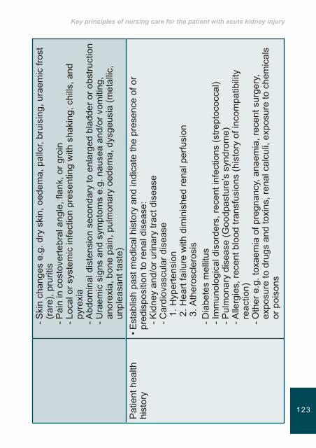
Key principles of nursing care for the patient with acute kidney injury
123122
- Ski
n ch
ange
s e.
g. d
ry s
kin,
oed
ema,
pal
lor,
brui
sing
, ura
emic
fros
t (r
are)
, pru
ritis
- Pai
n in
cos
tove
rtebr
al a
ngle
, flan
k, o
r gro
in- L
ocal
or s
yste
mic
infe
ctio
n pr
esen
ting
with
sha
king
, chi
lls, a
nd
pyre
xia
- Abd
omin
al d
iste
nsio
n se
cond
ary
to e
nlar
ged
blad
der o
r obs
truct
ion
- Ura
emic
sig
ns a
nd s
ympt
oms
e.g.
nau
sea
and/
or v
omiti
ng,
anor
exia
, bon
e pa
in, p
ulm
onar
y oe
dem
a, d
ysge
usia
(met
allic
, un
plea
sant
tast
e)P
atie
nt h
ealth
hi
stor
y• E
stab
lish
past
med
ical
his
tory
and
indi
cate
the
pres
ence
of o
r pr
edis
posi
tion
to re
nal d
isea
se:
- Kid
ney
and/
or u
rinar
y tra
ct d
isea
se- C
ardi
ovas
cula
r dis
ease
1. H
yper
tens
ion
2. H
eart
failu
re w
ith d
imin
ishe
d re
nal p
erfu
sion
3. A
ther
oscl
eros
is- D
iabe
tes
mel
litus
- Im
mun
olog
ical
dis
orde
rs, r
ecen
t inf
ectio
ns (s
trept
ococ
cal)
- Pul
mon
ary
dise
ase
(Goo
dpas
ture
’s s
yndr
ome)
- Alle
rgie
s, re
cent
blo
od tr
ansf
usio
ns (h
isto
ry o
f inc
ompa
tibili
ty
reac
tion)
- Oth
er e
.g. t
oxae
mia
of p
regn
ancy
, ana
emia
, rec
ent s
urge
ry,
expo
sure
to d
rugs
and
toxi
ns, r
enal
cal
culi,
exp
osur
e to
che
mic
als
or p
oiso
ns
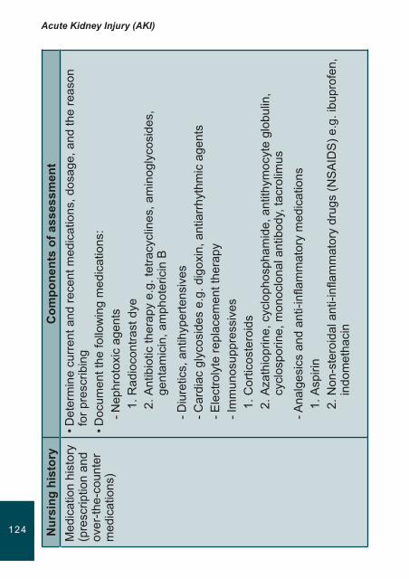
Acute Kidney Injury (AKI)
125124 Nursing
history
Com
pone
nts
of a
sses
smen
tM
edic
atio
n hi
stor
y (p
resc
riptio
n an
d ov
er-th
e-co
unte
r m
edic
atio
ns)
• Det
erm
ine
curr
ent a
nd re
cent
med
icat
ions
, dos
age,
and
the
reas
on
for p
resc
ribin
g• D
ocum
ent t
he fo
llow
ing
med
icat
ions
:- N
ephr
o tox
ic a
gent
s1.
Rad
ioco
ntra
st d
ye2.
Ant
ibio
tic th
erap
y e.
g. te
tracy
clin
es, a
min
ogly
cosi
des,
ge
ntam
icin
, am
phot
e ric
in B
- Diu
retic
s, a
ntih
yper
tens
ives
- Car
diac
gly
cosi
des
e.g.
dig
oxin
, ant
iarr
hyth
mic
age
nts
- Ele
ctro
lyte
repl
acem
ent t
hera
py- I
mm
unos
uppr
essi
ves
1. C
ortic
oste
roid
s2.
Aza
thio
prin
e, c
yclo
phos
pham
ide,
ant
ithym
ocyt
e gl
obul
in,
cycl
ospo
rine,
mon
oclo
nal a
ntib
ody,
tacr
olim
us
- Ana
lges
ics
and
anti-
infla
mm
ator
y m
edic
atio
ns1.
Asp
irin
2. N
on-s
tero
idal
ant
i-infl
amm
ator
y dr
ugs
(NS
AID
S) e
.g. i
bupr
ofen
, in
dom
etha
cin
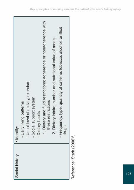
Key principles of nursing care for the patient with acute kidney injury
125124
Soc
ial h
isto
ry• I
dent
ify:
- Dai
ly li
ving
pat
tern
s- U
sual
leve
l of a
ctiv
ity, e
xerc
ise
- Soc
ial s
uppo
rt sy
stem
- D
ieta
ry h
abits
1. D
ieta
ry a
nd fl
uid
rest
rictio
ns; a
dher
ence
or n
onad
here
nce
with
th
ese
rest
rictio
ns2.
Die
tary
inta
ke, n
umbe
r and
nut
ritio
nal v
alue
of m
eals
- Fre
quen
cy, t
ype,
qua
ntity
of c
affe
ine,
toba
cco,
alc
ohol
, or i
llici
t dr
ugs
Ref
eren
ce: S
tark
(200
6)6 .

Acute Kidney Injury (AKI)
127126
With reference to Table 1, a detailed patient assessment is imperative in identifying the AKI cause and individual care priorities. Assessment will generate a unique set of clinical requirements that must be taken into consideration to prevent permanent renal damage or death5.
InvestigationsforAKIIn addition to a detailed patient assessment (Table 1), a series of laboratory investigations are required. Blood and urine testing are crucial to the management of AKI, in aiding diagnosis, monitoring AKI severity, and evaluating response to interventions7. Table 2 outlines the laboratory investigations for AKI.
Table 2: Laboratory investigations for AKI
Test Indication/abnormality
Urine testing• Urine dipstick
• Red cell casts on microscopy
• Mid-stream urine for microscopy, culture, and sensitivity
• Bence Jones protein
• Haematuria and proteinuria suggests glomerular disease
• Glomerular disease
• Urinary tract infection
• Monoclonal light chain mediated disease
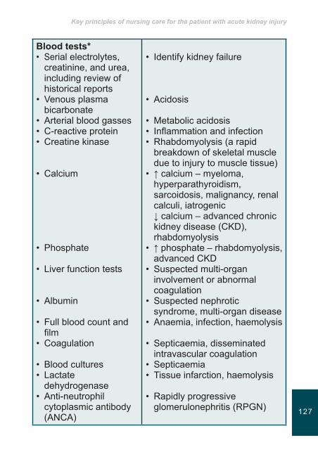
Key principles of nursing care for the patient with acute kidney injury
127126
Bloodtests*• Serial electrolytes,
creatinine, and urea, including review of historical reports
• Venous plasma bicarbonate
• Arterial blood gasses• C-reactive protein• Creatine kinase
• Calcium
• Phosphate
• Liver function tests
• Albumin
• Full blood count and film
• Coagulation
• Blood cultures• Lactate
dehydrogenase• Anti-neutrophil
cytoplasmic antibody (ANCA)
• Identify kidney failure
• Acidosis
• Metabolic acidosis• Inflammation and infection• Rhabdomyolysis (a rapid
breakdown of skeletal muscle due to injury to muscle tissue)
• ↑ calcium – myeloma, hyperparathyroidism, sarcoidosis, malignancy, renal calculi, iatrogenic ↓ calcium – advanced chronic kidney disease (CKD), rhabdomyolysis
• ↑ phosphate – rhabdomyolysis, advanced CKD
• Suspected multi-organ involvement or abnormal coagulation
• Suspected nephrotic syndrome, multi-organ disease
• Anaemia, infection, haemolysis
• Septicaemia, disseminated intravascular coagulation
• Septicaemia • Tissue infarction, haemolysis
• Rapidly progressive glomerulonephritis (RPGN)

Acute Kidney Injury (AKI)
129128
• Anti-glomerular basement membrane (anti-GBM) antibody
• Antinuclear antibodies (ANA)
• Complement components (C3, C4)
• Cryoglobulins• Serum uric acid• Serum protein
electrophoresis• Anti-streptolysin O
titre (ASOT)• Virology
• RPGN
• Systemic lupus erythematosus (SLE)
• Decreased concentrations in SLE, endocarditis, and cryoglobulinaemia
• Cryoglobulinaemia• Urate nephropathy• Myeloma
• Post-streptococcal glomerulonephritis
• Hepatitis B & C, human immunodeficiency virus
Reference: Lamb and Delaney (2009)7.*See Appendix 1 for reference ranges
Further to the laboratory investigations for AKI, imaging (chest and abdominal x-rays, renal ultrasonography) and electrocardiography facilitate diagnosis3. A kidney biopsy is reserved for cases where ultrasonography has excluded obstructed kidneys and the renal sizes are maintained (small kidneys indicate long standing disease and kidney biopsy is unlikely to change management)7.
Thefundamentalprinciplesofnursingcareforthepatientwith AKIIrrespective of the aetiology, the goals of nursing care for the patient with AKI encompass:
i Resolution of the AKIii Resumption of normal kidney function and urine output6

Key principles of nursing care for the patient with acute kidney injury
129128
Further to addressing the primary disorder, care focuses on correcting fluid and electrolyte imbalances, maintaining optimal nutritional status, preventing infection, preserving skin integrity, and educating and supporting the patient and family4,6. The latter is discussed in detail in Chapter 8 of this handbook.
1. Fluid management The nurse assumes a pivotal role in the assessment of fluid status in order to determine if the patient is hypo- or hypervolaemic8. The signs and symptoms of hypo- and hypervolaemia are summarised in Table 3.
Table 3: The signs and symptoms of fluid volume imbalances
Hypovolaemia (fluidvolumedeficit)
Hypervolaemia (fluidvolumeexcess)
• Dry skin and mucous membranes, decreased skin turgor
• Weight loss• Negative fluid balance
(fluid output exceeding fluid intake)
• Hypotension, orthostasis• Tachycardia• Absence of jugular venous
pressure (JVP) at 45˚• Raised haematocrit
• Oedema
• Weight gain• Positive fluid balance (fluid
intake exceeding fluid output)
• Hypertension • Bradycardia• Raised JVP
• Decreased haematocrit as a consequence of haemodilution
• Pulmonary congestion, dyspnoea
Reference: Litwack (2006)9.
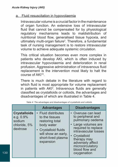
Acute Kidney Injury (AKI)
131130
a. Fluid resuscitation in hypovolaemia
Intravascular volume is a crucial factor in the maintenance of organ function. An extensive loss of intravascular fluid that cannot be compensated for by physiological regulatory mechanisms leads to maldistribution of nutritional blood flow, generalised tissue hypoxia, and ultimately multi-organ failure2. Therefore, a fundamental task of nursing management is to restore intravascular volume to achieve adequate systemic circulation.
This critical situation becomes even more complex in patients who develop AKI, which is often induced by intravascular hypovolaemia and deterioration in renal profusion. Aggressive administration of intravenous fluid replacement is the intervention most likely to halt the course of AKI10.
There is much debate in the literature with regard to which fluid is most appropriate for volume replacement in patients with AKI2. Intravenous fluids are generally classified as crystalloids or colloids, the advantages and disadvantages of which are illustrated in Table 4.
Table 4: The advantages and disadvantages of crystalloids and colloids
Advantages DisadvantagesCrystalloids e.g. 0.9% sodium chloride, 5% dextrose
• Fluid distributes to the tissues restoring total body water
• Crystalloid fluids will show an early, short-lived plasma expansion
• Overuse can lead to peripheral and pulmonary oedema
• Large volumes are required to replace intravascular losses
• Crystalloid therapy may adversely affect microcirculatory blood flow and oxygenation
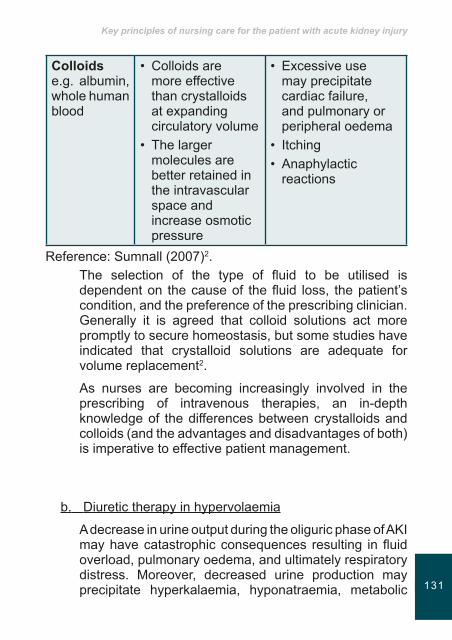
Key principles of nursing care for the patient with acute kidney injury
131130
Colloids e.g. albumin, whole human blood
• Colloids are more effective than crystalloids at expanding circulatory volume
• The larger molecules are better retained in the intravascular space and increase osmotic pressure
• Excessive use may precipitate cardiac failure, and pulmonary or peripheral oedema
• Itching• Anaphylactic
reactions
Reference: Sumnall (2007)2.The selection of the type of fluid to be utilised is dependent on the cause of the fluid loss, the patient’s condition, and the preference of the prescribing clinician. Generally it is agreed that colloid solutions act more promptly to secure homeostasis, but some studies have indicated that crystalloid solutions are adequate for volume replacement2.
As nurses are becoming increasingly involved in the prescribing of intravenous therapies, an in-depth knowledge of the differences between crystalloids and colloids (and the advantages and disadvantages of both) is imperative to effective patient management.
b. Diuretic therapy in hypervolaemia
A decrease in urine output during the oliguric phase of AKI may have catastrophic consequences resulting in fluid overload, pulmonary oedema, and ultimately respiratory distress. Moreover, decreased urine production may precipitate hyperkalaemia, hyponatraemia, metabolic
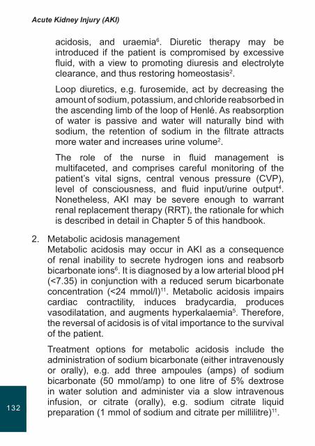
Acute Kidney Injury (AKI)
133132
acidosis, and uraemia6. Diuretic therapy may be introduced if the patient is compromised by excessive fluid, with a view to promoting diuresis and electrolyte clearance, and thus restoring homeostasis2.
Loop diuretics, e.g. furosemide, act by decreasing the amount of sodium, potassium, and chloride reabsorbed in the ascending limb of the loop of Henlé. As reabsorption of water is passive and water will naturally bind with sodium, the retention of sodium in the filtrate attracts more water and increases urine volume2.
The role of the nurse in fluid management is multifaceted, and comprises careful monitoring of the patient’s vital signs, central venous pressure (CVP), level of consciousness, and fluid input/urine output4. Nonetheless, AKI may be severe enough to warrant renal replacement therapy (RRT), the rationale for which is described in detail in Chapter 5 of this handbook.
2. Metabolic acidosis managementMetabolic acidosis may occur in AKI as a consequence of renal inability to secrete hydrogen ions and reabsorb bicarbonate ions6. It is diagnosed by a low arterial blood pH (<7.35) in conjunction with a reduced serum bicarbonate concentration (<24 mmol/l)11. Metabolic acidosis impairs cardiac contractility, induces bradycardia, produces vasodilatation, and augments hyperkalaemia5. Therefore, the reversal of acidosis is of vital importance to the survival of the patient.
Treatment options for metabolic acidosis include the administration of sodium bicarbonate (either intravenously or orally), e.g. add three ampoules (amps) of sodium bicarbonate (50 mmol/amp) to one litre of 5% dextrose in water solution and administer via a slow intravenous infusion, or citrate (orally), e.g. sodium citrate liquid preparation (1 mmol of sodium and citrate per millilitre)11.

Key principles of nursing care for the patient with acute kidney injury
133132
The role of the nurse in metabolic acidosis management is focused on the evaluation of acidosis-reversing measures, and encompasses arterial blood gas analysis12, pulse oximetry, oxygen therapy administration (via a face mask or nasal cannulae)4, and support of the patient on mechanical ventilation if specified. However, RRT will usually be required to treat severe acidosis (pH <7.1) in oligoanuric patients5 (see chapter 5).
3. Hyperkalaemia managementSevere hyperkalaemia is a medical emergency due to the risk for precipitating life threatening cardiac arrhythmias13. Consequently, the early recognition and management of hyperkalaemia is crucial4.
Hyperkalaemia may be managed by administering insulin and dextrose intravenously via a bolus injection or slow infusion4,6. It is pertinent for the nurse to monitor blood glucose levels when administering these infusions4, and to conduct strict electrocardiograph tracing in order to detect potentially fatal arrhythmias5. However, a refractory hyperkalaemia with serum potassium >6.5 mmol/l would necessitate the instigation of RRT4.
4. Nutritional managementAKI is associated with accelerated protein catabolism that predisposes to negative nitrogen balance and uncontrollably high blood urea nitrogen levels6. Protein calorie malnutrition is associated with muscle wastage and weakness3, and is implicated in the high mortality rates observed in patients with AKI4.
The literature denotes that supplementary nutritional support, with enteral feeding or total parenteral nutrition may enhance nutritional status and majorly contribute to the survival of the patient4,6. The role of the nurse in optimising nutritional status in AKI is discussed in detail in Chapter 7 of this handbook.
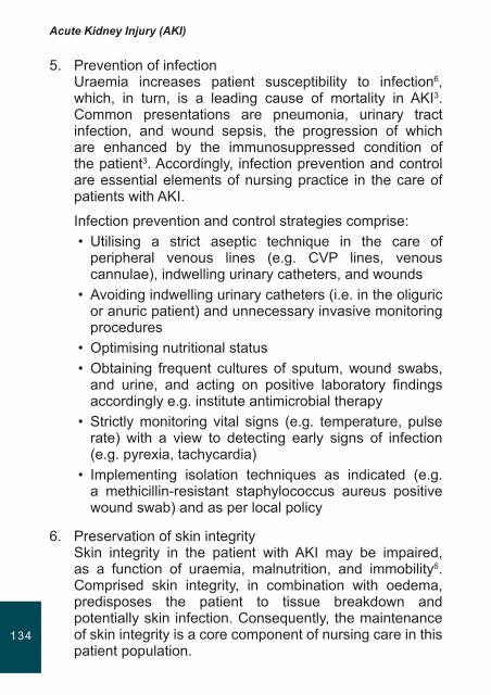
Acute Kidney Injury (AKI)
135134
5. Prevention of infectionUraemia increases patient susceptibility to infection6, which, in turn, is a leading cause of mortality in AKI3. Common presentations are pneumonia, urinary tract infection, and wound sepsis, the progression of which are enhanced by the immunosuppressed condition of the patient3. Accordingly, infection prevention and control are essential elements of nursing practice in the care of patients with AKI.
Infection prevention and control strategies comprise:• Utilising a strict aseptic technique in the care of
peripheral venous lines (e.g. CVP lines, venous cannulae), indwelling urinary catheters, and wounds
• Avoiding indwelling urinary catheters (i.e. in the oliguric or anuric patient) and unnecessary invasive monitoring procedures
• Optimising nutritional status• Obtaining frequent cultures of sputum, wound swabs,
and urine, and acting on positive laboratory findings accordingly e.g. institute antimicrobial therapy
• Strictly monitoring vital signs (e.g. temperature, pulse rate) with a view to detecting early signs of infection (e.g. pyrexia, tachycardia)
• Implementing isolation techniques as indicated (e.g. a methicillin-resistant staphylococcus aureus positive wound swab) and as per local policy
6. Preservation of skin integritySkin integrity in the patient with AKI may be impaired, as a function of uraemia, malnutrition, and immobility6. Comprised skin integrity, in combination with oedema, predisposes the patient to tissue breakdown and potentially skin infection. Consequently, the maintenance of skin integrity is a core component of nursing care in this patient population.

Key principles of nursing care for the patient with acute kidney injury
135134
Measures to preserve skin integrity encompass:• Assess for the uraemic effects on skin integrity e.g.
pruritis, dryness, ecchymosis, oedema• Ensure that skin is clean, dry, and intact in order to
prevent infection• Employ a strict aseptic technique when providing wound
care• Instigate regular pressure area care and pressure relief
especially if the patient is oedematous
PsychologicalcareofthepatientwithAKIThe experience of acute illness may have extensive and enduring consequences for patients14. Studies indicate that admission to an acute care environment may be associated with psychological distress within the context of anxiety, depression, and posttraumatic stress disorder (PTSD)15,16.
The acute care environment is ‘widely unfamiliar’ to the majority of individuals17. The noise, lay-out, machinery, technology, and pace of acute care may induce feelings of fear and anxiety. Moreover, experiencing a critical illness, its concomitant interventions, and being confronted with life and death scenarios, combine to create an often highly stressful episode14.
Studies also demonstrate that the incidence of PTSD and other psycho-affective disorders may be high not only in patients but also in relatives following intensive care18,19. Subsequently, psychological care constitutes a fundamental component of the care afforded to patients and their families in the acute care setting. The psychological implications of admission to an acute care environment are summarised in Table 5.
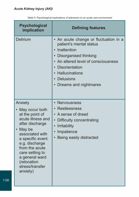
Acute Kidney Injury (AKI)
137136
Table 5: Psychological implications of admission to an acute care environment
Psychologicalimplication Definingfeatures
Delirium • An acute change or fluctuation in a patient’s mental status
• Inattention• Disorganised thinking• An altered level of consciousness• Disorientation• Hallucinations• Delusions• Dreams and nightmares
Anxiety
• May occur both at the point of acute illness and after discharge
• May be associated with a specific event e.g. discharge from the acute care setting to a general ward (relocation stress/transfer anxiety)
• Nervousness• Restlessness• A sense of dread• Difficulty concentrating• Irritability• Impatience• Being easily distracted

Key principles of nursing care for the patient with acute kidney injury
137136
PTSD
• Triggered by traumatic events and may last for years after the event
• Re-experiencing the event(s) through nightmares or flashbacks
• Avoidance of the stimuli associated with the event
• Hyperarousal symptoms e.g. hypervigilence, being easily startled
• Dissociative symptoms e.g. numbing, depersonalisation, amnesia
Depression • Continual low mood or sadness• Hopelessness, helplessness, and
tearfulness• Low self-esteem• Loss of motivation/interest in
everyday activities• Difficulty in making decisions• Suicidal thoughts/thoughts of self-
harm
Reference: Pattison (2005)14; Peris et al. (2011)20.With reference to Table 5, the psychological implications of admission to the acute care environment are multifaceted. As psychological factors impact on recovery post acute care episode14, it is imperative that nurses, when caring for patients with AKI, adequately assess for the presence of psychological symptoms and implement appropriate care strategies.
Research suggests that psychological interventions have the potential to promote recovery from PTSD, anxiety, and depression in patients who have experienced critical illness20. Interventions such as ensuring good pain control, maintaining excellent communication with both families and the patient about their situation, and minimising and coordinating
Acute Kidney Injury (AKI)
139138
interventions so that the patient, where possible, retains some control may assist in the management of anxiety14. Similarly, counselling strategies such as active listening, the use of open and closed questions, and paraphrasing may be valuable in addressing the symptoms of depression and PTSD.
Early psychological intervention in the acute care environment may be associated with positive patient outcomes20. Furthermore, nurses are pivotal to facilitating optimal recovery from AKI by providing psychological care and promoting well-being.
Keypoints
• Key principles of nursing care for the patient with AKI were examined.
• The experience of acute illness may have extensive and enduring consequences for patients.
• Nursing care management should focus on identifying the cause of the renal insult and the needs generated by that cause.
• Detailed patient assessment must be emphasise as a basis on which to plan, implement, and evaluate patient-focused care.
• Psychological care constitutes a fundamental component of the care afforded to patients and their families in the acute care setting.
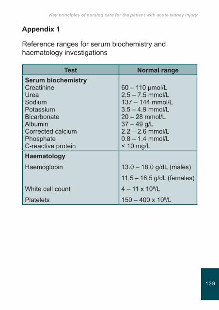
Key principles of nursing care for the patient with acute kidney injury
139138
Appendix1
Reference ranges for serum biochemistry and haematology investigations
Test NormalrangeSerumbiochemistryCreatinineUreaSodiumPotassiumBicarbonateAlbuminCorrected calciumPhosphateC-reactive protein
60 – 110 µmol/L2.5 – 7.5 mmol/L137 – 144 mmol/L3.5 – 4.9 mmol/L20 – 28 mmol/L37 – 49 g/L2.2 – 2.6 mmol/L0.8 – 1.4 mmol/L< 10 mg/L
HaematologyHaemoglobin
White cell count
Platelets
13.0 – 18.0 g/dL (males)
11.5 – 16.5 g/dL (females)
4 – 11 x 109/L
150 – 400 x 109/L
Acute Kidney Injury (AKI)
141140
References1. Jefferson JA and Schrier RW. Pathophysiology and etiology of
acute renal failure. In: Feehally J, Floege J, and Johnson RJ. Comprehensive Clinical Nephrology. 3rd ed. Mosby, 2007: 755-770.
2. Sumnall R. Fluid management and diuretic therapy in acute renal failure. NursinginCriticalCare. 2007; 12(1): 27-33.
3. Donald R. Caring for the renal system. In: Macintosh M and Moore T. Caringfortheseriouslyillpatient. 2nd ed. Hodder Arnold, 2011: 73-97.
4. Murphy F and Byrne G. The role of the nurse in the management of acute kidney injury. BritishJournalofNursing. 2010; 19(3): 146-152.
5. Fry AC and Farrington K. Management of acute renal failure. PostgraduateMedicalJournal. 2006; 82: 106-116.
6. Stark JL. The renal system. In: Alspach JG. CoreCurriculum forCriticalCareNursing. 6th ed. Saunders, 2006: 525-610.
7. Lamb E and Delaney M. KidneyDiseaseandLaboratoryMedicine. ACB Venture Publications, 2009: 77-93.
8. Byrne G and Murphy F. Acute kidney injury and its impact on the cardiac patient. British Journal of Cardiac Nursing. 2008; 3(9): 416-422.
9. Litwack K. The endocrine system. In: Alspach JG.CoreCurriculumforCriticalCareNursing. 6th ed. Saunders, 2006: 611-640.
10. Redmond A, McDevitt M, and Barns S. Acute renal failure: recognition and treatment in ward patients. NursingStandard. 2004; 18: 46-53.
11. Palmer BF and Alpern RJ. Metabolic acidosis. In: Feehally J, Floege J, and Johnson RJ. ComprehensiveClinicalNephrology. 3rd ed. Mosby, 2007: 147-157.
12. Ellstrom K. The pulmonary system. In: Alspach JG.CoreCurriculumforCriticalCareNursing. 6th ed. Saunders, 2006: 45-183.
13. Weiner ID, Linas SL, and Wingo CS. Disorders of potassium metabolism. In: Feehally J, Floege J, and Johnson RJ. ComprehensiveClinicalNephrology. 3rd ed. Mosby, 2007: 111-121.
14. Pattison N. Psychological implications of admission to critical care. BritishJournalofNursing. 2005; 14(13): 708-714.
Key principles of nursing care for the patient with acute kidney injury
141140
15. Griffiths J, Fortune G, Barber V, and Young JD. The prevalence of post traumatic stress disorder in survivors of ICU treatment: a systematic review. IntensiveCareMedicine. 2007; 33: 1506-1518.
16. Myhren H, Ekeberg O, Toien K, Karlsson S, and Stokland O. Posttraumatic stress, anxiety and depression symptoms in patients during the first year post intensive care unit discharge. CriticalCare. 2010; 14: R14.
17. Chew SL. Psychological reactions of intensive care patients. Care of theCriticallyIll. 1986; 2: 62-65.
18. Hewitt J. Psycho-affective disorder in intensive care units: a review. JournalofClinicalNursing.2002; 11: 575-584.
19. Cuthbertson BH, Hull A, Strachan M, and Scott J. Post-traumatic stress disorder after critical illness requiring general intensive care. IntensiveCareMedicine.2004; 30(3): 450-455.
20. Peris A, Bonizzoli M, Lozzelli D, Migliaccio ML, Zagli G, Bacchereti A, Debolini M, Vannini E, Solaro M, Balzi I, Bendoni E, Bacchi I, Giovannini V, and Belloni L. Early intra-intensive care unit psychological intervention promotes recovery from post traumatic stress disorders, anxiety and depression symptoms in critically ill patients. CriticalCare. 2011; 15: R41.

Notes

