acute infectious encephalopathy - roshini mathew,...
TRANSCRIPT

Acute Infectious
Encephalopathy
Roshini Mathew, RN, ACNP-BC(c)
Wright State University-Miami Valley College of
Nursing and Health
March 12, 2014

Objectives
1. Discuss the etiology and pathophysiology of acute
infectious encephalopathy.
2. Discuss the epidemiology of the condition.
3. Review the clinical presentation and the differential
diagnoses for the condition.
4. Examine the diagnostic tests specific for this
condition.
5. Determine treatment measures for acute infectious
encephalopathy.
6. Analyze the outcomes associated with the
treatments and other interventions.
7. Consider appropriate follow-up plan for patients.

Definition
• Presence of an inflammatory process of the brain
associated with signs and symptoms of neurological
dysfunction
• Often associated with evidence of meningeal
involvement
• Most cases are unusual complications of common
systemic viral infections

Epidemiology
• 3.5 to 7.4 per 100,000 persons per year
• 20,000 new cases each year in the U.S.
• Children (<12) and young adults are more commonly
affected, disease severity greatest in infants and
elderly patients
• Sex: Males > Females
• Transmission – inhalational, vector borne (mosquito,
tick), blood borne, gastrointestinal, or genital
• Etiology confirmed in ~30% of cases

Epidemiologic Clues
• To investigate for an etiologic diagnosis include the
following in assessment:
• Season of the year
• Geographic locale
• Prevalence in local community
• Travel history
• Recreational activities
• Occupational exposure
• Insect/animal contact
• Vaccination history
• Immune status of patient

Classification
• Infectious: viral (70%), bacterial (20%), prion (6%),
parasitic (3%), fungal (1%)
o Fungal, parasitic, or tuberculosis agents cause
chronic disease
o Host immune function critical for establishing an
infectious differential diagnosis
o Herpes simplex virus (HSV) – common cause of
viral encephalitis
• 2,000 cases in U.S. annually – as much as
28% mortality
o Cytomegalovirus (CMV) and varicella-zoster virus
(VZV) cause a more aggressive form of
encephalitis in immunocompromised hosts
Common Etiologies
Viral
• HSV, VZV, EBV, CMV, Hepatitis, Mumps,
Measles, Enterovirus, Adenovirus, Arboviruses
(e.g. West Nile Virus, St. Louis Encephalitis,
Eastern Equnie, Western Equine), HIV, Influenza
Bacterial
• Mycoplasma, Legionella, Listeria,
Mycobacterium tuberculosis
Parastic
• Toxoplasma, Trypanosoma, Echinococcus
Fungal
• Histoplasma, Crytococcus neoformans

Pathophysiology
• Etiologies:
1) Primary
• Direct viral invasion vs. virus replicates
outside of CNS Gains entry to the CNS by
hematogenous spread or by travel along
neural pathways
2) Immunologic Reaction
• The immune system attacks CNS antigens that
resemble proteins of the infectious agent
• Occurs 1-3 wks later of a viral infection or
vaccination
Pathophysiology
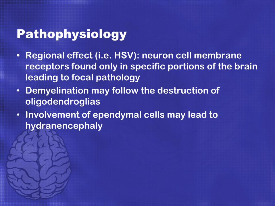
• Regional effect (i.e. HSV): neuron cell membrane
receptors found only in specific portions of the brain
leading to focal pathology
• Demyelination may follow the destruction of
oligodendroglias
• Involvement of ependymal cells may lead to
hydranencephaly

Pathophysiology
• Causing:
• Meningitis
• Cerebral edema
• Hemorrhage
• Apoptotic neuronal cell death
• End result: Inflammatory response with
neurological symptoms

Clinical Presentation
• Dependent on site of infection
• Constitutional – fever, fatigue, myalgias, malaise
• Neurologic – headache, stiff neck, irritability,
altered consciousness, incoherent speech, focal
neurologic findings, seizures, stupor, coma
• Neuropsychiatric – mood alterations,
hallucinations, depression
• Motor System – psychomotor hyperactivity,
retardation, tremor, hyperreflexia
• Dermatologic – skin rashes (Lyme disease,
typhus, rickettsial disease), skin lesions (VZV,
HSV), bite-site paresthesias (rabies)

Clinical Presentation
• Cardiac – autonomic dysfunction (sympathetic
overactivity), tachycardia, hypertension,
diaphoresis
• Gastroenterologic – nausea, emesis (enteroviral),
decreased appetite
• Pulmonary – cough, dyspnea (mycobacteria)
• Miscellaneous – incontinence, sleep/wake cycle
disturbance (fragmented sleep)

Differential Diagnoses
• Stroke
• Seizures
• Vasculitis
• Autoimmune disease (most commonly
systemic lupus erythematosus)
• Drug overdose
• Severe metabolic derangement (eg,
metabolic acidosis, hyperglycemia)
• Malignancy – primary or metastatic
• Nutritional deficit

Laboratory Testing
• CBC
o Leukocytosis – likely bacterial
o Lymphocytosis – likely viral
• Peripheral smear
• Electrolyte panel (e.g. Chem 10)
• Liver and renal function studies (rule out metabolic
encephalopathy); ammonia level
• Cerebrospinal fluid (CSF) studies
o Culture and Gram stain or other special stains (eg,
India ink for Cryptococcus, acid fast for TB)
o Cell count with differential – usually have
mononuclear pleocytosis
o Protein – usually elevated
o Glucose – low in bacterial, fungal, and mycobacterial
infections

Laboratory Testing
• Cultures – relatively poor sensitivity
o CSF fluid
o Blood – 2-3 sets from separate venipuncture sites
prior to the administration of antibiotics
o Other site cultures based on other organ system
involvement (sputum, urine, body fluid, tissue or
gastric aspirate)
• Other tests:
o Antibody titers for viruses – comparison of CSF
and serum antibody loads
Ratio ≥20 indicates intrathecal production
Intrathecal antibodies indicative of viral
etiology

Laboratory Testing
o PCR of Fluids
Greater sensitivity during first week of symptom
onset while viral agent present in CSF – yield
decreases rapidly after the first week
False negatives most common during the first 2
days of symptoms
In undiagnosed, severe cases, PCR should be
repeated after 3-7 days; Serology should be
repeated in 4-6 weeks
o Sedimentation rate/C-reactive protein for
suspected vasculitis
o For detailed lab tests for detection of specific
organism, see handout attached

Imaging Testing
• Head imaging (CT/MRI) – rule out structural lesions,
demyelination, and cerebral edema
• Temporal lobe enhancement suggestive of HSV-1

Diagnosis
• Correlate clinical findings with laboratory/imaging
findings
• Brain biopsy – rarely performed
• Incorporate:
1. Detailed patient history
2. List of prescribed, OTC, herbal medications
3. Environmental factors
4. Immune system functionality of patient

Empirical Treatment
• Initiate treatment early to prevent serious sequelae
(e.g. neurologic dysfunction, seizures, coma, death)
• Suspected viral encephalitis:
-Acyclovir 10 mg/kg IV every 8 hrs for 10-14 days
(e.g. HSV or VZV)
-Gancliclovir 5 mg/kg IV Q 12 hrs for 14-21 days
(e.g. CMV)

Empirical Treatment
• Initiate antimicrobials within 30 minutes if bacterial
meningitis suspected
-Vancomycin 1 g IV Q 12 hrs, Ceftriaxone 50 mg/kg
IV Q 12 hrs, Ampicillin 2 g IV Q 4 hrs (if listeria
suspected)
-For rickettsial or ehrlichial infections, add
doxycycline 100mg IV Q 12 hrs
-Other antimicrobials on the basis of clinical
factors
• Antifungal:
-Metronidazole 500 mg IV Q 6 hrs

Specific Treatment
• Following identification of the causative
microorganism, appropriate antimicrobial, antiviral,
and/or antifungal may be required
• Most viruses are managed with supportive care
• IDSA Recommendations (2008) for specific
organisms:
http://cid.oxfordjournals.org/content/47/3/303/F5.ex
pansion.html
• Corticosteroids may be used as an adjunctive
therapy
-Methylprednisolone, 1 g IV daily 3–5 days

Management
• Management of hydrocephalus and increased ICP
• Treatment of systemic complications
• Hypotension/shock
• Hypoxemia
• Hyponatremia
• Exacerbation of chronic diseases
• Antipsychotics:
• Haldol 0.5-5 mg IV
• Risperidone 0.25-3 mg/day
• Seroquel 12.5-100 mg/day
• Lorazepam 0.5-2mg IV/PO
• Start with low dose and minimize benzodiazapine
use, especially in geriatric population

Follow-up Care
• Monitor airway, breathing, circulation
• Frequent neurological assessments
• Monitor for seizures/subclinical seizures
• Monitor for cerebral edema/increased ICP
Prognosis
• Dependent on the virulence of the virus and the
patient’s health status
• 40% of survivors have residual deficits: learning
disabilities, memory impairment, neuropsychiatric
abnormalities, epilepsy, fine-motor-control deficits,
and dysarthria
• High mortality rates if not treated (50-100%); with
treatment mortality ranges from 2-20%
• Poor outcomes with:
• Extremes of age (< 1 y or >55 y)
• Immune-compromised status
• Preexisting neurologic conditions

Questions?

References• ARUP Laboratories. (2006). Encephalitis, infectious. The
Physician’s Guide to Laboratory Test Selection and
Interpretations. Retrieved from
http://www.arupconsult.com/Topics/Encephalitis.html#tabs=0
• Bloch, K.C. & Glaser, C. (2007). Diagnostic approaches for
patients with suspected encephalitis. Current Infectious Disease, 9(4), 315-322.
• Granerod, J., Cunningham, R., Zuckerman, M., Mutton, K.,
Davies, N., Walsh, A., & ... Crowcroft, N. (2010). Causality in
acute encephalitis: Defining aetiologies. Epidemiology & Infection, 138(6), 783-800
• Infectious Diseases Society of America (IDSA). (2008). The
management of encephalitis: Clinical practice guidelines.
Clinical Infectious Diseases, 47(3), 303-327.
• Katzung, B.G., Masters, S.B., & Trevor, A.J. (2012). Basic and clinical pharmacology. New York, NY: McGraw Hill Medical.

References
• Longo, D.L., Fauci, A.S., Kasper, D.L., Hauser, S.L.,
Jameson, J.L., & Loscalzo, J. (2012). Harrison’s Principles of Internal Medicine. New York, NY:
McGraw-Hill, Inc.
• Mailles, A., Broucker, T., Costanzo, P., Martinez-
Almoyna, L., Vaillant, V., & Stahl, Jean-Paul. (2012).
Long-term outcome of patients presenting with
acute infectious encephalitis of various causes.
Clinical Infectious Diseases, 54(10), 1455-1464.
• Stahl, J., & Mailles, A. (2010). Infectious causes of
encephalitis. Lancet Infectious Diseases, 10(12),
814-815.

Further Contact
• Roshini Mathew, RN, BSN, ACNP-BC (c)
• Wright State University-Miami Valley College of
Nursing and Health
Email: [email protected]
Mobile: 614.893.5407