1 running head: mathew clinical case study...
TRANSCRIPT

1
Running head: MATHEW CLINICAL CASE STUDY
Comprehensive Clinical Case Study: Acute Cholecystitis
Roshini Mathew, RN, BSN
Wright State University-Miami Valley College of Nursing and Health
NUR 7201
July 11, 2013

2
MATHEW CLINICAL CASE STUDY
Comprehensive Clinical Case Study: Acute Cholecystitis
History and Physical
Source of Information
Information was obtained from the patient, who is a reliable source of information.
Primary language is English.
Chief Compliant
“I have sharp pain along the right upper and middle part of my belly.”
History of Present Illness
This is a 66-year old Caucasian male presenting to the emergency department (ER) with
right upper quadrant pain that started earlier this afternoon. The pain started abruptly after lunch
at McDonald’s today, approximately at 1:00PM. The pain rating is a ten out of ten (on a pain
scale of zero to ten), described as “tender,” and it radiates to the right shoulder area. The
intensity of the pain is steady and does not fluctuate. The pain has been present for six hours now
and that is why the patient sought to seek medical advice. The patient took tramadol 50 mg PO
after the pain started in an attempt to relieve pain, but the intervention was not effective. The
pain is not aggravated by walking, lying, or coughing. “Nothing” seems to improve the pain. The
patient denies any trauma to the abdomen or a recent fall. He states the same type of pain
occurred about eight months ago, but the pain resolved spontaneously and he did not follow-up
with his physician. He believes the previous pain also occurred after eating a meal. The patient
states he felt “funny during the day” yesterday and had no appetite and was nauseous. He also
had one episode of vomiting earlier this morning, around 10:00 AM. He did not eat dinner
yesterday and breakfast today due to nausea. No other pain or discomfort is reported. He denies
recent illnesses or any changes to his medical regimen. Denies history of jaundice.

3
MATHEW CLINICAL CASE STUDY
Medications
Metoprolol 25 mg PO BID, trimterene-hydrochlorothiazide 37.5-25 mg PO daily,
simvastatin 40mg PO daily, glipizide 5 mg PO daily, omeprazole 20 mg PO daily, daily
multivitamin one tab PO, tramadol 50 mg PO every six hours PRN pain. Uses meditation and
music for relaxation. No other use of alternative or complementary therapies reported.
Medical History
Childhood illnesses. None
Adult illnesses. Hypertension diagnosed at age 40; diabetes diagnosed at age 43; peptic
ulcer disease diagnosed at age 42
Surgeries/Procedures. Tonsillectomy and adenoidectomy at age of 10; umbilical hernia
repair in 2008; bilateral knee arthroscopy in 2010
Allergies. No known food or drug allergies
Immunizations. Up-to-date on immunizations. Last tetanus-diphtheria booster received
2005. Flu and pneumonia vaccine received in January 2013.
Personal/Social History
The patient has been “happily” married for 45 years and has one son. He is a retired
paramedic, but currently works as a fire inspector on a part-time basis. No concern of financial or
work-related stress is shared. Denies use of alcohol, illicit drugs, or smoking. The patient is fairly
active and independent, although he is limited by his bilateral knee pain at times. He frequently
prefers to climb stairs rather than using the elevator and enjoys playing golf on the weekends.
Other leisure time activities include jogging, gardening, and reading self-improvement books. He
lives with his wife in a split-style two story home.
Family History

4
MATHEW CLINICAL CASE STUDY
The patient’s mother passed away at the age of 68 from congestive heart failure
complications. She had a history of hypertension, hyperlipidemia, diabetes, and cholelithiasis.
The father died of lung cancer at age of 74. Father’s medical history consists of hypertension,
cirrhosis, and renal failure. The patient has one brother who died from end-stage renal disease at
the age of 70. The patient does not know the cause of death of his grandparents.
Review of Symptoms
General. The patient states he is in good health and blood pressure is well controlled
with home medication. He checks his blood pressure daily. Independent with medication
management. States he had chills over night, but no fever. He does have a loss of appetite for the
past two days with nausea and vomiting. No recent weight loss or weight gain.
Neurological. Denies confusion, dizziness, tremors, numbness/tingling, or falls.
HEENT. Head: Denies headache or trauma to the head and face. Eyes/Ears: Denies
visual or hearing deficits, tinnitus, discharge, and pain. Nose: Having mild nasal congestion due
to sinus pain. Throat: Denies sore throat and difficulty swallowing.
Neck. Denies pain, stiffness, or swollen lymph nodes.
Respiratory. Denies dyspnea, orthopnea, shortness of breath, coughing, or pain during
inspiration/expiration.
Cardiovascular. No chest discomfort/pain, diaphoresis, palpitations, dizziness, light-
headedness or swelling.
Gastrointestinal/Abdominal. States has poor appetite, usual flatulence. Nausea past two
days, one episode of vomiting today. Mild heartburn at times after eating. Having right upper
abdomen pain that started abruptly this afternoon at 1:00 PM, after having lunch at McDonald’s.
Rating the pain ten out of ten, and describing it as “tender” and “sharp.” Denies hiccoughs,
5
MATHEW CLINICAL CASE STUDY
diarrhea, constipation, abdominal tenderness, jaundice, dark urine, light stools, or rectal bleeding.
Last bowel movement yesterday which was soft and formed brown stool.
Genitourinary. No urinary urgency, frequency, hesitancy, or burning. No history of
urinary tract infections. Denies bladder spasms, stress/night incontinence, flank pain. States urine
appearance is clear, straw colored, without odor.
Genitalia. Circumcised male. Sexually active with his wife. Denies any scrotal or penile
lumps, lesions, edema, or drainage. Performs self-testicular frequently.
Musculoskeletal. Reports having right shoulder pain that is radiating from his right
upper abdomen area which also started abruptly this afternoon at the time of right upper
abdomen pain. The pain in the right shoulder is “dull” with a pain rating of five out of ten. The
pain is not affected by activity or with movement. Denies history of trauma with or without falls,
fractures, or any other muscle or joint pain.
Integumentary. Denies itching, burning, open lesions, blistering, or thinning of skin or
nails.
Psychosocial. Denies depression, mood alteration, outbursts of anger, impulsive
behavior, anxiety, and social withdrawal.
Hematologic. No active bleeding issues. Denies history of anemia.
Endocrine. Denies confusion, weakness, diaphoresis, increased thirst or urination,
irritability, weight loss/gain, or thinning of hair.
Physical Examination
General. The patient is alert, appropriate, and pleasant. Patient is slightly anxious due to
pain in right upper quadrant (RUQ) of the abdomen. Height is five feet seven inches, weight is
210 pounds, Body Mass Index (BMI) is 32.9 kg/m2.

6
MATHEW CLINICAL CASE STUDY
Vital signs. Blood pressure is 158/84 mmHg, right arm sitting; apical pulse is 78 beats
per minute and regular; respiratory rate is 18 breaths per minute with normal depth and effort;
temperature is 99.5°F oral; oxygen level of 98% on room air.
Neurological. Alert and oriented to name, place, person, and time. following commands
appropriately. Cranial nerves II – XII intact. Face is symmetrical. Speech is clear, concise,
without aphasia or dysarthria. Bilateral pupils are equal (3mm), round, normal, reactive to light.
Corneal reflex present and vision intact. Short and long term memory intact.
HEENT. Head is normocephalic, no tenderness on palpation or trauma. Frontal and
maxillary sinuses are tender to palpation. Temporomandibular joint intact. No periorbital edema,
with clear conjunctiva. Extraocular movements intact. Ears in normal position, tympanic
membrane is gray. Nares with mild congestion and inflammation. Clear drainage noted. Lips and
mucosa pink and moist without lesions. Uvula and tongue midline, upper dentures in place.
Neck. No pain, distended veins, pulsations, or bruits. No thyromegaly or palpable
adenopathy. Neck supple, trachea midline. Bilateral +2 carotid pulses.
Chest. Non-tender on palpation. No masses, lesions, crepitus.
Respiratory. Symmetrical chest expansion with anteroposterior-to-transverse diameter of
1:2. Normal, effortless, regular, even breathing. Resonant lung fields, tactile fremitus present
bilateral lobes. Clear lungs upon auscultation without rhonci, wheezing, crackles, or rales. No
respiratory distress.
Cardiovascular. Bilateral +2 radial, brachial, posterior tibial, and dorsalis pedis pulses
with capillary refill of less than three seconds. Regular rate and rhythm, normal S1/S2, S3/S4
absent. No murmurs, clicks, snaps, or rubs. No peripheral edema in bilateral upper and lower
extremities.

7
MATHEW CLINICAL CASE STUDY
Abdomen. Umbilicus midline. No protrusion, pulsation, rigidity, or ascites.
Hepatomegaly and splenomegaly absent. Abdomen is round, non-distended, normal bowel
sounds in all four quadrants without bruits. Abdomen tender in right hypochondrium and
epigastrium regions with percussion and palpation. Positive Murphy’s sign without rebound
tenderness. No pain sensitivity at McBurney’s point. Guarding present with deep palpation.
Negative for tenderness at the costovertebral angle.
Musculoskeletal. Equal arm and leg lengths. No erythema, crepitation, spinal tenderness,
or edema. No kyphosis or scoliosis. Gross and fine sensory and motor strength intact in bilateral
upper and lower extremities. Full range of motion and 5/5 strength in bilateral upper extremities,
lower extremities, hip, and spine. Deep tendon reflexes intact. Negative Psoas sign, negative
obturator sign.
Integumentary. Skin warm and dry, pink skin tone. Intact without lesions, ecchymosis,
cyanosis or discoloration. Nail beds pink, no clubbing, no Lindsey’s nails.
Laboratory and Radiography Findings
To distinguish between the differential diagnoses of acute cholecystitis (AC), acute
pancreatitis, and acute appendicitis, laboratory tests of comprehensive metabolic panel (CMP),
complete blood count (CBC) with differential, pancreatic enzymes, and total and direct bilirubin
levels were obtained. Since the patient has a history of coronary artery disease, troponin levels,
B-type natriuretic peptide (BNP), chest x-ray, and an electrocardiogram (EKG) were attained to
eliminate atypical signs and symptoms of a cardiology etiology. A chest x-ray can also assist in
excluding non-biliary causes of right upper quadrant pain, such as right lower lobe pneumonia
(Watkins & Lemonovich, 2011). An abdominal ultrasound was acquired to assess pain in the
right hypochondrium and epigastrium regions. Ultrasonography is usually the initial imaging test
8
MATHEW CLINICAL CASE STUDY
of choice to investigate the right upper quadrant organs (e.g. gallbladder, liver, and pancreas). An
ultrasound can detect the presence of gallstones, but is not a sensitive test for AC (sensitivity of
67%, specificity of 82%) (Papadakis, McPhee, & Rabow, 2013; Tonolin, Ravelli, Villa, &
Bianco, 2012). A hepatobiliary imino-diacetic acid (HIDA) scan (sensitivity of 98%, specificity
of 81% if bilirubin less than 5 mg/dL) was obtained after reviewing equivocal abdominal
ultrasound results to confirm the suspicion of acute cholecystitis (Tonolini et al., 2012). A
radioactive dye, technetium-99m, is injected intravenously and is circulated in the liver and the
biliary tract. The test is negative if the gallbladder is visualized within one hour of dye injection.
A positive test for AC and the obstruction of the cystic duct illustrates the lack of gallbladder
visualization within four hours of the injection. The HIDA scan also allows the detection of the
severity of gallbladder dyskinesia by measuring the percentage of dye that is ejected from the
gallbladder. An ejection fraction (EF) of ≥ 50% is considered normal, whereas an EF of ≤ 30% is
illustrates severe obstruction and/or dyskinesia (an EF between 30-50% is considered
mild/moderate obstruction) (Kessenich, 2011).
To eliminate the diagnosis of complications such as perforated peptic ulcer, cholangitis,
and gangrene cholecystitis, a computed tomography (CT) scan was also ordered. Since the
increased laboratory values of serum aminotransferases (ALT and AST) and ultrasound results
suggested the possibility of dilatation of the common bile ducts, a magnetic resonance
cholangiopancreatography (MRCP) (sensitivity of 100%, specificity of 94%) was obtained to
examine the diagnosis of coexistent choledocholithiasis and cholangitis (Barak et al., 2009). The
MRCP explores the cystic, hepatic, and pancreatic ducts and assists in determining the cause,
location, and severity of obstruction. Furthermore, a MRCP allows detection of cholecystitis-
related complications of gangrene, pericholecystic abscess, perforation, and intrahepatic

9
MATHEW CLINICAL CASE STUDY
fistulization (Tonolini et al., 2012). The following are the results from laboratory and imaging
studies:
Table 1. Comprehensive Metabolic Panel (CMP) with Creatinine Clearance, Magnesium, and
Phosphorus
Lab Results Normal Values Lab Results Normal values
Sodium 137 mEq/L 135-148 mEq/L Globulin 2.2 g/dL 1.9-3.6 g/dL
Potassium 4.3 mEq/L 3.4-5.3 mEq/L A/G Ratio 1.9 0.8-2.6
Chloride 102 mEq/L 96-110 mEq/L Total Bilirubin 4.2 mg/dL 0.2-1.9 mg/dL
Carbon
Dioxide
26 mEq/L 23-29 mEq/L AST (SGOT) 229 U/L 0-45 U/L
Glucose 123 mg/dL 70-100 mg/dL ALT (SGPT) 326 U/L 0-40 U/L
BUN 17 mg/dL 7-20 mg/dL Alkaline
Phosphatase
(ALP)
150 U/L 23-150 U/L
Creatinine 1.0 mg/dL 0.8-1.4 mg/dL Anion Gap 16 10-20
BUN/Creat
Ratio
17.4:1 10:1-20:1 Magnesium 2.1 mEq/L 1.6-2.4 mEq/L
Calcium 9.6 mg/dL 8.5-10.5 mg/dL Phosphorus 3.9 mEq/L 2.5-5.2 mEq/L
Total
Protein
6.3 g/dL 6.0-8.3 g/dL Creatinine
Clearance
51.6
mL/min
97-137
mL/min (male)
Albumin 4.0 g/dL 3.5-5.2 g/dL
(Normal values from Pagana & Pagana, 2010)
Table 2. Complete Blood Count (CBC) with Differential
Lab Results Normal
Values
Lab Results Normal Values
WBC 16.9 k/mm3 4.5-10.5
k/mm3
Band
Neutrophils
2.7% 2-5%
RBC 4.8 m/mm3 4.7-6.1
m/mm3 (male)
Lymphocytes 14.0% 14.0-51.0%
Hemoglobin 12.8 g/dL 14.0-17.5 g/dL
(male)
Monocytes 2.0% 2-8%
Hematocrit 38.0 % 40.7-50.3%
(male)
Eosinophils 1.0% 1-3%
MCV 93.4 fL 80.0-100.0 fL Basophils 0.1% 0.0-1%
MCH 31.4 pG 27.0-31.0 pG Absolute
Segmented
Neutrophil
14 k/mm3 1.5-8.0 k/mm3
MCHC 33.7 g/dL 32.0-36.0 g/dL Absolute
Lymphocyte
1.8 k/mm3 0.9-4.1 k/mm3

10
MATHEW CLINICAL CASE STUDY
RDW 13.4% 9.0-15.0% Absolute
Monocyte
0.8 k/mm3 0.2-1.1 k/mm3
Platelets 215,000
k/mm3
150,000-
400,000
k/mm3
Absolute
Eosinophil
0.5 k/mm3 0.0-0.6 k/mm3
Segmented
Neutrophils
80.2% 40.0-76.0% Absolute
Basophil
0.2 k/mm3 0.0-0.3 k/mm3
(Normal values from Pagana & Pagana, 2010)
Table 3. Additional Laboratory Findings: Pancreatic Enzymes, Total and Direct Bilirubin,
Troponin, Serum Creatinine Kinase-MB (CK-MB), B-type natriuretic peptide (BNP)
Lab Results Normal Values Lab Results Normal
Values
Amylase 35 U/L 13.0-53.0 U/L Troponin <0.01 ng/mL <0.01 ng/mL
Lipase 39 U/L 15.0-65.0 U/L CK-MB 1.1 mcg/L 0-3 mcg/L
Direct
Bilirubin
1.8 mg/dL 0.0-0.4 mg/dL BNP 86 pg/mL <100 pg/mL
Total
Bilirubin
3.1 mg/dL 0.0-1.0 mg/dL
(Normal values from Pagana & Pagana, 2010)
Table 4. EKG Findings
EKG: Normal EKG. Normal sinus rhythm, heart rate: 92 beats per minute
Table 5. Radiography Findings
X-ray of Chest Normal chest x-ray without infiltrates or
atelectasis. No cardiomegaly.
Ultrasonography of Abdomen Gallbladder dilatation and wall thickening of
7mm, presence of pericholecystic fluid.
Positive Murphy’s sign. Dilatation of common
bile duct of 4mm. Limited study for detection
of stones and visualization of the pancreas. No
liver enlargement.
CT scan of Abdomen with Contrast Gallbladder distention, mural thickening with
mucosal hyperenhancement. Presence of
pericholecystic fluid. No free air identified.
Hepatomegaly not present.

11
MATHEW CLINICAL CASE STUDY
HIDA Scan Gallbladder not visualized, EF of 20%.
MRCP Presence of intraluminal sludge and 4.5mm
cystic duct stone. Transverse diameter of
gallbladder 8mm, mural thickening of 7mm.
Presence of moderate amount of
pericholecystic fluid. Dilatation of common
bile duct of approximately 3mm, but no stones
identified. No abscess detected. Normal
appearing pancreas.
Diagnosis
Acute cholecystitis secondary to cystic duct stone, or acute calculous cholecystitis, is the
primary diagnosis for this patient. The majority of stones consist of cholesterol as a result of
supersaturation, accelerated cholesterol crystal nucleation, and impaired gallbladder motility
(Bellows, Berger, & Crass, 2005; Gaby, 2009). The clinical presentation of AC is severe, abrupt,
steady sharp pain that has been present for six or more hours in the right hypochondrium and
epigastrium regions and that is accompanied with nausea and vomiting. The pain occurs usually
after a high-caloric meal when the gallbladder attempts to contract to release stored bile in order
to assist in digestion of food (Frossard, Steer, & Pastor, 2008). However, the obstruction in the
cystic duct prevents this process from occurring, leading to dilatation and inflammation of the
gallbladder and subsequent right upper quadrant pain. The pain may radiate to the right scapula
and/or back. A positive Murphy’s sign (cessation of inspiration during palpation of the
gallbladder) in AC has good sensitivity (86%), but low specificity (35%) (Summers et al., 2010).
Strong risk factors of cholelithiasis are obesity (BMI ≥ 30 kg/m2) (OR 3.7 [95% CI 2.3-5.3]) and
a family history of first-degree relatives with cholecystectomy (OR 2.22 [95% CI 1.5-3.0])
(Nakeeb et al., 2002).

12
MATHEW CLINICAL CASE STUDY
Leukocytosis results from inflammation of the gallbladder. Cystic duct obstruction causes
bile stasis and release of inflammatory enzymes (e.g. phospholipase-A) (Zaliekas & Munson,
2008). Increased levels of total bilirubin (>1 mg/dL), direct bilirubin (>0.4 mg/dL), ALT (>40
U/L), AST (>45 U/L), and ALP (>150 U/L) are associated with AC even in the absence of
common bile duct obstruction or liver injury, indicating inflammatory mediators (rather than
mechanical obstruction) affecting the liver. Serum aminotransferases (ALT and AST) values can
be as high as 300 U/L in AC, but can also indicate bile duct obstruction (Papadakis, McPhee, &
Rabow, 2013). All radiography findings suggest the diagnosis of AC with gallbladder dilatation,
mural thickening, and presence of pericholecystic fluid, sludge, and cystic duct stone. The
inflammatory process in AC damages mucosa resulting in wall thickening, sludge, and
development of exudate of pericholecystic fluid (Strasberg, 2008).
Differential Diagnosis
Acute pancreatitis, acute appendicitis, perforated peptic ulcer, and acute hepatitis are all
differential diagnoses (listed in order of highest to lowest probability) in the clinical scenario of
right upper quadrant pain. The clinical presentation of acute pancreatitis includes sudden mid-
epigastric pain that radiates to the back and is accompanied by severe nausea and vomiting,
weakness, diaphoresis, and anxiety. Usually, the pain is not associated with guarding, rigidity, or
rebound tenderness. The pancreatic pain is usually aggravated by walking and lying and
improves with sitting and leaning forward (Brisinda et al., 2011). The pathogenesis of acute
pancreatitis consists of choledocholithiasis (gallstone located in the biliary tree), reflux of bile
into the pancreatic duct due to a flawed sphincter of Oddi, or premature activation of pancreatic
enzymes (Penny, 2012). Serum amylase and lipase (pancreatic enzymes) are elevated by two to
three folds the normal value in acute pancreatitis (Frossard, Steer, & Pastor, 2008). Acute

13
MATHEW CLINICAL CASE STUDY
pancreatitis can be eliminated in this case scenario because the pancreatic enzymes are within the
normal range. Furthermore, the MRCP detected a normal-appearing pancreas without
inflammation.
The established frequency (10% of the population) and myriad presentation, appendicitis
is considered as a differential diagnosis in all patients with abdominal pain. Acute appendicitis is
an obstruction of the appendix caused by a fecalith, inflammation, or neoplasm (Vissers &
Lennarz, 2010). The peri-umbilical/right lower quadrant pain is the result of increased
intraluminal pressure, venous congestion, and thrombosis of intramural vessels. Maximum
sensitivity of the pain is over the McBurney’s point (located 2/3 the distance from the umbilicus
to the right anterior superior iliac spine). Nausea and vomiting is almost always present after the
onset of pain (Virmani et al., 2012). On physical examination, signs of peritoneal irritation of
abdominal guarding and rebound tenderness are present. A positive psoas sign (pain when right
hip is extended) and the obturator sign (pain when right hip is passively flexed and internally
rotated) can signify adjacent inflammation in appendicitis (Garst et al., 2013). Laboratory
findings illustrate moderate leukocytosis with neutrophila. Although a CT is fairly accurate in
diagnosis of appendicitis (sensitivity of 94%, specificity of 95%), a diagnostic laparotomy or
laparoscopy is indicated in some cases. Acute appendicitis is a surgical emergency and required
prompt treatment to prevent perforation and gangrene (Papadakis, McPhee, & Rabow, 2013).
The diagnosis of acute appendicitis is ruled out in this scenario because of the absence of
peritoneal inflammation (negative psoas and obturator sign, no pain indicated at the McBurney’s
point on physical examination). In addition, the pain associated with acute appendicitis is located
usually in the right lower quadrant of the abdomen.

14
MATHEW CLINICAL CASE STUDY
Since the patient has a history of peptic ulcer disease, perforated peptic ulcer is another
differential diagnosis in this scenario. Perforations occur on the anterior wall of the stomach or
duodenum and result in chemical peritonitis, causing severe abrupt generalized right abdominal
or mid-epigastric pain and tenderness. Ascites, nausea, vomiting, and chills may be present
(Tang & Chan, 2012). The incidence of perforations increases with non-steroidal anti-
inflammatory drugs (NSAIDs) or cocaine use. The diagnosis of perforated peptic ulcer is
eliminated from the results of the CT scan which revealed no free air (Koskensalo &
Leppäniemi, 2010).
Liver, located in the right upper quadrant region, also requires assessment to determine
the cause of the presenting symptom of pain. Acute viral hepatitis and drug-induced hepatitis are
two appropriate differential diagnoses for acute abdominal pain. Many hepatotoxic drugs exist,
but the most common include acetaminophen and certain antibiotics (e.g. amoxicillin-clavulanic
acid, azithromycin, isoniazid, nitrofurantoin) (Papadakis, McPhee, & Rabow, 2013). Since the
patient denied any recent illnesses or new medications, the diagnosis of drug-induced hepatitis in
this situation is unlikely. Clinical findings in hepatitis include mild, but constant right upper
quadrant or epigastric pain that is exacerbated by exertion. General malaise, arthralgia, upper
respiratory symptoms, nausea, and vomiting are usually present in the first phase (prodromal
phase). Jaundice usually occurs in the icteric phase, after 5-10 days of initial symptoms (Hawley,
2012). Hepatomegaly and splenomegaly is present in 50% and 15% of hepatitis cases,
respectively (Papadakis, McPhee, & Rabow, 2013). Although leukocytosis is not present in the
early stage of hepatitis, elevation of liver enzymes (ALT and AST) is strikingly as high as 1000
U/L due to hepatocellular necrosis and inflammation. Total bilirubinemia (>4 mg/dL) and direct
(conjugated) hyperbilirubinemia (>0.04 mg/dL) occurs as a resultant of impaired excretion of

15
MATHEW CLINICAL CASE STUDY
bilirubin from the liver due to hepatocellular dysfunction from epithelial damage or intrahepatic
cholestasis (caused by certain drugs or sepsis) (Mahboobi, Porter, Karayiannis, & Alavian,
2012). Lack of hepatomegaly on physical examination and imaging studies do not indicate a
diagnosis of hepatitis in this case scenario. The liver enzymes on laboratory findings are slightly
elevated and are not as high as 1000 U/L as usually seen in cases of hepatitis. If a high suspicion
of hepatitis is present, serum antibodies and/or antigens to specific viruses (i.e. hepatitis A, B, C,
D, E and G) may be obtained (Leung, 2010).
Plan
Pharmacological Treatment of AC
Treatment Goals. Treatment goals in AC consist of pain control, prevention of infection
and gallbladder perforation, and gastric decompression and intravenous alimentation.
Priority One: Pain Control. Managing acute pain is priority for patient well-being and
comfort. Meperidine (Demerol®) is the preferable choice for pain medication because of less
spasm of the sphincter of Oddi. The dysfunction of the sphincter of Oddi prevents pancreatic
enzymes and bile to enter the small intestine to assist with digestion. The accumulation of bile
and pancreatic enzymes can lead to further complications, such as acute pancreatitis and
cholangitis (Papadakis, McPhee, & Rabow, 2013).
Only one randomized, double-blind, placebo-controlled trial has been done to evaluate
the effect of opioid meperidine with another opioid (i.e. morphine). The small study with 36
patients (seven men, 29 women, mean age 50 with similar demographics), who were diagnosed
with acute cholecystitis with biliary pain, assessed the motility of sphincter of Oddi with
cumulative doses up to 10 mg of morphine or 100 mg of meperidine. Morphine resulted in
increased frequency of sphincter of Oddi contractions from an average of 2.4 to 7.9 (p < 0.001).

16
MATHEW CLINICAL CASE STUDY
However, meperidine inhibited the frequency of contractions from an average of 1.5 to 0. 8 (p <
0.05) (Thune et al., 1990). The American Pain Society (2012) recommends limiting the treatment
with meperidine to 48 hours and doses should be restricted to 600 mg daily. Adult dosing of
meperidine ranges from 50-150 mg intravenous every four hours as needed for pain (Lexi-Comp,
2013).
Another effective pain control adjunctive treatment option in AC consists of
administration of non-steroidal anti-inflammatory drugs (NSAIDs). A recent meta-analysis of 11
randomized controlled trials (RCTs) with 1076 participants assessing the use of NSAIDs with
biliary colic pain was conducted. The study supported that NSAIDs (e.g. diclofenac, tenoxicam,
flurbiprofen, ketorolac, ketoprofen) resulted in a higher analgesic effect than placebo and
spasmolytic drugs (i.e. hyoscine, scopolamine) in the treatment of cholelithiasis-related
complications (odds ratio [OR] 3.77 [95% CI 1.65-8.61], p = 0.002 and OR 1.47 [95% CI 1.03-
2.10], p = 0.03, respectively). However, NSAIDs did not illustrate higher efficacy in controlling
pain than opioids (i.e. meperidine and pentazocine) (OR 1.05 [95% CI 0.82-1.33], p = 0.71). The
analysis demonstrated significantly lower rate of complications and adverse effects such as
drowsiness, nausea, vomiting, dizziness, and hypotension with NSAIDs use than with all of other
pain medications studied (OR 0.53 [95% CI 0.31-0.89], p < 0.0001) (Colli et al., 2012). Due to
the inhibition of prostaglandins, NSAIDs favorably modify the progression of inflammation
(Qandil, 2012). The NSAID, diclofenac 75 mg PO two times a day, was the drug of choice for
effective pain control in six out of the 11 RCTs (Colli et al., 2012). Therefore, this medication
was prescribed as an adjunctive therapy to meperidine.
In the state of Ohio, an advanced practice nurse (APN) with a Certificate to Prescribe
(CTP) or with a Certificate to Prescribe Externship (CTP-E) and a drug enforcement

17
MATHEW CLINICAL CASE STUDY
administration (DEA) number is eligible to prescribe schedule II medications within APN
standards and scope of practice as an initial treatment for seven days. A physician consultation or
a physician initiation is mandated after the seven day period. An APN is able to prescribe
NSAIDs in the state of Ohio. Institutional policies should be followed if the medication is
prescribed in an institutional setting (Ohio Board of Nursing [OBN], 2013).
Priority Two: Reduce Inflammation. The inflammation in cholecystitis is initially
sterile, but secondary infection may occur with microorganisms from the Enterobacteriaceae
family. Complications such as necrosis and gangrene of the gallbladder can occur. Without
appropriate antibiotic treatment to treat inflammation, perforation of the gallbladder and
development of an abscess is possible (Mazeh et al., 2012). The Infectious Diseases Society of
America (IDSA) (2010) guidelines recommend initiating antimicrobial therapy if WBC is greater
than 12.5 k/mm3 or if the temperature is more than 101°F. Radiography findings of air in the
gallbladder also qualify for the antimicrobial treatment. Routine use of antibiotics is also
suggested for diabetic or immune-compromised patients and for prophylaxis treatment in
patients scheduled for cholecystectomy. The prophylactic antibiotic (cefazolin 1 g IV) is initiated
one hour prior to surgery, and continued every eight hours for 24 hours (IDSA, 2010).
According to the IDSA (2010) guidelines, anti-microbial therapy for acute cholecystitis
consists of coverage against Enterobacteriaceae organisms. Since this patient is considered a
high-risk for complications due to advanced age and a history of diabetes, a combination therapy
of ceftriaxone (Rocephin®) 1 g IV daily and metronidazole (Flagyl®) 500 mg IV every eight
hours is prescribed for seven days (IDSA, 2010). In a double-blind, randomized, placebo-
controlled study of 279 patients (93 men, 186 women, mean age 50, with similar demographics)
high-risk patients with symptomatic AC, a combination treatment with a second or third

18
MATHEW CLINICAL CASE STUDY
generation (e.g. ceftriaxone 1 g IV daily) and metronidazole 500 mg IV every eight hours
resulted in lower incidence of bacterial growth in bile cultures (23% vs. 6%) and gallbladder wall
cultures (30% vs. 17%) than those who were not given prophylaxis (p = 0.003 and p = 0.02
respectively). High-risk patients in the study were defined as individuals who were at an
increased risk of infective complications and included those over the age of 65 with a history of
diabetes. Symptomatic AC included patients with severe pain, nausea/vomiting, and/or
leukocytosis (Al-Abassi et al., 2001).
During surgery, culture of the bile and the gallbladder is recommended to guide the
selection of antibiotics in the setting of post-operative complications (IDSA, 2010). In the state
of Ohio, an APN with a CTP or with a CTP-E is eligible to prescribe antibiotics within APN
standards and scope of practice (OBN, 2013).
Priority Three: Treat Nausea and Vomiting/Intravenous Hydration. Gastric
decompression by a nasogastric tube is essential to prevent gallbladder stimulation and cease
nausea and vomiting. The patient should also refrain from eating and drinking by mouth.
Evacuation of gastric contents will also assist with surgery preparation (Strasberg, 2008). The
medication ondansetron (Zofran®) 4 mg IV every four to six hours will be added to the
medication regimen to alleviate nausea and vomiting. A double-blind, randomized controlled
trial with 110 participants (with AC and with similar demographics) compared the efficacy of
intravenous ondansetron (4 mg every six hours) with oral disintegrating ondansetron (8 mg every
six hours) and placebo. There was no statistically significant difference between the intravenous
and oral disintegrating ondansetron groups. However, administration of oral or intravenous
ondansetron resulted in less frequent nausea/vomiting episodes zero to 24 hours post
19
MATHEW CLINICAL CASE STUDY
administration period than the placebo group (p= 0.001 and p= 0.002 respectively) (Grover,
Mathew, & Hegde, 2009).
To prevent dehydration and maintain electrolyte balance, a crystalloid solution is ordered
(Ker, Perel, & Roberts, 2013). Based on the patient’s history of diabetes, the appropriate
crystalloid is 0.9% sodium chloride (versus a dextrose solution). Lactated Ringer’s solution is
usually indicated for severe, rapid blood loss, dehydration, burns, and loss of fluid in the lower
gastrointestinal tract (Ker, Perel, & Roberts, 2013). In the state of Ohio, an APN with a CTP or
with a CTP-E is eligible to prescribe crystalloid solutions and ondansetron within APN standards
and scope of practice (OBN, 2013).
Non- Pharmacological Therapy
Patient Education. The patient will be educated on a combination of verbal, written, and
teach-back methods. Providing sufficient disease-related education leads to increased adherence
to the treatment plan and improved patient outcomes (Mayoux-Benhamou et al., 2008).
Education material on AC and the potential risk factors associated with reoccurrences of stones
in the biliary tract will be provided. Maintaining a healthy body weight (BMI < 25 kg/m2) and
eating a diet low in cholesterol may help prevent the formation of recurrent calculi. Increased
intakes of saturated and trans-fatty acids are dietary factors that provoke cholelithiasis (Gaby,
2009). Exercising for at least 30 minutes a day five times a week and eating a balanced meal
with low in fat and calories will assist in a steady weight loss. Rapid weight loss (more than three
pounds a week) can lead to the development of gallstones by increasing bile cholesterol
saturation and bile stasis (Gaby, 2009; Ruhl & Everhart, 2011). The patient will be encouraged to
use the previous complimentary therapies of meditation and music to assist with pain alleviation.
20
MATHEW CLINICAL CASE STUDY
Surgical Therapy
Gastroenterology surgery team was consulted on the case and the patient underwent
laparoscopic cholecystectomy on hospital admission day three. Laparoscopic cholecystectomy is
the preferred treatment of choice in AC because 25% of patients develop recurrent gallstones
within five years with nonsurgical treatment with bile acid therapy (Barak et al., 2009).
Laparoscopic, rather than laparotomic (open), is the preferred treatment for cholecystectomy
because the procedure results in less tissue damage and post-operative pain, reduces the risk of
wound infection, and decreases overall mortality (Riall et al., 2010). However, some
laparoscopic procedures are converted to open cholecystectomy if biliary injury is detected or if
anatomical difficulty prevents successful completion of the procedure (Strasberg, 2008). Early
surgery (within five days) is preferred because local inflammation increases after 72 hours of the
initial onset of symptoms, inhibiting gallbladder exposure, prohibiting dissection, and increasing
the risk of developing surgical complications (i.e. bile leakage). Furthermore, delayed surgery
can lead to gangrenous cholecystitis and increases the likelihood of requiring an open
cholecystectomy and a longer hospitalization stay (Nikfarjam et al., 2011; Riall et al., 2010). A
Jackson-Pratt (JP) drain or a T-tube is inserted to drain excess fluid or bile, respectively, around
the surgical site (Cherng et al., 2012).
A large cohort study with 29, 818 Medicare patients (>65 years of age) supported the
benefit of performing laparoscopic cholecystectomy in the initial hospitalization. Gallstone or
surgery-related readmission rates were low in patients who underwent early laparoscopic
cholecystectomy (less than five days) than in patients who either did not receive the definitive
treatment or received late laparoscopic cholecystectomy (4.4% versus 38% (p < 0.0001).
Furthermore, the rate of performing open cholecystectomies increased by 8% for patients

21
MATHEW CLINICAL CASE STUDY
requiring readmission (p < 0.0001). The mortality rate was 56% higher in patients who were not
initially treated with laparoscopic cholecystectomy, even after controlling for patient
demographics and comorbidities (OR 1.56 [95% CI 1.47-1.65], p < 0.0001) (Riall et al., 2010).
Although cholecystectomy is the treatment of choice in AC, patients with severe chronic
illnesses, including cardiovascular or pulmonary disease, advanced malignancy, or sepsis have a
moderate to high surgical mortality rate (5-27%) (Ruhl & Everhart, 2011). Conservative
management for these patients includes a combination treatment of antibiotics and gallbladder
drainage by percutaneous cholecystostomy. The drainage allows removal of purulent material
from the obstructed gallbladder, resolution of edema, and unobstructed cystic duct. Surgical
cholecystectomy should be sought when the patient is medically stable, even in the setting of
resolved AC. If gangrenous cholecystitis has been suspected, hemodynamically unstable patients
may benefit from an open cholecystectomy. Another nonsurgical treatment consists of
percutaneous calculous extraction with or without mechanical lithotripsy (Cherng et al., 2012).
Follow-Up and Monitoring Parameters
Follow-up monitoring of AC consists of maintaining adequate pain control and hydration,
monitoring for infection, and assessing for post-procedure complications. Usually, the patient is
discharged within 24 hours of the laparoscopic cholecystectomy in the absence of complications
and if the procedure was not converted into an open cholecystectomy. Elderly patients (> 65
years of age) with comorbidities are observed overnight. A follow-up appointment is scheduled
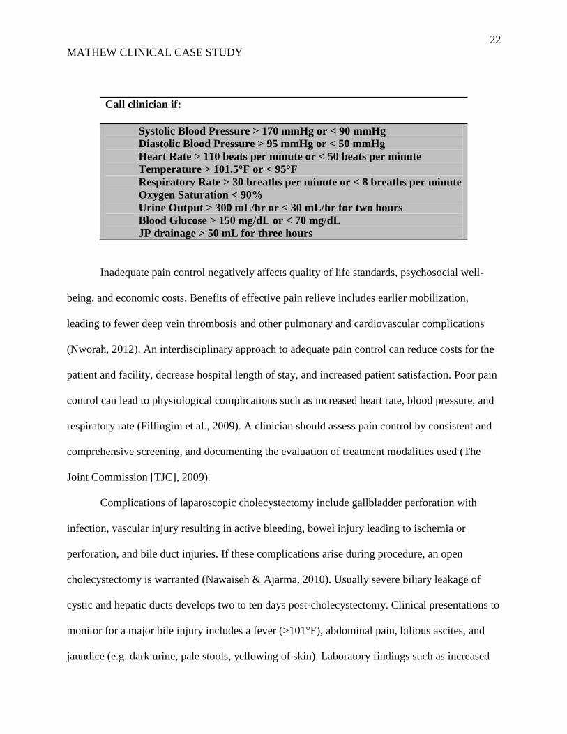
after two weeks of hospital discharge (Shuja, Bashir, & Rashid, 2011). The following monitoring
parameters are ordered based on patient’s medical history of hypertension and diabetes and in
order to monitor for possible post-procedure complications (Cherng et al., 2012; Girometti,
2010; Strasberg, 2008):
22
MATHEW CLINICAL CASE STUDY
Call clinician if:
Systolic Blood Pressure > 170 mmHg or < 90 mmHg
Diastolic Blood Pressure > 95 mmHg or < 50 mmHg
Heart Rate > 110 beats per minute or < 50 beats per minute
Temperature > 101.5°F or < 95°F
Respiratory Rate > 30 breaths per minute or < 8 breaths per minute
Oxygen Saturation < 90%
Urine Output > 300 mL/hr or < 30 mL/hr for two hours
Blood Glucose > 150 mg/dL or < 70 mg/dL
JP drainage > 50 mL for three hours
Inadequate pain control negatively affects quality of life standards, psychosocial well-
being, and economic costs. Benefits of effective pain relieve includes earlier mobilization,
leading to fewer deep vein thrombosis and other pulmonary and cardiovascular complications
(Nworah, 2012). An interdisciplinary approach to adequate pain control can reduce costs for the
patient and facility, decrease hospital length of stay, and increased patient satisfaction. Poor pain
control can lead to physiological complications such as increased heart rate, blood pressure, and
respiratory rate (Fillingim et al., 2009). A clinician should assess pain control by consistent and
comprehensive screening, and documenting the evaluation of treatment modalities used (The
Joint Commission [TJC], 2009).
Complications of laparoscopic cholecystectomy include gallbladder perforation with
infection, vascular injury resulting in active bleeding, bowel injury leading to ischemia or
perforation, and bile duct injuries. If these complications arise during procedure, an open
cholecystectomy is warranted (Nawaiseh & Ajarma, 2010). Usually severe biliary leakage of
cystic and hepatic ducts develops two to ten days post-cholecystectomy. Clinical presentations to
monitor for a major bile injury includes a fever (>101°F), abdominal pain, bilious ascites, and
jaundice (e.g. dark urine, pale stools, yellowing of skin). Laboratory findings such as increased

23
MATHEW CLINICAL CASE STUDY
WBC, decreased hemoglobin/hematocrit, elevated liver function tests (ALT, AST, ALP), and
increased total and direct bilirubin are monitored post-cholecystectomy (Papadakis, McPhee, &
Rabow, 2013). Fever accompanied by leukocytosis should prompt the clinician to adjust
antibiotics according to the culture results of the bile and the gallbladder that was obtained
intraoperatively (Strasberg, 2008). If clinical suspension of biliary leak is present, a
transabdominal ultrasonography or an abdominal CT with contrast is performed to evaluate the
extent of the possible leak. Positive results indicate the need for an endoscopic retrograde
cholangiopancreatography (ERCP) to determine the location of the leak and to directly treat the
complication (Tonolini et al., 2012). An endoprosthesis (i.e. a stent) or sphincterotomy with
insertion of percutaneous drains are the treatment of choice to decrease pressure in the biliary
system and to allow for bile drainage into the intestinal tract (Hsieh et al., 2012; Maekawa et al.,
2013). At two to four week follow-up, a HIDA scan and liver function tests are ordered. If the
scan reveals absence of bile leak and if the laboratory work-up is normal, the stent is removed
with a repeat ERCP (Maekawa et al., 2013).
Acute hemodynamic instability with hypotension and tachycardia are associated with
active bleeding, usually from arterial sources or the liver. Meticulous cardiovascular assessments
and frequent monitoring of vital signs (every 15 minutes for two hours, then hourly) are critical
in the post-operative period (Masood et al., 2012). The patients can also present with sepsis and
cardiovascular collapse from possible bowel injury/perforation during the procedure. Abdominal
pain or distention, constipation, peritonitis, and absent or hypoactive bowel sounds are related
clinical findings. A CT of the abdomen and pelvis with contrast is ordered for further evaluation.
In both of the above mentioned situations, an emergent open cholecystectomy may be indicated
for intervention (Nawaiseh & Ajarma, 2010).

24
MATHEW CLINICAL CASE STUDY
Another complication that may arise months to years after cholecystectomy is the Post-
cholecystectomy Syndrome (PCS). Persistence of symptoms of right upper quadrant pain,
flatulence, and dyspepsia after meals can be due to recurrent biliary stones, bile duct strictures,
gallbladder remnant, inflamed cystic duct, or biliary dyskinesia from dysfunction of sphincter of
Oddi (Papadakis, McPhee, & Rabow, 2013). Elevated liver function tests, fever, chills, or
jaundice suggest biliary tract disease. Extrabiliary causes of PCS include differential diagnoses
such as pancreatitis, hepatitis, peptic ulcer disease, diverticulitis, irritable bowel syndrome, or
esophagitis. Treatment is based on the underlying cause of the symptoms (Janes, Berry, &
Dijkstra, 2005). A CT of abdomen with contrast or a MRCP are noninvasive tests for evaluation
of biliary tract disease (Girometti et al., 2010).
Establishing monitoring parameters for the prescribed medications is also necessary.
When prescribing meperidine, clinician should observe the patient for respiratory depression,
hypotension, bradycardia, confusion, increased intracranial pressures, seizures, constipation,
nausea/vomiting, abdominal cramps and anaphylaxis (Lexi-Comp, 2013). Use of NSAIDs is
associated with renal insufficiency and increased risk of bleeding (Katzung, Masters, & Trevor,
2012). With NSAID, diclofenac, monitoring of CBC, CMP, liver enzymes, BUN/creatinine, and
urine output is essential (Lexi-Comp, 2013).
A possible complication that can arise with the use of antibiotics is Clostridium difficile,
presenting with leukocytosis and severe diarrhea (Howerton, Patra, & Abel-Santos, 2013). Side
effects of ceftriaxone include anaphylaxis, Stevens-Johnson syndrome, agranulocytosis,
thrombocytopenia or thrombocytosis, hemolytic anemia, and increased BUN and serum
transaminases. Adverse effects of metronidazole consist of T-wave flattening on an EKG,
syncope, flushing, confusion, Stevens-Johnson syndrome, aseptic meningitis, neutropenia, and

25
MATHEW CLINICAL CASE STUDY
thrombocytopenia. Routine follow-up with lab work (i.e. CBC with differential, CMP) and an
EKG may be required (Lexi-Comp, 2013).
Adverse effects of ondansetron consist of headache, malaise, fatigue, drowsiness, and
urinary retention. Less common side effects include hepatic failure and second-degree heart
block and ST-segment depression on an EKG (Lexi-Comp, 2013). Routine monitoring of liver
enzymes and cardiac tracing is suggested. Monitoring of electrolytes, vital signs, and signs and
symptoms of fluid retention is essential when administering intravenous crystalloid fluids (Ker,
Perel, & Roberts, 2013).
Discharge Planning/Health Promotion
The patient will be instructed to advance diet as tolerated, starting with a clear liquid diet.
Early ambulation and aggressive pulmonary toileting will be encouraged the post-operative
period to prevent cardiopulmonary and vascular complications (Drolet et al., 2013). If a large
umbilical incision was performed, the patient will be educated to limit heavy lifting for two
weeks (Strasberg, 2008). Due to the possible risk of infection after the surgical procedure, the
patient will be educated on signs and symptoms of infection, including increased temperature
greater than 101.5°F and purulent drainage, redness, warmth, or inflammation from the surgical
site, and instructed to promptly call the clinician if the signs and symptoms are present (Shuja,
Bashir, & Rashid, 2011). Other clinical symptoms to monitor and report are: dark urine, pale
stools, yellowing of skin, chills, dizziness, syncope, abdominal distention or severe sharp pain,
constipation or diarrhea, difficulty breathing, severe bleeding, and chest pain (Papadakis,
McPhee, & Rabow, 2013). The patient will be instructed on the importance of attending the
follow-up appointment scheduled in two weeks with the clinician in order to assess the patient’s
condition and obtain lab work (i.e. CBC, CMP) if necessary. Instructions on how to care for the

26
MATHEW CLINICAL CASE STUDY
JP and T-tube drains will be provided. Keeping the insertion site clean and dry is essential to
prevent infection. The patient will be instructed to wash the wound site with soap and water daily
(Cherng et al., 2012). Care must be taken to avoid accidental pulling or clamping of the drains.
The patient will be educated on medications’ signs and symptoms prescribed at discharge
and the importance of medication adherence. Common drug-to-drug interactions will be
verbalized in order to avoid possible concerning complications. The patient will continue current
home medications at discharge in addition to diclofenac (Zipsor®) 75 mg PO two times a day for
mild to moderate abdominal pain that may continue for up to a week after discharge (Riall et al.,
2012). If severe pain develops, the patient will be instructed to contact the clinician immediately.
The NSAID use will be restricted to one to two weeks due to the patient’s history of PUD. The
patient will be encouraged to use the medication only if pain present to avoid bleeding
complications (Katzung, Masters, & Trevor, 2012). If pain is not adequately relieved with
diclofenac, the patient will be asked to contact the clinician.
General health promotion strategies will be discussed with the patient. Since diabetes and
obesity are related factors in the development of recurrent biliary lithiasis, the patient will be
instructed on losing weight gradually and controlling blood glucose levels. The patient will be
encouraged to lose weight gradually to a BMI of < 25 kg/m2 with eating healthy, balanced meals
and exercising 30 minutes daily five days a week. Monitoring blood glucose before meals and at
bedtime is essential to avoid symptomatic hypoglycemia or hyperglycemia. Taking the
hypoglycemic medication as recommended and restricting foods that are high in glucose assists
in maintaining serum glucose levels in the target goal range between 70-110 mg/dL. Controlling
blood pressure (SBP < 120 mmHg, DBP < 80 mmHg) by limiting sodium intake and taking
medications as prescribed can prevent life-threatening complications such as heart attack and

27
MATHEW CLINICAL CASE STUDY
stroke (American College of Cardiology Foundation [ACCF]/American Heart Association
[AHA], 2009). Although the patient is currently up-to-date on immunizations, reiteration will be
provided on obtaining a flu vaccination yearly and a pneumonia vaccination every five years. An
annual physical health check with a primary care physician is essential to assess patient’s
condition and identify complications related to comorbidities early (Tsuboi, Hayakawa, Kanda,
& Fukushima, 2011).

28
MATHEW CLINICAL CASE STUDY
References
Al-Abassi, A. A., Farghaly, M. M., Ahmed, H. L., Mobasher, L. A., & Al-Manee, M. S. (2001).
Infection after laparoscopic cholecystectomy: Effect of infected bile and infected
gallbladder wall. European Journal of Surgery, 167(4), 268-273.
doi:10.1080/110241501300091426
American Pain Society. (2012). Types of Treatments. Retrieved from
www.americanpainsociety.org/uploads/pdfs/npc/section_3.pdf
Barak, O., Elazary, R., Appelbaum, L., Rivkind, A., & Almogy, G. (2009). Conservative
treatment for acute cholecystitis: Clinical and radiographic predictors of failure. The
Israel Medical Association Journal, 11(1), 739-743.
Bellows, C., Berger, D., & Crass, R. (2005). Management of gallstones. American Family
Physician, 72(4), 637-637-42, 567-9, 711 passim.
Brisinda, G., Vanella, S., Crocco, A., Mazzari, A., Tomaiuolo, P., Santullo, F., & ... Crucitti, A.
(2011). Severe acute pancreatitis: Advances and insights in assessment of severity and
management. European Journal of Gastroenterology & Hepatology, 23(7), 541-551.
doi:10.1097/MEG.0b013e328346e21e
Cherng, N., Witkowski, E. T., Sneider, E. B., Wiseman, J. T., Lewis, J., Litwin, D. M., & ...
Shah, S. A. (2012). Use of cholecystostomy tubes in the management of patients with
primary diagnosis of acute cholecystitis. Journal of The American College of Surgeons,
214(2), 196-201. doi:10.1016/j.jamcollsurg.2011.11.005
Colli, A. A., Conte, D. D., Valle, S., Sciola, V. V., & Fraquelli, M. M. (2012). Meta-analysis:
Nonsteroidal anti-inflammatory drugs in biliary colic. Alimentary Pharmacology &
Therapeutics, 35(12), 1370-1378. doi:10.1111/j.1365-2036.2012.05115.x

29
MATHEW CLINICAL CASE STUDY
Drolet, A., Dejuilio, P., Harkless, S., Henricks, S., Kamin, E., Leddy, E. A., & ... Williams, S.
(2013). Move to improve: The feasibility of using an early mobility protocol to increase
ambulation in the intensive and intermediate care settings. Physical Therapy, 93(2), 197-
207. doi:10.2522/ptj.20110400
Fillingim, R. B., King, C., Ribeiro-Dasilva, M., Rahim-Williams, B., Joseph, L., & Riley, L. J.
(2009). Sex, gender, and pain: A review of recent clinical and experimental findings.
Journal of Pain, 10(5), 447–485.
Frossard, J., Steer, M., & Pastor, C. (2008). Acute pancreatitis. Lancet, 371(9607), 143-152.
Gaby, A. R. (2009). Nutritional approaches to prevention and treatment of gallstones. Alternative
Medicine Review, 14(3), 258-267.
Garst, G., Moore, E., Banerjee, M., Leopold, D., Burlew, C., Bensard, D., & ... Sauaia, A.
(2013). Acute appendicitis: A disease severity score for the acute care surgeon. Journal
of Trauma & Acute Care Surgery, 74(1), 32-36. doi:10.1097/TA.0b013e318278934a
Girometti, R. R., Brondani, G. G., Cereser, L. L., Como, G. G., del Pin, M. M., Bazzocchi, M.
M., & Zuiani, C. C. (2010). Post-cholecystectomy syndrome: Spectrum of biliary
findings at magnetic resonance cholangiopancreatography. British Journal of Radiology,
83(988), 351-361. doi:10.1259/bjr/99865290
Grover, V. K., Mathew, P. J., & Hegde, H. H. (2009). Efficacy of orally disintegrating
ondansetron in preventing postoperative nausea and vomiting after laparoscopic
cholecystectomy: A randomized, double-blind placebo controlled study. Anaesthesia,
64(6), 595-600. doi:10.1111/j.1365-2044.2008.05860.x
Hawley, C. (2012). Understanding viral hepatitis: As easy as A, B, C. British Journal of Primary
Care Nursing: Cardiovascular Disease, Diabetes & Kidney Care, 16-21.

30
MATHEW CLINICAL CASE STUDY
Howerton, A., Patra, M., & Abel-Santos, E. (2013). A new strategy for the prevention of
clostridium difficile infection. Journal of Infectious Diseases, 207(10), 1498-1504.
Hsieh, Y., Chen, C., Su, C., Chan, C., Huo, T., Liu, C., & ... Lin, H. (2012). Outcome after
percutaneous cholecystostomy for acute cholecystitis: A single-center experience.
Journal of Gastrointestinal Surgery, 16(10), 1860-1868. doi:10.1007/s11605-012-1965-8
Janes, S., Berry, L. L., & Dijkstra, B. B. (2005). Management of post cholecystectomy mirizzi's
syndrome. Journal of Minimal Access Surgery, 1(1), 34-36.
Jessup, M., Abraham, W., Casey, D., Feldman, A., Francis, G., Ganiats, T., & ... Yancy, C.
(2009). 2009 focused update: ACCF/AHA Guidelines for the diagnosis and management
of heart failure in adults. A report of the American College of Cardiology
Foundation/American Heart Association task force on practice guidelines. Circulation,
119(14), 1977-2016.
Katzung, B.G., Masters, S.B., & Trevor, A.J. (2012). Basic and clinical pharmacology. New
York, NY: McGraw Hill Medical.
Ker, K., Perel, P., & Roberts, I. (2013). Colloids versus crystalloids for fluid resuscitation in
critically ill patients. Cochrane Library, 3, 1-73. doi:10.1002/14651858.CD000567.pub6
Kessenich, C. R. (2011). Cholecystitis and HIDA scan. Nurse Practitioner, 36(9), 11-12.
doi:10.1097/01.NPR.0000403295.82092.20
Koskensalo, S., & Leppäniemi, A. (2010). Perforated duodenal ulcer: Has anything changed?.
European Journal of Trauma & Emergency Surgery, 36(2), 145-150.
doi:10.1007/s00068-010-9128-7
Leung, N. (2010). Viral hepatitis lab testing. Advance For Administrators Of The Laboratory,
19(2), 42.
31
MATHEW CLINICAL CASE STUDY
Lexi-Comp, Inc. (2013). Lexi-Drugs™: [Smart-phone application]. Accessed July 14, 2013.
Maekawa, S., Nomura, R., Murase, T., Ann, Y., Oeholm, M., & Harada, M. (2013). Endoscopic
gallbladder stenting for acute cholecystitis: A retrospective study of 46 elderly patients
aged 65 years or older. BMC Gastroenterology, 13(1), 1-7. doi:10.1186/1471-230X-13-
65
Mahboobi, N., Porter, S., Karayiannis, P., & Alavian, S. (2012). Oral fluid and hepatitis A, B and
C: A literature review. Journal Of Oral Pathology & Medicine, 41(7), 505-516.
doi:10.1111/j.1600-0714.2011.01123.x
Masood, R., Samillah, Afridi, Z., Masood, K., Khan, B., & Khurshied, F. (2012). Laparoscopic
cholecystectomy in acute gallbladder. JPMI: Journal of Postgraduate Medical Institute,
26(2), 212-217.
Mayoux-Benhamou, A., Giraudet-Le Quintrec, J., Ravaud, P., Champion, K., Dernis, E., Zerkak,
D., & ... Dougados, M. (2008). Influence of patient education on exercise compliance in
rheumatoid arthritis: A prospective 12-month randomized controlled trial. Journal of
Rheumatology, 35(2), 216-223.
Mazeh, H., Mizrahi, I., Dior, U., Simanovsky, N., Shapiro, M., Freund, H., & Eid, A. (2012).
Role of antibiotic therapy in mild acute calculus cholecystitis: A prospective randomized
controlled trial. World Journal of Surgery, 36(8), 1750-1759. doi:10.1007/s00268-012-
1572-6
Nakeeb, A., Comuzzie, A.G., Martin, L., Sonnenberg, G.E., Swartz-Basile, D., & Kissebah A.H.
(2002). Gallstones: Genetics versus environment. Annals of Surgery, 235(12), 842-849.
Nawaiseh, K., & Ajarma, K. (2010). Laparoscopic versus open cholecystectomy in the treatment
of acute cholecystitis. Middle East Journal of Internal Medicine, 3(1), 38-41.

32
MATHEW CLINICAL CASE STUDY
Nikfarjam, M., Niumsawatt, V., Sethu, A., Fink, M. A., Muralidharan, V., Starkey, G., & ...
Christophi, C. (2011). Outcomes of contemporary management of gangrenous and non-
gangrenous acute cholecystitis. HPB: The Official Journal of The International Hepato-
Pacreato-Biliary Association, 13(8), 551-558. doi:10.1111/j.1477-2574.2011.00327.x
Nworah, U. (2012). From documentation to the problem: Controlling postoperative pain.
Nursing Forum, 47(2), 91-99. doi:10.1111/j.1744-6198.2012.00262.x
Ohio Board of Nursing (OBN). (2013). The formulary developed by the Committee on
Prescriptive Governance. Retrieved from
http://www.nursing.ohio.gov/PDFS/AdvPractice/1-20-13_Formulary.pdf
Pagana, K. D. & Pagana, T. J. (2010). Mosby’s manual of diagnostic and laboratory tests. St.
Louis, MI: Elsevier, Inc.
Papadakis, M., McPhee, S. J., & Rabow, M.W. (2013). Current medical diagnosis and
treatment. New York, NY: McGraw-Hill Companies, Inc.
Penny, S. M. (2012). Clinical signs of pancreatitis. Radiologic Technology, 83(6), 561-581.
Qandil, A. M. (2012). Prodrugs of nonsteroidal anti-inflammatory drugs (NSAIDs), more than
meets the eye: A critical review. International Journal of Molecular Sciences, 13(12),
17244-17274. doi:10.3390/ijms131217244
Riall, T. S., Zhang, D., Townsend, C. M., Kuo, Y., & Goodwin, J. S. (2010). Failure to perform
cholecystectomy for acute cholecystitis in elderly patients is associated with increased
morbidity, mortality, and cost. Journal of The American College of Surgeons, 210(5),
668-677. doi:10.1016/j.jamcollsurg.2009.12.031
Ruhl, C.E. & Everhart, J.E. (2011). Gallstone disease is associated with increased mortality in
the United States. Gastroenterology, 140(12), 508-516.

33
MATHEW CLINICAL CASE STUDY
Shuja, A., Bashir, A., & Rashid, A. (2011). Optimal timing for surgery in acute gallstone disease.
Professional Medical Journal, 18(2), 237-242.
Solomkin, J. S., Mazuski, J. E., Bradley, J. S., Rodvold, K. A., Goldstein, E. C., Baron, E. J., &
... Bartlett, J. G. (2010). Diagnosis and management of complicated intra-abdominal
infection in adults and children: Guidelines by the Surgical Infection Society and the
Infectious Diseases Society of America. Clinical Infectious Diseases, 50(2), 133-164.
doi:10.1086/649554
Strasberg, S. (2008). Clinical practice. Acute calculous cholecystitis. New England Journal of
Medicine, 358(26), 2804-2811.
Summers, S., Scruggs, W., Menchine, M., Lahham, S., Anderson, C., Amr, O., & ... Fox, J.
(2010). A prospective evaluation of emergency department bedside ultrasonography for
the detection of acute cholecystitis. Annals of Emergency Medicine, 56(2), 114-122.
doi:10.1016/j.annemergmed.2010.01.014
Tang, R. S., & Chan, F. K. (2012). Therapeutic management of recurrent peptic ulcer disease.
Drugs, 72(12), 1605-1616. doi:10.2165/11634850
The Joint Commission (TJC). (2012). TJC Standards for Pain Management. Comprehensive
Accreditation Manual for Critical Access Hospitals (CAMCAH): The official handbook.
Retrieved from
http://www.jointcommission.org/standards_information/cah_requirements.aspx
Thune, A., Baker, A., Saccone, G.T., Owen, H., & Toouli, J. (1990). Differing effects of
pethidine and morphine on human sphincter of oddi motility. British Journal of Surgery,
77(9), 992-995.

34
MATHEW CLINICAL CASE STUDY
Tonolini, M., Ravelli, A., Villa, C., & Bianco, R. (2012). Urgent MRI with MR
cholangiopancreatography (MRCP) of acute cholecystitis and related complications:
Diagnostic role and spectrum of imaging findings. Emergency Radiology, 19(4), 341-348.
doi:10.1007/s10140-012-1038-z
Tsuboi, S., Hayakawa, T., Kanda, H., & Fukushima, T. (2011). Physical activity in the context of
clustering patterns of health-promoting behaviors. American Journal of Health
Promotion, 25(6), 410-416.
Virmani, V., Papadatos, D., Fasih, N., Kielar, A., Gulati, A., & Sethi, V. (2012). Rule out
appendicitis: "To be or not to be". Canadian Association of Radiologists Journal, 63(1),
47-60. doi:10.1016/j.carj.2010.06.001
Vissers, R., & Lennarz, W. (2010). Pitfalls in appendicitis. Emergency Medicine Clinics of North
America, 28(1), 103. doi:10.1016/j.emc.2009.09.003
Watkins, R., & Lemonovich, T. (2011). Diagnosis and management of community-acquired
pneumonia in adults. American Family Physician, 83(11), 1299-1306.
Zaliekas, J., & Munson, L. (2008). Complications of gallstones: The Mirizzi syndrome, gallstone
ileus, gallstone pancreatitis, complications of "lost" gallstones. Surgical Clinics of North
America, 88(6), 1345-1368.