acute cva and tia -...
TRANSCRIPT
Acute CVA and TIA
Robert Dachs, MD, FAAFP
Clinical Associate Professor and Director of Research
Ellis Hospital Family Medicine Residency Program
Albany Medical College, Albany, New York

Disclosure StatementIt is the policy of the AAFP that all individuals in a position to control content disclose any relationships with commercial interests upon nomination/invitation of participation. Disclosure documents are reviewed for potential conflicts of interest. If conflicts are identified, they are resolved prior to confirmation of participation. Only participants who have no conflict of interest or who agree to an identified resolution process prior to their participation were involved in this CME activity.
All individuals in a position to control content for this session have indicated they have no relevant financial relationships to disclose.

Learning Objectives
1. Identify patients with underlying risk factors for stroke.2. Employ cost-effective measures to decrease stroke in patients at
risk.3. State the 2009 AHA/ASA definition of TIA and describe the
recommended evaluation.4. Propose appropriate treatment options to improve outcomes in
patients who suffer a stroke.
In 30 Minutes … The Plan
• CVA risk factors and 10 prevention
• Acute CVA care
• 20 prevention
• TIA – everything has changed

A. Acute thrombosisB. Acute embolic event C. Acute intracerebral hemorrhageD. Acute subarachnoid hemorrhage
Stroke Pathogenesis1. Acute stroke events are most often the result of which of the following pathological processes?
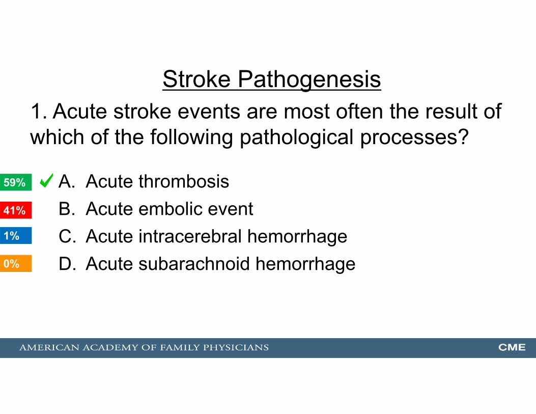
A. Acute thrombosisB. Acute embolic event C. Acute intracerebral hemorrhageD. Acute subarachnoid hemorrhage
Stroke Pathogenesis1. Acute stroke events are most often the result of which of the following pathological processes?
0%
59%
41%
1%1
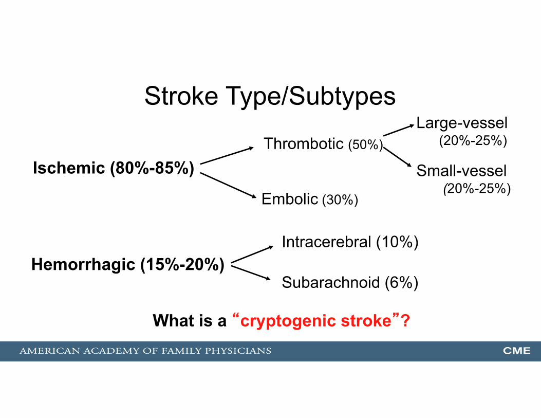
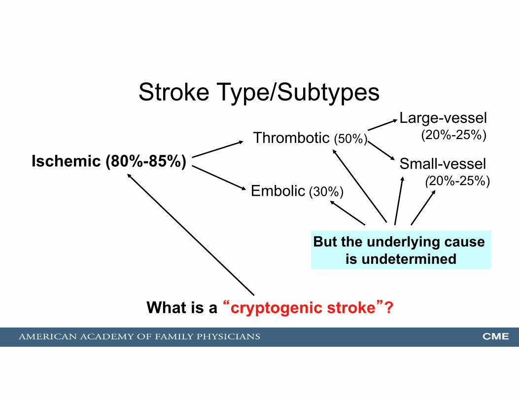
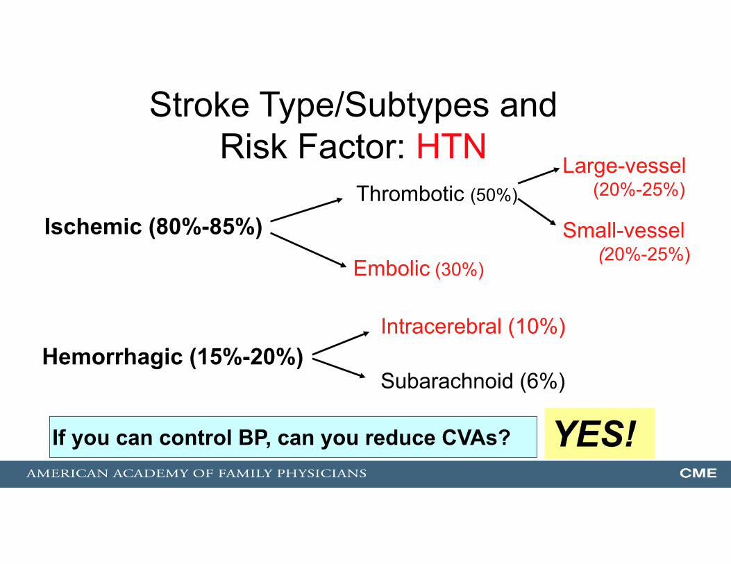
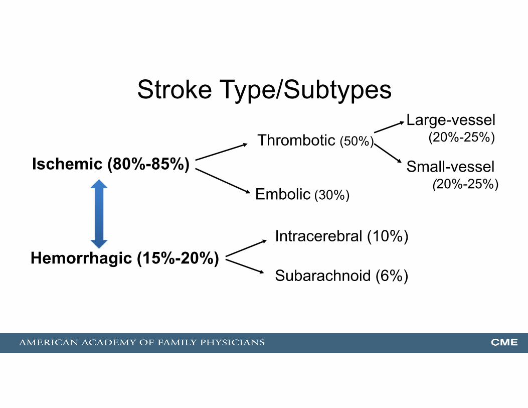
Stroke Type/Subtypes
Ischemic (80%-85%)Thrombotic (50%)
Embolic (30%)
Large-vessel(20%-25%)
Small-vessel(20%-25%)
Hemorrhagic (15%-20%)Intracerebral (10%)
Subarachnoid (6%)
What is a “cryptogenic stroke”?
Stroke Type/Subtypes
Ischemic (80%-85%)Thrombotic (50%)
Embolic (30%)
Large-vessel(20%-25%)
Small-vessel(20%-25%)
But the underlying cause is undetermined
What is a “cryptogenic stroke”?
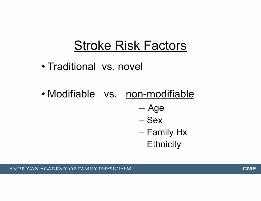
Stroke Risk Factors• Traditional vs. novel
• Modifiable vs. non-modifiable– Age– Sex– Family Hx– Ethnicity
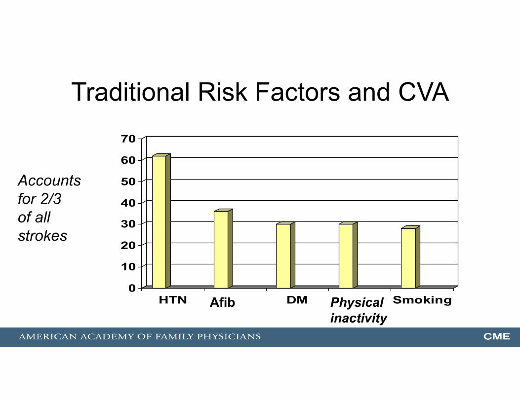
Traditional Risk Factors and CVA
0
10
20
30
40
50
60
70
HTN DM Smoking
Accounts for 2/3 of all strokes
Afib Physical inactivity
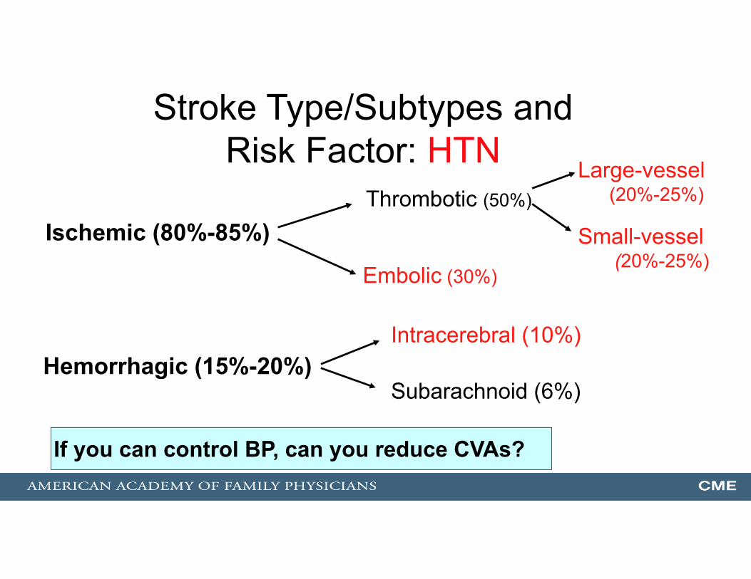
Ischemic (80%-85%)Thrombotic (50%)
Embolic (30%)
Large-vessel(20%-25%)
Small-vessel(20%-25%)
Hemorrhagic (15%-20%)Intracerebral (10%)
Subarachnoid (6%)
Stroke Type/Subtypes and Risk Factor: HTN
If you can control BP, can you reduce CVAs?
Ischemic (80%-85%)Thrombotic (50%)
Embolic (30%)
Large-vessel(20%-25%)
Small-vessel(20%-25%)
Hemorrhagic (15%-20%)Intracerebral (10%)
Subarachnoid (6%)
Stroke Type/Subtypes and Risk Factor: HTN
YES!If you can control BP, can you reduce CVAs?
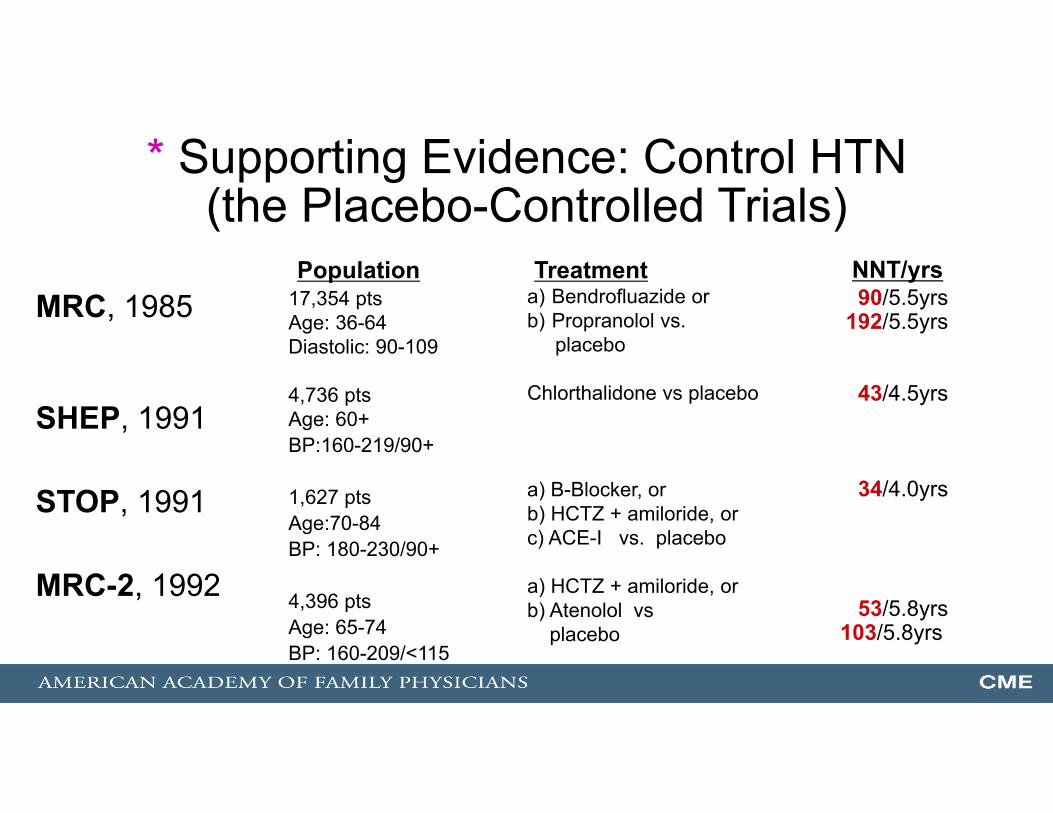
* Supporting Evidence: Control HTN(the Placebo-Controlled Trials)
MRC, 1985
SHEP, 1991
STOP, 1991
MRC-2, 1992
17,354 ptsAge: 36-64Diastolic: 90-109
4,736 ptsAge: 60+BP:160-219/90+
1,627 ptsAge:70-84BP: 180-230/90+
4,396 ptsAge: 65-74BP: 160-209/<115
a) Bendrofluazide orb) Propranolol vs.
placebo
Chlorthalidone vs placebo
a) B-Blocker, orb) HCTZ + amiloride, orc) ACE-I vs. placebo
a) HCTZ + amiloride, orb) Atenolol vs
placebo
90/5.5yrs192/5.5yrs
43/4.5yrs
34/4.0yrs
53/5.8yrs103/5.8yrs
Population Treatment NNT/yrs

Ischemic (80%-85%)Thrombotic (50%)
Embolic (30%)
Large-vessel(20%-25%)
Small-vessel(20%-25%)
Hemorrhagic (15%-20%)Intracerebral (10%)
Subarachnoid (6%)
Stroke Type/Subtypes and Risk Factor: DM

Ischemic (80%-85%)Thrombotic (50%)
Embolic (30%)
Large-vessel(20%-25%)
Small-vessel(20%-25%)
Hemorrhagic (15%-20%)Intracerebral (10%)If anticoagulated
Subarachnoid (6%)
Stroke Type/Subtypes and Risk Factor: Afib

Atrial Fibrillation and Stroke
• The older the patient with atrial fibrillation, the higher the risk of cardioembolic stroke.
• Strokes due to Afib have higher mortality and morbidity.• Warfarin decreases absolute annual risk from 4.5% --> 1.4% (NNT=30).
0
1
2
3
4
5
6
7
8
< 65 yrs 65-75yrs > 75 yrs
CVA rate(% per yr)
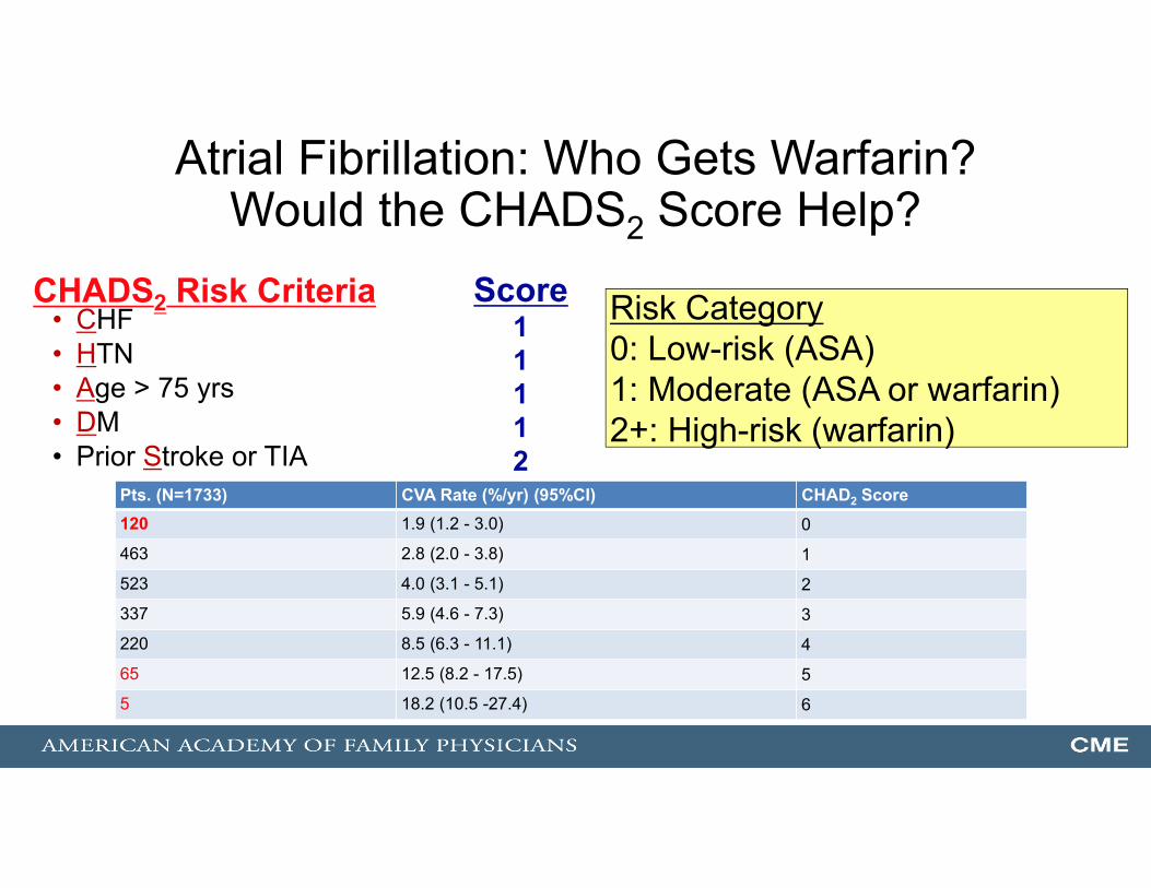
Atrial Fibrillation: Who Gets Warfarin? Would the CHADS2 Score Help?
• CHF• HTN• Age > 75 yrs• DM• Prior Stroke or TIA
CHADS2 Risk Criteria Score11112
Risk Category0: Low-risk (ASA)1: Moderate (ASA or warfarin)2+: High-risk (warfarin)
Pts. (N=1733) CVA Rate (%/yr) (95%CI) CHAD2 Score120 1.9 (1.2 - 3.0) 0
463 2.8 (2.0 - 3.8) 1
523 4.0 (3.1 - 5.1) 2
337 5.9 (4.6 - 7.3) 3
220 8.5 (6.3 - 11.1) 4
65 12.5 (8.2 - 17.5) 5
5 18.2 (10.5 -27.4) 6
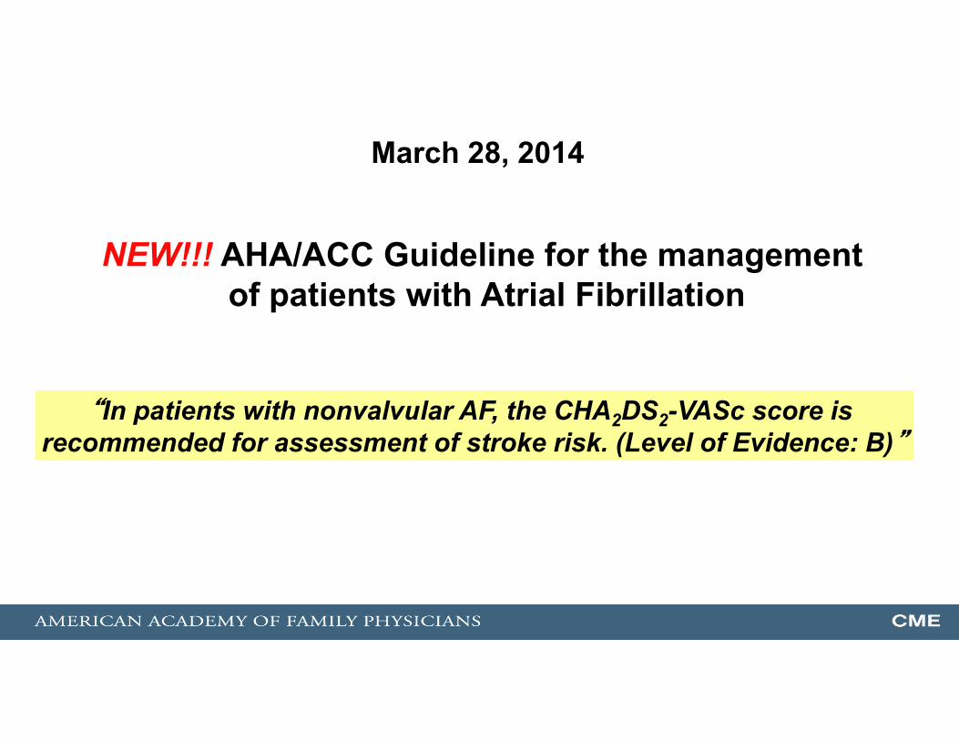
“In patients with nonvalvular AF, the CHA2DS2-VASc score is recommended for assessment of stroke risk. (Level of Evidence: B)”
March 28, 2014
NEW!!! AHA/ACC Guideline for the management of patients with Atrial Fibrillation
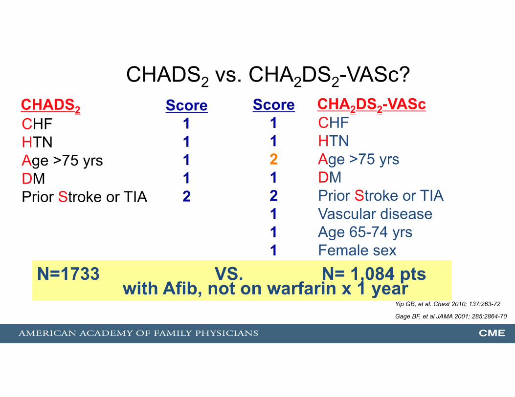
CHADS2 vs. CHA2DS2-VASc?
CHFHTN Age >75 yrsDMPrior Stroke or TIA
CHADS2 Score11112
Score11212111
CHA2DS2-VAScCHFHTN Age >75 yrsDMPrior Stroke or TIAVascular diseaseAge 65-74 yrsFemale sex
N=1733 VS. N= 1,084 ptswith Afib, not on warfarin x 1 year
Yip GB, et al. Chest 2010; 137:263-72
Gage BF, et al JAMA 2001; 285:2864-70
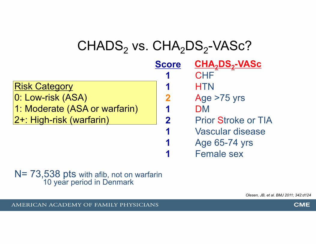
CHADS2 vs. CHA2DS2-VASc?Score
11212111
CHA2DS2-VAScCHFHTN Age >75 yrsDMPrior Stroke or TIAVascular diseaseAge 65-74 yrsFemale sex
Risk Category0: Low-risk (ASA)1: Moderate (ASA or warfarin)2+: High-risk (warfarin)
N= 73,538 pts with afib, not on warfarin 10 year period in Denmark
Olesen, JB, et al. BMJ 2011; 342:d124

Atrial Fibrillation: WarfarinPotential Harms vs. Potential Benefits
• Decreases CVA by 64% (vs. ASA 22%)–Absolute reduction approx. 3%/yr
• Rate of ICH 0.1 - 0.6%–Increased with advanced age, HTN
• Major bleeding rates: 1.2%/yr
Which of the following new oral anticoagulants is approved for the management of non-valvular Afib?
A. Dabigatran (Pradaxa) - BIDB. Rivaroxaban (Xarelto) - qdC. Apixaban (Eliquis) - BIDD. All of the above
Which of the following new oral anticoagulants is approved for the management of non-valvular Afib?
A. Dabigatran (Pradaxa) - BIDB. Rivaroxaban (Xarelto) - qdC. Apixaban (Eliquis) - BIDD. All of the above

If you were asked…
Which of the following new oral anticoagulants is approved for the management of DVT/PE?
A. Dabigatran (Pradaxa)B. Rivaroxaban (Xarelto)C. Apixaban (Eliquis)D. All of the above
If you were asked…
Which of the following new oral anticoagulants is approved for the management of DVT/PE?
A. Dabigatran (Pradaxa)B. Rivaroxaban (Xarelto)C. Apixaban (Eliquis)D. All of the above

Traditional Risk Factors and CVA
0
10
20
30
40
50
60
70
HTN Afib DM Physicalinactivity
Smoking
Accounts for 2/3 of all strokes
_____

The Last “Traditional” Risk Factor: What About Family History?
• Documented parental stroke by 65 yrs of age is associated with a 3-fold increase in stroke in offspring.
Based on 8-year follow-up of 3,443 stroke-free Framingham offspring
Seshadri S, et al. Circulation. 2010
Stroke Risk Factors
• Traditional vs. novel
• Modifiable vs. non-modifiable– Age– Sex– Family Hx– Ethnicity
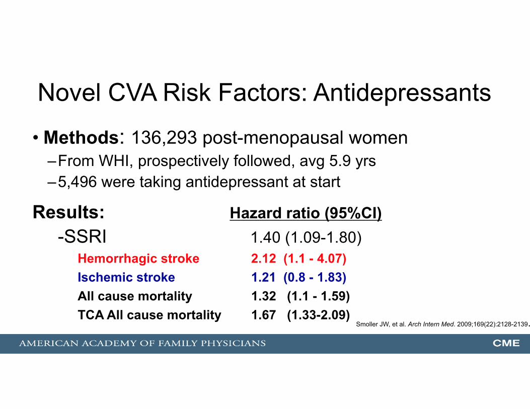
Novel CVA Risk Factors: Antidepressants
• Methods: 136,293 post-menopausal women–From WHI, prospectively followed, avg 5.9 yrs–5,496 were taking antidepressant at start
Results: Hazard ratio (95%CI)-SSRI 1.40 (1.09-1.80)
Hemorrhagic stroke 2.12 (1.1 - 4.07)Ischemic stroke 1.21 (0.8 - 1.83)All cause mortality 1.32 (1.1 - 1.59)TCA All cause mortality 1.67 (1.33-2.09)
Smoller JW, et al. Arch Intern Med. 2009;169(22):2128-2139.

Risk Factors: Intracerebral Hemorrhage• Hypertension• Amyloid angiopathy• AVMs• Brain tumors• Bleeding disorders• Vasculitis• CNS infection• Septic embolism
• High-risk groups– Older age– Ethnicity(African American, Asian, Mexican American)
• Drugs– Anticoagulants– Cocaine– Amphetamines– SSRIs
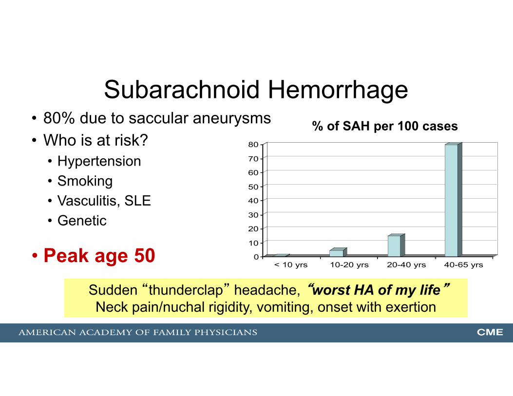
Subarachnoid Hemorrhage• 80% due to saccular aneurysms• Who is at risk?
• Hypertension• Smoking• Vasculitis, SLE• Genetic
• Peak age 50 0
10
20
30
40
50
60
70
80
< 10 yrs 10-20 yrs 20-40 yrs 40-65 yrs
% of SAH per 100 cases
Sudden “thunderclap” headache, “worst HA of my life”Neck pain/nuchal rigidity, vomiting, onset with exertion

In 30 Minutes … The Plan
• CVA risk factors and prevention
• Acute CVA care
• TIA – everything has changed
2. An 82 y/o male developed sudden dysarthria and RUE weakness.Onset at 8am. He arrives at the ED at 11 am. IV is inserted, labs sent.
Which of the following imaging studies should be ordered STAT?
A. Non-contrast head CT
B. Contrast head CT
C. CT-A (angiogram) of the head
D. Non-contrast MRI of head
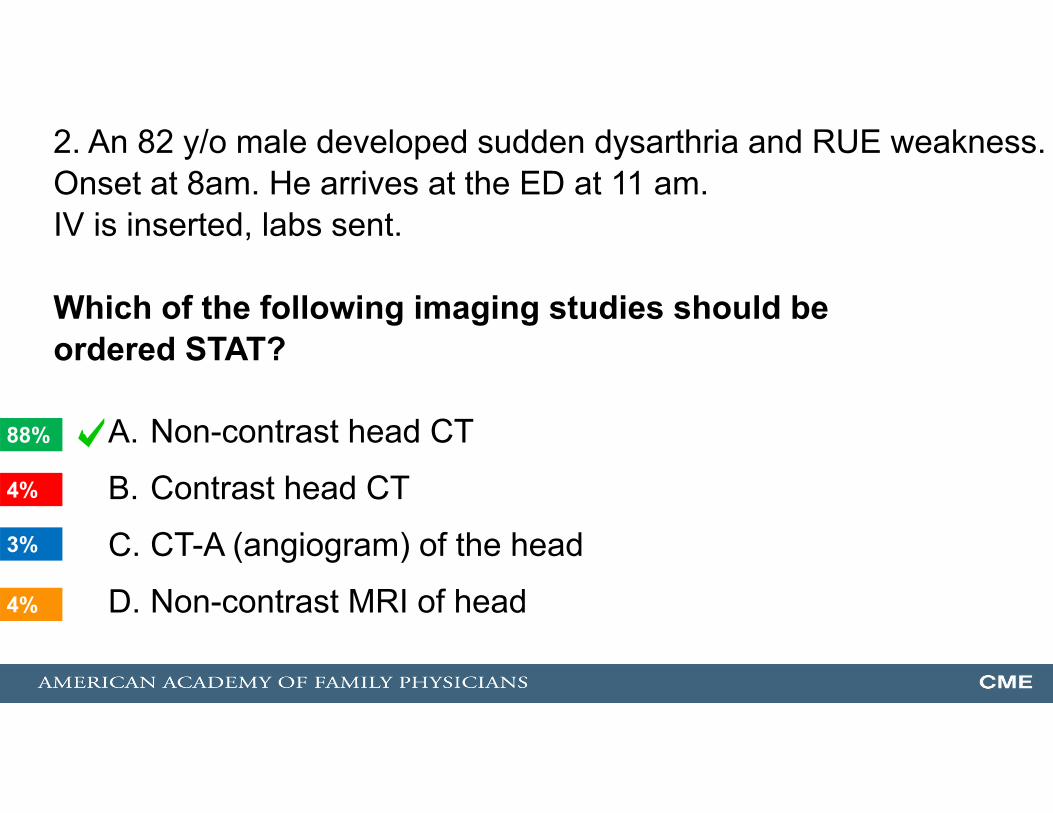
2. An 82 y/o male developed sudden dysarthria and RUE weakness.Onset at 8am. He arrives at the ED at 11 am. IV is inserted, labs sent.
Which of the following imaging studies should be ordered STAT?
A. Non-contrast head CT
B. Contrast head CT
C. CT-A (angiogram) of the head
D. Non-contrast MRI of head4%
88%
4%
3%
1
Stroke Type/Subtypes
Ischemic (80%-85%)Thrombotic (50%)
Embolic (30%)
Large-vessel(20%-25%)
Small-vessel(20%-25%)
Hemorrhagic (15%-20%)Intracerebral (10%)
Subarachnoid (6%)

Stroke Type/Subtypes
Ischemic (80%-85%)
Hemorrhagic (15%-20%)
<--------------------------------------------------------------------------------------------------------------------------------------->
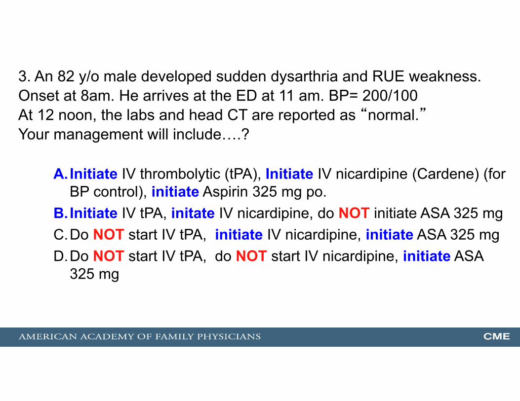
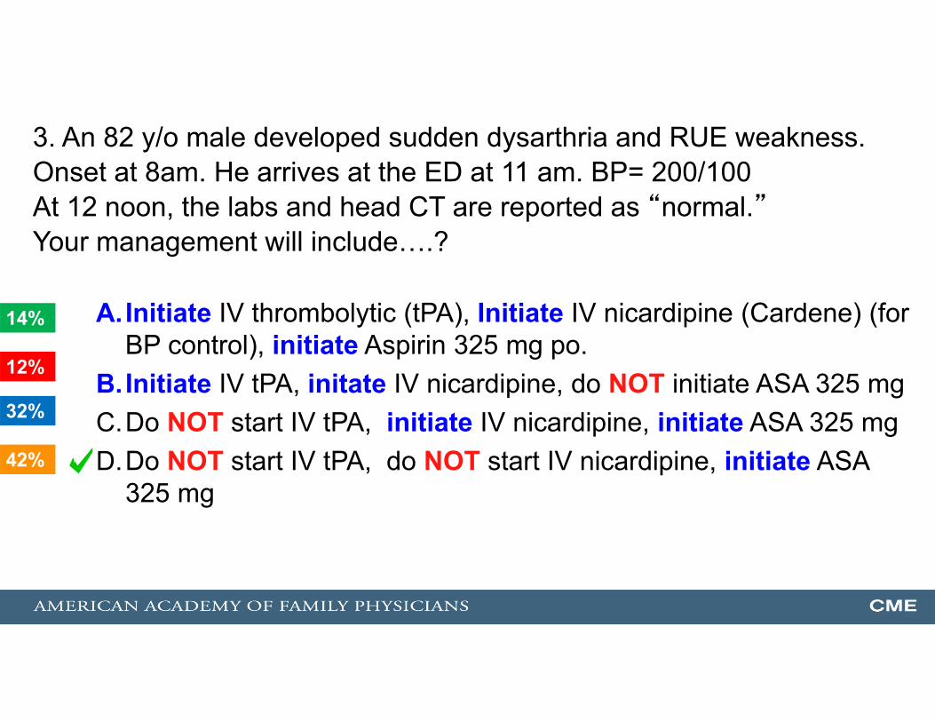
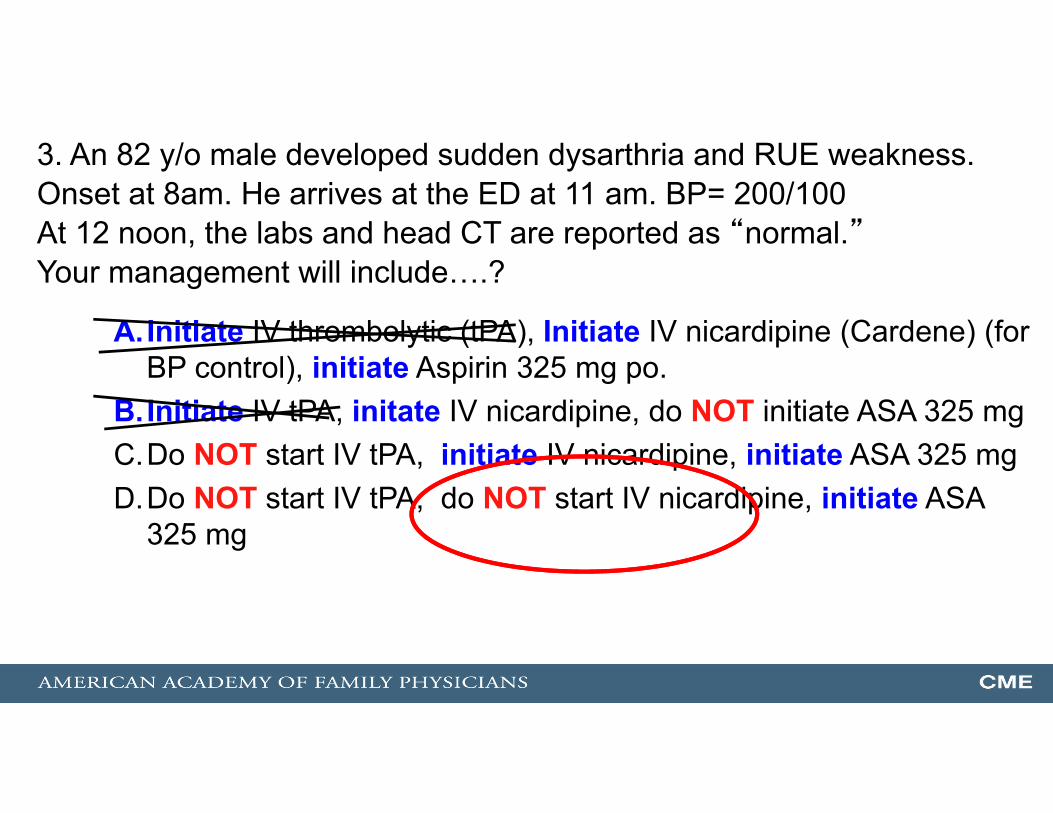
3. An 82 y/o male developed sudden dysarthria and RUE weakness.Onset at 8am. He arrives at the ED at 11 am. BP= 200/100At 12 noon, the labs and head CT are reported as “normal.”Your management will include….?
A.Initiate IV thrombolytic (tPA), Initiate IV nicardipine (Cardene) (for BP control), initiate Aspirin 325 mg po.
B.Initiate IV tPA, initate IV nicardipine, do NOT initiate ASA 325 mgC.Do NOT start IV tPA, initiate IV nicardipine, initiate ASA 325 mgD.Do NOT start IV tPA, do NOT start IV nicardipine, initiate ASA
325 mg
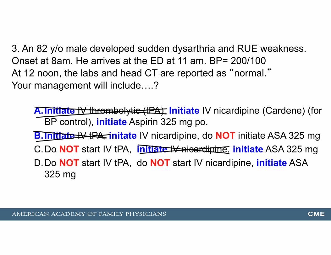
3. An 82 y/o male developed sudden dysarthria and RUE weakness.Onset at 8am. He arrives at the ED at 11 am. BP= 200/100At 12 noon, the labs and head CT are reported as “normal.”Your management will include….?
A.Initiate IV thrombolytic (tPA), Initiate IV nicardipine (Cardene) (for BP control), initiate Aspirin 325 mg po.
B.Initiate IV tPA, initate IV nicardipine, do NOT initiate ASA 325 mgC.Do NOT start IV tPA, initiate IV nicardipine, initiate ASA 325 mgD.Do NOT start IV tPA, do NOT start IV nicardipine, initiate ASA
325 mg42%
14%
12%
32% 1
3. An 82 y/o male developed sudden dysarthria and RUE weakness.Onset at 8am. He arrives at the ED at 11 am. BP= 200/100At 12 noon, the labs and head CT are reported as “normal.”Your management will include….?
A.Initiate IV thrombolytic (tPA), Initiate IV nicardipine (Cardene) (for BP control), initiate Aspirin 325 mg po.
B.Initiate IV tPA, initate IV nicardipine, do NOT initiate ASA 325 mgC.Do NOT start IV tPA, initiate IV nicardipine, initiate ASA 325 mgD.Do NOT start IV tPA, do NOT start IV nicardipine, initiate ASA
325 mg

What Is the Data Supporting tPA for Stroke?
• 12 randomized trials of thrombolytics vs placebo for acute ischemic CVA
• 2 were positive• 10 were negative or neutral
The NNT.com

The NINDS TrialLocation # of Pts Time of CVA Drug and Dose
United States 624 3 hours 1) tPA 0.9 mg/kg vs.2) Placebo
RESULTSA. Mortality: No difference!!!B. Part 1: At 24-hour neurologic assessment (291 pts)
No difference!!!C. Part 2: At 3-month neurologic assessment (333 pts)
(+) Significant difference***
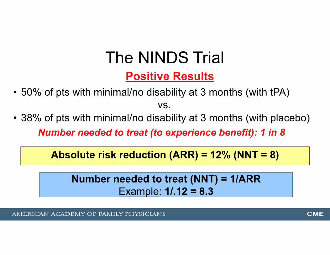
The NINDS TrialPositive Results
• 50% of pts with minimal/no disability at 3 months (with tPA)vs.
• 38% of pts with minimal/no disability at 3 months (with placebo)Number needed to treat (to experience benefit): 1 in 8
Absolute risk reduction (ARR) = 12% (NNT = 8)
Number needed to treat (NNT) = 1/ARRExample: 1/.12 = 8.3
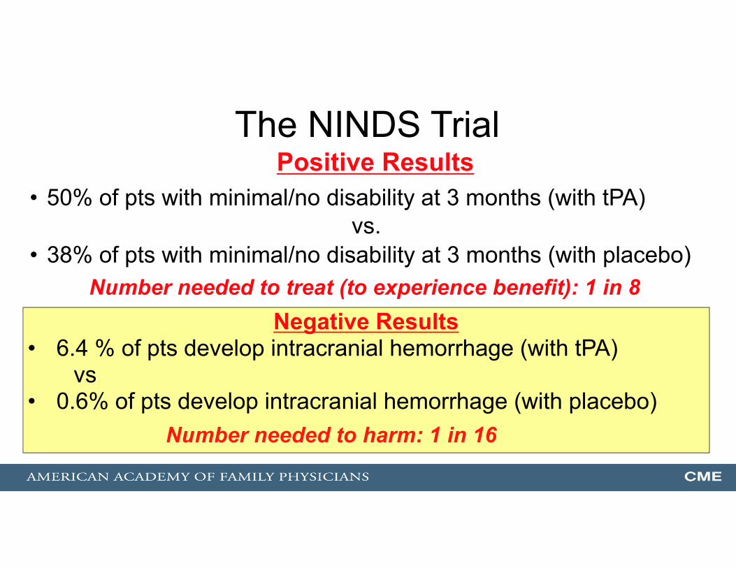
The NINDS TrialPositive Results
• 50% of pts with minimal/no disability at 3 months (with tPA)vs.
• 38% of pts with minimal/no disability at 3 months (with placebo)Number needed to treat (to experience benefit): 1 in 8
Negative Results• 6.4 % of pts develop intracranial hemorrhage (with tPA)
vs• 0.6% of pts develop intracranial hemorrhage (with placebo)
Number needed to harm: 1 in 16
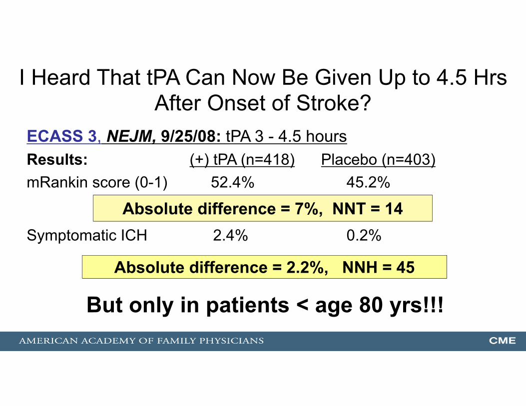
I Heard That tPA Can Now Be Given Up to 4.5 Hrs After Onset of Stroke?
ECASS 3, NEJM, 9/25/08: tPA 3 - 4.5 hoursResults: (+) tPA (n=418) Placebo (n=403)mRankin score (0-1) 52.4% 45.2%
Symptomatic ICH 2.4% 0.2%
Absolute difference = 7%, NNT = 14
Absolute difference = 2.2%, NNH = 45
But only in patients < age 80 yrs!!!
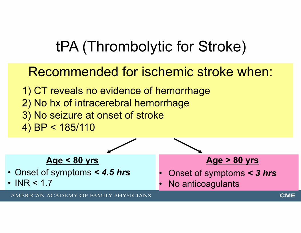
tPA (Thrombolytic for Stroke)
Age < 80 yrs• Onset of symptoms < 4.5 hrs• INR < 1.7
Recommended for ischemic stroke when:1) CT reveals no evidence of hemorrhage2) No hx of intracerebral hemorrhage3) No seizure at onset of stroke4) BP < 185/110
Age > 80 yrs• Onset of symptoms < 3 hrs• No anticoagulants
3. An 82 y/o male developed sudden dysarthria and RUE weakness.Onset at 8am. He arrives at the ED at 11 am. BP= 200/100At 12 noon, the labs and head CT are reported as “normal.”Your management will include….?
A.Initiate IV thrombolytic (tPA), Initiate IV nicardipine (Cardene) (for BP control), initiate Aspirin 325 mg po.
B.Initiate IV tPA, initate IV nicardipine, do NOT initiate ASA 325 mgC.Do NOT start IV tPA, initiate IV nicardipine, initiate ASA 325 mgD.Do NOT start IV tPA, do NOT start IV nicardipine, initiate ASA
325 mg

Blood Pressure Control: CAUTION in Acute (ischemic) CVA!!!
• Elevated BP is body’s desire to maintain cerebral perfusion
• AHA guidelines: Treat BP systolic > 220(2003, 2007) Treat BP diastolic >120
• Recommended meds:1. Labetalol: 10 mg q 10-20 min2. Nicardipine: 5 mg/hr, titrate q 5 min
AHA Stroke Guideline, 2007, 2013
Goal: 15% decrease in BP

Blood Pressure Control:A. For Acute (Hemorrhagic) CVA!!!
• If systolic BP 150-220 - lower BP sys to < 140 Class I, Level of Evidence A, AHA guideline, 5/2015
Blood Pressure Control:B. For Subarachnoid Hemorrhage!!!
• Decrease systolic BP < 160 Class IIa, Level of Evidence C, AHA guideline, 2012
------------------------------------------------------------------------------------------------------------------------------------------------------------------------------------------
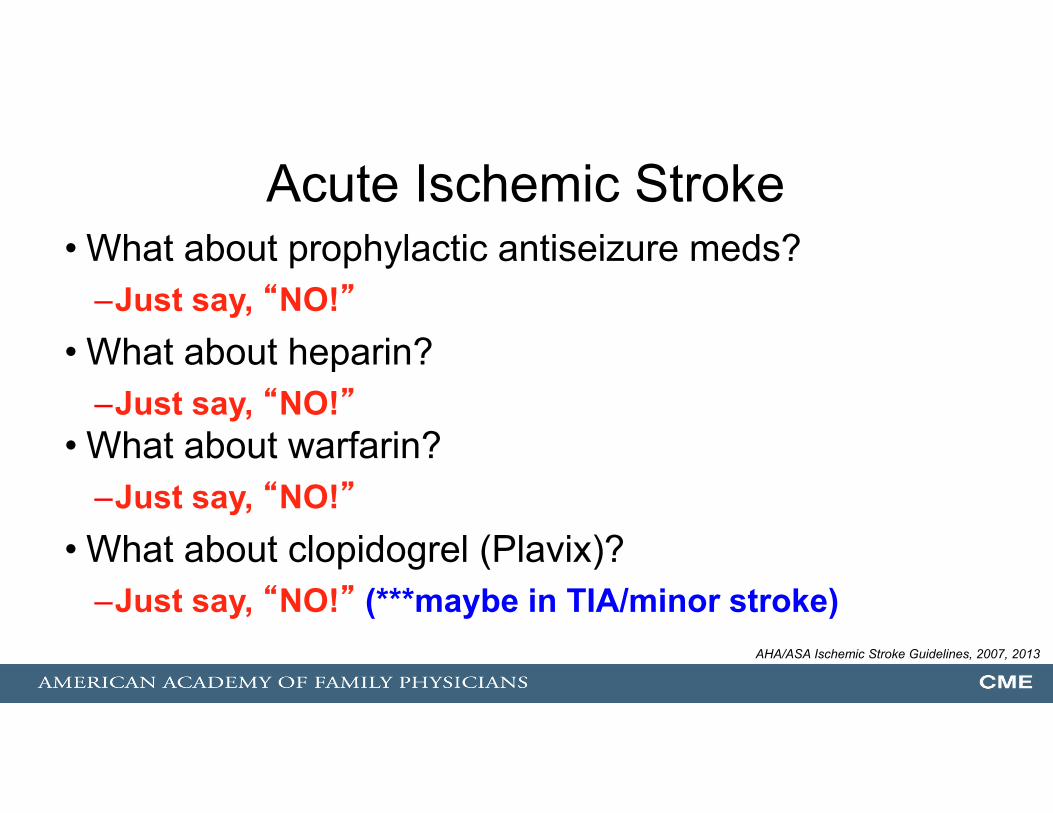
Acute Ischemic Stroke• What about prophylactic antiseizure meds?
–Just say, “NO!”• What about heparin?
–Just say, “NO!”• What about warfarin?
–Just say, “NO!”• What about clopidogrel (Plavix)?
–Just say, “NO!” (***maybe in TIA/minor stroke)AHA/ASA Ischemic Stroke Guidelines, 2007, 2013
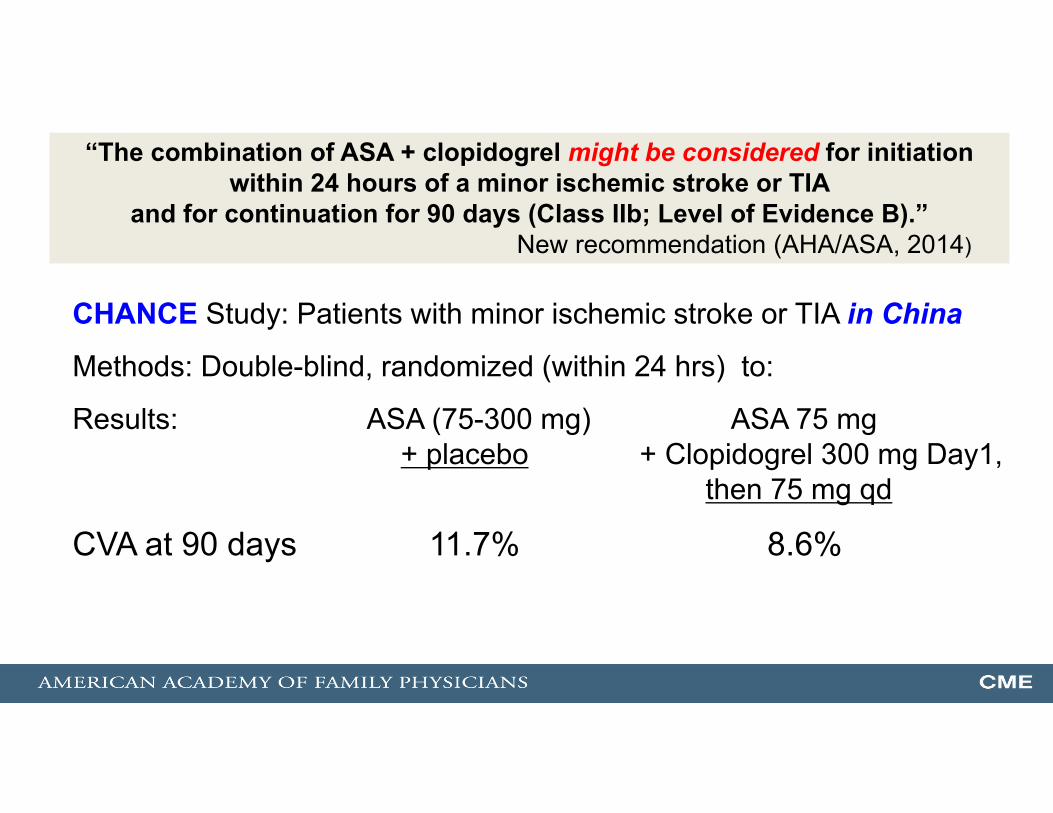
“The combination of ASA + clopidogrel might be considered for initiation within 24 hours of a minor ischemic stroke or TIA
and for continuation for 90 days (Class IIb; Level of Evidence B).”New recommendation (AHA/ASA, 2014)
CHANCE Study: Patients with minor ischemic stroke or TIA in China
Methods: Double-blind, randomized (within 24 hrs) to:
Results: ASA (75-300 mg) ASA 75 mg + placebo + Clopidogrel 300 mg Day1,
then 75 mg qd
CVA at 90 days 11.7% 8.6%

Acute Ischemic Stroke• What about Aspirin?
–Just say, “YES!”
AHA/ASA Ischemic Stroke Guidelines, 2007, 2013
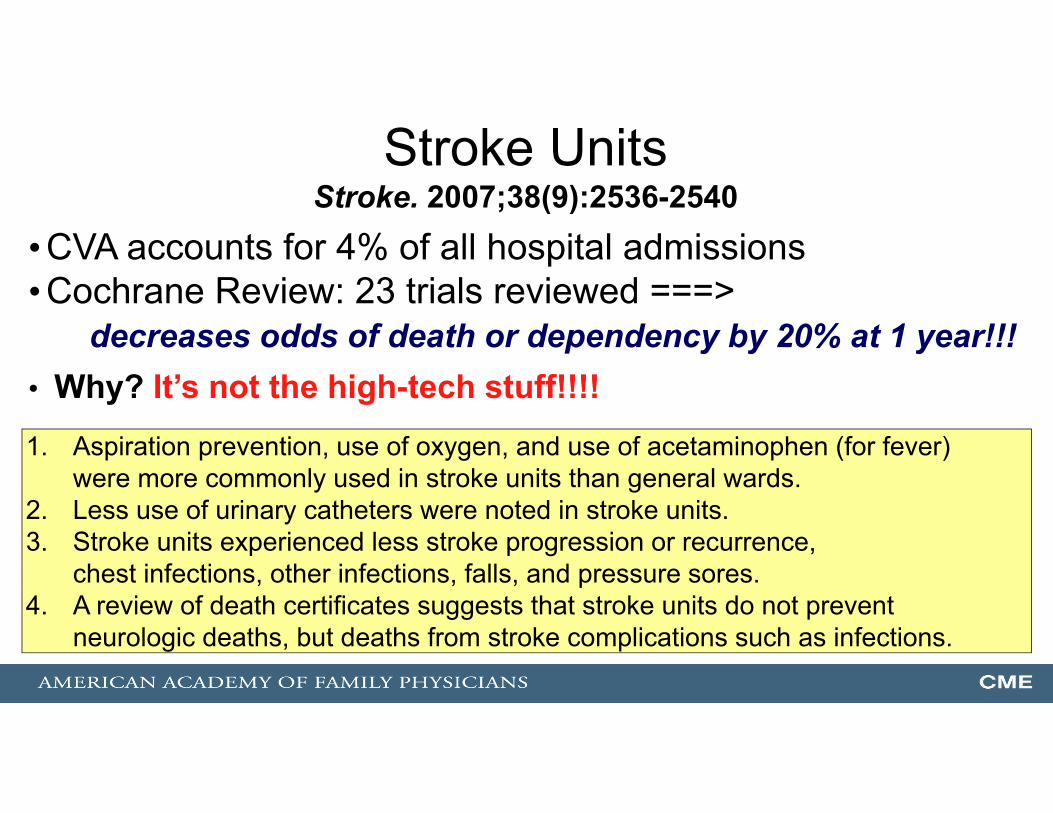
Stroke Units Stroke. 2007;38(9):2536-2540
•CVA accounts for 4% of all hospital admissions•Cochrane Review: 23 trials reviewed ===>
decreases odds of death or dependency by 20% at 1 year!!!• Why? It’s not the high-tech stuff!!!!1. Aspiration prevention, use of oxygen, and use of acetaminophen (for fever)
were more commonly used in stroke units than general wards. 2. Less use of urinary catheters were noted in stroke units.3. Stroke units experienced less stroke progression or recurrence,
chest infections, other infections, falls, and pressure sores.4. A review of death certificates suggests that stroke units do not prevent
neurologic deaths, but deaths from stroke complications such as infections.

The post-CVA patient now returns to your care (office/rehab). Note the following recommendations for secondary
prevention of ischemic stroke or TIA:
• Hypertension: start or resume BP meds “beyond the first several days”Class I, Level of evidence A
- Goal: < 140/90 (Class IIA), ? < 130 systolic for lacunar CVA (Class IIb)
• Lipids: (+) statins - for LDL-C > 100 mg/dL Class I, Level of evidence B - for LDL-C < 100 mg/dL Class I, Level of evidence C
• Glucose/DM: Screen for DM Class IIa, Level of evidence C
AHA/ASA guideline 2014

The post-CVA patient now returns to your care (office/rehab). Note the following recommendations for secondary
prevention of ischemic stroke or TIA:
• Obesity: get the BMI Class I, Level of evidence C• Physical Activity: increase it. Class IIA, Level of evidence C • Nutrition: assess it. Class IIA, Level of evidence C • Sleep study: consider it. Class IIB, Level of evidence B • Homocysteine, Antiphospholipid antibody: – do not routinely test!• Hypercoagulation: ????? – “unknown”
• Carotid artery evaluation: See later in TIA sectionAHA/ASA guideline 2014

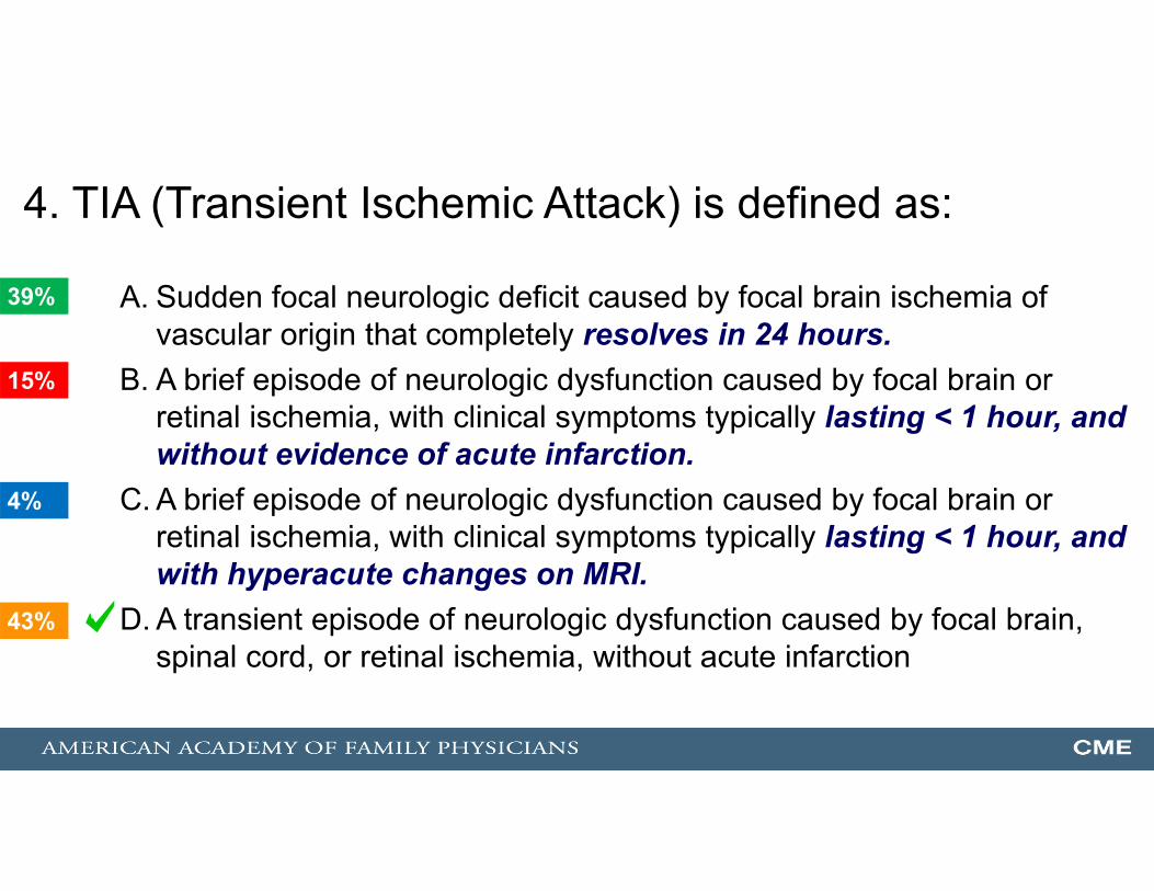
4. TIA (Transient Ischemic Attack) is defined as:
A. Sudden focal neurologic deficit caused by focal brain ischemia of vascular origin that completely resolves in 24 hours.
B. A brief episode of neurologic dysfunction caused by focal brain or retinal ischemia, with clinical symptoms typically lasting < 1 hour, and without evidence of acute infarction.
C. A brief episode of neurologic dysfunction caused by focal brain or retinal ischemia, with clinical symptoms typically lasting < 1 hour, and with hyperacute changes on MRI.
D. A transient episode of neurologic dysfunction caused by focal brain, spinal cord, or retinal ischemia, without acute infarction
43%
39%
15%
4%
1

TIA: The Definition Has Changed!!!!• Classic definition: sudden focal neurologic deficit caused by focal brain ischemia of vascular origin that completely resolves in 24 hours
• 2002 TIA Working Group“A brief episode of neurologic dysfunction caused by focal brain or retinal ischemia, with clinical symptoms typically lasting less than 1 hour, and without evidence of acute infarction”
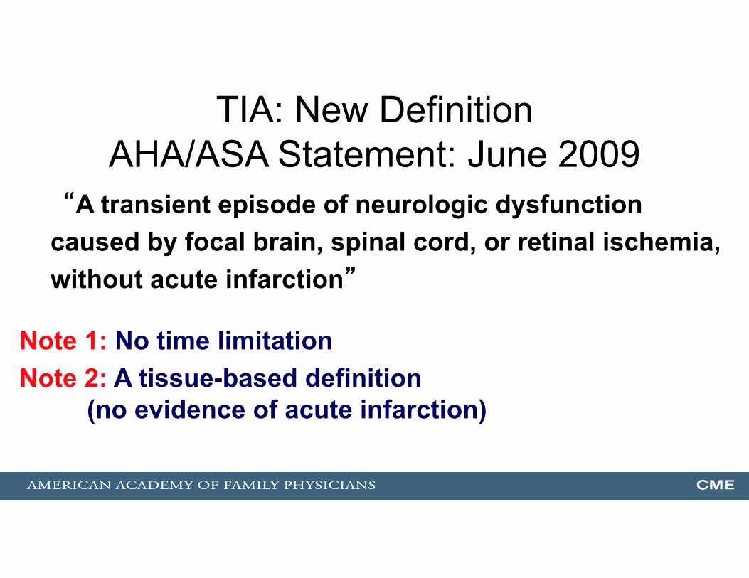
TIA: New DefinitionAHA/ASA Statement: June 2009
“A transient episode of neurologic dysfunction caused by focal brain, spinal cord, or retinal ischemia, without acute infarction”
Note 1: No time limitationNote 2: A tissue-based definition
(no evidence of acute infarction)
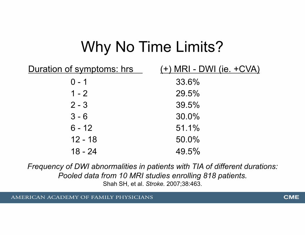
Why No Time Limits?
0 - 1 33.6%1 - 2 29.5%2 - 3 39.5%3 - 6 30.0%6 - 12 51.1%12 - 18 50.0%18 - 24 49.5%
Duration of symptoms: hrs (+) MRI - DWI (ie. +CVA)
Frequency of DWI abnormalities in patients with TIA of different durations: Pooled data from 10 MRI studies enrolling 818 patients.
Shah SH, et al. Stroke. 2007;38:463.
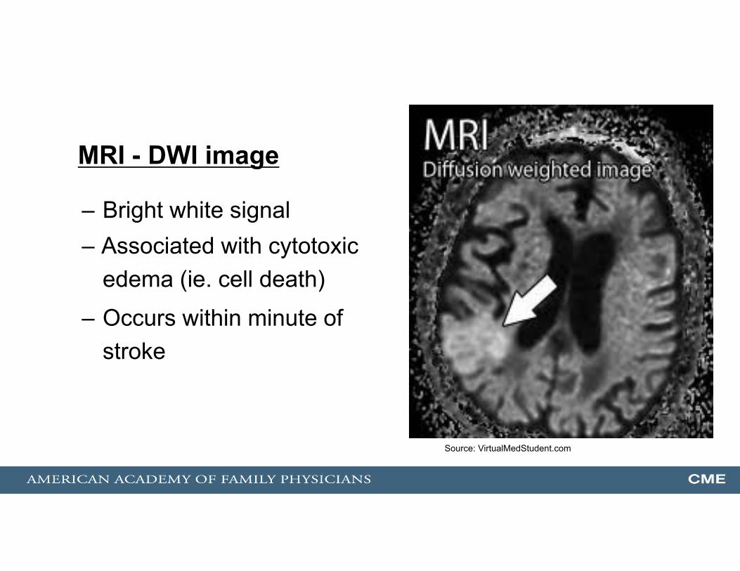
MRI - DWI image
– Bright white signal– Associated with cytotoxic
edema (ie. cell death)
– Occurs within minute of stroke
Source: VirtualMedStudent.com
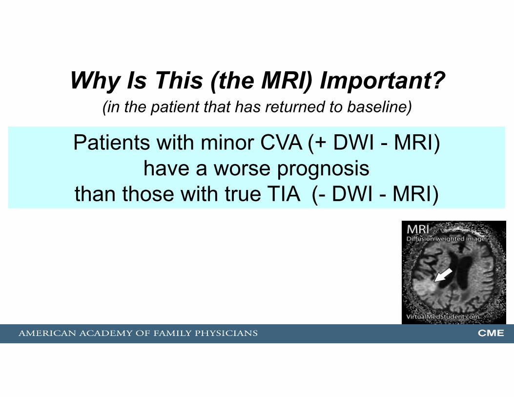
Why Is This (the MRI) Important?(in the patient that has returned to baseline)
Patients with minor CVA (+ DWI - MRI)have a worse prognosis
than those with true TIA (- DWI - MRI)

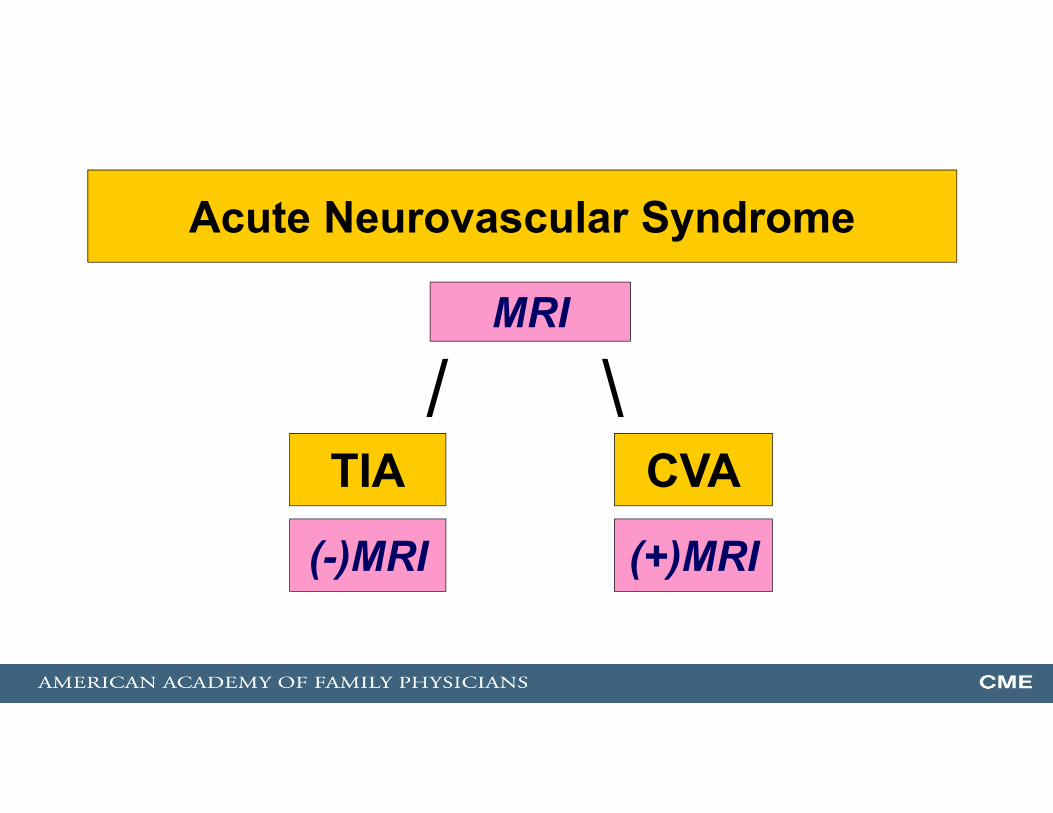
TIA CVA/ \
MRI
(+)MRI(-)MRI
Transient Neurologic Changesand has returned to baseline
TIA CVA/ \
MRI
(+)MRI(-)MRI
Acute Neurovascular Syndrome
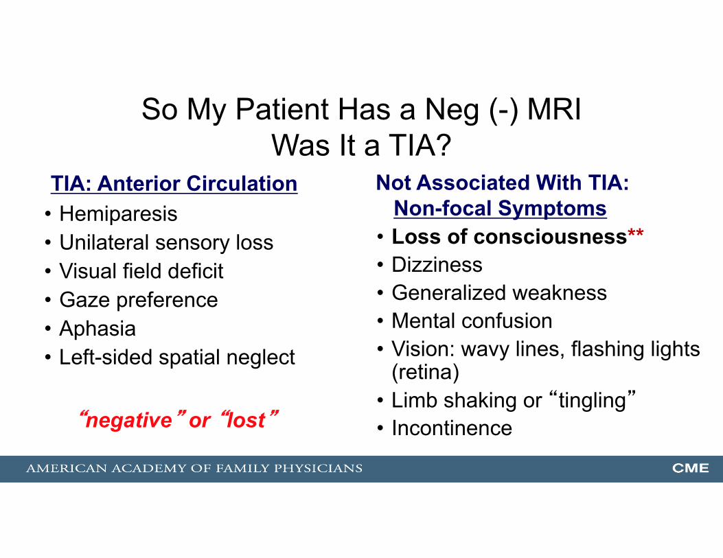
So My Patient Has a Neg (-) MRIWas It a TIA?
• Hemiparesis• Unilateral sensory loss• Visual field deficit• Gaze preference• Aphasia• Left-sided spatial neglect
• Loss of consciousness**• Dizziness• Generalized weakness• Mental confusion• Vision: wavy lines, flashing lights
(retina) • Limb shaking or “tingling”• Incontinence
TIA: Anterior Circulation Not Associated With TIA:Non-focal Symptoms
“negative” or “lost”

TIA/Stroke Mimics(The Differential Diagnosis of TIA/CVA)
• Structural brain lesion (tumor, hemorrhage, AVM, aneurysm)• Infection (focal abscess, septic emboli)• Seizure/Todd’s paralysis• Complicated migraine• Hypoglycemia• Syncope from any cause (especially arrhythmia)• Labyrinthine disorders• Temporal arteritis• Multiple sclerosis (flare)
TIA Management: Risk Assessment
Who is going on to acute CVA? Who has such high-risk that they need hospitalization?
The AHA/ASA recommends the ABCD2 score to calculate a patient’s short-term risk
of developing a CVA

ABCD2 Score• Age: greater than or equal to 60 (1 pt)• Blood pressure: SBP > 140 or DBP > 90 (1 pt)• Clinical Features:
– Focal weakness (2 pt) or – Speech impairment without focal weakness (1 pt)
• Duration of symptoms:– > 60 minutes (2 pt) or– < 59 minutes (1 pt)
• Diabetes (1 pt)
Risk of CVA at 2 days0-3 points = 1% risk 4-5 points = 4.1% risk 6-7 points = 8.1% risk
Johnston SC, et al. Lancet. 2007;369:283-292.
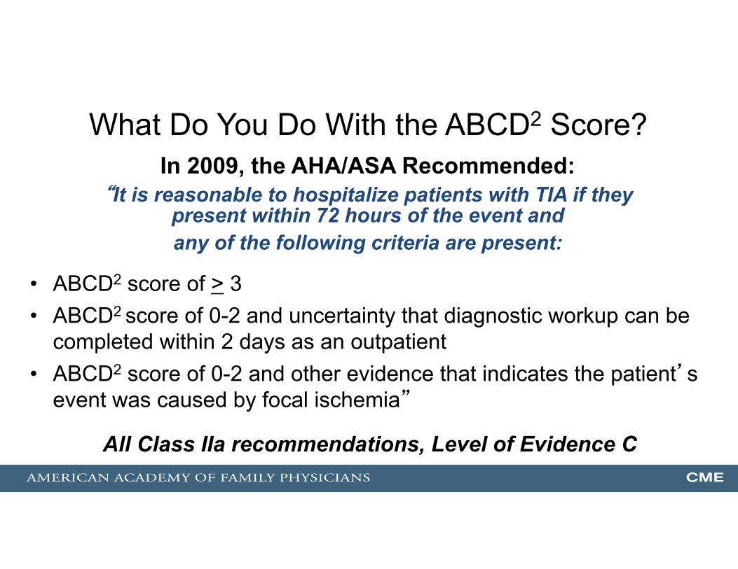
What Do You Do With the ABCD2 Score?
• ABCD2 score of > 3 • ABCD2 score of 0-2 and uncertainty that diagnostic workup can be
completed within 2 days as an outpatient• ABCD2 score of 0-2 and other evidence that indicates the patient’s
event was caused by focal ischemia”
All Class IIa recommendations, Level of Evidence C
In 2009, the AHA/ASA Recommended:“It is reasonable to hospitalize patients with TIA if they
present within 72 hours of the event andany of the following criteria are present:

AHA/ASA 2011 Guideline on Carotid and Vertebral Disease Released 1/31/11
• “Duplex US is recommended to detect carotid stenosis in pts. who develop focal neurological symptoms corresponding to the territory supplied by the L or R internal carotid artery”
• “…MRA or CTA is indicated to detect carotid stenosis when US either cannot be obtained or yields equivocal or …nondiagnostic results”
Class I recommendation, Level of Evidence C
The TIA Diagnostic Evaluation:Step 1: MRI – done… nextStep 2: Duplex US of carotids
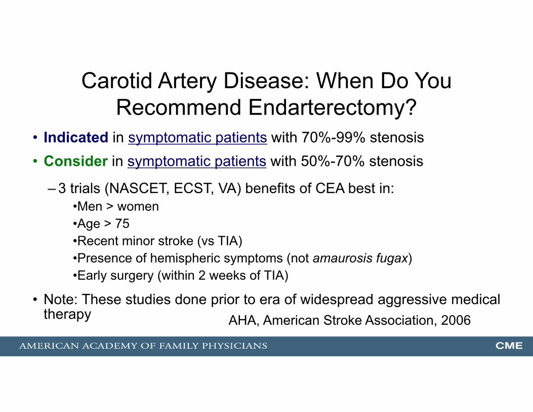
Carotid Artery Disease: When Do You Recommend Endarterectomy?
• Indicated in symptomatic patients with 70%-99% stenosis• Consider in symptomatic patients with 50%-70% stenosis
– 3 trials (NASCET, ECST, VA) benefits of CEA best in:•Men > women•Age > 75•Recent minor stroke (vs TIA) •Presence of hemispheric symptoms (not amaurosis fugax) •Early surgery (within 2 weeks of TIA)
• Note: These studies done prior to era of widespread aggressive medical therapy AHA, American Stroke Association, 2006

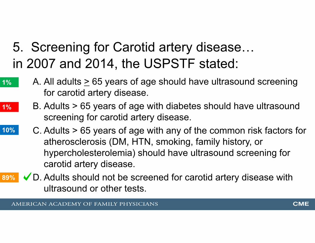
5. Screening for Carotid artery disease…in 2007 and 2014, the USPSTF stated:
A. All adults > 65 years of age should have ultrasound screening for carotid artery disease.
B. Adults > 65 years of age with diabetes should have ultrasound screening for carotid artery disease.
C. Adults > 65 years of age with any of the common risk factors for atherosclerosis (DM, HTN, smoking, family history, or hypercholesterolemia) should have ultrasound screening for carotid artery disease.
D. Adults should not be screened for carotid artery disease with ultrasound or other tests.
89%
1%
1%
10%
1

Carotid Artery Screening …(Asymptomatic Population): D recommendation
• The bottom line:Prevalence of disease in age > 65 = 1%
• 4,348 persons would need screening to prevent 1 CVA in 5 years.
• 8,696 persons would need screening to prevent 1 disabling CVA in 5 years.
Answers1. A2. A3. D4. D5. D
Thank you!

Physical Activity: Just Do It!• Methods: Women’s Health Study
- 39,315 women, reported physical activity at baseline, followed 11.9 yrs
• Results: compared to sedentary (nonwalkers) - Walk > 2 hrs/week ==> lowered CVA risk 30%-… and speed (ie, vigorous) did not matter!!
Sattelmair, JR, Kurth T, Buring JE, et al. Physical activity and risk of stroke in women.Stroke. 2010 [Epub ahead of print].
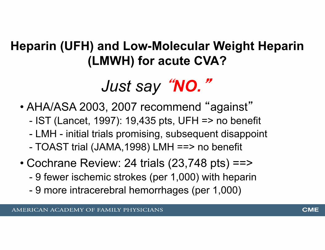
Heparin (UFH) and Low-Molecular Weight Heparin (LMWH) for acute CVA?
• AHA/ASA 2003, 2007 recommend “against”- IST (Lancet, 1997): 19,435 pts, UFH => no benefit- LMH - initial trials promising, subsequent disappoint- TOAST trial (JAMA,1998) LMH ==> no benefit
• Cochrane Review: 24 trials (23,748 pts) ==>- 9 fewer ischemic strokes (per 1,000) with heparin- 9 more intracerebral hemorrhages (per 1,000)
Just say “NO.”

What About Aspirin and Ischemic Stroke? Just Say “YES”
• Cochrane Review, Issue 3, 2008- 12 trials (43,041 pts) - 13 pts; alive and independent (per 1,000) with ASA- 10 more pts: made complete recovery (per 1,000) - 2 more pts: intracerebral hemorrhage (per 1,000)
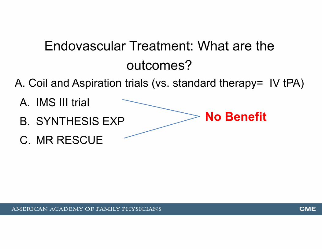
Endovascular Treatment: What are the outcomes?
A. Coil and Aspiration trials (vs. standard therapy= IV tPA)
A. IMS III trial
B. SYNTHESIS EXP
C. MR RESCUE
No Benefit
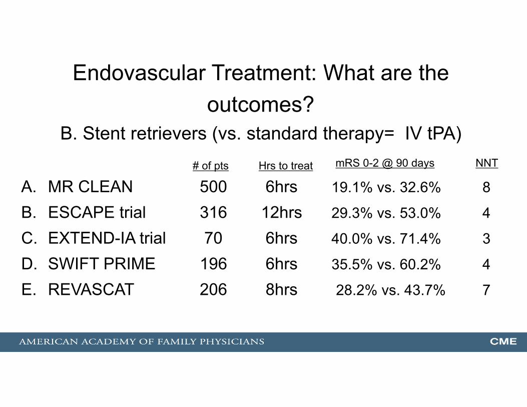
Endovascular Treatment: What are the outcomes?
B. Stent retrievers (vs. standard therapy= IV tPA)
A. MR CLEAN 500 6hrs 19.1% vs. 32.6% 8
B. ESCAPE trial 316 12hrs 29.3% vs. 53.0% 4
C. EXTEND-IA trial 70 6hrs 40.0% vs. 71.4% 3
D. SWIFT PRIME 196 6hrs 35.5% vs. 60.2% 4
E. REVASCAT 206 8hrs 28.2% vs. 43.7% 7
# of pts mRS 0-2 @ 90 daysHrs to treat NNT

AHA/ASA Guideline 6/30/151. Patients eligible for IV r-tPA should receive IV r-tPA even if endovascular
treatments are being considered (Class I; Level of Evidence A). 2. Patients should receive endovascular therapy with a stent retriever if they meet
all the following criteria (Class I; Level of Evidence A). (a) prestroke mRS score 0 to 1,(b) acute ischemic stroke receiving IV r-tPA within 4.5 hours of onset per
guidelines(c) causative occlusion of the internal carotid artery or proximal MCA (M1),(d) age ≥18 years,(e) NIHSS score of ≥6,(f) ASPECTS of ≥6, and(g) treatment can be initiated (groin puncture) within 6 hours of symptom onset
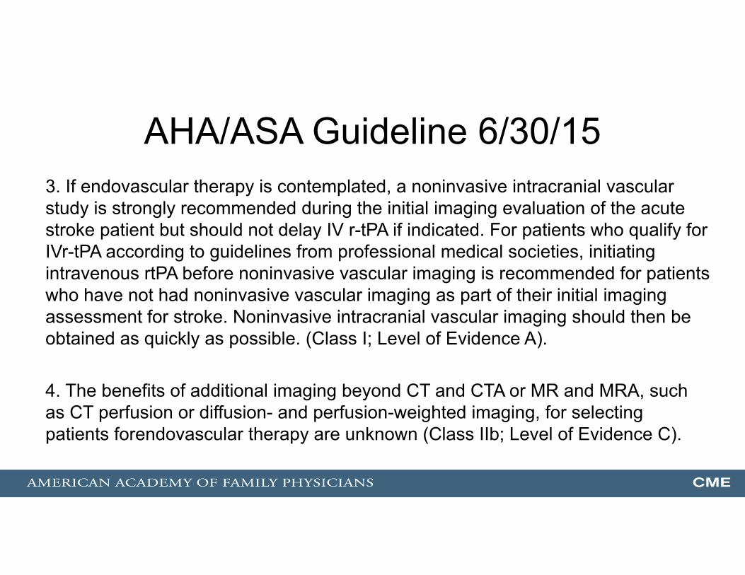
AHA/ASA Guideline 6/30/153. If endovascular therapy is contemplated, a noninvasive intracranial vascular study is strongly recommended during the initial imaging evaluation of the acute stroke patient but should not delay IV r-tPA if indicated. For patients who qualify for IVr-tPA according to guidelines from professional medical societies, initiating intravenous rtPA before noninvasive vascular imaging is recommended for patients who have not had noninvasive vascular imaging as part of their initial imaging assessment for stroke. Noninvasive intracranial vascular imaging should then be obtained as quickly as possible. (Class I; Level of Evidence A).
4. The benefits of additional imaging beyond CT and CTA or MR and MRA, such as CT perfusion or diffusion- and perfusion-weighted imaging, for selecting patients forendovascular therapy are unknown (Class IIb; Level of Evidence C).
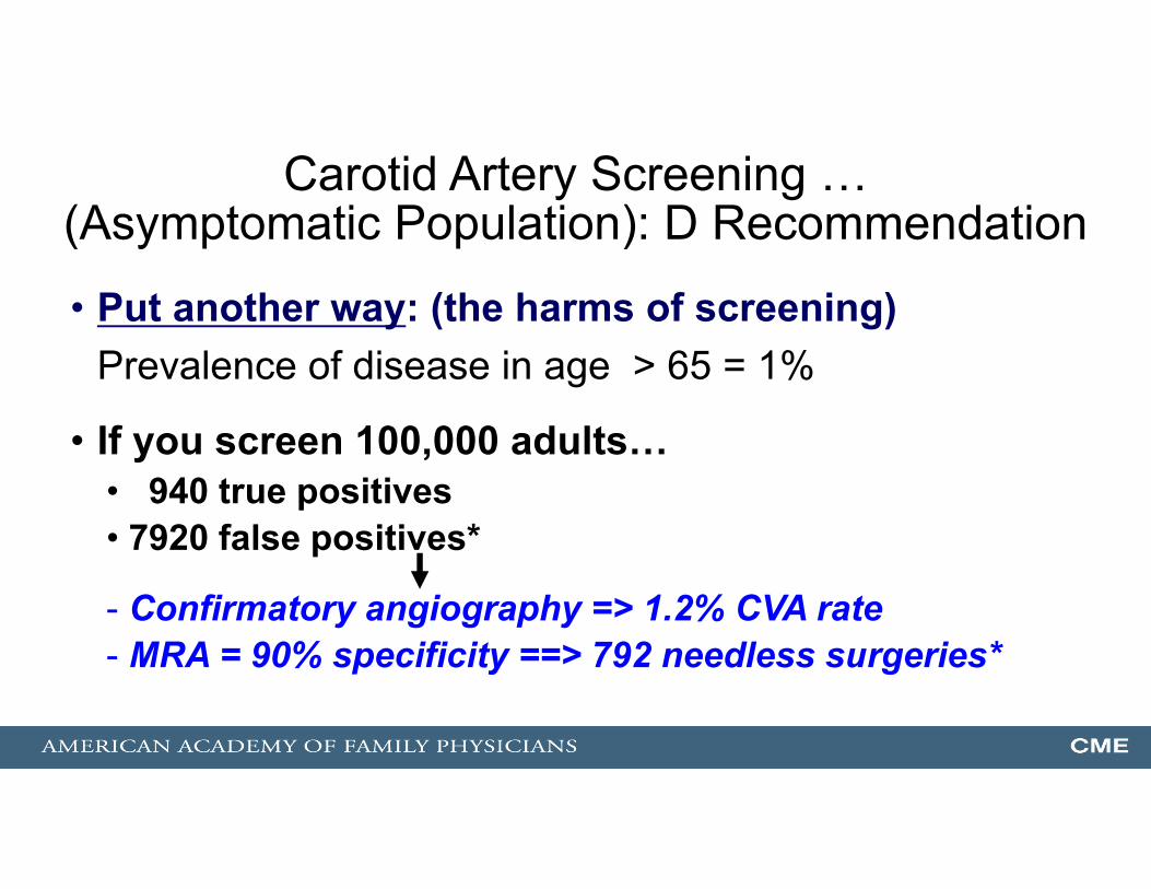
Carotid Artery Screening …(Asymptomatic Population): D Recommendation
• Put another way: (the harms of screening)Prevalence of disease in age > 65 = 1%
• If you screen 100,000 adults…• 940 true positives• 7920 false positives*
- Confirmatory angiography => 1.2% CVA rate- MRA = 90% specificity ==> 792 needless surgeries*