acral pseudolymphomatous angiokeratoma – a case report ... · se das lesões com resolução...
TRANSCRIPT

An Bras Dermatol. 2006;81(5 Supl 3):S277-80.
Received on May 12, 2003.Approved by the Consultive Council and accepted for publication on August 07, 2006.* Work done at Department Dermatology of Santa Casa de Misericórdia de Vitória (ES), Brazil.Conflict of interests: None
1 Dermatologist.2 Head of the Department of Dermatology at Santa Casa de Misericórdia de Vitória - Vitória (ES), Brazil.3 Professor of Pathology at the Escola de Medicina da Santa Casa de Misericórdia de Vitória - Vitória (ES), Brazil.4 Assistant Professor of Dermatology at the Serviço de Dermatologia da Santa Casa de Misericórdia de Vitória - Vitória (ES), Brazil.
©2006 by Anais Brasileiros de Dermatologia
Acral pseudolymphomatous angiokeratoma – a case report*
Angioqueratoma pseudolinfomatoso acral – relato de caso*
Fernanda Maria Zucoloto Freire1 João Basilio de Souza Filho2
Luiz Calice Cintra3 Lucia Martins Diniz4
Abstract: A 29-year-old female patient with asymptomatic red-brown papules that had arisenfour years before in her right forearm. Two lesions were removed for histopathological andimmunohistochemical exams. Intralesional corticosteroid injection resulted in no improve-ment and the lesions were subsequently surgically excised with complete resolution of thecondition. Keywords: Immunohistochemistry; Pseudolymphoma; Skin
Resumo: Paciente de 29 anos, do sexo feminino, com aparecimento há quatro anos depápulas assintomáticas, eritêmato-acastanhadas, no antebraço direito. Duas das lesõesforam biopsiadas para exames histopatológico e imuno-histoquímico. A paciente foi inicial-mente tratada com corticoterapia intralesional, sem melhora. Procedeu-se, então, à exére-se das lesões com resolução completa do quadro.Palavras-chave: Imuno-histoquímica; Pele; Pseudolinfoma
Case Report
S277
INTRODUCTION Acral pseudolymphomatous angiokeratoma is
a benign subcutaneous tissue condition that, in spiteof histopathological similarities with lymphomatousprocesses, must be included in the group of pseu-dolymphomas of the skin. It was described by Ramsayin 1988, after the observation of five children (onemale and four females) who presented multiple vio-let erythematic papules, located unilaterally, in thefoot in four cases, and in the hand in one case, the-reby the denomination acral. It is known by itsacronym Apache – Acral PseudolymphomatousAngiokeratoma of Children.1-3
Apache etiology remains unknown; however,some authors believe in the possibility that it is ahypersensitivity reaction to insect bites, in virtue ofthe histopathological picture and acral location of thelesions.3-5
It is clinically characterized by the presence of10 to 40 violet-erythematic papules, whose diameter
ranges from 1 to 4 mm, and which are asymptomatic,located unilaterally, generally with acral distributionand occurring more often between 2 and 13 years ofage. It should be distinguished upon histopathologi-cal examination from the following clinically similardiseases: Mibelli’s angiokeratoma, hemangioma,melanocytic nevus, basocellular carcinoma,lymphocytoma cutis and cutaneous lymphocytoma.1,2
Histological studies of Apache reveal an epider-mis of normal aspect and a dense well-differentiatedlymphocyte infiltrate in the dermis, amongst connec-tive tissue structures, not affecting skin annexes. Suchhistopathological aspect configures lymphocyte proli-feration, albeit with no nuclear atypies, which requi-res an immunohistochemical study in order to diffe-rentiate from lymphomas.1-3
Recommended therapies for Apache includetotal exeresis of skin lesions, Intralesional steroids orradiation therapy.2,3
An Bras Dermatol. 2006;81(5 Supl 3):S277-80.
S278 Freire FMZ, Filho JBS, Cintra LC, Diniz LM.
CASE REPORTTwenty-nine-year-old brow-skinned female
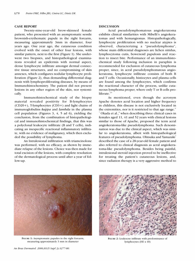
patient, who presented with an asymptomatic sessilebrownish-erythematic papule in the right forearm,measuring approximately 3mm in diameter, fouryears ago. One year ago, the cutaneous conditionevolved with the onset of other four lesions, withsimilar pattern, next to the first (Figure 1). She under-went two biopsies, and histopathological examina-tions revealed an epidermis with normal aspect,dense lymphocyte infiltrate amongst dermal connec-tive tissue structures, and no compromising of skinannexes, which configures nodular lymphocyte proli-feration (Figure 2), thus demanding differential diag-nosis with lymphoproliferating diseases, by means ofImmunohistochemistry. The patient did not presentlesions in any other region of the skin, nor systemi-cally.
Immunohistochemical study of the biopsymaterial revealed positivity for B-lymphocytes(CD20+), T-lymphocytes (CD3+) and light chains ofimmunoglobulins kappa and lambda in the plasmacell population (Figures 3, 4, 5 ad 6), yielding theconclusion, from the combination of histopathologi-cal and immunohistochemical findings, that this wasa polyclonal leukocyte infiltrate (B and T cells), indi-cating an inespecific reactional inflammatory infiltra-te, with no evidence of malignancy, which then exclu-ded the possibility of lymphoma.
An Intralesional infiltration with triamcinolonewas performed, with no efficacy, as shown by imme-diate relapse of the lesions. Choice was then made fortotal excision of the lesions, with complete resolutionof the dermatological process until after a year of fol-low-up.
DISCUSSIONAcral pseudolymphomatous angiokeratoma
exhibits clinical similarities with Mibelli’s angiokera-tomas and with hemangiomas. Histopathologically,lymphocyte proliferation with no nuclear atypies isobserved, characterizing a “pseudolymphoma”,whose main differential diagnoses are lichen nitidus,lymphocytoma cutis, bowenoid papulosis and reac-tion to insect bite. Performance of an immunohisto-chemical study following inclusion in paraphin isrecommended for exclusion of cutaneous lymphomaproper, since, in acral pseudolymphomatous angio-keratoma, lymphocyte infiltrate consists of both Band T cells. Occasionally, histiocytes and plasma cellsare found among the lymphocytes, which confirmsthe reactional character of the process, unlike cuta-neous lymphoma proper, where only T or B cells pro-liferate.1-5
As mentioned, even though the acronymApache denotes acral location and higher frequencyin children, this disease is not exclusively located inthe extremities, nor is it restricted to that age range.1-
7 Okada et al.,6 when describing three clinical cases infemales aged 11, 41 and 52 years with clinical lesionssimilar to those of Apache, proposed the term acralangiokeratoma-like pseudolymphoma. Such denomi-nation was due to the clinical aspect, which was simi-lar to angiokeratoma, albeit with histopathologicalfeatures of pseudolymphoma. Ohtsuka and Yamazaki7
described the case of a 28-year-old female patient andalso referred to clinical diagnosis as acral angiokera-toma-like pseudolymphoma. Besides being painful,intralesional steroid injection proved to be ineffectivefor treating the patient’s cutaneous lesions, and,since radiation therapy is a very aggressive method to
FIGURE 1: Asymptomatic papules in the right forearm,measuring approximately 3 mm in diameter
FIGURE 2: Leukocyte infiltrate with predominance oflymphocytes (HE x 40)

An Bras Dermatol. 2006;81(5 Supl 3):S277-80.
be used upon such a benign condition, the authorschose to carry out a total excision of the lesion, whichwas eventually the best therapeutic option, as thepatient is still cured after one year of follow-up.3
In brief, acral pseudolymphomatous angioke-ratoma is a rare, benign, cutaneous pseudolympho-ma, characterized by the presence of unilateral violet-
erythematic papules, usually acral and often betweenthe ages of 2 and 13 years, which requires both histo-pathological and immunohistochemical evaluations,in order to make differential diagnosis with cuta-neous lymphomas. Although uncommon, there is apossibility of occurrence in adults and outside extre-mities, as observed in the case reported here.1-5 �
FIGURE 4: Immunohistochemistry: CD3 – complex associatedwith T-positive lymphocyte receptors, in dark brown (HE x 40)
FIGURE 5: Immunohistochemistry: light chain of kappa-positiveimmunoglobulin, in brown (HE x 40)
FIGURE 6: Immunohistochemistry: light chain of lambda-positiveimmunoglobulin, in brown (HE x 40)
FIGURE 3: Immunohistochemistry: CD20 – positive,in dark brown, in B-lymphocytes (HE x 40)
Acral pseudolymphomatous angiokeratoma – a case report S279

girl. J Eur Acad Dermatol Venereol. 2001;15:160-1.6. Okada M, Funayama M, Tanita M. Acral angiokeratoma-
like pseudolymphoma: one adolescent and two adults. J Am Acad Dermatol. 2001;45(Suppl):S209-11.
7. Ohtsuka T, Yamazaki S. Acral angiokeratoma-like pseudolymphoma in a 28-year-old Japanese woman. Dermatology. 2003;207:77-8.
REFERENCES1. Kaddu S, Cerroni L, Pilatti A. Acral pseudolymphomanus
angiokeratoma. A Variant of the Cutaneous Pseudolymphomas. Am J Dermatopathol. 1994;16:130-3.
2. Ramsay B, Dahl MCG, Malcolm AJ. Acral pseudolym-phomatous angiokeratoma of children. Arch Dermatol. 1990;126:1524-5.
3. Hara M, Matsunaga J, Tagami H. Acral pseudolymphoma-tous angiokeratoma of children (APACHE): a case report and immunohistological study. Br J Dermatol. 1991; 124:387-8.
4. Fillus Neto J. Angioqueratoma pseudolinfomatoso acral (APACHE) em adolescente de 16 anos. A pele. [Informativo da Fundação Nacional do Câncer da Pele]. 2002;16:8-9.
5. Barbosa JM, Fillus Neto J, Werner B. Acral pseudolym-phomatous angiokeratoma occuring in a 16-year-old
MAILING ADDRESS:Fernanda Maria Zucoloto Freire Av. Antônio Borges, 225 - Bairro Mata da Praia 29065–250 - Vitória - ES - BrazilTels: +55 (27) 3235-9607 / +55 (27) 9836-9205E-mail: [email protected]
An Bras Dermatol. 2006;81(5 Supl 3):S277-80.
S280 Freire FMZ, Filho JBS, Cintra LC, Diniz LM.