achilles tendon rupture; assessment of non- … medical journal 3 that the mid portion of the...
TRANSCRIPT

PHD THESIS DANISH MEDICAL JOURNAL
DANISH MEDICAL JOURNAL 1
This review has been accepted as a thesis together with four previously published papers by University of Copenhagen 15th of November 2013 and defended on 10th of January 2014 Tutor(s): Anders Troelsen & Steffen Jacobsen. Official opponents: Per Hölmich, Professor Martin Lind & Michael Möller. Correspondence: Department of Orthopedic Surgery, Copenhagen University Hospi-tal Hvidovre, Denmark. E-mail: [email protected]
Dan Med J 2014;61(4)B4837
The four original papers are:
I. Barfod KW, Nielsen F, Helander KN, Mattila VM, Tingby O, Boesen A, Troelsen A. Treatment of acute Achilles tendon rupture in Scandinavia does not adhere to evidence based guidelines. A cross-sectional questionnaire-based study of 138 departments. American Journal of Foot and Ankle Surgery. 2013;52(5):629–633.
II. Barfod KW, Bencke J, Lauridsen HB, Ban I, Ebskov L, Troelsen A. Non-operative, dynamic treatment of acute Achilles tendon rupture: The influence of early weight-bearing on clinical out-come. A blinded, randomized, controlled trial. Submitted manu-script.
III. Barfod KW, Bencke J, Lauridsen HB, Dippmann C, Ebskov L, Troelsen A. Non-operative, dynamic treatment of acute Achilles tendon rupture: The influence of early weight-bearing on biome-chanical properties of the plantar-flexor muscle-tendon complex. A blinded, randomized, controlled trial. Submitted manuscript.
IV. Barfod KW, Riecke AF, Boesen A, Hansen P, Maier JF, Døssing S, Troelsen A. Validation of a Novel Ultrasound Measurement of Achilles tendon Length and Elongation. Submitted manuscript. ABBREVIATIONS ASA American Society of Anesthesiologists ATRS Achilles tendon Total Rupture Score ICC Intraclass correlation Coefficient MDC Minimal Detectable Change MRI Magnetic Resonance Imaging PROM Patient Reported Outcome Measure RCT Randomized Controlled Trial RSA Roentgen Stereophotogrammetric Analysis SEM Standard Error of the Measurement US Ultrasound
1 INTRODUCTION
‘This tendon, if bruised or cut, causes the most acute fevers,
induces choking, deranges the mind, and at length brings death’
Hippocrates
The Achilles tendon was named after the Greek hero Achilles, the central character and greatest warrior of Homer's Iliad. To protect Achilles from harm, his mother dipped him into the River Styx. However, his heel was left vulnerable, as it was not covered by water. During the Trojan War, Achilles was struck on his unpro-tected heel by a poisoned arrow and died43. The Achilles tendon is also called the calcaneal tendon. The oldest known written record using the term ‘Achilles tendon’ is found in the work Corporis Humani Anatomia, published in 1693 by the Dutch anatomist Philip Verheyen29.
Figure 1: As a young man Philippe Verheyen had his lower limb amputated due to illness. It was preserved to be buried with him. In his older days he investigated his own limb to see if his phantom pain in fact originated from the actual amputated limb. Title: Philippe Verheyen Dissecting His Amputated Limb (1715-1730), Artist unknown. Source: Collection of Pieter Deheijde
Acute Achilles tendon rupture is, and has always been, a problem for the affected person and for society. It affects people in their most active years and follows them for the rest of their life. The area has been the focus of intense research over the past decades and treatment protocols have been continuously debated. Over the past five to ten years a shift towards non-operative treatment has been observed8,74, notoriously raising the question: ‘What is the optimal non-operative treatment protocol for acute
Achilles tendon rupture; Assessment of non-operative treatment
Kristoffer Weisskirchner Barfod

DANISH MEDICAL JOURNAL 2
Achilles tendon rupture?’ It is my hope that this thesis will aid in illuminating this question. 2 THE ACHILLES TENDON
“If you can’t explain it simply,
you don’t understand it well enough”
Albert Einstein
2.1 ANATOMY The Achilles tendon is the strongest and thickest tendon in the body. It transfers energy from the leg to the foot and is essential for walking, running and postural control. It serves to transmit force from the suralis muscle to the calcaneal bone. 2.1.1 The suralis muscle As the name indicates, the suralis muscle consist of three parts: the gastrocnemius muscle containing two superficial heads and the soleus muscle containing the profound head (figure 2 and 3). The gastrocnemius muscle is two headed originating from the medial and lateral femur epicondyles, respectively, making the suralis muscle span two joints: the knee and ankle joint.
Figure 2: The anatomy of the Achilles tendon and the suralis muscle.
The muscle fibers are mainly type II and 6-8cm long, making them capable of explosive contractions used for jumping and running. They insert in a profound, gathered tendon sheet. The medial head is the most distal. The soleus muscle originates from the posterior site of tibia. The muscle fibers are 2-3 cm long and organized in a multipennat pattern. The soleus muscle consist mainly of type I fibers making it the workhorse in postural control and walking. The soleus muscle inserts in a superficial tendon sheet that gathers with the tendon sheet of the gastrocnemius muscle half way down the calf to form the Achilles tendon (figure 2 and 3). 1,16,137
Figure 3: A panoramic ultrasound picture of the calf: 1) Calcaneus, 2) The Achilles tendon, 3) the convergence of the tendon sheets of the suralis and soleus muscles, 4) the gastrocnemius muscle, and 5) the soleus muscle.
2.1.2 The Achilles tendon The Achilles tendon has a cross sectional area of approximately ½ cm2 (figure 2 and 3). It rotates 180° in supination before inserting at the calcaneal bone16. This spiraling of the tendon contributes to the elastic recoil of the tendon93. The insertion in the calcaneal bone, the enthesis, has been estimated to be four times as strong as the mid substance of the tendon75. The Achilles tendon is covered by a tendon sheet on the posterior/superior side of the tendon, but not at the anterior/inferior side. The plantaris tendon is found within the tendon sheet just medial to the Achilles ten-don. 2.1.3 Architecture of the tendon The Achilles tendon is built of collagen molecules in a complex matrix of left and right turned helices bound together by pro-teoglycans16. Type I collagen constitutes 95% of the total collagen. The remaining 5% consist of type III and V, mainly located to the enthesis and the epitenon. The tendon is a hierarchical structure composed of collagen molecules, fibrils, fiber bundles, fascicles and tendon units that run parallel to the tendon’s axis (figure 4)116. Fibers and fascicles are enclosed by the epitenon, which is a fine, loose connective-tissue sheath containing the vascular, lymphatic, and nerve supply to the tendon137. The dominant cell type is the fibroblast (teno-blasts and tenocytes), which align in rows between collagen fiber bundles and produce the collagen matrix.
Figure 4: A schematic drawing of the tendon as a multi-unit hierarchical structure (modified from Wang et al., 2006)
2.1.4 The plantar flexor muscle-tendon complex While the suralis muscle is by far the strongest plantar flexing muscle, also the plantaris muscle, the flexor hallucis longus, the flexor digitorum longus and the tibialis posterior muscle contrib-ute to plantar flexion as they run behind the rotational axis of the ankle joint16. Due to this, some people are able to plantar flex the ankle after a total Achilles tendon rupture. Together, these five muscles and their respective tendons constitute the plantar flexor muscle-tendon complex. 2.1.5 Circulation Vascularization of the Achilles tendon can be divided into three areas: the upper third, the middle third and the lower third; cor-respondent to the areas from where the tendon receives its blood supply: the musculotendinous junction, the paratenon and the osseotendinous junction1,128. The upper and lower third are mainly supplied by the posterior tibial artery1, whereas the mid-dle third is supplied by the peroneal artery128. It has been argued

DANISH MEDICAL JOURNAL 3
that the mid portion of the Achilles tendon is especially suscepti-ble to rupture due to its poor blood supply1, however, newer studies using micro dialysis have shown blood supply in the Achil-les tendon to increase proportionally with muscle tissue137. 2.1.6 Innervation Innervation of the Achilles tendon is found in the epitenon. The sensory branches originate from the contributing muscles and from the nearby cutaneous nerves93. The suralis and the saphenous nerves run lateral and medial to the Achilles tendon leaving them exposed to injury during surgery on the Achilles tendon. The suralis muscle is innervated from the tibial nerve. 2.2 BIOMECHANICAL PROPERTIES The Achilles tendon is remarkably strong and can withstand stresses that by far exceed those transmitted during daily activi-ties and sports. It has been estimated that the peak force trans-mitted through the Achilles tendon during running is 9 kN, which is equivalent to 12.5 times the body weight59. Our knowledge on biomechanical properties of tendons is mainly derived from animal models and cadaveric studies16. A typical tendon stress-strain curve is seen in figure 5.
Figure 5: A schematic drawing of the stress-strain curve for the Achilles tendon (modified from Wang et al., 2006)
The first 2% of elongation represents the stretching-out of the collagen fibers137. As all fibers become stretched the stress-strain curve becomes linear until the tendon starts failing and micro-scopic tearing occurs. Beyond a 8–10% strain, macroscopic failure occurs. The slope of the curve represents the tendon stiffness and is referred to as Young’s modulus. Stiffness has been shown to be associated with the tendons efficiency in storing and releasing energy135. The area under the loading stress-strain curve repre-sents the tendons ability to store energy.
Figure 6: A schematic drawing of energy absorption in a tendon and energy lost during the coil-recoil process.
The area under the un-loading curve represents the tendons ability to release energy. The area between the two curves repre-sents the energy lost in the coil-recoil process (figure 6). This ability to store and release energy is extremely important for the stretch-shortening cycle22, as it has been demonstrated that up to 60% of the work involved in repetitive jumping exercises is gener-ated in the Achilles tendon135. If the tendon is held maximally stretched, the tension will decline over time, a phenomenon referred to as torque relaxation (figure7)18.
Figure 7: A schematic drawing of the phenomenon torque relaxaion.
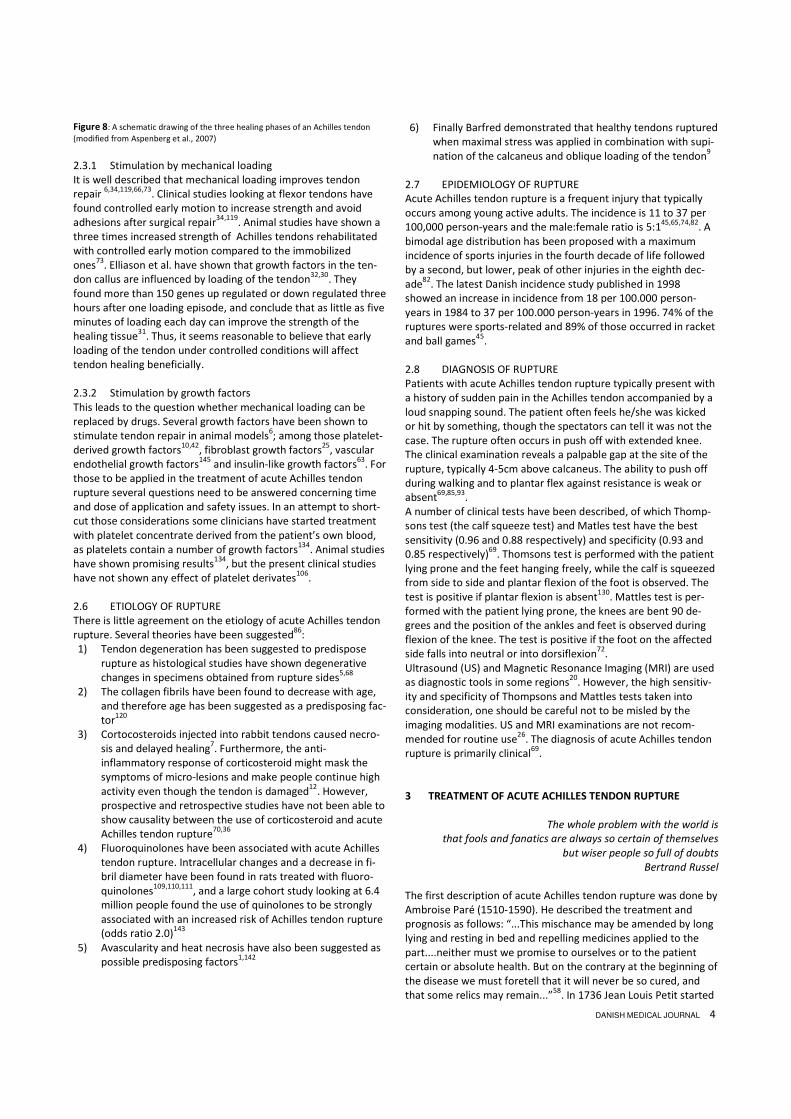
2.3 HEALING AFTER RUPTURE Tissue turnover in the Achilles tendon is an extremely slow proc-ess. It has been shown that the core of the Achilles tendon is formed before the age of 20 and essentially not renewed thereaf-ter40. In contrast, the periphery of the tendon is able to respond to mechanical forces by altering its structure, composition, and mechanical properties. An adaptation mediated by the fibroblasts through biochemical signaling137. Tendon repair can be described in three overlapping phases: 1) the inflammatory phase, 2) the proliferative phase, and 3) the remodeling phase (figure 8). In the inflammatory phase, the bleeding caused by the rupture leads to hematoma and activation of platelets and neutrophils, which again leads to the release of growth factors, chemotactic factors and vasoactive factors. The vascular permeability is increased, inflammatory cells are re-cruited and a tendon granuloma is produced6,32. In the prolifera-tive phase, angiogenesis allows for vascular and neuronal in-growth in the granuloma. The fibroblasts produce collagen (mainly type 3) and the mechanical strength of the granuloma gradually increases. After 10 to 14 days a tendon callus has been produced gluing the torn tendon ends together. Production of collagen type 1 gradually takes over and the callus reaches its largest size. The large transverse area of the tendon compensates for its weak composition. In the remodeling phase, the randomly deposited collagen fibers are resorbed and replaced to produce better architecture and cross-linking. The remodeling phase starts one to three months after rupture and lasts for several years6,32,66,112.
DANISH MEDICAL JOURNAL 4
Figure 8: A schematic drawing of the three healing phases of an Achilles tendon (modified from Aspenberg et al., 2007)
2.3.1 Stimulation by mechanical loading It is well described that mechanical loading improves tendon repair 6,34,119,66,73. Clinical studies looking at flexor tendons have found controlled early motion to increase strength and avoid adhesions after surgical repair34,119. Animal studies have shown a three times increased strength of Achilles tendons rehabilitated with controlled early motion compared to the immobilized ones73. Elliason et al. have shown that growth factors in the ten-don callus are influenced by loading of the tendon32,30. They found more than 150 genes up regulated or down regulated three hours after one loading episode, and conclude that as little as five minutes of loading each day can improve the strength of the healing tissue31. Thus, it seems reasonable to believe that early loading of the tendon under controlled conditions will affect tendon healing beneficially. 2.3.2 Stimulation by growth factors This leads to the question whether mechanical loading can be replaced by drugs. Several growth factors have been shown to stimulate tendon repair in animal models6; among those platelet-derived growth factors10,42, fibroblast growth factors25, vascular endothelial growth factors145 and insulin-like growth factors63. For those to be applied in the treatment of acute Achilles tendon rupture several questions need to be answered concerning time and dose of application and safety issues. In an attempt to short-cut those considerations some clinicians have started treatment with platelet concentrate derived from the patient’s own blood, as platelets contain a number of growth factors134. Animal studies have shown promising results134, but the present clinical studies have not shown any effect of platelet derivates106. 2.6 ETIOLOGY OF RUPTURE There is little agreement on the etiology of acute Achilles tendon rupture. Several theories have been suggested86: 1) Tendon degeneration has been suggested to predispose
rupture as histological studies have shown degenerative changes in specimens obtained from rupture sides5,68
2) The collagen fibrils have been found to decrease with age, and therefore age has been suggested as a predisposing fac-tor120
3) Cortocosteroids injected into rabbit tendons caused necro-sis and delayed healing7. Furthermore, the anti-inflammatory response of corticosteroid might mask the symptoms of micro-lesions and make people continue high activity even though the tendon is damaged12. However, prospective and retrospective studies have not been able to show causality between the use of corticosteroid and acute Achilles tendon rupture70,36
4) Fluoroquinolones have been associated with acute Achilles tendon rupture. Intracellular changes and a decrease in fi-bril diameter have been found in rats treated with fluoro-quinolones109,110,111, and a large cohort study looking at 6.4 million people found the use of quinolones to be strongly associated with an increased risk of Achilles tendon rupture (odds ratio 2.0)143
5) Avascularity and heat necrosis have also been suggested as possible predisposing factors1,142
6) Finally Barfred demonstrated that healthy tendons ruptured when maximal stress was applied in combination with supi-nation of the calcaneus and oblique loading of the tendon9
2.7 EPIDEMIOLOGY OF RUPTURE Acute Achilles tendon rupture is a frequent injury that typically occurs among young active adults. The incidence is 11 to 37 per 100,000 person-years and the male:female ratio is 5:145,65,74,82. A bimodal age distribution has been proposed with a maximum incidence of sports injuries in the fourth decade of life followed by a second, but lower, peak of other injuries in the eighth dec-ade82. The latest Danish incidence study published in 1998 showed an increase in incidence from 18 per 100.000 person-years in 1984 to 37 per 100.000 person-years in 1996. 74% of the ruptures were sports-related and 89% of those occurred in racket and ball games45. 2.8 DIAGNOSIS OF RUPTURE Patients with acute Achilles tendon rupture typically present with a history of sudden pain in the Achilles tendon accompanied by a loud snapping sound. The patient often feels he/she was kicked or hit by something, though the spectators can tell it was not the case. The rupture often occurs in push off with extended knee. The clinical examination reveals a palpable gap at the site of the rupture, typically 4-5cm above calcaneus. The ability to push off during walking and to plantar flex against resistance is weak or absent69,85,93. A number of clinical tests have been described, of which Thomp-sons test (the calf squeeze test) and Matles test have the best sensitivity (0.96 and 0.88 respectively) and specificity (0.93 and 0.85 respectively)69. Thomsons test is performed with the patient lying prone and the feet hanging freely, while the calf is squeezed from side to side and plantar flexion of the foot is observed. The test is positive if plantar flexion is absent130. Mattles test is per-formed with the patient lying prone, the knees are bent 90 de-grees and the position of the ankles and feet is observed during flexion of the knee. The test is positive if the foot on the affected side falls into neutral or into dorsiflexion72. Ultrasound (US) and Magnetic Resonance Imaging (MRI) are used as diagnostic tools in some regions20. However, the high sensitiv-ity and specificity of Thompsons and Mattles tests taken into consideration, one should be careful not to be misled by the imaging modalities. US and MRI examinations are not recom-mended for routine use26. The diagnosis of acute Achilles tendon rupture is primarily clinical69.
3 TREATMENT OF ACUTE ACHILLES TENDON RUPTURE
The whole problem with the world is
that fools and fanatics are always so certain of themselves
but wiser people so full of doubts
Bertrand Russel
The first description of acute Achilles tendon rupture was done by Ambroise Paré (1510-1590). He described the treatment and prognosis as follows: “...This mischance may be amended by long lying and resting in bed and repelling medicines applied to the part....neither must we promise to ourselves or to the patient certain or absolute health. But on the contrary at the beginning of the disease we must foretell that it will never be so cured, and that some relics may remain...”58. In 1736 Jean Louis Petit started

DANISH MEDICAL JOURNAL 5
treating patients in equinus position. The patient was treated prone with the knees flexed and the feet plantar flexed. A slipper on the foot was attached to the thigh with pins to maintain plan-tar flexion. The bandages were removed and reapplied after eight and 15 days. Healing was advanced at 22 days and weight-bearing commenced ten days later58. The first published series describing acute Achilles tendon rupture was published by Qenu and Stoianovitch in 1929100. They com-pared the results of 29 non-operatively treated and 39 opera-tively treated patients, thereby starting an ongoing discussion of whether to treat operatively or non-operatively. During the past fifty years an increased awareness of acute Achilles tendon rup-ture and treatment modalities developed, and a range of differ-ent treatment protocols have been described57. The historical diversity of published results and recommendations has led to considerable variation in treatment protocols between depart-ments treating this injury8.
Figure 9: A flow diagram to visualize the different treatment and rehabilitation protocols after acute Achilles tendon rupture.
Treatment of acute Achilles tendon rupture can be divided into two main categories: Operative and non-operative treatment (figure 9). Rehabilitation can be immobilizing or mobilizing. In this thesis, treatment protocols allowing mobilization before the end of the fourth week are categorized as mobilizing. Mobilization can be divided into controlled early motion and early weight-bearing. Some confusion exist concerning terminology as the term dy-namic rehabilitation by some authors refers to the overall term ‘mobilization’ and by others to ‘controlled early motion’. In paper I-III dynamic rehabilitation refers to controlled early motion. Over the past decade a change in treatment of acute Achilles tendon rupture from operative and immobilizing treatment to-wards non-operative treatment using controlled early motion has taken place74. 3.1 OPERATIVE VS. NON-OPERATIVE TREATMENT The first known open repair of an acute Achilles tendon rupture was performed in 1888 by Polaillon58. Since then a variety of open, minimally invasive, and percutaneous surgical techniques have been developed and described3. Surgical techniques are, however, not addressed in this thesis. Since the early 80ies, 12 randomized controlled trials (RCT’s) and 7 meta-analyses comparing operative and non-operative treat-ment have been published (table 1).
Table 1
Most of the trials published before 2005 suggested better out-come after surgery due to a higher rate of re-rupture in the non-surgical group23,84,107. Since then a number of high quality RCT’s have opted in favor of non-operative treatment due to a non-significant difference in the rate of re-rupture and a significantly increased risk of other complications in the surgically treated group55,87,132,141. In the same period two large prospective cohort studies looking at 945 and 487 patients, respectively, showed re-rupture rates of 2.8% and 6.6% in non-operatively treated pa-tients11,136. Looking at the meta-analyses we find compelling evidence that operative treatment yields a risk of re-rupture of 3-5% and non-operative treatment of 9-13%. Likewise operative treatment yields a risk of other complications of 27-34% and non-operative treatment of 3-8%. Looking at functional results some RCT ‘s have been able to show a slightly improved function after operative treatment23,87, but this is not reflected in the meta-analyses48,49,57,117,139. 3.2 MOBILIZATION VS. IMMOBILIZATION The role of mobilization has been discussed since the early 1980s and has gained increasing popularity over the past dec-ade8,27,74,80,104,122,132. All RCT’s comparing operative and non-operative treatment published after 2007 have used mobilization in both study groups. In 2007 Twaddle & Poon hypothesized that use of mobilization might be the most important factor in opti-mizing outcome in patients with Achilles tendon rupture and that surgery makes no difference to the outcome apart from increas-ing the risk of local infection132. Looking at the literature very little evidence exists concerning the use of mobilization in the treatment of acute Achilles tendon rupture. The shift towards mobilization has been driven by RCT’s comparing operative and non-operative treatment protocols and not by trials comparing mobilization and immobilization55,78,87,92,
117,132, 141. The low rate of re-rupture in both the operatively and non-operatively treated groups has been attributed to the early controlled rehabilitation regimes, though this has not been the research area of the trials.
DANISH MEDICAL JOURNAL 6
In order to investigate the effect of mobilization one should look at trials designed for this purpose. Operatively treated and non-operatively treated patients must be investigated separately, as the baseline situation for the healing process is different. Also controlled early motion must be distinguished from controlled early weight-bearing, as it is unknown how the two variables affect tendon healing and how they interact. 3.2.1 Controlled early motion Three RCT’s have investigated the effect of controlled early mo-tion in the treatment of acute Achilles tendon rupture (table 2)24,51,80. The quality of the three trials was assessed to be me-dium by two independent reviewers using the Jadad score47,90. No statistically significant differences were found in rate of re-rupture and other complications between the immobilized group and the group treated with controlled early motion. The studies argue for shorter sick leave, less tendon elongation, and better strength in the group treated with controlled early motion. Table 2
3.2.2 Controlled early weight-bearing One RCT has investigated the influence of controlled early weight-bearing in the treatment of acute Achilles tendon rupture (table 3)121. The study looked at health-related quality of life assessed with use of the RAND 36-Item Health Survey (RAND-36) as its primary outcome. Secondary outcomes were activity level, calf strength, ankle range of motion, return to sports and work, and complications. 98 patients (89%) completed the six-month follow-up. At six weeks the weight-bearing group had significantly better scores than the non-weight-bearing group in the RAND-36 do-mains of physical functioning, social functioning, role-emotional, and vitality scores (p<0.05). Patients in the weight-bearing group also reported fewer limitations of daily activities at six weeks postoperative (p < 0.001). Table 3
At six months, no significant differences between the groups were seen in any outcome. Suchak et al. concludes that no detri-mental effect of weight-bearing from the second postoperative week was found in operatively treated patients. 3.2.3 Controlled early motion and weight-bearing Three RCT’s have investigated the combined influence of con-trolled early motion and weight-bearing in the treatment of acute Achilles tendon rupture (table 4). Saleh et al.104 developed a new orthosis, the Sheffield splint, allowing both controlled early mo-tion and weight-bearing which they tested against immobilization in cast. They found that the group treated with mobilization regained mobility and range of motion significantly quicker than the immobilized group. Costa et al.27 also tested a new orthosis allowing both controlled early motion and weight-bearing against immobilization in cast. No significant differences were found. The trials were influenced by selection bias, as active people were operated and inactive people were treated non-operatively.
Table 4
3.3 REHABILITATION PROTOCOLS The following are examples of rehabilitation protocols being used after both operative and non-operative treatment of acute Achil-les tendon rupture: 3.3.1 Immobilizing rehabilitation protocols A typical immobilizing treatment protocol has been described by Cetti et al.23: A below-the-knee plaster cast with the foot in 20° of plantar flexion is worn for eight weeks. During this period no weight-bearing is allowed on the injured leg. After eight weeks the cast is removed and weight-bearing allowed. Alternatively the cast is changed after three to four weeks bringing the ankle in a neutral position or a brace is worn throughout the treatment period gradually bringing the ankle into neutral position. 3.3.2 Mobilizing rehabilitation protocols Mobilizing treatment must be divided into 1) controlled early motion, 2) controlled early weight-bearing and 3) controlled early motion and weight-bearing. 3.3.2.1 Controlled early motion
Controlled early motion can be performed using a dynamic brace, allowing movement of the ankle, or using a removable brace, instructing the patient to remove the brace and exercise. A treatment protocol using a dynamic brace has been described by Nilsson-Helander et al.87: Patients are treated with a below-the-knee cast with the foot in equinus position for two weeks, fol-lowed by an adjustable brace for the next six weeks. The brace is set at free plantar flexion motion with dorsiflexion limited to -30° the first two weeks, -10° the next two weeks, and +10° the last two weeks. Weight-bearing as tolerated is allowed after six to eight weeks. A treatment protocol using a removable brace has been described by Twaddle et al.132: Patients are treated with a removable brace for eight weeks. The foot is initially placed in equinus position by application of three wedges. Every second week a wedge is re-moved, slowly bringing the ankle to neutral. Patients are in-structed to remove the brace for five minutes of every hour and, while sitting with the injured leg hanging, practice active ankle dorsiflexion and passive plantar flexion, letting the foot fall down as far as was comfortable. Particular emphasis was made of the importance of not dorsiflexing the ankle beyond the neutral position and remaining non-weight-bearing. 3.3.2.2 Controlled early weight-bearing
Controlled early weight-bearing without movement of the ankle is a widely used treatment protocol in Scandinavia8. Yet, it has not been possible to find a published description of this protocol. The following treatment protocol is used in hospitals in the area of Copenhagen: The patient is treated with a brace for six weeks when operatively treated and eight weeks when non-operatively treated. The foot is initially placed in equinus position by applica-tion of wedges in the brace and slowly brought to neutral by removal of the wedges. Full weight-bearing is allowed from day one.

DANISH MEDICAL JOURNAL 7
3.3.2.3 Controlled early motion and weight-bearing
A treatment protocol using both controlled early motion and weight-bearing has been described by Möller et al.84: The patient is treated with a below-the-knee plaster cast with the ankle in equinus for two weeks. After two weeks the cast is replaced with a brace and full weight-bearing and range of motion exercises are encouraged during weeks three to eight. 4 OUTCOME ASSESMENT
‘If you can measure that of which you speak, and can express it by
a number, you know something of your subject, but if you cannot
measure it, your knowledge is meager and unsatisfactory.’
William Thomson
Outcome denotes whether or not a patient benefits from the medical care provided37. In order to evaluate patient outcome and compare treatment regimens valid, broadly accepted and clinically relevant outcome parameters are required. 4.1 PATIENT REPORTED OUTCOME MEASURES Patient reported outcome measures (PROM’s) have added a new dimension to clinical outcome evaluation. PROM’s are important both for assessing individual patients in the clinic, for quality monitoring and research purposes. Traditional clinical methods for assessing outcome after Achilles tendon rupture may fail to detect the patient’s perception of disability33,38,55,88,123. Over the past decades numerous PROM’s have been developed, but only the Achilles tendon Total Rupture Score is developed and vali-dated for use after acute Achilles tendon rupture54. The most widely used scores are presented in the following paragraphs. 4.1.1 Achilles tendon Total Rupture Score (ATRS) The Achilles tendon Total Rupture Score consists of 10 items reflecting symptoms and physical activity after treatment of acute Achilles tendon rupture. The items are assessed using a Likert scale (range 0-10; 10 being the best possible score). It was devel-oped and validated in 2007 by Nilsson-Helander et al.88 It has since been translated to English and Danish and both versions have been validated33,53. The ATRS was found to be a good and reliable PROM for measuring differences of more than 7 points between groups of patients. However, for assessing individual patients in the clinic, the ATRS showed serious limitations, as it cannot be expected to detect changes of less than 19 points when reassessing individual patients at several points33. 4.1.2 Victorian Institute of Sports Assessment – Achilles (VISA-A) The VISA-A questionnaire is a PROM validated for Achilles tendi-nopathy based on symptoms. It consists of eight questions that measure the domains of pain, function in daily living and sporting activity. The results range from 0 to 100, where 100 represents the ideal score103. VISA-A has been used in validations of the ATRS88,53 and has been translated to and validated in Danish46. 4.1.3 Physical Activity Scale (PAS) The physical activity scale has been developed to be a simple alternative to measuring physical activity by diary. It is a self-reported questionnaire estimating the total physical activity on an average week-day. It is developed and validated in Danish146.
4.1.4 Health related quality of life A wide range of patient reported questionnaires to assess quality of life have been developed and validated. The most widely used are listed below this paragraph. Inspired from those a purpose made questionnaire was developed to investigate the health related quality of life during the initial eight weeks of treatment in study II (Annex A). Five items were assessed using a Likert scale (range 0-10; 10 being the best possible score). The items re-flected: 1) limitations in daily living, 2) limitations in work situa-tions, 3) limitations in social life, 4) affection of the patient mood, and 5) level of pain. 4.1.4.1 Short Form 36 (SF-36) and RAND-36
The SF-36 and RAND-36 are identical multi-purpose, short-form health surveys containing 36 questions39,124,138. They measure eight health concepts summarized in two main scores: the physi-cal component score and the mental component score. The ques-tionnaires are thoroughly tested and validated39,124,138. 4.1.4.2 EuroQOL (EQ-5D)
The EQ-5D essentially consists of a descriptive system comprising five dimensions (mobility, self-care, usual activities, pain/discomfort and anxiety/depression) and a visual analogue scale where the person is to give an overall rating of self-rated health127. The questionnaire is thoroughly tested and validated97. 4.2 FUNCTIONAL TESTING Functional testing is widely used in trials assessing the outcome after acute Achilles tendon rupture. 4.2.1 Heel-rise test The heel-rise test is an endurance test where the number of standing unilateral heel raises, at a rate of 40/minute, is counted. Only heel lifts above 5 cm are counted83,85. 4.2.2 Heel-rise work test The heel-rise work test is an endurance test where the patient stands on one leg and lifts the heel up and down until exhaustion. The participants are allowed to place two fingertips per hand, at shoulder height, against the wall for balance. A frequency of 30 heel-rises per minute is kept. The participants are instructed to go as high as possible on each heel-rise and then lower the heel to the starting position before next heel-rise. The participants are asked to perform as many heel-rises as possible. The test is ter-minated when the patient stops, cannot maintain the frequency, or does not perform a proper heel-rise (<2cm)114. Silbernagel et al. have found a good validity of the heel-rise work test and greater ability to detect differences between the injured and the uninjured sides compared to the heel-rise test114. 4.2.3 Heel-rise height The heel-rise height is measured under the same setting as the Heel-rise work test. The maximum heel-rise is recorded114. The decrease in heel-rise height has been hypothesized to be corre-lated to tendon lengthening115. 4.2.3 Single heel-rise test The patient stands with the foot in neutral position. It is recorded whether the patient is able to make a single heel-rise on the injured leg. The heel-rise is acknowledged if the heel can be lifted at least 2 cm with stretched knee. It has been shown that the ability to do a heel-rise 12 weeks after rupture is correlated to

DANISH MEDICAL JOURNAL 8
outcome. The heel-rise ability appears to be an important early achievement and reflects the general level of healing, which influences patient-reported outcome and physical activity91. 4.2.4 Strength tests Strength testing has been performed in different settings. Siber-nagel et al. had the patients do concentric and eccentric toe-raise against increasing weight113. Möller et al. tested concentric and eccentric strength with the patient sitting with 90 degrees flexion in the hip and knee using an isokinetic dynamometer. Angular velocities of 30°/sec and 180°/sec were used. The reliability was found to be good83. McNair et al. tested concentric strength at an angular velocity of 240°/sec76. 4.2.5 Jump tests In 2005 Silbernagel et al. described a test battery consisting of three jump tests: a counter movements jump, a drop counter movement jump and hopping113. The test battery was found to be reliable and able to detect differences in lower leg function be-tween the injured and non-injured limb87,113. 4.3 BIOMECHANICAL TESTING Biomechanical testing is typically done using an isokinetic dyna-mometer76,83. The patient is seated or lying with the hip and knee joint in a specified position. Positions between 0 and 90 degrees have been described for each joint76,83. The subjects foot is strapped to the pedal of the dynamometer and he/she is asked to relax and not activate the lower limb muscles while the isokinetic dynamometer moves the ankle joint in dorsiflexion at an angular velocity of typically 5˚/sec76,83. The resistant torque is sampled by the isokinetic dynamometer and the matching torque and angle values are recorded. 4.3.1 Stiffness Stiffness is the slope of the stress strain curve. Stiffness of the plantar flexor muscle-tendon complex increases with increasing dorsiflexion. It is, therefore, often reported in intervals like early, middle and late dorsiflexion76. 4.3.2 Peak passive torque Peak passive torque is the maximum torque exerted when stretching the passive ‘plantar flexion muscle-tendon complex’18,76. It is usually reached in maximum dorsiflexion. 4.3.3 Energy stored during loading The energy stored during loading is the area under the loading curve. It represents the energy absorbed by the plantar flexor muscle-tendon complex76. 4.3.4 Torque relaxation The torque relaxation is representative of the shock absorbing quality of the plantar flexor muscle-tendon complex. It is the fraction of energy stored after a period of relaxation, and can be expressed as a percentage: the persisting passive torque after a period of relaxation divided by the peak passive torque18. 4.3.5 Dissipation coefficient The dissipation coefficient is another measure of shock absorbing quality. It is the fraction of energy released compared to the energy stored and can be calculated by dividing the energy lost in the coil-recoil process (the area between the loading and unload-
ing curves) by the energy stored during loading (the area under the loading curve)76. 4.4 LENGTH MEASURES A variety of methods have been developed to measure Achilles tendon length and elongation4,23,50,64,95,99,102,105,118,144. The meth-ods can be divided into four groups depending on modality: clini-cal, X-ray, ultrasound (US), and Magnetic Resonance Imaging (MRI). 4.4.1 Clinical Clinically, the length of the Achilles tendon can be estimated by the sponta-neous position of the ankle joint in the relaxed leg (figure 10). An Achilles ten-don of normal length positions the ankle joint in 10-20 degrees of plantar flexion when the patient is lying prone with the knees flexed 90 degrees. An elongation of the tendon will make the foot fall into neutral or dorsiflexion72. Likewise, the height of a maximal heel-rise has been used to estimate elonga-tion of the Achilles tendon91,115. 4.4.2 Radiological X-ray allows for measurement of displacement of the torn tendon ends by implantation of radio-logical markers (figure 11)23,50. The latest and best validated method is Roentgen Stereopho-togrammetric Analysis (RSA) using tantalum beads as mark-ers64,105. Implantation of the markers is invasive and so far the technique has only been used to evaluate elongation after operative treatment. 4.4.3 Ultrasound Ultrasound (US) has been established as an important and cost-effective tool in the diagnosis of tendon problems. It provides a fast investigation that can be performed by the surgeon. Three types of US based length measures have been described for the Achilles tendon. Rees et al. developed a method combining video-based motion capture and US imaging102. Achilles tendon length was defined as the distance between the gastrocnemius muscle tendon junction and the tendon insertion at the calcaneus. Two retroreflective markers were placed on the back of the US probe and the mark-ers were used to track the position of the probe. The measure-ment was validated by Silbernagel et al.115 It showed high repro-ducibility and a measurement error of < 1%. Panoramic ultrasound imaging allows the transducer to be moved along the patients Achilles tendon, blending multiple images together to form one long image with an extremely wide field of view99 (figure 3). The method is easy to perform and clinically applicable, but no validation of the measurement has been pub-lished95,118,144. The authors of study IV have tested reliability of the measurement in a setting similar to the one used in study IV. The data has not been published yet, but showed an average
Figure 10: Matles test
Figure 11: RSA markers im-planted in the Achilles tendon

DANISH MEDICAL JOURNAL 9
intra-rater reliability of ICC 0.84, SEM 0.94 and MDC 2.62 and inter-rater reliability of ICC 0.89, SEM 0.78 and MDC 2.15. Finally, Amlang et al. reported an US classification system to be used for individual treatment selection in patients with acute Achilles tendon rupture, looking at the degree of overlap of the torn tendon ends as a surrogate for tendon displacement and elongation4. This classification system is yet to be validated and correlated to the final outcome of treatment. 4.4.4 MRI Magnetic Resonance Imaging (MRI) is also a non-invasive modal-ity for tendon length measurement60. In relation to US the setup is more demanding and expensive, as radiological assistance is needed to perform MRI measurements. 4.4.5 Strength in plantar flexion Strength in the terminal part of plantar flexion can be used as a surrogate for tendon elongation. If tendon elongation is present one would expect strength to decline as plantar flexion increases and the suralis muscle loses its ability to contract further. 4.5 COMPLICATIONS All published RCT’s have reported the rate of complications23,
55,84,87,107,132,141. Some studies have reported all complications and others only selected complications. 4.5.1 Re-rupture The rate of re-rupture is the most common primary endpoint in the literature23, 55,84,87,107,132,141. However, a clear definition of how re-rupture is defined is seldom given. 4.5.2 Infection Infections can be divided into superficial and deep infections. Superficial infections are related to the skin, whereas deep infec-tions involve the Achilles tendon. 4.5.3 Other complications Other reported complications involve damage to the suralis nerve, adhesions between the tendon and the surrounding con-nective tissue, scar problems and deep vein thrombosis79,94. 4.6 RETURN OF FUNCTION In 2006 Costa et al.27 concluded that the return to normal activi-ties was the most important outcome measure as viewed by the patient and therefore claimed that it also should be the most important outcome measure for researchers. 4.6.1 Sick leave The length of sick leave is of fundamental importance for the patient as well as for society. The measurement is usually self-reported through questionnaires or structured questioning at follow up27,84. 4.6.2 Return to sport Most acute Achilles tendon ruptures are acquired through sport-ing activities. The return to sports, and preferably a level of sport-ing activity similar to the one before the injury, is therefore an obvious outcome. The measurement is usually self-reported through questionnaires or structured questioning at follow up23,57.
4.7 VALIDITY, RELIABILITY AND AGREEMENT OF MEASURE-MENTS Valid and reliable outcome parameters are cornerstones in proper research. Invalid outcome parameters may lead to wrong conclusions and unreliable outcome parameters may keep actual differences from being detected. Terminology within the field is confusing and imprecise, as it differs between papers and text-books14, 44,52,62,123,133. The following chapter is an outline of the terminology and concepts as they are understood and used in this thesis. 4.7.1 Validity Validity can be divided into content, criterion and construct valid-ity. Further description of this subdivision is out of the scope of this thesis. Generally speaking validity is the extent to which a measurement measures what it claims to measure (figure 12). 4.7.2 Assessment of validity In order to assess if a measurement instrument actually measures what it claims to measure, one needs to know the real value of the object being measured. For example, if one would like to assess if an ultrasound measurement measures distances of tendons correctly, one needs to know the actual length of the measured tendons. If the actual length is not known, an alterna-tive is to define a gold standard of measurement to validate the new measurement against. Validity can be expressed as the measurement error115:
4.7.3 Reliability and agreement The term reliability, as used in the previous chapter (4.7.1), refers to the spread of data. It can be further subdivided into the terms reliability and agreement133. Agreement is the extent to which a measurement measures the same at different occasions; it is related to the measurement error of the measurement itself. Reliability is the extent to which study objects can be distin-guished from each other, despite measurement errors; it relates the measurement error of the measurement to the natural vari-ability of the study populaTion62,133.
Figure 12: Diagram showing the difference between validity and reliability. (modi-fied from Nevit Dilmen)

DANISH MEDICAL JOURNAL 10
Figure 13 illustrates an example of 3 subjects (�, � and �) being measured five times. The distance between the five identical symbols represents the agreement of the measurement (the measurement error), which is not affected by the variability of the population. In contrast, reliability of a measurement is influenced by the variability of the study population. In a population repre-sented by the subjects � and � the measurement will have a good reliability, as the measurement error will not affect dis-crimination of the subjects. In a population represented by the subjects � and � the measurement will have a poor reliability, as the measurement error will affect the ability to discriminate the subjects.
Figure 13: Diagram showing the difference between reliability and agreement. (Modified from de Vet et al. 2006)
Reliability is a characteristic of the performance of an instrument in a certain population sample. Agreement is rather a characteris-tic of the measurement instrument itself133. 4.7.4 Assessment of agreement and reliability 4.7.4.1 Kappa-statistics
Agreement between categorical data is often assessed using kappa-statistics. The kappa-value tells the observed agreement as a percentage of agreement expected just by chance. A value of 1 indicates perfect agreement and a value of 0 indicates no agree-ment2.
4.7.4.2 Intraclass correlation coefficient (ICC)
Reliability of a measurement can be assessed using the Intraclass Correlation Coefficient (ICC). The basic formula of the ICC is:
If the measurement error is small, compared to the variability between persons, the ICC approaches 1. This means that the discrimination of the persons is hardly affected by the measure-ment error, and thus the reliability is high. If the measurement error is large, compared to the variability between persons, the ICC value becomes smaller133. 4.7.4.3 Standard error of measurement (SEM) and minimal de-
tectable change (MDC)
Agreement of a measurement can be assessed by calculating the standard error of the measurement (SEM) and the minimal de-tectable change (MDC). The SEM is used to assess the agreement between groups of data. It can be derived from the ICC: SEM = standard deviation x √(1 – ICC)133. The MDC is used to assess agreement between data of the individual subjects. It can be derived from the SEM: MDC = 1.96 x √2 x SEM. Agreement pa-rameters are expressed on the actual scale of measurement, and are therefore easily interpretable133.
4.7.4.4 The Bland-Altman method
Agreement can be assessed graphically using the Bland-Altman plot14,15. The plot shows the difference between measurements plotted against the average of measurements (figure 14).
Figure 14: Bland-Altman plot showing difference against mean
The mean shows if there is a systematic difference between measurements and the 95% limits of agreement show the spread of data in the sampled distribution. The limits of agreement are calculated as the mean ± 1.96 standard deviations. The visual presentation of data makes it possible to detect skewed distribu-tions and correlation between measurement error and the scale of the measurement.
5 AIMS
Aim at the sun, and you may not reach it;
but your arrow will fly far higher
than if aimed at an object on a level with yourself.
Joel Hawes
Over the past decade a change in treatment of acute Achilles tendon rupture away from operative and immobilizing treatment towards non-operative treatment using controlled early motion occured8,74. This thesis addresses the question: ‘What is the opti-mal non-operative treatment protocol for acute Achilles tendon rupture?’ 5.1 AIM OF THE THESIS The aim of this PhD thesis was to evaluate non-operative treat-ment of acute Achilles tendon rupture, in order to pave the way for national and international treatment guidelines concerning acute Achilles tendon rupture. 5.2 AIMS OF THE STUDIES 5.2.1 Study I: Treatment of acute Achilles tendon rupture in Scandinavia The aim of this survey was to investigate whether departments treating acute Achilles tendon rupture in Scandinavia adhere to the latest evidence.

DANISH MEDICAL JOURNAL 11
5.2.2 Study II: The influence of early weight-bearing on clinical outcome The aim of this blinded, randomized, controlled trial was to com-pare patient reported and functional outcome in patients ran-domized to immediate weight-bearing or non-weight-bearing in a non-operative treatment protocol for acute Achilles tendon rup-ture using controlled early motion. 5.2.3 Study III: The influence of early weight-bearing on biome-chanical outcome The aim of this blinded, randomized, controlled trial was to com-pare the biomechanical properties of the plantar flexor muscle-tendon complex of both limbs in patients randomized to early weight-bearing or non-weight-bearing. Both groups were treated non-operatively using controlled early mobilization. 5.1.4 Study IV: A Novel Ultrasound Measurement of Achilles tendon Length and Elongation The aim of this study was to develop and validate a method which can determine Achilles tendon length and elongation accurately in a clinical setting using standard US equipment. In order to do so we had the following specific aims:
1) US identification of the anatomical landmarks and de-velopment of the specific measurement
2) Assessment of the intra-rater reliability 3) Assessment of the inter-rater reliability 4) Assessment of the validity by comparison with MRI
Comparison of the tendon length of both legs of the same subject
6 SUBJECTS AND METHODS
‘I hate definitions!’
Benjamin Disraeli
In this chapter the methodology used in study I-IV is described. Outcome parameters and statistical methods are discussed and critically assessed. A thorough discussion of methodological con-siderations is done in chapter 7. 6.1 STUDY DESIGN, SUBJECTS, MATERIAL AND ETHICAL CON-SIDERATIONS 6.1.1 Study I: Treatment of acute Achilles tendon rupture in Scandinavia 6.1.1.1 Study design
The study was conducted as a questionnaire based cross-sectional study investigating: 1) the use of surgical treatment, 2) the use of dynamic rehabilitation, and 3) the use of weight-bearing. 6.1.1.2 Subjects
All orthopedic departments treating acute Achilles tendon rup-ture in Denmark, Sweden, Norway and Finland were identified. Only public hospitals were included as private hospitals according to the experience of the authors play a negligible role in treat-ment of acute Achilles tendon rupture in Scandinavia. Data were collected in the period from October 2011 to October 2012. 6.1.1.3 Material
A questionnaire was developed in Danish and translated by bilin-gual speakers into Swedish, Norwegian and Finish (Annex B). The questionnaire was divided into four parts. Part one investigated
Achilles tendon rupture diagnostics, how choice of treatment was made and to what degree the patients were involved in the deci-sion making process (four questions). Part two investigated which patients were recommended surgical treatment, which surgical technique was used and what kind of post-surgical regimen was offered (seven questions). Part three investigated what kind of non-surgical treatment was offered (three questions). Part four consisted of three patient cases where preference of surgical or non-surgical treatment had to be chosen. 6.1.1.4 Ethical considerations
Our survey did not make use of patients or animals and as such no ethical consideration were made in regard to those. 6.1.2 Study II: The influence of early weight-bearing on clinical outcome 6.1.2.1 Study design
This was a blinded, randomized, controlled, superiority trial with participants individually randomized to one of two parallel groups. The trial was designed in accordance with the Consort requirements; no changes were made to the trial design after commencement of the trial. 6.1.2.2 Subjects
Sixty patients were included in the trial. Patients with acute Achil-les tendon rupture referred to the orthopedic department of Copenhagen University Hospital Hvidovre from April 2011 to March 2012 were assessed for eligibility. Diagnosis was based on a medical history with a clear snap of the Achilles tendon and a clinical examination with a palpable gap and positive Thompsons test130. All patients aged 18 to 60 years who were expected to be able to follow the treatment protocol and give written consent in Danish were eligible for inclusion if randomization could be done within 4 days of the rupture. Exclusion criteria were previous Achilles tendon injury, corticosteroid injections within the last 6 months, ASA-score of three or more, medical history of arterial insuffi-ciency in the legs and rupture within 1 cm of calcaneus. 29 patients from the intervention group and 27 from the control group were included in the one-year analysis, all belonging to their original assigned groups. The groups were comparable at all parameters except for height with the non-weight-bearing group being three cm taller. 6.1.2.3 Material
All patients were treated with controlled early motion and ran-domized to either immediate weight-bearing or non-weight bear-ing. Intervention The intervention group was allowed full weight-bearing from day one. Crutches were recommended but not obligatory the first two weeks of treatment. The control group was instructed not to bear weight for the first six weeks of treatment. The last two weeks full weight-bearing was allowed Standard treatment protocol used in both groups In the emergency department an ankle orthosis (DJO Nextep Contour2 Walker) with three wedges of 1.5cm was applied, fixat-

DANISH MEDICAL JOURNAL 12
ing the ankle in equinus position (20-30 degrees of plantar flex-ion). The orthosis was worn for eight weeks gradually bringing the ankle to neutral position by removing a wedge every second week (table 5).
Patients were seen in the outpatient clinic after two and eight weeks. The patients were instructed not to remove the orthosis at any time during the first two weeks. After two weeks the first wedge was removed and controlled early motion begun: Patients were instructed to remove the orthosis at least five times a day sitting on the edge of a table with both legs hanging (figure 15). Gravity plantar flexed the foot and the patient actively dorsiflexed the foot to a horizontal position. Patients were instructed to do this in series of 25 repetitions. The orthosis was not to be re-moved inbetween exercises from week three to six. The last two weeks of treatment the orthosis could be removed at night.
Figure 15: Controlled early motion. Gravity plantar flexed the foot, where upon the patient actively dorsi flexed the foot to a horizontal position. Patients were in-structed to do this in series of 25 repetition five times a day. Drawing by Ilija Ban, MD
6.1.2.4 Ethical considerations
We believe that the potential benefits for the patients enrolled in the study (better functional outcomes, improved health-related well-being and economic savings) exceed the potential inconven-
iences (additional follow-up audits and possible risks/side ef-fects). The study was conducted in accordance with the principles of the Helsinki Declaration. The study was approved by the Regional Ethical Review Board. All participants received oral and written information concerning the trial before written consent was obtained. Patients were covered by Hvidovre University Hospital patient insurance. 6.1.3 Study III: The influence of early weight-bearing on biome-chanical outcome 6.1.3.1 Study design
This study is based on data from a blinded, randomized, con-trolled trial with participants individually randomized to one of two parallel groups (study II). 6.1.3.2 Subjects
The study population was the same as described in study II (chap-ter 6.1.2.2). 26 patients from the intervention group and 20 from the control group were included in the one-year analysis, all belonging to their original assigned groups. 6.1.3.3 Material
The treatment protocol and the intervention was the same as described in study II (chapter 6.1.2.3). Follow up was done in the Gait analysis laboratory at Hvidovre Hospital. Measurement of passive tension in the plantar flexor muscle-tendon complex was performed using an isokinetic dynamome-ter. A motor (Caldercraft Electric Motors, model 26) was driven by a DC power amplifier (model 2708; Brüel & Kjær, Naerum, Den-mark) and could deliver maintained torques up to 80Nm and peak torques up to 120Nm. An electro-goniometer, connected to the footplate, measured the angle of the ankle joint and a torque meter measured the torque exerted on the footplate. This appa-ratus was developed and found to produce highly reproducible measurements of passive and reflex-mediated torque around the ankle joint by Sinkjær et al.131,140. Before testing in the isokinetic dynamometer the patient warmed up on a stationary bicycle for 5 minutes. For the measurement, the patient was positioned with the foot strapped to a pedal in 10 degrees of plantar flexion and the knee and hip fixed in 90 degrees flexion. Patients were asked to relax and not activate their calf muscles as the ankle was pas-sively moved into dorsiflexion by the pedal at an angular velocity of 5 °/sec. The isokinetic dynamometer rested for 2 seconds at maximal dorsiflexion before the pedal was returned to the start-ing position at the same velocity. The resistant torque was sam-pled at a frequency of 1000Hz. Patients who had a ROM exciding 20 degrees of dorsiflexion were tested in the range from 10 de-grees plantar-flexion to 20 degrees dorsiflexion. Seven patients had a range of motion below 20 degrees of dorsiflexion, they were tested in the range from 20 degrees plantar-flexion to 10 degrees dorsiflexion. Ten repetitions were performed on each limb. The unaffected limb was tested before the affected. Strength was measured using the same isokinetic dynamometer setting. The patient was asked to relax as the dynamometer dorsi-flexed their ankle to 10 or 20 degrees of dorsiflexion, respectively. The dynamometer rested for three seconds and the patient was encouraged to deliver maximal plantar-flexion power without activating the thigh while the dynamometer plantar flexed the foot 40 degrees from the starting position with a speed of 6.7°/s. Five repetitions were performed on each limb. The unaffected

DANISH MEDICAL JOURNAL 13
limb was tested before the affected. Standardized verbal encour-agement was provided to ensure maximal effort77. 6.1.3.4 Ethical considerations
The ethical considerations were the same as described in study II (chapter 6.1.2.4). 6.1.4 Study IV: A Novel Ultrasound Measurement of Achilles tendon Length and Elongation 6.1.4.1 Study design
The study was done as a cross-sectional study comparing the investigated measures twice within a certain period of time. 6.1.4.2 Subjects
Both legs of 19 uninjured persons (8 males and 11 females) were studied. Their mean age was 43.4 years (SD 10.7, range 26-63). Average height was 175cm (SD 9, range 158-192) and average weight was 76.8kg (SD 12.9, range 58-110). They all had right as their dominant side. 6.1.4.3 Material
Definition of landmarks Achilles tendon length was defined as the distance between the tendon insertion at the calcaneus and the medial gastrocnemius muscle tendon junction, as previously described by Rees et al.102 (figure 16).
Figure 16: The blue line represents the distance measured by the novel ultra sound measure. (A) A posterior view of the calf. (B) A panoramic ultrasound picture of the calf: 1) Calcaneus, 2) The Achilles tendon, 3) the tendon sheet of the suralis muscle, 4) the gastrocnemius musle, and 5) the soleus muscle.
First, the anatomical landmarks were identified and marked, then the distance between them was measured. The distal landmark was the posterior and most superior corner of the calcaneus in the midline, which on sagittal US examination was identified as the point where the cortical bone and its underlying shadow ended (figure 17). The proximal landmark was the distal tip of the medial gastrocnemius head, which was defined as the most distal point where the muscular fibers inserted into the v-shaped con-vergence of the deep crural fascia (figure 17).
Figure 17: Sagittal US pictures showing (A) the insertion of the Achilles tendon at the calcaneus and (B) the muscle-tendon junction. (A) The distal landmark, the posterior-superior corner of calcaneus, is seen as the point where the cortical bone and its underlying shadow ends. (B) The proximal landmark is the distal tip of the medial gastrocnemius head (the insertion of the most distal muscle fibers into the deep fascia). 1) Calcaneus, 2) The Achilles tendon, 3) the tendon sheet of the suralis muscle, 4) the gastrocnemius musle, 5) the soleus muscle, and 6) the posterior
acoustic shadowing of the needle of a 21 gauge needle projecting the landmark to the skin.
Setup Participants were positioned in a prone position with the knee flexed 10 degrees. Anteriorly to (below) the ankle joint a triangle shaped foam pad was placed with the feet resting relaxed against it (figure 18). Using a goniometer the ankle joint was positioned in 10 degrees of plantar flexion by adjustment of the foam pad. Participants were positioned identically for US and MRI investiga-tions. The predefined anatomical landmarks were identified using longi-tudinal imaging. A needle was placed between the probe and the surface of the skin, and the posterior acoustic shadow created by the needle on the US image was centered over the landmark (figure 17). The position of the needle, thus representing the landmark, was then marked on the skin (figure 18). The direct distance between landmarks was measured with a tape measure following the curves of the leg.
Reliability-testing of the US measurement was performed us-ing three different US scanners: GE Healthcare Logiq S8, Logiq 9 and logiq P5. Frequency was set to 15 MHz and focus was dy-namically adjusted by the US operator. After each scan the marks on the skin were removed in order to secure blinding of results between investigators.
The novel US measurement was validated by three independ-ent US-investigators with two to five years of experience within musculoskeletal US. MR-images were evaluated by two inde-pendent investigators, both specialized in musculoskeletal MRI. All investigators were blinded to the results of the other investi-gators and to their own previous results. Inter- and intra-rater reliability for the novel US measurement was determined for the three independent US investigators on two occasions with three weeks between scans. Within the fol-lowing two months, MRI examinations were performed.
Figure 18: Positioning of the study subjects. Participants were positioned in a prone position with the knee flexed 10 – 20 degrees. The ankles rested on a triangular foam pad. Using a goniometer the ankle joint was positioned in 10 degrees of plantar flexion by adjustment of the foam pad (A). With a marker the point where the US probe and the needle crossed was marked on the skin (B+C). The distance between landmarks was measured with a tape measure following the curves of the leg (D).
6.1.4.4 Ethical considerations

DANISH MEDICAL JOURNAL 14
There are no known side-effects or risks associated with the performed UL and MRI investigations. We believe that the poten-tial benefits for coming generations of patients with acute Achil-les tendon rupture exceed the potential inconveniences for the study subject. The study was conducted in accordance with the principles of the Helsinki Declaration. The study was approved by the Regional Ethical Review Board. All participants received oral and written information concerning the trial before written consent was obtained. 6.2 CRITICAL ASSESSMENT OF OUTCOMES A variety of outcomes have been used for assessment of acute Achilles tendon rupture. The following guidelines for choosing appropriate outcomes have been suggested37: 1) the measure must be quantifiable, 2) the measure should be relatively easy to define and use, 3) the measure should lend itself to standardiza-tion and validation, and 4) the measure should be clinical rele-vant. 6.2.1 Study I: Treatment of acute Achilles tendon rupture in Scandinavia 6.2.1.1 Outcome parameters in study I
The following four outcomes were assessed in the study: 1) The use of surgical treatment Departments were asked which of the following statements was most correct:
a) In principle all patients are recommended surgery b) Healthy and active people under the age of 60 are rec-
ommended surgery c) Active sportsmen and people with excessive demands
to their Achilles tendons are recommended surgery d) Only patients with delayed diagnosis, patients treated
with corticoid-steroid, patients with sharp lesions and a few other cases are recommended surgery
2) The use of controlled early motion Departments were asked if their treatment protocol allowed movement of the ankle joint from week three of treatment. 3) The use of controlled early weight-bearing Departments were asked when partial and full weight-bearing was allowed. 4) The level of experience of the performing surgeons Departments were asked if the operations were performed by specialist in orthopedic surgery or by any surgeon.
6.2.1.2 Critical assessment of the outcome parameters in study I
The chosen outcomes were investigated using a purpose made questionnaire. The questionnaire was developed in Danish and translated to Swedish, Norwegian and Finish. The Swedish and Norwegian translations were approved by the first author who is able to read and understand both languages. The questionnaire was not validated concerning construct, content or criterion validity123. Nor was the reliability and agreement of the question-naire tested62. As such, we do not know if the questionnaire measured what it intended to measure. 6.2.2 Study II: The influence of early weight-bearing on clinical outcome Most RCT’s presented in chapter 3 have used the re-rupture rate as their primary outcome parameter. It is debatable if the re-rupture rate is a good primary outcome, as it does not contain information concerning the functional outcome and the patient’s
own perception of the outcome27,33,87,121. In 2002 Pajala et al. investigated the outcome after 23 re-ruptures and nine deep infections and concluded that the outcome after a simple re-rupture without infection is satisfactory, whereas the outcome after a deep infection often is devastating94.
6.2.2.1 Outcome parameters in study II
We decided to follow the recommendation by Silbernagel et al.114 and use a combination of patient reported outcome and func-tional outcome. Our primary outcome parameter was the Achilles tendon Total Rupture Score (ATRS) at one year follow up. Secon-dary outcomes were 1) the Heel-rise work test, 2) re-rupture rate, 3) length of sick leave, 4) time to return to sports, and 5) quality of life during treatment.
6.2.2.2 Critical assessment of the outcome parameters in study II
Whether to use doctor reported or patient reported outcomes is an ongoing discussion. Re-rupture and infection rate are doctor reported outcomes; they are quantifiable, easy to use and easily standardized. The ATRS is a patient reported outcome; it is quan-tifiable, easily standardized and relatively easy to use. The ques-tion is which of the outcome measures is the most clinical rele-vant. We have chosen to use the ATRS as our primary outcome as we find the patients’ perception of his/her injury to be the most important criteria of success of a given treatment. Critics of PROM-assessment argue that results are influenced by the so-ciocultural context, the shape and application of the question-naire, by the patients’ familiarity in answering the questionnaire and by the patients’ relationship with the clinician/researcher67,98. The ATRS it is developed and validated for use after acute Achilles tendon rupture and found to have good validity, reliability, inter-nal consistency and responsiveness33,53,88. Still, the construction of the questionnaire can be questioned. The questionnaire inves-tigates 10 items with a possible score between zero and 10, it is thus possible to achieve a total sum score of maximum 10088. It is common practice in sociology and psychology to add together ordered items to sum-scores and then use these sum-scores for subsequent analyses. It is however questionable if the added item-scores represent the same value, and as such if the sum-score is a valid measurement21. Another problem concerning the ATRS is the lack of clear analysis guidelines. Finally, the Danish validation of the ATRS showed an SEM of 7 and an MDC of 19; meaning that differences below 7 points cannot be measured with the ATRS. Sick leave and time-to-return-to-sport was registered retrospec-tively at six and 12 months follow up; though there is a risk of recall bias. Quality of life during treatment was measured using a purpose made 5 item questionnaire. The questionnaire was not validated concerning construct, content or criterion validity123. Nor was the reliability and agreement of the questionnaire tested62. It would have added a dimension of validity to the study if a validated quality of life questionnaire had been used. 6.2.3 Study III: The influence of early weight-bearing on biome-chanical outcome No primary outcome was determined a priori as the study was based on data from study II. We did, however, suggest a primary outcome post-hoc, based on the availability of data from previous studies18,76.

DANISH MEDICAL JOURNAL 15
6.2.3.1 Outcome parameters in study III
We suggested peak passive torque as primary outcome parame-ter at one year follow up. Secondary endpoints were 1) stiffness in early, middle and late dorsiflexion, 2) energy stored, 3) torque relaxation and 3) the maximal strength.
6.2.3.2 Critical assessment of the outcome parameters in study III
The used outcome parameters have all been described and used in a range of experimental and clinical studies17,18,76,81. The ques-tion is, however, to what degree we are measuring the biome-chanical property of the Achilles tendon and to what degree the surrounding tissue (the plantar flexor muscles, the ankle joint and the loose connective tissue) influences the measurements. 6.2.4 Study IV: A Novel Ultrasound Measurement of Achilles tendon Length and Elongation
6.2.4.1 Outcome parameters sin study IV
Intra- and inter-rater reliability and agreement was tested using the ICC, SEM and MDC. Furthermore a graphical evaluation using the Bland-Altman method was used. Validity of the novel ultra-sound was tested against MRI measurements and the measure-ment error was calculated. Comparison of the two limbs was done using ICC, SEM and MDC.
6.2.4.2 Critical assessment of the outcome parameters in study IV
It is an ongoing discussion whether to use reliability parameters like ICC or to use the Bland-Altman method14,62,133. We chose to present both, which some might consider unnecessary additional material. Proportions of agreement could have given additional informa-tion. It would have been interesting to know the proportion of measurements lying within e.g. 5mm from each other and it would be interesting to know the proportion of persons having a discrepancy between the length of the left and right Achilles tendons above e.g. 10mm. Validity was assessed by comparing the UL-measurements with the MRI-measurements. The actual length of the tendon is not known and as such the calculated measurement error might be misleading. 6.3 STATISTICS The Statistical methods used in this thesis are outlined in table 6. 6.3.1 Paper I - statistical considerations Two-way tables with Fisher’s exact test were used for analysis as cells had expected values below 5. 6.3.2 Paper II - statistical considerations The ATRS was considered a continuous score. One could argue that it is a categorical, ordinal score, but it does not affect the statistical testing as the results were not normally distributed and the Wilcoxon signed rank test was used. 6.3.3 Paper III - statistical considerations The paired t-test was used for evaluation within groups of data. It was also used for comparison between the right and left leg of subjects, as data from the two sides of a person were considered to be connected.
6.3.4 Paper IV - statistical considerations The statistical considerations concerning the use of the Bland-Altman method and ICC, SEM and MDC are described in chapter 4.7. Tabel 6
7 METHODOLOGICAL CONSIDERATIONS
There are only a handful of ways to do a study properly but a
thousand ways to do it wrong
Sackett
In the attempt to produce research of the highest quality, it is of outmost importance to choose the correct study design and setup in order to avoid bias and fulfill the aim of the study2,35. The fol-lowing chapter is an attempt to 1) assess the methodological problems encountered in this thesis, 2) discuss the possible in-duced bias, and 3) explain how bias has been or should be con-trolled. 7.1 BIAS Bias refers to the presence of systematic error in the design, conduct, analysis or publication of a study35,37,96. More than 70 different types of bias have been described and several classifica-tion systems proposed, the most common distinguishing between information, selection and confounding bias28,35. Information bias refers to errors of assessment and measurement of the studied population (figure 19). Selection bias refers to factors influencing who is included in the study population. Confounding bias refers to factors that determine who is exposed to the studied interven-tion and influences the generalizability of the study results35,37. The terminology and division of bias into categories varies by discipline and among authors.

DANISH MEDICAL JOURNAL 16
Figure 19: Schematic illustration of the relationship between information, selection and confounding bias and the study, source and background population. Modified from Gerhard et al.
7.1.1 The randomized controlled trial The RCT has been developed to produce groups who are identical in all maters except for the intervention under investigation. Ideally it would lead to unbiased conclusions. In reality bias is always present in research studies, the question is how much bias exists and to what extend it influences the drawn conclusion35. The Cochrane group has published guidelines for design of RCT’s in order to minimize risk of bias41, the main com-ponents are shown in table 7. Table 7
7.2 STUDY I: TREATMENT OF ACUTE ACHILLES TENDON RUP-TURE IN SCANDINAVIA The aim of study I was to investigate the used treatment proto-cols in Scandinavia at a given time. For that an observational, prospective, cross-sectional study design was chosen, and data were collected through a questionnaire based survey. This design introduces a range of information bias. The risk of recall bias; that the person who filled out the ques-tionnaire did not recall or due to positive or negative emotions did over or under estimate the answer. It is an inherent part of a
questionnaire based cross-sectional study. In order to control for recall bias a prospective design investigating the actual treatment over a given period of time could be chosen. Also the risk of language bias was present. Translation and cul-tural differences might have led to different interpretation of the questions in different parts of Scandinavia. To better control for language bias a proper validation of the questionnaire could be made in each country. Alternatively an English questionnaire could have been used. However, it might also be differently inter-preted across Scandinavia and a validation would be needed to investigate this matter. Some of the questions were designed as ordinal scales and others as nominal scales. Misclassification bias might have been intro-duced if the scales were not intuitively understood and the ques-tions overlapping. Again the solution to better control for misclas-sification bias would be a proper validation of the questionnaire. The survey was designed to investigate the used treatment pro-tocols in orthopedic departments across Scandinavia and did not consider the size of the respective departments. As such, the design did not allow for quantitative considerations concerning the number of patients receiving a given treatment. If one was interested in the number of patients receiving a given treatment a retrospective or prospective register study could be chosen. The prospective being the least biased of the two in this matter. The aim of the study was to investigate the treatment in Scandi-navia and as all departments in Scandinavia were included in the source population it equals the background population. Therefore no confounding bias was present. If one would like to generalize the results outside of Scandinavia an analysis of possible con-founding bias should be performed. The high response rate of 93% minimizes the risk of selection bias. Still it is possible that the remaining 7% did not respond due to a systematic difference in their treatment protocols, which would lead to unrecognized selection bias. 7.3 STUDY II AND III: THE INFLUENCE OF EARLY WEIGHT-BEARING ON CLINICAL AND BIOMECHANICAL OUTCOME The aim of study II and III was to investigate the effect of an inter-vention. For that an experimental, prospective, longitudinal de-sign was chosen: a blinded, randomized, controlled, clinical trial. The randomized study design is the best design to prevent bias as unknown factors affecting the trial are expected to be equally distributed between groups. The randomized setup is logistically demanding and time consuming. The following is a systematic review of the components listed in table 7. Inclusion and exclusion criteria were chosen according to existing literature. Age, delayed presentation, treatment with corticoster-oid and severe medical illness were considered to be probable confounding variables and controlled for. Thus, the study can be generalized to a population of 18 to 60 year old persons without severe medical illness and without prior use of corticosteroids. Randomization was done by the book and no selection bias was recognized. The procedure would have had more credibility if the randomization sequence had been computer generated. No baseline imbalance was found except for a small difference in height. A larger sample size would most probably have eliminated this difference. The investigators were blinded to the intervention, whereby we have controlled for detection bias. Patients and healthcare per-sonal could not be blinded due to the type of intervention and as such performance bias might be present. The intervention taken into consideration, it is not possible to control for performance

DANISH MEDICAL JOURNAL 17
bias as blinding of patients and healthcare personnel is not possi-ble. In paper II data form 56 of 60 patients were analyzed, in paper III data from 46 of 60 patients were analyzed. In order to reduce attrition bias, available-case-analysis was performed. In paper II and III it is stated that intention-to-treat-analysis was used. This is incorrect as no imputation of data was performed, only the avail-able cases were analyzed. All outcomes stated in the trial protocols are reported and sought published. No reporting bias was present. The trial was stopped according to the trial protocol thereby minimizing the risk of early stopping bias. The research group had no previous publication concerning the topic of interest. The risk for Academic bias is therefore low. The trial was funded by Hvidovre University Hospital, though a minor contribution was received from a company fabricating orthoses. The company had no role in study design, data collec-tion and analysis, decision to publish, or preparation of the manu-script. Furthermore the study result, whether positive or nega-tive, did not have substantial implication for the firm. The risk of bias due to the source of funding is therefore considered minimal. It should also be considered if the lack of difference found be-tween the intervention and control groups could be due to lack of compliance. We intended to register the extent to which the allocated regimens were followed by implantation of pressure sensors in the orthoses. The sensors were custom made to meas-ure the number of steps taken. Unfortunately the pressure sen-sors broke during treatment and we were not able to measure the difference in weight-bearing between groups. Patients and health personnel were not informed about the malfunction of the pressure sensors. A degree of sham-effect is therefore expected. The large variation of data in study III makes one consider if the setup was imprecise. If seating of subjects in the isokinetic dyna-mometer lead to unrecognized bias or the isokinetic dynamome-ter functioned probably. However, the isokinetic dynamometer was used according to standard instructions and other studies performed on the same machine in the same period of time did not lead to excess spread of data. We therefore conclude that the spread of data is caused by an actual diversity in study popula-tion. 7.4 STUDY IV: A NOVEL ULTRASOUND MEASUREMENT OF ACHILLES TENDON LENGTH AND ELONGATION The aim of study IV was to develop and validate a novel UL meas-urement. For that an observational, prospective, cross-sectional study design was used. We consider the study population to be static and data to be collected at a prolonged point in time. One can discuss the optimal time interval between test and re-test. The interval should be long enough for the investigators to forget their previous measurement but at the same time sufficiently short for the study subject to remain static. We decided for an interval between tests of three weeks, as we expected the length of an uninjured Achilles tendon to remain constant over time. When designing an observational study one would like the study population to reflect the background population in order to be able to generalize the results. The size of the study population in paper IV taken into consideration a kind of selection bias must be expected. All the investigated persons were health personal, aged 26 to 63 years, without prior Achilles tendon problems who vol-unteered to participate. They probably differ from the back-ground population in a range of unrecognized ways, but it is of
less importance in the chosen setup where the left and the right sides were compared. The next source of possible bias was the study setup. Positioning of the study subjects and execution of the measurements were done according to a written study protocol and agreed upon by the investigators just before measurement. Still some kind of bias must be expected as the measurement was personalized by the investigators and a learning curve effect is possible. Study sub-jects rotated between investigators positioned in the same room using three different US-scanners. Though no communication concerning the measurements was allowed between investigators they might have influenced each other. Finally, the measurement required visible marks to be made on the leg with a marker. The marks were removed between tests, but in some cases a vague mark persisted which might have influence the measurement of the following investigators. It is difficult to control for the bias connected to the study setup. However, the risk of bias could possibly have been reduced study subjects had been positioned by the same person for all investiga-tors. The risk of learning curve could have been reduced by a thorough training in the measurement before the study period. Finally the same scanner could have been used by all three inves-tigators and scans could have been done in isolated rooms with time enough between scans to allow for proper removal of the marks.
8 SUMMARY OF RESULTS
Experience is that marvelous thing that enables you
to recognize a mistake when you make it again.
Franklin Jones
8.1 STUDY I: TREATMENT OF ACUTE ACHILLES TENDON RUP-TURE IN SCANDINAVIA The questionnaire was returned by 138 of 148 departments (re-sponse rate: 93%). In Norway 73% of departments recommend operative treatment to all patients. This is significantly different from the remaining Scandinavian countries (P < 0.001) where only 9 – 18% recommends surgery to principally all patients. Some 92% of departments in Norway, 83% in Denmark and 65% in Sweden recommend surgical treatment for active people under the age of 60; this is only the case in 30% of departments in Finland (P < 0.001). Controlled early motion was used significantly less in Denmark 22% (5/23), Norway 38% (17/45) and Sweden 28% (11/40) compared to Finland 58% (15/26) (P=0.015). Two weeks after surgery 57% – 92% of departments across Scandina-via allow partial or full weight-bearing and it increases to 82% - 100% after 4 weeks. There is a significant difference between countries in educational level of performing surgeons. In Sweden 73% of departments answered that surgery was performed by a specialist in orthopedic surgery in the vast majority of cases. This is significantly different (p = 0.001) from Denmark (22%), Norway (19%) and Finland (41%). 8.2 STUDY II: THE INFLUENCE OF EARLY WEIGHT-BEARING ON CLINICAL OUTCOME Thirty patients were randomized to each group; 29 and 27, re-spectively, were analyzed. There were no statistically significant differences between the weight-bearing and the non-weight-bearing groups at 12 months, except from a better health-related quality of life in the weight-bearing group (p=0.009). Mean ATRS at 12 months was 73 weight-bearing and 74 non-weight-bearing

DANISH MEDICAL JOURNAL 18
(p=0.81). The Heel-rise-work-test showed a total work performed of the injured limb compared to the uninjured limb of 53% weight-bearing and 58% non-weight-bearing (p=0.37) at 12 months. There were three re-ruptures in the weight-bearing group and two re-ruptures in the non-weight-bearing (p=1.0). The weight-bearing group returned to work averagely six days earlier and resumed sport 38 days earlier compared to the non-weight-bearing group, this was not statistically significant. 8.3 STUDY III: THE INFLUENCE OF EARLY WEIGHT-BEARING ON BIOMECHANICAL OUTCOME Thirty patients were randomized to each group; 26 and 20, re-spectively, were analyzed. There were no significant differences between the weight-bearing and the non-weight-bearing groups. Compared to the unaffected limb, peak passive torque was sig-nificantly lower for the affected limb at six months (91%, p=0.01), but not at 12 months (98%, p=0.51). Stiffness was significantly lower for the affected limb during the early part of dorsiflexion at six months (67%, p<0.001), and remained inferior at 12 months (77%, p<0.001). Irrespectively of group, a statistically significant decrease in the ability to store energy was seen in the affected limbs at both six (74%, p<0.001) and 12 months (82% p<0.001) follow up. Likewise, a statistically significant increase in torque relaxation was found in the affected limbs at both six (114%, p<0.001) and 12 months (111% p<0.001) follow up. The strength was significantly lower in the affected compared to the unaf-fected limb at both six (82%, p<0.001) and 12 months (93%, p=0.009). As a surrogate for tendon elongation we looked at strength throughout range of motion. If tendon elongation was present one would expect strength to decline in the end of plantar flex-ion. As seen from figure 20 no difference between groups were found.
Figure 20: Strength throughout range of motion in the affected leg of the weight-bearing (WB) group and the non-weight-bearing (NWB) group.
8.4 STUDY IV: A NOVEL ULTRASOUND MEASUREMENT OF ACHILLES TENDON LENGTH AND ELONGATION The intra-rater evaluation of the novel US measurement showed excellent reliability (ICC 0.96) and an acceptable agreement (SEM 3.7mm and MDC 10.3mm). The inter-rater evaluation showed a systematic difference between US observers of 2.1mm – 4.5mm (p=0.001-0.036); reliability was excellent (ICC 0.97) and agree-ment acceptable (SEM 3.3mm and MDC 9.3mm). Validity was evaluated by comparison with MRI measurements. UL measure-ments were on average 3.8mm longer than US (p=0.001); the measurement error was 2%. No statistically significant difference in length was found between the left and the right Achilles ten-
don. However, a rather large spread of data was found; agree-ment SEM 4.1mm and MDC 11.5mm.
9 DISCUSSION
All who drink of this treatment recover in a short time,
except those whom it does not help, who all die.
It is obvious, therefore, that it fails only in incurable cases.
Galen
The aim of this PhD thesis was to evaluate non-operative treat-ment of acute Achilles tendon rupture, addressing the question: ‘What is the optimal non-operative treatment protocol for acute Achilles tendon rupture?’ Through the four papers of this thesis we have investigated 1) the status of treatment across Scandinavia and if departments adhere to the latest evidence, 2) the influence of immediate weight-bearing on patient reported and functional outcome, 3) the influ-ence of immediate weight-bearing on biomechanical properties of the plantar flexor muscle-tendon complex, and 4) the validity and reliability of a novel ultrasound measurement of Achilles tendon Length and Elongation. 9.1 TREATMENT 9.1.1 Operative vs. non-operative treatment There is increasing evidence supporting non-operative treatment as a safe and recommendable treatment for acute Achilles tendon rupture (chapter 3.1)55,87,132,141. The meta-analyses reveal convinc-ing evidence that non-operative treatment yields a 2-3 times increased risk of re-rupture and a 4-5 times decreased risk of other complications48,49,57,117,139. Looking at functional results no clinical relevant differences are found in the meta-analyses48,49,57,117,139. The few studies that have assessed the patient perception through a PROM have not found any differ-ences either55,87. However, no evidence of difference is not equivalent with evi-dence of no difference. All studies performed so far have been superiority studies, and as such they are not able to claim non-inferiority of a given treatment. Furthermore, one could argue that the trials lack ability to show the actual differences between operative and non-operative treatment due to imprecise out-come parameters. Also it could be argued that the actual differ-ences between treatment protocols are hidden in the heterogenic study populations. It is likely that a subgroup of the study popula-tions would benefit from operative treatment and another sub-group from non-operative treatment. Dynamic ultrasound and Amlangs ultrasound classification have been suggested for use as selection tools to divide patients in subgroups, but no assessment of the measurements ability to predict final outcome has been made4,61. Considering the present literature the decision to treat opera-tively or non-operatively relies on an assessment of the severity of complications, in particular re-rupture and deep infection. The only published assessment of severity of complications was per-formed by Pajala et al.94 They concluded that the outcome after a simple re-rupture without infection was satisfactory, whereas the outcome after a deep infection often was devastating. The severity of complications taken into consideration, our hum-ble conclusion is that the literature favors non-operative treat-ment as the standard treatment for acute Achilles tendon rup-ture.

DANISH MEDICAL JOURNAL 19
9.1.2 Departments in Scandinavia treat operatively In Norway 3 out of 4 departments treat all patients operatively. In Denmark and Norway 9 out of 10 departments, in Sweden 7 out of 10 and in Finland 3 out of 10 departments use operative treatment as their standard protocol for active people under the age of 60. According to the literature, this puts patients in in-creased risk of complications without clear functional bene-fits48,49,57,117,139 and as such does not adhere to the latest evi-dence. 9.1.3 Controlled early motion vs. immobilization Controlled early motion has been investigated in a number of experimental trials6,19,31 and a few RCT’s24,51,80 (chapter 3.2.1). Furthermore controlled early motion has been used as standard treatment protocol in a number of RCT’s testing operative vs. non-operative treatment55,78,87,92, 117,132, 141. The trials have not found clear positive or negative effects of controlled early mo-tion, but tend to favor the mobilizing regimen. Also, the argu-ments used in chapter 9.1.1 concerning precision of the used outcome parameters and heterogeneity of the study population are valid here. 9.1.4 Departments in Scandinavia use of controlled early mo-tion to some extend Controlled early motion is used in 20% – 60% of departments in Scandinavia; the least in Denmark and the most in Finland. One could argue that the spread use of controlled early motion is in accordance with the emerging evidence which might favor con-trolled early motion. 9.1.5 Controlled early weight-bearing vs. immobilization Controlled early weight-bearing has been investigated in one RCT looking at operatively treated patients121 and been part of the standard treatment protocol in a number of other trials27,104 (chapter 3.2.2). The RCT found a positive impact on health related quality of life during treatment. No other effects were found. Also, the arguments used in chapter 9.1.1 concerning precision of the used outcome parameters and heterogeneity of the study population are valid here. 9.1.6 Departments in Scandinavia use controlled early weight-bearing Partial or full weight-bearing after surgical and non-surgical treatment is allowed in the majority of departments across Scan-dinavia within the first 2 – 4 weeks. Considering the little evi-dence on the subject no conclusion can be drawn in regard to whether departments adhere to the latest evidence. 9.2 CONTROLLED EARLY WEIGHT-BEARING The role of weight-bearing is of fundamental importance as it influences not only the quality of treatment but also the patient’s ability to take care of him/herself. This ability is an essential part of most health related quality of life scores39,124,127 and as such, when using a PROM, it is expectable to find an improved quality of life during treatment in the weight-bearing group, as it was done by Suchak et al.121 The evidence concerning controlled early weight-bearing, as discussed in chapter 9.1.5-6, does not allow for proper conclu-sions; especially concerning non-operative treatment, where no RCT’s have been performed. Anyway, controlled early weight-bearing is used as the standard treatment in the majority of hos-pitals across Scandinavia, probably due to the obvious advantages concerning the patient’s ability to take care of him/herself and
the patients improved quality of life. The wide-spread use of controlled early rehabilitation can to some extend be considered a rough test of safety, as major complications most probably would have been recognized. However, the most probable com-plication associated with controlled early weight-bearing, namely tendon elongation, cannot be expected to be recognized outside a controlled clinical setup. In this thesis we investigated the influence of immediate weight-bearing on patient reported and functional outcome in study II and on biomechanical properties of the plantar flexor muscle-tendon complex in study III. No differences were found between the weight-bearing and the non-weight-bearing groups except for the expected difference in quality of life. Again it is important to emphasize that no evidence of difference is not equivalent with evidence of no difference. The trials were not designed as a non-inferiority trials and as such they cannot claim immediate- and non-weight-bearing to be equally effective in the treatment of acute Achilles tendon rupture. Also the argu-ments used in chapter 9.1.1 concerning precision of the used outcome parameters and heterogeneity of the study population are valid here. 9.3 LENGTH-MEASUREMENT Elongation of the Achilles tendon after acute rupture is well known and associated with inferior clinical outcome50,78,81,115. Therefore, it is desirable to identify the group of patients in high risk of elongation in the acute or early phase after acute Achilles tendon rupture in order to individualize and thus optimize their treatment. A clinically applicable, accurate and easy to perform method for evaluating Achilles tendon length and elongation is needed for this purpose. A range of length measures have been described in chapter 4.44,23,50,64,95,99,102,105,118,144, but they do not fulfill the above men-tioned criteria. The aim of this study was to develop and validate a method which can determine Achilles tendon length and elon-gation accurately in a clinical setting using standard US equip-ment. The first question that arises is whether it makes sense to investi-gate the length of a ruptured tendon. Does a ruptured tendon have a length or is it merely two flowing tendon ends. With the novel UL measurement we chose two anatomical landmarks at the periphery of the tendon. One could say that instead of inves-tigating the tendon itself, we investigated if changes were made to the working length of the suralis muscle in relation to the calcaneal bone. When the tendon ruptures, the position of the proximal end of the Achilles tendon varies with knee flexion and contraction of the Triceps surae muscle and the position of the distal end of the Achilles tendon varies with the position of the ankle joint101,108. It is therefore paramount for a measurement of the ruptured Achilles tendon to use a standardized position of the ankle and knee joint. The validity of the measurement compared to MRI was accept-able (error 2%). The length assessment methods are different in US and MRI and as such a difference must be expected. MRI has not been validated for this specific length measure, thus it could be argued that our validity test is invalid. However, MRI was considered to be the best available non-invasive modality to use as gold standard concerning Achilles tendon length. The reliability and agreement of the novel US measurement was comparable with the more advanced and logistically more de-manding methods described in chapter 4.4102,115. For comparison between groups of non-injured subjects, differences of more than

DANISH MEDICAL JOURNAL 20
4mm can be detected. For repeated assessment of individual subjects, differences of more than 10mm can be detected. If the findings by Silbernagel et al.115,125 of an average elongation after ATR of 2.5-3.5cm holds to be true, the precision of the novel US measurement is fully acceptable. Finally, the large variation in length between the right and the left side in healthy people introduces an error in all measurements using the healthy leg as reference. We do not see an alternative reference value, but it is important for clinicians and researchers to be aware of this additional error.
10 CONCLUSION
Life is the art of drawing sufficient conclusions
from insufficient premises.
Samuel Butler
Non-operative treatment using controlled early weight-bearing after acute Achilles tendon rupture is safe. It may be the standard of care in national and international treatment guidelines con-cerning acute Achilles tendon rupture. 10.1 TREATMENT DOES NOT ADHERE TO EVIDENCE BASED GUIDELINES. Operative treatment and controlled early weight-bearing is the preferred treatment protocol for acute Achilles tendon rupture in Scandinavia. This is in contrast with the latest evidence, which may favor non-operative treatment as the standard treatment for acute Achilles tendon rupture. 10.2 CONTROLLED EARLY WEIGHT-BEARING IS SAFE It seems reasonable to recommend immediate weight bearing in a non-operative, dynamic treatment protocol as a safe treatment modality for acute Achilles tendon rupture. Immediate weight-bearing improves health related quality of life during treatment and does not seem to have detrimental effect on patient reported and functional outcome and on biomechanical properties of the plantar flexor muscle-tendon complex. 10.3 THE NOVEL UL MEASUREMENT IS VALID AND RELIABLE The novel US measurement showed good validity, reliability and agreement. For comparison between groups of non-injured sub-jects differences of more than 4mm can be detected. For re-peated assessment of individual subjects differences of more than 10mm can be detected. A measurement error of 2% was found. No systematic difference in length of the right and left Achilles tendon was found, but a rather large variation. Due to this an error of 5mm is introduced when looking at groups of patients using the uninjured side as reference and an error of 13mm for repeated assessment of individual subjects. 11 PERSPECTIVE AND FUTURE RESEARCH
‘The more you know, the more you realize you know nothing.’
Socrates The best treatment of acute Achilles tendon rupture has been discussed for a century. This thesis is a small step on the road of evidence.
An unacceptable diversity of treatment protocols is seen in Scan-dinavia. Future research should aim at developing a uniform treatment algorithm. RCT’s have focused on operative vs. non-operative treatment. There is a need for better investigation of the influence of reha-bilitation. Future trials should investigate the area of controlled early motion and the combination of controlled early motion and weight-bearing. An unacceptable fraction of patients acquiring an acute Achilles tendon rupture do not return to their premorbid level of function. We do not know why and we are not able to predict who will re-rupture and who will heal in elongation. If this subgroup could be identified in the acute phase of rupture or early phase of treat-ment, e.g. by US measurements, we might be able to individualize and improve their treatment. Future research should focus on identifying measures to identify this subgroup. The novel US measurement should be tested in a population with Achilles tendon rupture and it should be correlated to final out-come. The same should be done for other US measurements like Amlangs US classification system4. All RCT’s published to date have considered patients the same and used re-rupture as the primary outcome. Future research could stratify people according to their risk profiles and use vali-dated outcome measures. SUMMERY
Background: Acute Achilles tendon rupture is a frequent and potentially disabling injury. Over the past decade a change in treatment of acute Achilles tendon rupture away from operative towards non-operative treatment has taken place. However, the optimal non-operative treatment protocol remains to be clarified, particularly the role of weight-bearing during early rehabilitation. Also, there is a need for a clinically applicable and accurate meas-urement to detect patients in risk of developing Achilles tendon elongation.
Purpose: The aim of this PhD thesis was to evaluate non-operative treatment of acute Achilles tendon rupture.
Methods: In study I, a cross sectional survey was performed investigating the chosen treatment protocols across Scandinavia. In study II, the effect of immediate weight-bearing on patient reported and functional outcomes was investigated in a random-ized controlled trial (RCT). In study III, the effect of immediate weight-bearing on the biomechanical properties of the plantar flexor muscle-tendon complex was investigated in an RCT. In study IV, validity, reliability and agreement of a novel ultrasound measurement of Achilles tendon length and elongation was te-sted.
Results: Study I found surgery to be the preferred treatment in 83% of departments in Denmark, 92% in Norway, 65% in Swe-den, and 30% in Finland (p < 0.001). Study II and III showed no statistically significant effects of controlled early weight-bearing at one year follow up except from a better health-related quality of life in the weight-bearing group (p=0.009). Compared to the unaffected limb, the affected limb had decreased stiffness (77%, p<0.001) and strength (93%, p=0.009) of the plantar flexor mus-cle-tendon complex. Study IV showed excellent intra-rater reli-ability (ICC 0.96, SEM 3.7mm and MDC 10.3mm), inter-rater reli-ability (ICC 0.97, SEM 3.3mm and MDC 9.3mm) and validity (measurement error 2%). Conclusion: Treatment algorithms across Scandinavia sho-wed considerable variation, though operative treatment and controlled early weight-bearing was the preferred treatment in

DANISH MEDICAL JOURNAL 21
Denmark, Norway and Sweden. Immediate weight-bearing was found to be safe and recommendable in non-operative treatment of acute Achilles tendon rupture. The novel ultrasound measure-ment showed excellent reliability and acceptable validity and agreement.
12 REFERENCES 1. Ahmed IM, Lagopoulos M, McConnell P, Soames RW,
Sefton GK. Blood supply of the Achilles tendon. J Orthop
Res. 1998;16(5):591–6. Available at: http://www.ncbi.nlm.nih.gov/pubmed/9820283.
2. Altman DG. Practical Statistics For Medical Research. First. Chapman & Hall; 1991.
3. American Academy of Orthopaedic Surgeons. The
diagnosis and treatment of acute Achilles tendon
rupture: guideline and evidence report.; 2009:http://www.aaos.org/research/guidelines/ATRguideli.
4. Amlang MH, Zwipp H, Friedrich A, Peaden A, Bunk A, Rammelt S. Ultrasonographic Classification of Achilles Tendon Ruptures as a Rationale for Individual Treatment Selection. ISRN Orthop. 2011;2011:1–10. Available at: http://www.hindawi.com/isrn/orthopedics/2011/869703/. Accessed April 6, 2013.
5. Arner O, Lindholm Å, Orell SR. No Histological changes in in subcutaneus rupture of the Achilles tendon. A study of 74 cases. Acta Chir Scand. 1959;116(5-6):484–490.
6. Aspenberg P. Stimulation of tendon repair: mechanical loading, GDFs and platelets. A mini-review. Int Orthop. 2007;31(6):783–9. Available at: http://www.pubmedcentral.nih.gov/articlerender.fcgi?artid=2266668&tool=pmcentrez&rendertype=abstract. Accessed November 14, 2012.
7. Balasubramaniam P, Prathap K. The effect of injection of injection of hydrocortisone into rabbit calcaneal tendons. J bone Jt Surg. 1972;54-B:729–734.
8. Barfod KW, Nielsen F, Helander KN, et al. Treatment of Acute Achilles Tendon Rupture in Scandinavia Does Not Adhere to Evidence-based Guidelines: A Cross-sectional Questionnaire-based Study of 138 Departments. J Foot
Ankle Surg. 2013;52(5):629–633. Available at: http://www.ncbi.nlm.nih.gov/pubmed/23731942. Accessed August 24, 2013.
9. Barfred T. Experimental rupture of the Achilles tendon. Comparison of various types of experimental rupture in rats. Acta Orthop Scand. 1971;42(6):528–543.
10. Batten ML, Hansen JC, Dahners LE, Hill C, Carolina N. Influence of Dosage and Timing of Application of Platelet-Derived Growth Factor on Early H e a h g of the Rat Medial Collateral Ligament. 1996:736–741.
11. Bergkvist D, Astr I, Josefsson P-O, Dahlberg LE. Acute Achilles Tendon Rupture. A Questionnaire Follow-up of 487 Patients. J Bone Jt Surg Am. 2012;94(13):1229–1233.
12. Beskin JL, Sanders R a, Hunter SC, Hughston JC. Surgical repair of Achilles tendon ruptures. Am J Sports Med. 15(1):1–8. Available at: http://www.ncbi.nlm.nih.gov/pubmed/3544884.
13. Bhandari M, Guyatt G, Siddiqui F, et al. Treatment of acute Achilles tendon ruptures: a systematic overview and metaanalysis. Clin Orthop Relat Res. 200AD;(400):190–200.
14. Bland JM, Altman DG. Applying the right statistics: analyses of measurement studies. Ultrasound Obstet
Gynecol. 2003;22(1):85–93. Available at: http://www.ncbi.nlm.nih.gov/pubmed/12858311. Accessed November 7, 2012.
15. Bland JM, Altman DG. Statistical methods for assessing agreement between two methods of clinical measurement. Lancet. 1986;8476(1):307–310.
16. Bojsen-Møller F. Bevægeapparatets anatomi. Gylendal; 1996.
17. Bressel E, Larsen BT, McNair PJ, Cronin J. Ankle joint proprioception and passive mechanical properties of the calf muscles after an Achilles tendon rupture: a comparison with matched controls. Clin Biomech (Bristol,
Avon). 2004;19(3):284–91. Available at: http://www.ncbi.nlm.nih.gov/pubmed/15003344. Accessed October 2, 2013.
18. Bressel E, McNair PJ. Biomechanical behavior of the plantar flexor muscle-tendon unit after an Achilles tendon rupture. Am J Sports Med. 2001;29(3):321–6. Available at: http://www.ncbi.nlm.nih.gov/pubmed/11394603.
19. Bring DK-I, Reno C, Renstrom P, Salo P, Hart D a, Ackermann PW. Joint immobilization reduces the expression of sensory neuropeptide receptors and impairs healing after tendon rupture in a rat model. J Orthop Res. 2009;27(2):274–80. Available at: http://www.ncbi.nlm.nih.gov/pubmed/18655130. Accessed July 11, 2012.
20. Calleja M, Connell DA, Uk FFSEM. The Achilles Tendon. 2010:307–322.
21. Cano SJ, Hobart JC. The problem with health measurement. Patient Prefer Adherence. 2011;5:279–90. Available at: http://www.pubmedcentral.nih.gov/articlerender.fcgi?artid=3140310&tool=pmcentrez&rendertype=abstract. Accessed November 5, 2013.
22. Cavagna GA. Storage and utilization of elastic energy in skeletal muscle. Exerc Sport Sci Rev. 1977;(5):89–129.
23. Cetti R, Christensen S-E, Ejsted R, Jensen NM, Jorgensen U. Operative versus nonoperative treatment of Achilles tendon rupture. A prospective randomized study and review of the literature. Am J Sport Med. 1993;21(6):791–799.
24. Cetti R, Henriksen LO, Jacobsen KS. A new treatment of ruptured Achilles tendons. A prospective randomized study. Clin Orthop Relat Res. 1994;(308):155–65.
25. Chan BP, Fu S, Qin L, Lee K, Rolf CG, Chan K. Effects of basic fibroblast growth factor (bFGF) on early stages of tendon healing: a rat patellar tendon model. Acta Orthop
Scand. 2000;71(5):513–8. Available at: http://www.ncbi.nlm.nih.gov/pubmed/11186411.
26. Chiodo CP, Glazebrook M, Bluman EM, Cohen BE. Diagnosis and treatment of acute Achilles tendon rupture. J Am Acad Orthop Surg. 2010;18(8):503–10.
27. Costa ML, MacMillan K, Halliday D, et al. Randomised controlled trials of immediate weight-bearing mobilisation for rupture of the tendo Achillis. J Bone Joint
Surg Br. 2006;88(1):69–77. Available at: http://www.ncbi.nlm.nih.gov/pubmed/16365124. Accessed June 5, 2013.

DANISH MEDICAL JOURNAL 22
28. Delgado-Rodríguez M, Llorca J. Bias. J Epidemiol
Community Health. 2004;58(8):635–41. Available at: http://www.pubmedcentral.nih.gov/articlerender.fcgi?artid=1732856&tool=pmcentrez&rendertype=abstract. Accessed November 7, 2013.
29. Van Dijk CN, van Sterkenburg MN, Wiegerinck JI, Karlsson J, Maffulli N. Terminology for Achilles tendon related disorders. Knee Surg Sports Traumatol Arthrosc. 2011;19(5):835–41. Available at: http://www.pubmedcentral.nih.gov/articlerender.fcgi?artid=3076576&tool=pmcentrez&rendertype=abstract. Accessed October 20, 2013.
30. Eliasson P, Andersson T, Aspenberg P. Rat Achilles tendon healing : mechanical loading and gene expression. j Appl Physiol. 2009;(14):399–407.
31. Eliasson P, Andersson T, Hammerman M, Aspenberg P. Primary gene response to mechanical loading in healing rat Achilles tendons. J Appl Physiol. 2013;114(11):1519–26. Available at: http://www.ncbi.nlm.nih.gov/pubmed/23519232. Accessed October 28, 2013.
32. Eliasson P. Response to mechanical loading in healing tendons. 2011.
33. Ganestam A, Barfod K, Klit J, Troelsen A. Validity and Reliability of the Achilles Tendon Total Rupture Score. J Foot Ankle Surg. 2013:1–4. Available at: http://www.ncbi.nlm.nih.gov/pubmed/23965178. Accessed August 24, 2013.
34. Gelberman RH, Woo SL-Y. The physiological basis for application of controlled stress in the rehabilitation of flexor tendon injuries. J Hand Ther. 1989;2(2):66–70. Available at: http://linkinghub.elsevier.com/retrieve/pii/S0894113089800444. Accessed January 7, 2013.
35. Gerhard T. Bias: considerations for research practice. Am
J Health Syst Pharm. 2008;65(22):2159–68. Available at: http://www.ncbi.nlm.nih.gov/pubmed/18997149. Accessed November 9, 2013.
36. GILL SS, GELBKE MK, MATTSON SL, ANDERSON MW, HURWITZ SR. Fluoroscopically Guided Corticosteroid Injection for Achilles Tendinopathy. J Bone Joint Surg Am. 2004;86-A(4):802–6.
37. Gordis L. Epidemiology. Fourth edi. Saunders Elsevier; 2009.
38. Guyatt G, Bombardier C, Tugwell P. Measuring disease-specific quality of life in clinical trials. Can Med Assoc J. 1986;134(8):889 –895.
39. Hays RD, Morales LS. The RAND-36 measure of health-related quality of life. Ann Med. 2001;33(5):350–7. Available at: http://www.ncbi.nlm.nih.gov/pubmed/11491194.
40. Heinemeier KM, Schjerling P, Heinemeier J, Magnusson SP, Kjaer M. Lack of tissue renewal in human adult Achilles tendon is revealed by nuclear bomb 14C. FASEB
J. 2013:1–6. Available at: http://www.ncbi.nlm.nih.gov/pubmed/23401563. Accessed February 21, 2013.
41. Higgins JP, Green S, eds. Cochrane Handbook for
Systematic Reviews of Interventions. Version 5. The Cochrane Collaboration; 2011.
42. Hildebrand K a, Woo SL, Smith DW, et al. The effects of platelet-derived growth factor-BB on healing of the
rabbit medial collateral ligament. An in vivo study. Am J
Sports Med. 1998;26(4):549–54. Available at: http://www.ncbi.nlm.nih.gov/pubmed/9689377.
43. Homer. Iliaden. 1. udgave. Thaning & Appel; 1955. 44. Hopkins WG. Measures of reliability in sports medicine
and science. Sports Med. 2000;30(1):1–15. Available at: http://www.ncbi.nlm.nih.gov/pubmed/10907753.
45. Houshian S, Tscherning T, Riegels-Nielsen P. The epidemiology of Achilles tendon rupture in a Danish county. Injury. 1998;29(9):651–4. Available at: http://www.ncbi.nlm.nih.gov/pubmed/10211195.
46. Iversen JV, Bartels EM. The victorian institute of sports assessment - achilles questionnaire (visa-a) - a reliable tool for measuring achilles tendinopathy. Int J Sports
Phys Ther. 2012;7(1):76–84. 47. Jadad a R, Moore R a, Carroll D, et al. Assessing the
quality of reports of randomized clinical trials: is blinding necessary? Control Clin Trials. 1996;17(1):1–12. Available at: http://www.ncbi.nlm.nih.gov/pubmed/8721797.
48. Jiang N, Wang B, Chen A, Dong F, Yu B. Operative versus nonoperative treatment for acute Achilles tendon rupture: a meta-analysis based on current evidence. Int
Orthop. 2012;36(4):765–73. Available at: http://www.ncbi.nlm.nih.gov/pubmed/22159659. Accessed October 19, 2012.
49. Jones MP, Khan RJK, Smith RLC. Surgical Interventions for Treating Acute Achilles Tendon Rupture: Key Findings from a Recent Cochrane Review. J Bone Joint Surg Am. 2012;88(1):1–6.
50. Kangas J, Pajala A, Ohtonen P, Leppilahti J. Achilles tendon elongation after rupture repair: a randomized comparison of 2 postoperative regimens. Am J Sports
Med. 2007;35(1):59–64. Available at: http://www.ncbi.nlm.nih.gov/pubmed/16973901. Accessed November 15, 2012.
51. Kangas J, Pajala A, Siira P, Hämäläinen M, Leppilahti J. Early functional treatment versus early immobilization in tension of the musculotendinous unit after Achilles rupture repair: a prospective, randomized, clinical study. J Trauma. 2003;54(6):1171–80; discussion 1180–1. Available at: http://www.ncbi.nlm.nih.gov/pubmed/12813340. Accessed June 5, 2013.
52. Karlsson J. ISAKOS Scientific Committee Research
Methods Handbook: A Practical Guide to Research:
Design, Execution, and Publication. Elsevier; 2011. 53. Kearney RS, Achten J, Lamb SE, Parsons N, Costa ML. The
Achilles tendon total rupture score: a study of responsiveness, internal consistency and convergent validity on patients with acute Achilles tendon ruptures. Health Qual Life Outcomes. 2012;10:24. Available at: http://www.pubmedcentral.nih.gov/articlerender.fcgi?artid=3305449&tool=pmcentrez&rendertype=abstract.
54. Kearney RS, Achten J, Lamb SE, Plant C, Costa ML. A systematic review of patient-reported outcome measures used to assess Achilles tendon rupture management: What’s being used and should we be using it? Br J Sports Med. 2011:1–10. Available at: http://www.ncbi.nlm.nih.gov/pubmed/22089078. Accessed October 15, 2012.
55. Keating JF, Will EM. Operative versus non-operative treatment of acute rupture of tendo Achillis: a

DANISH MEDICAL JOURNAL 23
prospective randomised evaluation of functional outcome. J Bone Joint Surg Br. 2011;93(8):1071–8. Available at: http://www.ncbi.nlm.nih.gov/pubmed/21768631. Accessed December 12, 2012.
56. Khan RJ, Fick D, Keogh A, Crawford J, Brammar T PM. Treatment of Acute Achilles Tendon Ruptures. A META-ANALYSIS OF RANDOMIZED, CONTROLLED TRIALS. J Bone
Joint Surg Am. 2005;87(10):2202–2210. 57. Khan RJK CSR. Surgical interventions for treating acute
Achilles tendon ruptures ( Cochrane Review ). Cochrane
Libr. 2010. 58. Klenerman L. The early history of tendo Achillis and its
rupture. J Bone Joint Surg Br. 2007;89(4):545–7. Available at: http://www.ncbi.nlm.nih.gov/pubmed/17463129. Accessed October 30, 2013.
59. Komi P, Fukashiro S, Järvinen M. Biomechanical loading of Achilles tendon during normal locomotion. Clin Sports
Med. 1992;11(3):521–531. 60. Kongsgaard M, Aagaard P, Kjaer M, Magnusson SP.
Structural Achilles tendon properties in athletes subjected to different exercise modes and in Achilles tendon rupture patients. 2005:1965–1971.
61. Kotnis R, David S, Handley R, Willett K, Ostlere S. Dynamic ultrasound as a selection tool for reducing achilles tendon reruptures. Am J Sports Med. 2006;34(9):1395–400. Available at: http://www.ncbi.nlm.nih.gov/pubmed/16801690. Accessed September 3, 2011.
62. Kottner J, Audigé L, Brorson S, et al. Guidelines for Reporting Reliability and Agreement Studies (GRRAS) were proposed. J Clin Epidemiol. 2011;64(1):96–106. Available at: http://www.ncbi.nlm.nih.gov/pubmed/21130355. Accessed November 10, 2012.
63. Kurtz C a, Loebig TG, Anderson DD, DeMeo PJ, Campbell PG. Insulin-like growth factor I accelerates functional recovery from Achilles tendon injury in a rat model. Am J
Sports Med. 1999;27(3):363–9. Available at: http://www.ncbi.nlm.nih.gov/pubmed/10352775.
64. Kärrholm J. Roentgen stereophotogrammetry. Review of orthopedic applications. Acta Orthop Scand. 1989;60(4):491–503. Available at: http://www.ncbi.nlm.nih.gov/pubmed/2683567.
65. Levi N. The incidence Copenhagen of Achilles tendon rupture in. Injury. 1997;28(4):311–313.
66. Lin TWTW, Cardenas L, Soslowsky LJLJ. Biomechanics of tendon injury and repair. J Biomech. 2004;37(6):865–77. Available at: http://www.ncbi.nlm.nih.gov/pubmed/15111074. Accessed October 31, 2012.
67. Lohr KN, Zebrack BJ. Using patient-reported outcomes in clinical practice: challenges and opportunities. Qual Life
Res. 2009;18(1):99–107. Available at: http://www.ncbi.nlm.nih.gov/pubmed/19034690. Accessed November 5, 2013.
68. Maffulli N, Longo UG, Maffulli GD, Rabitti C, Khanna A, Denaro V. Marked pathological changes proximal and distal to the site of rupture in acute Achilles tendon ruptures. Knee Surg Sports Traumatol Arthrosc. 2011;19(4):680–7. Available at:
http://www.ncbi.nlm.nih.gov/pubmed/20563556. Accessed October 28, 2013.
69. Maffulli N. The clinical diagnosis of subcutaneous tear of the Achilles tendon. A prospective study in 174 patients. Am J Sports Med. 1998;26(2):266–70. Available at: http://www.ncbi.nlm.nih.gov/pubmed/9548122.
70. Mahler F, Med DS, Fritschy D. Literature review Partial and complete ruptures of the Achilles tendon and local corticosteroid injections. 1992;26(1):7–14.
71. Majewski M, Rickert M, Steinbrück K. Die frische Achillessehnenruptur. Orthopade. 2000;29(7):670–676. Available at: http://link.springer.com/10.1007/PL00003753.
72. Matles AL. Rupture of the tendo achilles: another diagnostic sign. Bull Hosp Jt Dis. 1975;36(1):48–51.
73. Matsumoto F, Trudel G, Uhthoff HK, Backman DS. Mechanical effects of immobilization on the Achilles[apos ] tendon. Arch Phys Med Rehabil. 2003;84(5):662–667. Available at: http://www.mosby.com/scripts/om.dll/serve?action=searchDB&searchDBfor=art&artType=abs&id=as0003999303048347. Accessed January 8, 2013.
74. Mattila VM, Huttunen TT, Haapasalo H, Sillanpaa P, Malmivaara a., Pihlajamaki H. Declining incidence of surgery for Achilles tendon rupture follows publication of major RCTs: evidence-influenced change evident using the Finnish registry study. Br J Sports Med. 2013. Available at: http://bjsm.bmj.com/cgi/doi/10.1136/bjsports-2013-092756. Accessed October 16, 2013.
75. McGonagle D, Marzo-Ortega H, Benjamin M, Emery P. Report on the Second international Enthesitis Workshop. Arthritis Rheum. 2003;48(4):896–905. Available at: http://www.ncbi.nlm.nih.gov/pubmed/12687531. Accessed October 21, 2013.
76. McNair P, Nordez A, Olds M, Young SW, Cornu C. Biomechanical Properties of the Plantar Flexor Muscle-Tendon Complex 6 Months Post-Rupture of the Achilles Tendon. J Orthop Res. 2013:1–6. Available at: http://www.ncbi.nlm.nih.gov/pubmed/23649780. Accessed May 29, 2013.
77. Mcnair PJ, Depledge J, Brettkelly M, Stanley SN. Verbal encouragement : effects voluntary muscle action maximum effort. 1996;(December 1995):243–245.
78. Metz R, Verleisdonk E-JMM, van der Heijden GJ-M-G, et al. Acute Achilles tendon rupture: minimally invasive surgery versus nonoperative treatment with immediate full weightbearing--a randomized controlled trial. Am J
Sports Med. 2008;36(9):1688–94. Available at: http://www.ncbi.nlm.nih.gov/pubmed/18645042. Accessed July 22, 2011.
79. Molloy A, Wood E V. Complications of the treatment of Achilles tendon ruptures. Foot Ankle Clin. 2009;14(4):745–59. Available at: http://www.ncbi.nlm.nih.gov/pubmed/19857846. Accessed July 6, 2013.
80. Mortensen NHM, Skov O, Jensen PE. Early Motion of the Ankle After Operative Treatment of a Rupture of the Achilles Tendon. J bone Jt Surg. 1999;81(7):983–990.
81. Mullaney MJ, McHugh MP, Tyler TF, Nicholas SJ, Lee SJ. Weakness in end-range plantar flexion after Achilles tendon repair. Am J Sports Med. 2006;34(7):1120–5.
DANISH MEDICAL JOURNAL 24
Available at: http://www.ncbi.nlm.nih.gov/pubmed/16476917. Accessed September 3, 2011.
82. Möller A, Astron M, Westlin N. Increasing incidence of Achilles tendon rupture. Acta Orthop Scand. 1996;67(5):479–81. Available at: http://www.ncbi.nlm.nih.gov/pubmed/8948254.
83. Möller M, Lind K, Styf J, Karlsson J. The reliability of isokinetic testing of the ankle joint and a heel-raise test for endurance. Knee Surg Sports Traumatol Arthrosc. 2005;13(1):60–71. Available at: http://www.ncbi.nlm.nih.gov/pubmed/14634720. Accessed November 1, 2013.
84. Möller M, Movin T, Granhed H, Lind K, Faxén E, Karlsson J. Acute rupture of tendo Achillis. A prospective, randomized study of comparision between surgical and non-surgical treatment. J bone Jt Surg. 2001;83(August).
85. Möller M. On The Treatment Of Achilles Tendon Rupture. 2001.
86. Niek van Dijk C, Karlsson J, Maffulli N, Thermann H, eds. Achilles tendon rupture. Current concepts. Second edi. DJO Publications; 2008.
87. Nilsson-Helander K, Silbernagel KG, Thomeé R, et al. Acute achilles tendon rupture: a randomized, controlled study comparing surgical and nonsurgical treatments using validated outcome measures. Am J Sports Med. 2010;38(11):2186–93. Available at: http://www.ncbi.nlm.nih.gov/pubmed/20802094. Accessed November 5, 2012.
88. Nilsson-Helander K, Thomeé R, Silbernagel KG, et al. The Achilles tendon Total Rupture Score (ATRS): development and validation. Am J Sports Med. 2007;35(3):421–6. Available at: http://www.ncbi.nlm.nih.gov/pubmed/17158277. Accessed September 3, 2011.
89. Nistor L. Surgical and non-surgical treatment of Achilles Tendon rupture. A prospective randomized study. J Bone
Joint Surg Am. 1981;63(3):394–9. Available at: http://www.ncbi.nlm.nih.gov/pubmed/15241626.
90. Olivo SA, Macedo LG, Gadotti IC, Fuentes J, Stanton T, Magee DJ. Scales to assess the quality of randomized controlled trials: a systematic review. Phys Ther. 2008;88(2):156–75. Available at: http://www.ncbi.nlm.nih.gov/pubmed/18073267. Accessed October 31, 2013.
91. Olsson N, Karlsson J, Eriksson BI, Brorsson a, Lundberg M, Silbernagel KG. Ability to perform a single heel-rise is significantly related to patient-reported outcome after Achilles tendon rupture. Scand J Med Sci Sports. 2012;(2006):1–7. Available at: http://www.ncbi.nlm.nih.gov/pubmed/22716232. Accessed November 13, 2012.
92. Olsson N, Silbernagel KG, Eriksson BI, et al. Stable Surgical Repair With Accelerated Rehabilitation Versus Nonsurgical Treatment for Acute Achilles Tendon Ruptures: A Randomized Controlled Study. Am J Sports
Med. 2013:1–10. Available at: http://www.ncbi.nlm.nih.gov/pubmed/24013347. Accessed October 17, 2013.
93. Olsson N. ACUTE ACHILLES TENDON RUPTURE. 2013. 94. Pajala A, Kangas J, Ohtonen P, Leppilahti J. Rerupture and
deep infection following treatment of total Achilles
tendon rupture. J Bone Joint Surg Am. 2002;84-A(11):2016–21. Available at: http://www.ncbi.nlm.nih.gov/pubmed/12429764.
95. Pang BSF, Ying M. Sonographic Measurement of Achilles Tendons in Asymptomatic Subjects. Ultrasound. 2006:1291–1296.
96. Pannucci CJ, Wilkins EG. Identifying and avoiding bias in research. Plast Reconstr Surg. 2010;126(2):619–25. Available at: http://www.pubmedcentral.nih.gov/articlerender.fcgi?artid=2917255&tool=pmcentrez&rendertype=abstract. Accessed November 9, 2013.
97. Paulsen A, Pedersen AB, Overgaard S, Roos EM. Feasibility of 4 patient-reported outcome measures in a registry setting. Acta Orthop. 2012;83(4):321–7. Available at: http://www.pubmedcentral.nih.gov/articlerender.fcgi?artid=3427620&tool=pmcentrez&rendertype=abstract. Accessed November 1, 2013.
98. Peláez-ballestas I. Patient Reported Outcome Measures : What Is Their Importance? 2012;8(3):105–106.
99. Powers J, Kremkau F. Medical ultrasound systems. Interface Focus. 2011;1(4):477–89. Available at: http://www.pubmedcentral.nih.gov/articlerender.fcgi?artid=3262275&tool=pmcentrez&rendertype=abstract. Accessed August 21, 2013.
100. Qenu J, Stoianovich. Les ruptures du tendon d’Achille. Rev Chir. 1929;67:647–78.
101. Qureshi A a, Ibrahim T, Rennie WJ, Furlong A. Dynamic ultrasound assessment of the effects of knee and ankle position on Achilles tendon apposition following acute rupture. J Bone Joint Surg Am. 2011;93(24):2265–70. Available at: http://www.ncbi.nlm.nih.gov/pubmed/22258772. Accessed July 25, 2013.
102. Rees JD, Lichtwark G a, Wolman RL, Wilson a M. The mechanism for efficacy of eccentric loading in Achilles tendon injury; an in vivo study in humans. Rheumatology
(Oxford). 2008;47(10):1493–7. Available at: http://www.ncbi.nlm.nih.gov/pubmed/18647799. Accessed March 4, 2013.
103. Robinson JM. The VISA-A questionnaire: a valid and reliable index of the clinical severity of Achilles tendinopathy. Br J Sports Med. 2001;35(5):335–341. Available at: http://bjsm.bmj.com/cgi/doi/10.1136/bjsm.35.5.335. Accessed November 1, 2013.
104. Saleh M, Marshall PD, Senior R. The Sheffield splint for controlled early mobilisation after rupture of the calcaneal tendon. A prospective, randomised comparison with plaster treatment. J bone Jt Surg. 1992;74(2):206–209.
105. Schepull T, Kvist J, Andersson C, Aspenberg P. Mechanical properties during healing of Achilles tendon ruptures to predict final outcome: a pilot Roentgen stereophotogrammetric analysis in 10 patients. BMC
Musculoskelet Disord. 2007;8:116. Available at: http://www.pubmedcentral.nih.gov/articlerender.fcgi?artid=2244624&tool=pmcentrez&rendertype=abstract. Accessed June 3, 2013.
106. Schepull T, Kvist J, Norrman H, Trinks M, Berlin G, Aspenberg P. Autologous platelets have no effect on the

DANISH MEDICAL JOURNAL 25
healing of human achilles tendon ruptures: a randomized single-blind study. Am J Sports Med. 2011;39(1):38–47. Available at: http://www.ncbi.nlm.nih.gov/pubmed/21051425. Accessed October 27, 2013.
107. Schroeder D, Lehmann M, Steinbrueck K. American Orthopaedic Society for Sports Medicine. Am Orthop Soc
Sport Med. 1997;21:1228. Available at: http://ieeexplore.ieee.org/lpdocs/epic03/wrapper.htm?arnumber=1414516.
108. Sekiya JK, Evensen KE, Jebson PJ, Kuhn JE. The effect of knee and ankle position on displacement of Achilles tendon ruptures in a cadaveric model. Implications for nonoperative management. Am J Sports Med. 1999;27(5):632–5. Available at: http://www.ncbi.nlm.nih.gov/pubmed/10496582.
109. Shakibaei M, Pfister K, Schwabe R, Vormann J, Stahlmann R. Ultrastructure of Achilles tendons of rats treated with ofloxacin and fed a normal or magnesium-deficient diet. Antimicrob Agents Chemother. 2000;44(2):261–6. Available at: http://www.pubmedcentral.nih.gov/articlerender.fcgi?artid=89668&tool=pmcentrez&rendertype=abstract.
110. Shakibaei M, Stahlmann R. Ultrastructural changes induced by the des-F(6)-quinolone garenoxacin (BMS-284756) and two fluoroquinolones in Achilles tendon from immature rats. Arch Toxicol. 2003;77(9):521–6. Available at: http://www.ncbi.nlm.nih.gov/pubmed/12811465. Accessed October 28, 2013.
111. Shakibaei M, Stahlmann R. Ultrastructure of Achilles tendon from rats after treatment with fleroxacin. Arch
Toxicol. 2001;75(2):97–102. Available at: http://link.springer.com/10.1007/s002040000203. Accessed October 28, 2013.
112. Sharma P, Maffulli N. Biology of tendon injury: healing, modeling and remodeling. J Musculoskelet Neuronal
Interact. 2006;6(2):181–90. Available at: http://www.ncbi.nlm.nih.gov/pubmed/16849830.
113. Silbernagel KG, Gustavsson A, Thomeé R, Karlsson J. Evaluation of lower leg function in patients with Achilles tendinopathy. Knee Surg Sports Traumatol Arthrosc. 2006;14(11):1207–17. Available at: http://www.ncbi.nlm.nih.gov/pubmed/16858560. Accessed November 1, 2013.
114. Silbernagel KG, Nilsson-Helander K, Thomeé R, Eriksson BI, Karlsson J. A new measurement of heel-rise endurance with the ability to detect functional deficits in patients with Achilles tendon rupture. Knee Surg Sports
Traumatol Arthrosc. 2010;18(2):258–64. Available at: http://www.ncbi.nlm.nih.gov/pubmed/19690833. Accessed June 4, 2013.
115. Silbernagel KG, Steele R, Manal K. Deficits in heel-rise height and achilles tendon elongation occur in patients recovering from an Achilles tendon rupture. Am J Sports
Med. 2012;40(7):1564–71. Available at: http://www.ncbi.nlm.nih.gov/pubmed/22593092. Accessed July 22, 2013.
116. Silver FH, Freeman JW, Seehra GP. Collagen self-assembly and the development of tendon mechanical properties. J Biomech. 2003;36(10):1529–1553. Available at:
http://linkinghub.elsevier.com/retrieve/pii/S0021929003001350. Accessed October 21, 2013.
117. Soroceanu A, Sidhwa F, Aarabi S, Kaufman A, Glazebrook M. Surgical Versus Nonsurgical Treatment. J Bone Joint
Surg Am. 2012;94(23):2136–2143. 118. Stokes OM, Theobald PS, Pugh ND, Nokes LDM.
Panoramic ultrasound to measure in vivo tendo Achilles strain. Foot Ankle Int. 2010;31(10):905–9. Available at: http://www.ncbi.nlm.nih.gov/pubmed/20964970. Accessed June 5, 2013.
119. Strickland JW. The Scientific Basis for Advances in Flexor Tendon Surgery. J Hand Ther. 2005;18(2):94–110. Available at: http://linkinghub.elsevier.com/retrieve/pii/S0894113005000402. Accessed December 5, 2012.
120. Strocchi R, De Pasquale V, Guizzardi S, Govoni P, Facchini A, Raspanti M. Human Achilles tendon: morphological and morphometric variations as a function of age. Foot
Ankle. 1991;12:100–104. 121. Suchak A a, Bostick GP, Beaupré L a, Durand DC, Jomha
NM. The influence of early weight-bearing compared with non-weight-bearing after surgical repair of the Achilles tendon. J Bone Joint Surg Am. 2008;90(9):1876–83. Available at: http://www.ncbi.nlm.nih.gov/pubmed/18762647. Accessed November 30, 2011.
122. Suchak A a, Spooner C, Reid DC, Jomha NM. Postoperative rehabilitation protocols for Achilles tendon ruptures: a meta-analysis. Clin Orthop Relat Res. 2006;445(445):216–21. Available at: http://www.ncbi.nlm.nih.gov/pubmed/16456316. Accessed August 17, 2013.
123. Suk M, Hanson B, Norvel D, Helfet D. Musculoskeletal
Outcomes Measures and Instruments. 2nd Revise. AO Publishing; 2009.
124. Sullivan M, Karlsson J, Ware JE. The Swedish SF-36 Health Survey--I. Evaluation of data quality, scaling assumptions, reliability and construct validity across general populations in Sweden. Soc Sci Med. 1995;41(10):1349–58. Available at: http://www.ncbi.nlm.nih.gov/pubmed/8560302.
125. Suydam SM, Buchanan TS, Manal K, Silbernagel KG. Compensatory muscle activation caused by tendon lengthening post-Achilles tendon rupture. Knee Surg
Sports Traumatol Arthrosc. 2013. Available at: http://www.ncbi.nlm.nih.gov/pubmed/23609529. Accessed July 24, 2013.
126. Tengberg PT, Barfod K, Krasheninniko M, Ebskov L, Troelsen A. Lovende resultater ved konservativ behandling med dynamisk mobilisering efter akillesseneruptur. :2–8.
127. The Euroqol Group. EuroQol--a new facility for the measurement of health-related quality of life. Health
Policy. 1990;16(3):199–208. Available at: http://www.ncbi.nlm.nih.gov/pubmed/10109801.
128. Theobald P, Benjamin M, Nokes L, Pugh N. Review of the vascularisation of the human Achilles tendon. Injury. 2005;36(11):1267–72. Available at: http://www.ncbi.nlm.nih.gov/pubmed/16214470. Accessed October 21, 2013.
129. Thermann H. Funktionelles Behandlungskonzept der frischen Achillessehnenruptur. Unfallchirurg. 1995;98.

DANISH MEDICAL JOURNAL 26
130. Thompson TC, Doherty JH. Spontaneous rupture of tendon of Achilles: a new clinical diagnostic test. J Trauma. 1962;2:126–129.
131. Toft E, Sinkjaer T, Andreassen S, Larsen K. Mechanical and electromyographic responses to stretch of the human ankle extensors. J Neurophysiol. 1991;65(6):1402–10. Available at: http://www.ncbi.nlm.nih.gov/pubmed/1875249.
132. Twaddle BC, Poon P. Early motion for Achilles tendon ruptures: is surgery important? A randomized, prospective study. Am J Sports Med. 2007;35(12):2033–8. Available at: http://www.ncbi.nlm.nih.gov/pubmed/17885221. Accessed November 8, 2012.
133. De Vet HCW, Terwee CB, Knol DL, Bouter LM. When to use agreement versus reliability measures. J Clin
Epidemiol. 2006;59(10):1033–9. Available at: http://www.ncbi.nlm.nih.gov/pubmed/16980142. Accessed October 30, 2012.
134. Virchenko O, Aspenberg P. How can one platelet injection after tendon injury lead to a stronger tendon after 4 weeks? Interplay between early regeneration and mechanical stimulation. Acta Orthop. 2006;77(5):806–12. Available at: http://www.ncbi.nlm.nih.gov/pubmed/17068715. Accessed October 23, 2013.
135. Voigt M, Bojsen-Møller F, Simonsen EB, Dyhre-Poulsen P. The influence of tendon Youngs modulus, dimensions and instantaneous moment arms on the efficiency of human movement. J Biomech. 1995;28(3):281–91. Available at: http://www.ncbi.nlm.nih.gov/pubmed/7730387.
136. Wallace RGH, Heyes GJ, Michael a LR. The non-operative functional management of patients with a rupture of the tendo Achillis leads to low rates of re-rupture. J Bone
Joint Surg Br. 2011;93(10):1362–6. Available at: http://www.ncbi.nlm.nih.gov/pubmed/21969435. Accessed November 1, 2011.
137. Wang JH-C. Mechanobiology of tendon. J Biomech. 2006;39(9):1563–82. Available at: http://www.ncbi.nlm.nih.gov/pubmed/16000201. Accessed October 19, 2013.
138. Ware JE, Kosinski M, Gandek B, Aaronson NK. The Factor Structure of the SF-36 Health Survey in 10 Countries : Results from the IQOLA Project. J Clin Epidemiol. 1998;51(11):1159–1165.
139. Wilkins R, Bisson LJ. Operative versus nonoperative management of acute Achilles tendon ruptures: a quantitative systematic review of randomized controlled trials. Am J Sports Med. 2012;40(9):2154–60. Available at: http://www.ncbi.nlm.nih.gov/pubmed/22802271. Accessed October 30, 2013.
140. Willerslev-Olsen M, Lorentzen J, Sinkjaer T, Nielsen JB. Passive muscle properties are altered in children with cerebral palsy before the age of 3 years and are difficult to distinguish clinically from spasticity. Dev Med Child
Neurol. 2013;55(7):617–23. Available at: http://www.ncbi.nlm.nih.gov/pubmed/23517272. Accessed October 28, 2013.
141. Willits K, Amendola A, Bryant D, et al. Operative versus nonoperative treatment of acute Achilles tendon ruptures: a multicenter randomized trial using
accelerated functional rehabilitation. J Bone Joint Surg
Am. 2010;92(17):2767–75. Available at: http://www.ncbi.nlm.nih.gov/pubmed/21037028. Accessed July 14, 2011.
142. Wilson a M, Goodship a E. Exercise-induced hyperthermia as a possible mechanism for tendon degeneration. J Biomech. 1994;27(7):899–905. Available at: http://www.ncbi.nlm.nih.gov/pubmed/8063840.
143. Wise BL, Peloquin C, Choi H, Lane NE, Zhang Y. Impact of Age, Sex, Obesity, and Steroid Use on Quinolone-associated Tendon Disorders. Am J Med. 2012;125(12):1228.e23–8. Available at: http://www.ncbi.nlm.nih.gov/pubmed/23026288. Accessed December 11, 2012.
144. Ying M, Sin M-H. Comparison of extended field of view and dual image ultrasound techniques: accuracy and reliability of distance measurements in phantom study. Ultrasound Med Biol. 2005;31(1):79–83. Available at: http://www.ncbi.nlm.nih.gov/pubmed/15653233. Accessed July 25, 2013.
145. Zhang F, Liu H, Stile F, et al. Effect of vascular endothelial growth factor on rat Achilles tendon healing. Plast
Reconstr Surg. 2003;112(6):1613–9. Available at: http://www.ncbi.nlm.nih.gov/pubmed/14578792. Accessed October 28, 2013.
146. Aadahl M, Jørgensen T. Validation of a new self-report instrument for measuring physical activity. Med Sci
Sports Exerc. 2003;35(7):1196–202. Available at: http://www.ncbi.nlm.nih.gov/pubmed/12840642. Accessed November 14, 2011.