acetylation of vgll4 regulates hippo-yap signaling and postnatal cardiac growth€¦ · ·...
TRANSCRIPT
Developmental Cell, Volume 39
Supplemental Information
Acetylation of VGLL4 Regulates Hippo-YAP
Signaling and Postnatal Cardiac Growth
Zhiqiang Lin, Haidong Guo, Yuan Cao, Sylvia Zohrabian, Pingzhu Zhou, Qing Ma, NathanVanDusen, Yuxuan Guo, Jin Zhang, Sean M. Stevens, Feng Liang, Qimin Quan, Pim R.van Gorp, Amy Li, Cristobal dos Remedios, Aibin He, Vassilios J.Bezzerides, and William T. Pu
Supporting Information for:
Acetylation of VGLL4 Regulates Hippo-YAP signaling andpostnatal cardiac growth
Zhiqiang Lin1*, Haidong Guo1,2, Yuan Cao1,3, Sylvia Zohrabian1, Pingzhu Zhou1, QingMa1, Nathan VanDusen1, Yuxuan Guo1, Jin Zhang1, Sean Stevens1, Feng Liang4,
Qimin Quan4, Amy Lai5, Cris dos Remedios5, Aibin He6, Vassilios Bezzerides1, andWilliam T. Pu1,7,*
Supporting InformationA. Detailed Materials and MethodsB. Supplemental ReferencesC. Supplementary Figures.
1
A. Detailed Materials and Methods.Animal experiments
All animal procedures were approved by the Boston Children’s Hospital Animal Care and Use Committee. Rosa26BirA and Rosa26mTmG mice were previously de-scribed (Driegen et al., 2005; Muzumdar et al., 2007) and were obtained from Jack-son Labs. Tead1fb knock-in mice were generated by targeting the C-terminus of Tead1 in murine embryonic stem cells to introduce FLAG and Bio epitope tags, fol-lowed by embryonic stem cell blastocyst injection. After establishing germline trans-mission, the Frt-neo-Frt resistance cassette was removed using FLP expressing mice. These mice are available through the mutant mouse resource (MMRRC: 037514-JAX). Echocardiography was performed in conscious mice by investigators blinded to genotype or treatment group on a VisualSonics Vevo 2100 with Vevostrainsoftware.Human myocardium
Human left ventricular myocardium was obtained from unused donor hearts with-out known heart disease, under protocols approved by the Institutional Review Boards of the University of Sydney and St. Vincent's Hospital. Myocardial samples from the left ventricle were snap frozen in liquid nitrogen within 2 hours of organ harvest. Cardiomyocyte proliferation Clonal analysis
1-day-old Rosa26 Confetti/+ mouse (Livet et al., 2007) pups were administrated with AAV9.Cre together with AAV.Luciferase, AAV9. Vgll4 or AAV9. Vgll4[M], respec-tively. 7 days after virus injection, hearts were collected and processed for cryosec-tioning. To quantify the different color clones for each heart, whole heart cross-sec-tion images were taken using the Nikon TE2000 epifluorescent microscope equippedwith Velocity stitching program. Clone numbers were counted virus type blinded. Histology and measurement of cardiomyocyte proliferation, apoptosis, andnecrosis
Hearts were fixed in 4% PFA, washed in PBS, equilibrated with 30% sucrose,and embedded in OCT. 10 µm cryosections were used for H&E staining, Sirius Red-Fast Green staining and immunostaining.
Antibodies used for immunostaining are listed below:
Antigen Company (catalog #) Origin Working dilution Primary antibodies
Cardiac troponin I (TNNI3)
Abcam (ab56357) Goat 1:200 for IF
Flag Sigma (F3165) Rabbit 1:1000 for western blot
GAPDH Sigma (WH0002597M1) Mouse 1:200,000 for WB
WGA-647 Life technology (W32466) NA 1:250 for IF YAP Sigma (Y4770) Rabbit 1:1000 for WB
HA tag CST(2367) Mouse 1:1000 for WBHis tag Life technology (R93025) Mouse 1:1000 for WBGFP Memorial-Sloan Kettering
Monoclonal Ab FacilityMouse IP 1:100 for Co-IP
Phospho Histone 3 Upstate (06-570) Rabbit 1:200 for IF
2
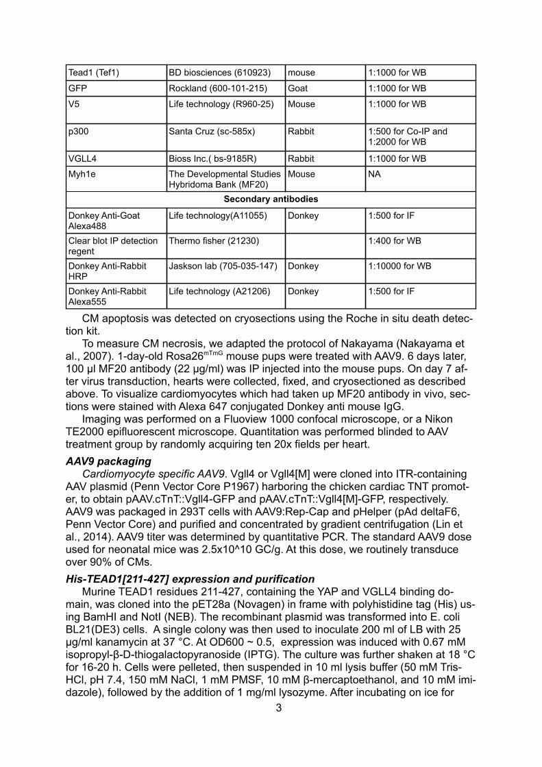
Tead1 (Tef1) BD biosciences (610923) mouse 1:1000 for WB
GFP Rockland (600-101-215) Goat 1:1000 for WB
V5 Life technology (R960-25) Mouse 1:1000 for WB
p300 Santa Cruz (sc-585x) Rabbit 1:500 for Co-IP and 1:2000 for WB
VGLL4 Bioss Inc.( bs-9185R) Rabbit 1:1000 for WB
Myh1e The Developmental StudiesHybridoma Bank (MF20)
Mouse NA
Secondary antibodies
Donkey Anti-Goat Alexa488
Life technology(A11055) Donkey 1:500 for IF
Clear blot IP detection regent
Thermo fisher (21230) 1:400 for WB
Donkey Anti-Rabbit HRP
Jaskson lab (705-035-147) Donkey 1:10000 for WB
Donkey Anti-Rabbit Alexa555
Life technology (A21206) Donkey 1:500 for IF
CM apoptosis was detected on cryosections using the Roche in situ death detec-tion kit.
To measure CM necrosis, we adapted the protocol of Nakayama (Nakayama et al., 2007). 1-day-old Rosa26mTmG mouse pups were treated with AAV9. 6 days later, 100 µl MF20 antibody (22 µg/ml) was IP injected into the mouse pups. On day 7 af-ter virus transduction, hearts were collected, fixed, and cryosectioned as described above. To visualize cardiomyocytes which had taken up MF20 antibody in vivo, sec-tions were stained with Alexa 647 conjugated Donkey anti mouse IgG.
Imaging was performed on a Fluoview 1000 confocal microscope, or a Nikon TE2000 epifluorescent microscope. Quantitation was performed blinded to AAV treatment group by randomly acquiring ten 20x fields per heart. AAV9 packaging
Cardiomyocyte specific AAV9. Vgll4 or Vgll4[M] were cloned into ITR-containing AAV plasmid (Penn Vector Core P1967) harboring the chicken cardiac TNT promot-er, to obtain pAAV.cTnT::Vgll4-GFP and pAAV.cTnT::Vgll4[M]-GFP, respectively. AAV9 was packaged in 293T cells with AAV9:Rep-Cap and pHelper (pAd deltaF6, Penn Vector Core) and purified and concentrated by gradient centrifugation (Lin et al., 2014). AAV9 titer was determined by quantitative PCR. The standard AAV9 dose used for neonatal mice was 2.5x10^10 GC/g. At this dose, we routinely transduce over 90% of CMs.His-TEAD1[211-427] expression and purification
Murine TEAD1 residues 211-427, containing the YAP and VGLL4 binding do-main, was cloned into the pET28a (Novagen) in frame with polyhistidine tag (His) us-ing BamHI and NotI (NEB). The recombinant plasmid was transformed into E. coli BL21(DE3) cells. A single colony was then used to inoculate 200 ml of LB with 25 µg/ml kanamycin at 37 °C. At OD600 ~ 0.5, expression was induced with 0.67 mM isopropyl-β-D-thiogalactopyranoside (IPTG). The culture was further shaken at 18 °Cfor 16-20 h. Cells were pelleted, then suspended in 10 ml lysis buffer (50 mM Tris-HCl, pH 7.4, 150 mM NaCl, 1 mM PMSF, 10 mM β-mercaptoethanol, and 10 mM imi-dazole), followed by the addition of 1 mg/ml lysozyme. After incubating on ice for
3

30 min, the suspension was sonicated (5 reps, 10 sec on, 30 sec off, amplitude 75) with Branson Digital Sonifier equipped with a microtip and subsequently centrifuged at 20,000g for 20 min at 4°C. The supernatant was incubated with 2 ml Ni-NTA Agarose (Qiagen) for 3 h at 4°C. The resin was washed with 20 column volumes of wash buffer (50 mM Tris-HCl, pH7.4, 150 mM NaCl, 1 mM PMSF, 10 mM β-mercap-toethanol and 20 mM imidazole). The protein was eluted with 4-column volumes of elution buffer (50 mM Tris-HCl, pH7.4, 150 mM NaCl, 1 mM PMSF, 10 mM β-mer-captoethanol and 300 mM imidazole). His-TEAD1[211-427] was concentrated and further purified by size exclusion chromatography with a Superdex 200 increase 10/300 GL column (GE Health Sciences) pre-equilibrated in a buffer of 50 mM Tris-HCl, pH7.4, 150 mM NaCl, 1 mM PMSF, 10 mM β-mercaptoethanol.Synthetic VGLL4 TDU domain peptides
Wild type and acetylated peptide containing VGLL4 Tdu domain and V5 epitope were synthesized in LifeTein LLC. The synthesized peptides were purified with HPLCto reach 95% purity and their molecular weight analyzed by electrospray ionization (ESI) mass spectrometry. The sequences are shown in Figure S3.Affinity measurement by photonic crystal nanobeam sensor.
Affinity measurement was performed using nanobeam photonic sensors consist-ing of photonic crystal nanobeam cavities for protein sensing and polymer spot-size converters for efficient on-and-off chip light coupling (Liang et al., 2013; Quan et al., 2010). Polydimethylsiloxane (PDMS) microfluidic channels were integrated on the sensor chip for sample delivery. The photonic crystal nanobeam cavities confine the optical energy into nanoscale dimensions, and build up high quality factor (Q-factor) resonances. The nanobeam cavity consists of a tapered array of holes with periodici-ty 330 nm along a 600 nm wide ridge waveguide. The radii of the holes were taperedfrom 240 nm in the center of the cavity to 100 nm to both ends of the cavity, de-signed by the deterministic method described in (Quan and Loncar, 2011). The de-vice was fabricated as described (Quan and Loncar, 2011). Protein binding was measured by monitoring the resonance shift of the nanobeam cavity.
100 ng/mL His-TEAD1[211-427] was first flowed to the nanobeam sensor via PDMS microfluidic channels, together with 4 mM sodium cyanoborohydride (Sigma) in PBS. After 2 hour-incubation at room temperature, the nanobeam sensor was washed by PBS flow for 10 min. Different concentrations of VGLL4 or acetylated VGLL4 were consecutively injected into the channel. We used the tunable laser (Santec) to scan the input wavelength and collected the signal transmitted through the cavity. We obtained the resonance shift by fitting the resonance with Lorentz curve.mVGLL4 K216 acetylation-specific antibody generation.
The antigen design and antibody generation was carried out by Yenzym antibod-ies, LLC. The antigen used to raise VGLL4 antibody is synthesized mouse VGLL4 (209-222, EHFRRSLGKNYKEPE) peptide, in which K216 was acetylated. Rabbits were given four immunizations and acetylation-specific antibodies were isolated by affinity purification. TEAD1-Dendra2 merge protein time lapse imaging
293T cells were cultured on glass bottom 35mm dishes. One day after plasmidtransfection, cells were treated with Dox in the absence or presence of E64. GreenDendra2 protein was partially converted into red fluosrecenese protein with 30 sec-onds 405nm light illuminating. Images were taken 3 minutes after illuninating. Timelapse imaging was carried out with Nikon TE2000 epifluorescent microscope at a
4
speed of 1 image/min. For each group, 6 different regions of interest were used forquantifying red fluorescence intensity (RFI). Gene Expression
Real time PCR was performed with Syber Green or Taqman detection using Bio-Rad CFX96 Real time system. PCR primers are listed below: PrimersGene* Forward Reverse mCTGF CCACCCGAGTTACCAATGAC GACAGGCTTGGCGATTTTAGmCCNA2 GCCTTCACCATTCATGTGGAT TTGCTCCGGGTAAAGAGACAGmCDC20 TTCGTGTTCGAGAGCGATTT
GACCTTGGAACTAGATTTGCCAG
mAurka GGGTGGTCGGTGCATGCTCCA
GCCTCGAAAGGAGGCATCCCCACTA
mMyh6 CTCTGGATTGGTCTCCCAGC GTCATTCTGTCACTCAAACTCTGGmGapdh CAGGTTGTCTCCTGCGACTT GGCCTCTCTTGCTCAGTGTCmTead1 TACTGCCATCCACAACAAGC TGCTGCACAAAGGGCTTGACABI Taqmanassays
Gene Assay Number
mNppa PN4453320mGapdh 4352339E
StatisticsValues are expressed as mean ± SEM. For two group comparisons, Student’s t-
test was used to test for statistical significance. To analyze data containing morethan two groups, we used ANOVA with the Tukey HSD post-hoc test. Both tests wereperformed using JMP 10.0 (SAS).
Supplemental References
Driegen, S., Ferreira, R., van Zon, A., Strouboulis, J., Jaegle, M., Grosveld, F., Philipsen, S., and Meijer, D. (2005). A generic tool for biotinylation of tagged proteins in transgenic mice. Transgenic Res 14, 477-482.
Liang, F., Clarke, N., Patel, P., Loncar, M., and Quan, Q. (2013). Scalable photon-ic crystal chips for high sensitivity protein detection. Opt Express 21, 32306-312.
Lin, Z., von Gise, A., Zhou, P., Gu, F., Ma, Q., Jiang, J., Yau, A.L., Buck, J.N., Gouin, K.A., et al. (2014). Cardiac-specific YAP activation improves cardiac function and survival in an experimental murine MI model. Circ Res 115, 354-363.
Livet, J., Weissman, T.A., Kang, H., Draft, R.W., Lu, J., Bennis, R.A., Sanes, J.R.,and Lichtman, J.W. (2007). Transgenic strategies for combinatorial expression of fluorescent proteins in the nervous system. Nature 450, 56-62.
Muzumdar, M.D., Tasic, B., Miyamichi, K., Li, L., and Luo, L. (2007). A global dou-ble-fluorescent Cre reporter mouse. Genesis 45, 593-605.
Nakayama, H., Chen, X., Baines, C.P., Klevitsky, R., Zhang, X., Zhang, H., Jaleel,N., Chua, B.H.L., Hewett, T.E., et al. (2007). Ca2+- and mitochondrial-dependent cardiomyocyte necrosis as a primary mediator of heart failure. J Clin Invest 117, 2431-444.
5

Quan, Q., and Loncar, M. (2011). Deterministic design of wavelength scale, ultra-high Q photonic crystal nanobeam cavities. Opt Express 19, 18529-542.
Quan, Q., Deotare, P.B., and Loncar, M. (2010). Photonic crystal nanobeam cavi-ty strongly coupled to the feeding waveguide. arXiv preprint arXiv:1002.1319
6
2 3 1 4 5 6 7 8 9 10 11 12
Exon 12
Flagbio-Stop FRT sequence
Neo 3’UTR
Exon 12
Flagbio-Stop
ActB::Flpe
ES cell genetargeting
Tead1 genomic
Tead1flagbio-Neo
Tead1flagbio
3’UTR
78KD
54KD
TEAD1fb
TEAD1fb
A
DC
B
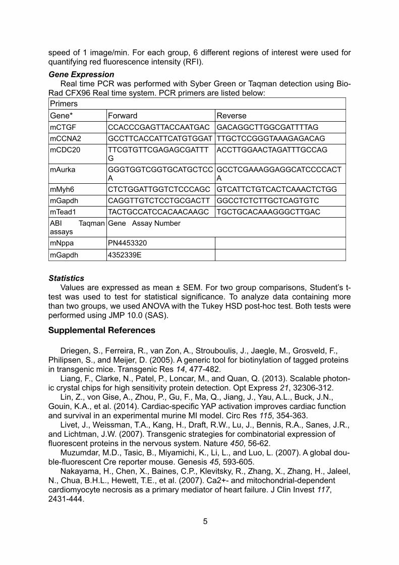
Figure S1. Construction and validation of Tead1fb allele (supporting Figure 1). A. Gene targeting strategy for generation of Tead1flagbio knock-in mice (Tead1fb/+). Flag and bio epitope tags were placed on the Tead1 C-terminus. Tead1flagbio-Neo mouse was mated to ActB::Flpe mouse to removed Neomycin. B. Homologous recombination in embryonic stem (ES) cells was confirmed by Southern Blotting. Arrow indicates the wildtype allele, and arrowhead indicates the targeted allele. Two independent Tead1flagbio-Neo ES clones(#48 and #49) were tested. NC, negative control, no gemomic DNA included. WT, wild type ES genomic DNA. C. After removal of the Frt-neo-Frt cassette by ActB::Flpe, Tead1fb/+ mice were intercrossed. Tead1fb/fb mice survived normally. D. Echocar-diography measurement of Tead1fb/fb mice heart function. 4 months old willd type mice and Tead1fb/fb mice were used for heart function test. NS, no signifi-cant difference. N=4. E. Western blot with adult heart tissue from indicated mice. F. Western blot with E14.5 heart tissue. Sreptavidin HRP was used to detect biotinylated Tead1fb, demonstrating in vivo biotinylation.
Streptavidin HRP blot
E F
TEAD1 immunoblot
TEAD1
Tead
1fb/f
b
Tead1+/+;RosaBirA/+
Tead1fb/+;RosaBirA/+
Tead1+/+
Tead
1+/+
+/+fb/+
fb/fbn=62
Frequency at weaning
WT
2363bp
4312bp
fb-neo48 49 NC
010
20
30
40
50
FS%
NS
Input
YAP
TEAD1
Load
GFP Ab(VGLL4-GFP)
VGLL4
TEAD1fb
(SA) Co-IPGFP Vgll4 GFP Vgll4
GFP Vgll4Input
GFP Vgll4
TEAD1fb
(SA) Co-IP
GFP Ab(VGLL4)
GFP Ab(VGLL4-GFP)
GAPDH TEAD1 Ab
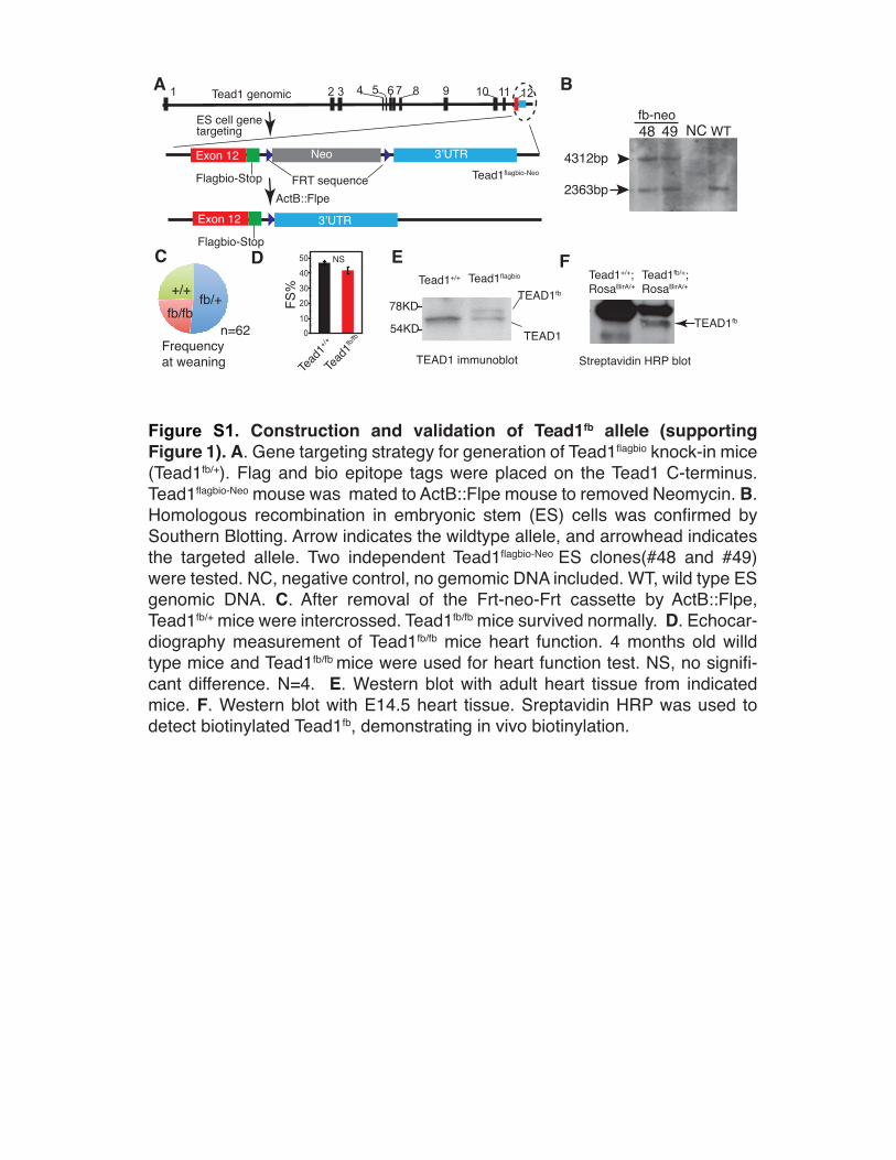
Figure S2. TEAD1 interacts with VGLL4-GFP in the adult heart (supporting Figure 2). AAV-GFP (GFP) or AAV-VGLL4-GFP (Vgll4) were administered to 1 day old Tead1fb/+;R26BirA/+
pups. Mouse hearts were collected at either P8 (A) or at 1 month after AAV administration (B). Tead1fb was pulled down on SA beads, and co-precipitated VGLL4-GFP was analyzed by western blotting. Ponceau S (A) or GAPDH (B) were used as loading controls.
A B
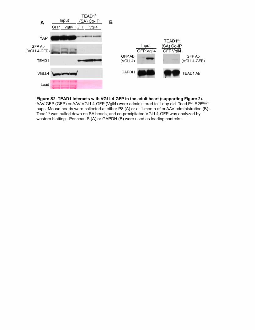
Figure S3. Recombinant TEAD1 and synthetic VGLL4-TDU peptides; Validation of VGLL4 and TEAD1 antibody for immunofluores-cence staining;p300 and VGLL4 interaction analysis in adult heart. Related to Figure 3.
A. Sequence of synthesized VGLL4 TDU domain peptide. Underlined characters indicate V5 peptide sequence. The acetylated lysine is shown in red. B. TEAD1 YBD domain was fused to His tag, and expressed in E.Coli. Soluble proteins were run through Ni resin to purify TEAD1-YBD-His(T-YBD-His). FPLC peaks are labeled with number. The elution volume for peaks 1, 2, 3, 4, 5, 6, are 8.8, 10.6, 15, 17, 20, 21ml, respectively. Peak “N” not included in the western blot. C. Commassie blue staining and western blot. Arrow indicate the T-YBD-His protein. His tag antibody was used to detect His-TEAD1 in the western blot. Peak 3 was run in two lanes, lane 5 and lane 9. In lane 5, samples from peak 3 were diluted 10 times. D. Validation of TEAD1 and VGLL4 antibodies for immunofluoresence staining. NRVMs were fixed with PFA and stained with the indicated anitbodies. TNNI3 was used as a cardiomyocyte marker. Bar = 20 μm. E. Validation of VGLL4-K225Ac antibody. Acetylated or non-acetylated synthetic VGLL4 peptides were bound to PVDF membranes and then probed with antibody directed against total or K225Ac VGLL4. Bound antibody was visualized with HRP-conjugated secondary antibody. F. Validation of VGLL4-K255Ac antibody in cell lysates. 293T cells were co-transfected with p300 and VGLL4 or VGLL4[R] expression constructs. Lysates were immunoblotted wtih total VGLL4 and VGLL4-K225-Ac antibodies. G. Expression of p300 in neonatal and adult heart. H, p300 does not interact with VGLL4 in the adult heart. AAV9-GFP (GFP) and AAV9-V-GFP (V) were delivered into the P1 mouse pups, respectively. At P60, hearts were collected for p300 Co-IP assay. Arrow indicates non-specific IgG band. VGLL4-GFP did not detectably co-immunoprecipitate with p300.
CB
A
D E
Input
P1
2
P2 P3(10x
dil)
P3 (no
dil)
P4 P5 P6
His-TEAD1[211-427](His Ab)
P1
P3
P2
P4
P5
P6N
A 280
Vol (ml)27KD
27KD
19KD
19KD
(Peak number)
TDU-V5TDU-KAc-V5
DPVVEEHFRRSLGKNYKEPEPAPNSVSITGSVDDHFAKALGDTWLQIKAAKDGAGKPIPNPLLGLDST
DPVVEEHFRRSLGKNYKEPEPAPNSVSITGSVDDHFAKALGDTWLQIKAAKDGAGKPIPNPLLGLDST
DAPI
DAPI
TEAD1 TNNI3 Merge
MergeTNNI3VGLL4
Ac
V5
VGLL4-TDUnot acetylated
VGLL4-TDUK225Ac
VGLL4 Ab
VGLL4 K225Ac Ab
GAPDH
VGLL4
VGLL4 K225 Ac
p300
p300F VGLL4 VGLL4[R]
+– +–HG
GAPDH
Inputp300 Ab
Co-IP
V-GFP
AAV9: V VGFPGFP
p300
P6 P60
p300
TetO Vgll4
VGLL4
HA
EF1a rtTA
Dox
TetO Vgll4HA
EF1a rtTApEF1α::rtTA
pTetO::Vgll4
No Dox With DoxA
C
B
D
pEF1α::rtTA + pTetO::HA-Vgll4
HA-VGLL4
GAPDH
0 2 4 6 8 10Dox(Hours)
0 2 4 6 8Dox(Hours)
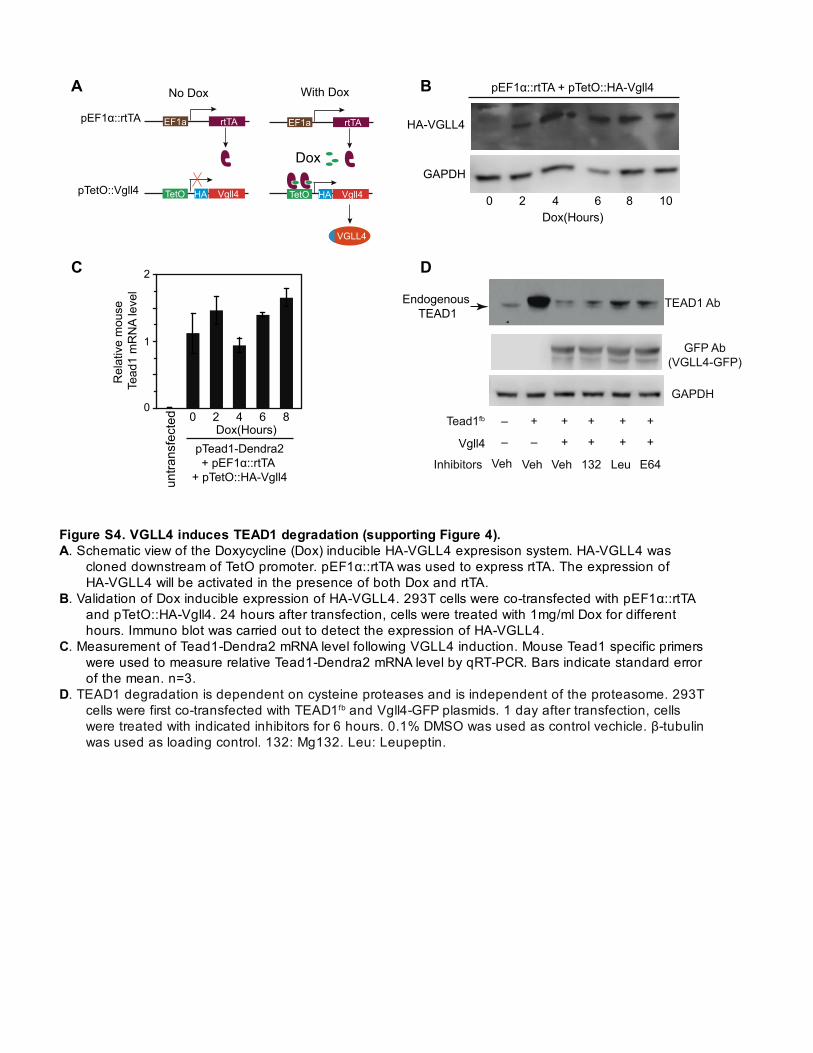
Figure S4. VGLL4 induces TEAD1 degradation (supporting Figure 4). A. Schematic view of the Doxycycline (Dox) inducible HA-VGLL4 expresison system. HA-VGLL4 was
cloned downstream of TetO promoter. pEF1α::rtTA was used to express rtTA. The expression of HA-VGLL4 will be activated in the presence of both Dox and rtTA.
B. Validation of Dox inducible expression of HA-VGLL4. 293T cells were co-transfected with pEF1α::rtTA and pTetO::HA-Vgll4. 24 hours after transfection, cells were treated with 1mg/ml Dox for different hours. Immuno blot was carried out to detect the expression of HA-VGLL4.
C. Measurement of Tead1-Dendra2 mRNA level following VGLL4 induction. Mouse Tead1 specific primers were used to measure relative Tead1-Dendra2 mRNA level by qRT-PCR. Bars indicate standard error of the mean. n=3.
D. TEAD1 degradation is dependent on cysteine proteases and is independent of the proteasome. 293T cells were first co-transfected with TEAD1fb and Vgll4-GFP plasmids. 1 day after transfection, cells were treated with indicated inhibitors for 6 hours. 0.1% DMSO was used as control vechicle. β-tubulin was used as loading control. 132: Mg132. Leu: Leupeptin.
E64VehVeh Veh Leu132
– – + + + +
– + + + + +
TEAD1 Ab
Tead1fb
GFP Ab(VGLL4-GFP)
Inhibitors
GAPDH
Rel
ativ
e m
ouse
Te
ad1
mR
NA
leve
l
pTead1-Dendra2+ pEF1α::rtTA
+ pTetO::HA-Vgll4untra
nsfe
cted
0
1
2
Vgll4
Endogenous TEAD1
Body w
eig
ht
(g)
Heart
weig
ht
(mg)
P8 P12 P8 P120 0
20
40
60
2
4
6
8
AAV9.GFP
AAV9.VGLL4
AAV9.VGLL4[R]
A B
C
*
*
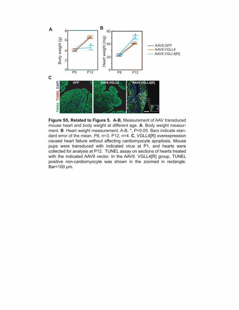
Figure S5, Related to Figure 5. A-B, Measurement of AAV transduced
mouse heart and body weight at different age. A. Body weight measur-
ment. B. Heart weight measurement. A-B, *, P<0.05. Bars indicate stan-
dard error of the mean. P8, n=3. P12, n=4. C, VGLL4[R] overexpression
caused heart failure without affecting cardiomyocyte apoptosis. Mouse
pups were transduced with indicated virus at P1, and hearts were
collected for analysis at P12. TUNEL assay on sections of hearts treated
with the indicated AAV9 vector. In the AAV9. VGLL4[R] group, TUNEL
positive non-cardiomyocyte was shown in the zoomed in rectangle.
Bar=100 µm.
GFP AAV9.VGLL4 AAV9.VGLL4[R]
TNN
I3 T
UN
EL D
API
Clo
ne p
erc
enta
ge
Monocrh
om
atic
/section
2x10⁹ GC/g
2x10⁹ GC/g
1x10⁸ GC/g
1x10⁸ GC/g
5x10⁸ GC/g
5x10⁸ GC/g
A
B C
2 color
clones
1 color
clones
YFP RFP
0
2
4
6
8
10
0
50
100
150
200
250
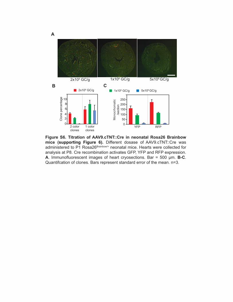
Figure S6. Titration of AAV9.cTNT::Cre in neonatal Rosa26 Brainbow mice (supporting Figure 6). Different dosase of AAV9.cTNT::Cre was
administered to P1 Rosa26Brainbow/+ neonatal mice. Hearts were collected for
analysis at P8. Cre recombination activates GFP, YFP and RFP expression.
A. Immunofluorescent images of heart cryosections. Bar = 500 µm. B-C.
Quantifcation of clones. Bars represent standard error of the mean. n=3.