hippo component yap promotes focal adhesion and tumour
TRANSCRIPT

RESEARCH Open Access
Hippo component YAP promotes focaladhesion and tumour aggressiveness viatranscriptionally activating THBS1/FAKsignalling in breast cancerJie Shen1,2, Beibei Cao1,2, Yatao Wang1,2, Chenshen Ma1,2, Zhuo Zeng1,2, Liang Liu1,2, Xiaolan Li2, Deding Tao2,Jianping Gong1,2* and Daxing Xie1,2*
Abstract
Background: Focal adhesion plays an essential role in tumour invasiveness and metastasis. Hippo component YAPhas been widely reported to be involved in many aspects of tumour biology. However, its role in focal adhesionregulation in breast cancer remains unexplored.
Methods: Tissue microarray was used to evaluate YAP expression in clinical breast cancer specimens byimmunohistochemical staining. Cell migration and invasion abilities were measured by Transwell assay. A celladhesion assay was used to measure the ability of cell adhesion to gelatin. The focal adhesion was visualizedthrough immunofluorescence. Phosphorylated FAK and other proteins were detected by Western blot analysis.Gene expression profiling was used to screen differently expressed genes, and gene ontology enrichment wasperformed using DAVID software. The gene mRNA levels were measured by quantitative real-time PCR. The activityof the THBS1-promoter was evaluated by dual luciferase assay. Chromatin immunoprecipitation (ChIP) was used toverify whether YAP could bind to the THBS1-promoter region. The prediction of potential protein-interaction wasperformed with the String program. The ChIP sequence data of TEAD was obtained from the ENCODE databaseand analysed via the ChIP-seek tool. The gene expression dataset (GSE30480) of purified tumour cells from primarybreast tumour tissues and metastatic lymph nodes was used in the gene set enrichment analysis. Prognosticanalysis of the TCGA dataset was performed by the SurvExpress program. Gene expression correlation of the TCGAdataset was analysed via R2: Genomics Analysis and Visualization Platform.
Results: Our study provides evidence that YAP acts as a promoter of focal adhesion and tumour invasiveness viaregulating FAK phosphorylation in breast cancer. Further experiments reveal that YAP could induce FAKphosphorylation through a TEAD-dependent manner. Using gene expression profiling and bioinformatics analysis,we identify the FAK upstream gene, thrombospondin 1, as a direct transcriptional target of YAP-TEAD. SilencingTHBS1 could reverse the YAP-induced FAK activation and focal adhesion.
Conclusion: Our results unveil a new signal axis, YAP/THBS1/FAK, in the modulation of cell adhesion andinvasiveness, and provides new insights into the crosstalk between Hippo signalling and focal adhesion.
Keywords: Breast cancer, Focal adhesion, YAP, THBS1, FAK
* Correspondence: [email protected]; [email protected];[email protected] Medicine Center, Tongji Hospital, Tongji Medical College,Huazhong University of Science and Technology, 1095 Jiefang Av., Wuhan,Hubei 430030, People’s Republic of ChinaFull list of author information is available at the end of the article
© The Author(s). 2018 Open Access This article is distributed under the terms of the Creative Commons Attribution 4.0International License (http://creativecommons.org/licenses/by/4.0/), which permits unrestricted use, distribution, andreproduction in any medium, provided you give appropriate credit to the original author(s) and the source, provide a link tothe Creative Commons license, and indicate if changes were made. The Creative Commons Public Domain Dedication waiver(http://creativecommons.org/publicdomain/zero/1.0/) applies to the data made available in this article, unless otherwise stated.
Shen et al. Journal of Experimental & Clinical Cancer Research (2018) 37:175 https://doi.org/10.1186/s13046-018-0850-z
BackgroundAlthough great achievements have been made in theareas of screening, diagnosis and therapy, breast canceris still the leading cause of cancer-related deaths inwomen worldwide [1]. In breast cancer patients, metas-tasis at distant sites, rather than primary tumour, is themajor obstacle of treatment and the main cause of can-cer lethality [2]. Metastasis is a long, sequential process,in which the interaction between cancer cells and thetumour extracellular matrix (ECM) is essential [3].Cell-ECM crosstalk plays a key role in regulating tumourcell motility and invasiveness through numerous cellularbiomechanics, such as focal adhesion, membrane remod-elling, actin protrusion, actomyosin contraction, and cellmotility signalling pathways [4]. Among these, focal ad-hesion has been revealed to be a crucial determinant ofcell migration and plays an important role in promotingtumour cell invasion [5].Focal adhesion (FA) is a subcellular structure which pro-
vides strong adhesion to the ECM and acts as a scaffoldfor many signalling pathways involving integrin or themechanical force exerted on cells [6]. Recent studies haverevealed the dynamic cycle of “FA assembly–cytoskeletonremodelling–FA disassembly”, which allows cells toachieve motility, and the dysregulation of FA is consideredto be an essential step in tumour invasion [5, 7]. Manycomponents of FA are tyrosine kinases and their sub-strates, of which focal adhesion kinase (FAK, also knownas PTK2) has been demonstrated to be a major participantin FA dynamics [8]. After integrin engagement, FAK is re-cruited and phosphorylated at Tyr397 [9]; the phosphory-lated FAK leads to the recruitment of other signallingmolecules and promotes the assembly of FA complexes[8]. In addition, there is also evidence showing that FAK isnecessary in FA disassembly [10]. As a key regulator ofFA, FAK plays an oncogenic role in a wide range of hu-man cancers [11]. Increased FAK expression and activityare often correlated with metastasis and poor prognosis[12–14]. Previous research has proven the correlation be-tween FAK activation and metastasis in breast cancer [15].Disrupting FAK could slow metastasis formation of mam-mary tumours [16, 17]; thus, it has been selected as a po-tential therapeutic target for aggressive breast cancers(reviewed in [18]). Although the significance of focal adhe-sion and FAK in breast malignancy metastasis has beenwidely reported, it is still unclear how FA is regulated intumour progression.Over the past decade, Hippo signalling has been proven
to be a master regulator network in many aspects oftumour biology [19, 20]. Yes-associated protein (YAP) actsas the main effector of the Hippo pathway and triggersdownstream biological effects through inducing targetgene transcription via interacting with related transcrip-tion factors, especially TEA domain family members
(TEADs) [21]. YAP has been considered to be an onco-gene in breast cancer, and its dysregulation often leads totumour aggressiveness and metastasis [22, 23]. Recentstudies have uncovered the critical role of YAP in theregulation of actin dynamics and cell motility [24, 25].This evidence indicates a potential relationship betweenHippo signalling and tumour metastasis; however, theconcrete mechanism still remains to be explored.This current study focuses on the role of YAP in FA
regulation and tumour metastasis in breast cancer. Inthis research, we have revealed the potential relationshipbetween YAP activation and tumour metastasis in clin-ical breast tumour specimens. Through in vitro experi-ments we have observed that YAP could significantlypromote FA formation and FAK activation in breast can-cer cell lines. Furthermore, we have validated that theYAP-TEAD interaction is essential for these biologicaleffects. Using gene expression profiling and the EN-CODE database, we have identified Thrombospondin 1(THBS1), a previously reported FAK stimulator [26–28],as a direct transcriptional target of Hippo signalling. Wehave further demonstrated that YAP/TEAD could in-crease THBS1 expression to promote FAK phosphoryl-ation and FA formation, leading to the activation oftumour cell migration and invasiveness. Collectively, ourfindings revealed a novel function of Hippo signalling ininducing FAK activation and focal adhesion formation topromote breast cancer aggressiveness and metastasis.
Materials and methodsTissue microarray and immunohistochemistry (IHC)A human breast cancer tissue microarray of 104 cases ofpaired primary lesion/lymphatic metastasis (US Biomax,Cat. #BR20837a) was used to evaluate the expression ofYAP in primary and metastatic tissue. The slide wasdewaxed, rehydrated and heated in sodium citrate buffer(0.01 M, pH 6.0) for antigen retrieval. Endogenous per-oxidase was then inhibited with 3% hydrogen peroxidewith 0.1% sodium azide for 30 min and non-specificstaining was blocked by incubation in 5% bovine serumalbumin for 2 h. The slide was then incubated in 1:100diluted YAP antibody (Cell Signaling Technology, Cat.#4912) at 4 °C overnight and subsequently with biotinyl-ated secondary antibody for 2 h. The DAB HorseradishPeroxidase Color Development Kit (Wuhan BosterBioCo. Ltd., Cat. #AR1022) was used for immunostaining,and counterstain was performed by haematoxylin stain-ing. The results were analysed under a microscope.The expression level of YAP was evaluated by the IHC
score, which was calculated by multiplying a proportionscore and an intensity score, and was categorized as level1 (IHC score 0–3), level 2 (IHC score 4–6) or level 3 (IHCscore greater than 6). The proportion score reflected thefraction of positive-stained cells (0, none; 1, ≤10%; 2, 10%
Shen et al. Journal of Experimental & Clinical Cancer Research (2018) 37:175 Page 2 of 17
to ≥25%; 3, > 25 to 50%; 4, > 50%), and the intensity scorerevealed the staining intensity (0, no staining; 1, weak; 2,intermediate; 3, strong). The nucleus localization of YAPwas measured by a nucleus score. The nucleus scorerepresented the fraction of positive-stained nuclei (0 = 0–10%; 1 = 11–30%; 2 = 31–70%; 3 = 71–100%). The cyto-plasm expression of YAP was evaluated by a cytoplasmicscore. The cytoplasmic score was calculated by multiply-ing the intensity of cytoplasmic staining (no staining = 0,weak staining = 1, moderate staining = 2, strong staining =3) and the extent of stained cells (0 = 0–10%; 1 = 11–30%;2 = 31–70%; 3 = 71–100%).
Cell culture and transfectionHuman breast cancer cell lines MDA-MB-231, MCF7 andhuman embryonic kidney cell line HEK293T were pur-chased from American Type Culture Collection (ATCC).The HEK293T and MCF7 cell lines were routinely cul-tured in Dulbecco’s modified Eagle medium (DMEM,KeyGEN), and the MDA-MB-231 cell line was maintainedin Leibovitz’s L-15 medium (L15, KeyGEN). DMEM andL15 culture media were supplemented with 10% foetal bo-vine serum (MULTICELL, Cat. #086–150) and 1% penicil-lin/streptomycin (KeyGEN). MCF7 and HEK293T cellswere cultured at 37 °C in a 5% CO2 incubator, whileMDA-MB-231 cells were cultured at 37 °C in a 100% airincubator, according to the ATCC instructions.Small-interfering RNAs (siRNAs) targeting YAP and
THBS1 were designed and synthesized by Guangzhou Ribo-Bio Co. Ltd. The sequences of the siRNAs are described inAdditional file 1: Table S1. The siRNA transfections wereperformed using Lipofectamine® 2000 transfection reagent(Thermo Fisher, Cat. #11668019), according to the manufac-turer’s protocol. Non-targeting siRNAs (siNCs) were usedfor the negative control. After 48 h, cell biological and bio-chemical experiments were performed.The plasmids pcDNA3.1-YAP, pcDNA3.1-YAP-S127A
(FLAG-tagged) and pcDNA3.1-YAP-S94A (GFP-tagged)were previously constructed and used for the overex-pression of YAP and its mutants. After transfection, thecells were treated with 500 μg/ml G418 (Santa Cruz Bio-technology, Cat. #sc-29,065) for 4 weeks to obtain stablecell lines. Empty vector was used as a negative control.
Transwell migration and invasion assayTranswell plates (24-well, pore size 8 μm (Corning, Cat.#3422)) were used for the transwell assay. For the migra-tion assay, 1*105 cells were harvested in 100 μl ofserum-free culture medium and added into the upperchamber, without Matrigel. For the invasion assay, trans-well filters were pre-coated with 30 μl of 1:8 dilutedMatrigel (BD, Cat. #356234) prior to the addition of thecell suspension. Next, 600 μl of 30% foetal bovine serummedium was placed into the bottom compartment of
the chamber as a source of chemo-attractant. After 24 hof culturing, the cells that crossed the inserts were fixedand strained with crystal-violet. Migrated cells werephotographed and counted via an inverted microscope(100X magnification).
Cell adhesion assayThe cell adhesion assay was performed as previously de-scribed [29]. Briefly, 2*105 cells per well were seeded into24-well plates on coverslips that were pre-coated with1% sterile gelatin (Sigma, Cat. #G-2500) and exposed todifferent treatments. After 2 h of culturing, the plateswere gently washed with PBS to remove thenon-adherent cells. The attached cells were then fixedwith 4% formaldehyde and stained with Wright’s-Giemsa. Attached cells were photographed and countedusing a microscope with 100X magnification.
ImmunofluorescenceTwenty thousand cells per well were seeded into 24-wellplates on 12 mm coverslips that were pre-coated with 1%sterile gelatin (Sigma, Cat. #G-2500) and exposed to dif-ferent treatments. After 24 h of culturing, the cells werefixed with 4% paraformaldehyde and permeabilized with0.1% Triton X-100/PBS. Blocking of nonspecific stainingwas achieved by incubation in 5% bovine serum albumin/PBS for 2 h. Subsequently, the cells were incubated over-night at 4 °C with anti-paxillin antibody (Abcam, Cat.#ab32084) at a dilution of 1:100, followed by incubation ina solution of fluorescently labelled secondary antibody(1:100) (Abbkine, Cat. #A24221, A23620) and 1:100 phal-loidin (Life Technologies, Cat. #A22287) for 2 h. Nucleiwas strained by DAPI, and coverslips were placed facedown onto a drop of anti-fading mounting medium on amicroscope slide. Images were captured via a confocallaser scanning microscope with 400X magnification. Eachexperiment was performed in triplicate.
Western blot assayTotal protein was extracted with NP40 lysis buffer with theaddition of phenylmethylsulfonyl fluoride and protein phos-phatase inhibitor cocktail (Cell Signaling Technology, Cat.#5870) and was separated on 10% SDS-PAGE gels. Afterelectrophoresis, the separated protein bands were transferredonto polyvinylidene fluoride membranes (Millipore, Cat.#IPVH00010) and blocked in 5% non-fat milk for 1 h atroom temperature. The membranes were then incubatedwith the primary antibodies against YAP (Cell SignalingTechnology, Cat. #4912), FAK (Abclonal, Cat. #A11131),pY397-FAK (Abclonal, Cat. #AP0302), THBS1 (Abclonal,Cat. #A2125) and GAPDH (Santa Cruz Biotechnology, Cat.#sc-32,233) at a diluted ratio of 1:1000 overnight at 4 °C.After washing three times, the membranes were incubatedin 1:5000 horseradish peroxidase-linked secondary antibodies
Shen et al. Journal of Experimental & Clinical Cancer Research (2018) 37:175 Page 3 of 17
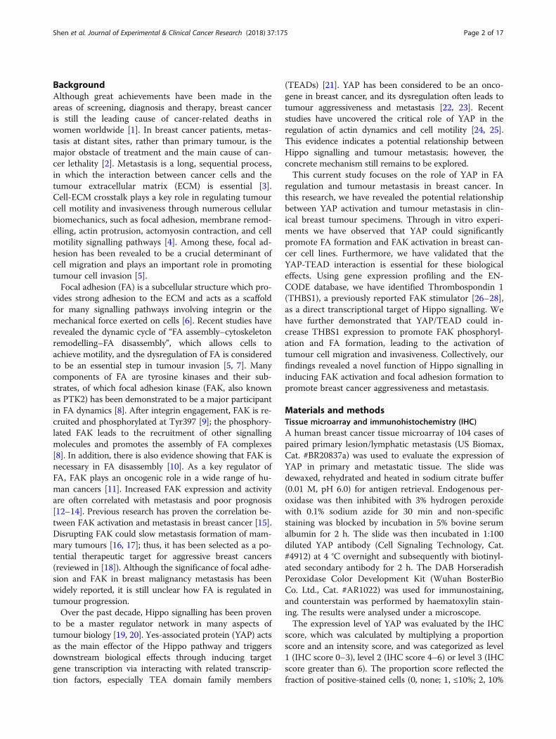
Fig. 1 YAP overexpression and activation were associated with lymphatic metastasis and poor prognosis in breast cancer patients. (a)Immunohistochemistry staining of YAP protein in paired primary and lymphatic metastatic specimens from one breast cancer patient. Lymphaticmetastasis revealed a higher expression level of YAP. Scale bar: 20 μm. (b) Immunohistochemistry staining score (IHC score) of YAP expression in101 paired primary/lymphatic metastatic breast cancer specimens from a breast cancer tissue microarray. The expression level of YAP wassignificantly higher in lymphatic metastases than in primary lesions (**p < 0.01 by paired Student’s t-test). Primary: primary lesion; Metastasis:lymphatic metastasis. (c) Immunohistochemistry cytoplasm expression (cytoplasmic score, left panel) and nucleus accommodation (nucleus score,right panel) of YAP in the 101 paired primary/lymphatic metastatic breast cancer specimens. The cytoplasm expression and nucleus accumulationof YAP was significantly higher in lymphatic metastases than in primary lesions (*p < 0.05 by paired Student’s t-test). Primary: primary lesion;Metastasis: lymphatic metastasis. (d) Analysis of TCGA breast invasive carcinoma dataset (n = 962) via SurvExpress program. Left: Heat mapsummarizing the expression values of YAP and its target genes (CTGF, CYR61, AXL and MYC) in breast cancer specimens from the TCGA dataset.Patients were sorted by prognostic index and divided into “Low Risk” and “High Risk” groups, according to the “Maximized Risk Groups” algorithm(see reference [32]). Middle: patients in the “High Risk” group presented a significantly higher expression level of YAP and its downstream genes(p < 0.01). Right: Kaplan-Meier analysis revealed that patients in the “High Risk” group suffered from poor prognosis (p < 0.01)
Shen et al. Journal of Experimental & Clinical Cancer Research (2018) 37:175 Page 4 of 17
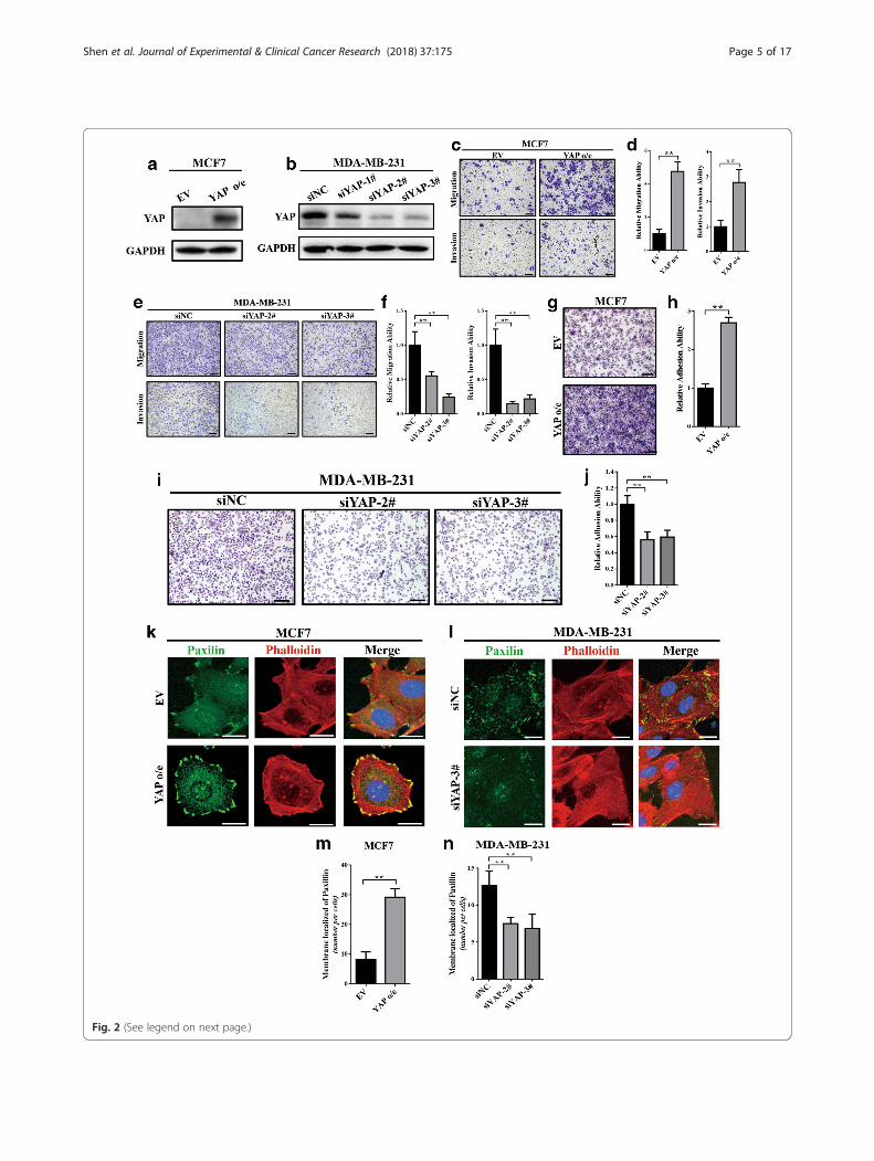
Fig. 2 (See legend on next page.)
Shen et al. Journal of Experimental & Clinical Cancer Research (2018) 37:175 Page 5 of 17

at room temperature for 2 h. Finally, the membranes werewashed three times and were visualized using an ECL Kit(Thermo Fisher, Cat. #34096).
Gene expression profilingTotal RNA of MCF7-vector and MCF7-YAP1-S127Awere freshly extracted using TRIzol reagent (Takara, Cat.#9108). RNA quantity and integrity were assessed usinga NanoDrop ND-2000 (Thermo Scientific) and an Agi-lent Bioanalyzer 2100 (Agilent Technologies). The geneexpression profiling was conducted by Shanghai Oebio-tech Corporation using the Agilent SurePrint G3 HumanGene Expression v3 Panel (Agilent, CA, USA). All datawere analysed according to the manufacturer’s protocol.Differentially expressed genes were then identified byfold change. The threshold set for up- and downregu-lated genes was a fold change greater than 2.
Quantitative real-time PCR (qRT-PCR)Total RNA was extracted using TRIzol (Takara, Cat.#9108), according to the manufacturer’s protocol.Reverse-transcription was performed to obtain cDNAusing a PrimeScript™ RT Master Mix Reagent Kit(Takara, Cat. #RR036A), and quantitative real-time PCRwas carried out using a TB Green™ Premix Ex Taq™ IIKit (Takara, Cat. #RR820A) according to the manufac-turer’s protocol. GAPDH gene expression was used asan endogenous control, and the results from qRT-PCRwere analysed though the comparative Ct method(2-ΔΔCt). The primer sequences used in this research areprovided in Additional file 2: Table S2. Each experimentwas performed in triplicate.
Dual luciferase assayA total of 100 ng of pGL3-Basic plasmid (Promega, Cat.#E1751) with inserts of the THBS1 promoter sequence(TSS: − 2000 ~ + 50) were co-transfected into HEK293Tand MCF7 cells using Lipofectamine® 2000 transfectionreagent (Thermo Fisher, Cat. #11668019) along with
200 ng of YAP/YAP-mutant construct and 10 ng ofRenilla luciferase pRL-TK plasmid (Promega, Cat.#E2241). After 48 h, the dual luciferase assay was per-formed using the Dual-Luciferase® Reporter Assay System(Promega, Cat. #E1910), according to the manufacturer’sprotocol. Luciferase activity was measured as the ratio offirefly luciferase signal to Renilla luciferase signal. All mea-surements were normalized to the control group alone.Each experiment was performed in triplicate.
Chromatin immunoprecipitation (ChIP)After transfecting with empty vector or YAP1-S127Aplasmid for 48 h, the MCF7 cells were harvested, andChIP experiments were performed using the SimpleChIPEnzymatic Chromatin IP Kit (Cell Signaling Technology,Cat. #9003), according to the manufacturer’s protocol. Atotal of 500 μl of diluted cross-linked chromatin was in-cubated overnight with 5 μg of mouse monoclonalanti-YAP antibody (Cell Signaling Technology, Cat.#14074) or with 1 μg of normal mouse IgG (Cell Signal-ing Technology, Cat. #2729) at 4 °C. The THBS1 pro-moter sequence (primers: F: ACCGACTTTTCTGAGAAG, R: GCAACTTTCCAGCTAGAA) were quanti-fied by PCR and analysed by 2% agarose gel electrophor-esis with a 100 bp DNA marker.
Public database and bioinformatics analysisThe gene expression dataset (GSE30480, [30]) of purifiedtumour cells from 14 primary breast tumour tissues and 6metastatic lymph nodes was obtained from the Gene Expres-sion Omnibus database and was used in gene set enrichmentanalysis (GSEA) (http://software.broadinstitute.org/gsea/)[31]. The prognostic analysis of TCGA breast invasive carcin-oma dataset was performed by the SurvExpress program(http://bioinformatica.mty.itesm.mx:8080/Biomatec/SurvivaX.jsp) [32]. The ChIP-sequence data of TEAD4 in the MCF7cell line was downloaded from the ENCODE project (http://genome.ucsc.edu/ENCODE/downloads.html) (GSM1010860)and was analysed via the ChIPseek online tool (http://
(See figure on previous page.)Fig. 2 YAP was able to induced cell migration, invasion and focal adhesion in breast cancer cell lines. (a) Western blot verified the overexpressionof YAP in MCF7 cells. EV: empty vector; o/e: overexpression. (b) Western blot verified the knockdown of YAP in MDA-MB-231 cells via a collectionof siRNAs; siYAP-#2 and siYAP-#3 has relatively high knockdown efficiency, thus these two siRNAs were used in this research. (c, d) Transwell assayshowing that overexpression of YAP induced cell migration and invasion ability in MCF7 cells. The experiment was performed in triplicate.**p < 0.01 by Student’s t-test. Scale bar: 100 μm. (e, f) Transwell assay showing that knockdown of YAP significantly inhibited cell migration andinvasion ability in MDA-MB-231 cells. The experiment was performed in triplicate. ** p < 0.01 by ANOVA test. Scale bar: 100 μm. (g, h)Overexpression of YAP induced MCF7 cell adhesion to gelatin. The attached cells were stained with Wright’s-Giemsa and are shown in (g). Theexperiment was performed in triplicate. ** p < 0.01 by Student’s t-test. Scale bar: 100 μm. (i, j) Knockdown of YAP significantly inhibitedMDA-MB-231 cell adhesion to gelatin. The attached cells were stained with Wright’s-Giemsa and are shown in (i). The experiment was performedin triplicate. ** p < 0.01 by Student’s t-test. Scale bar: 100 μm. (k) Overexpression of YAP induced focal adhesions in MCF7 cells. Focal adhesionswere visualized by co-localization of paxilin (green) and F-actin (stained with phalloidin, red). Nuclei were counterstained with DAPI (blue). Scalebar: 20 μm. (l) Knockdown of YAP expression inhibited focal adhesions in MDA-MB-231 cells. Scale bar: 20 μm. (m) Quantification of themembrane-localized paxilin in (k). The experiment was performed in triplicate. ** p < 0.01 by Student’s t-test. (n) Quantification of themembrane-localized paxilin in (l). The experiment was performed in triplicate. ** p < 0.01 by ANOVA test
Shen et al. Journal of Experimental & Clinical Cancer Research (2018) 37:175 Page 6 of 17

Fig. 3 (See legend on next page.)
Shen et al. Journal of Experimental & Clinical Cancer Research (2018) 37:175 Page 7 of 17

chipseek.cgu.edu.tw/) [33]. The String database (http://www.string-db.org/) [34] was used for protein interactionanalysis. The gene expression correlations were revealed bythe R2: Genomics Analysis and Visualization Platform(http://r2.amc.nl) using the TCGA invasive carcinoma data-set. Gene ontology enrichment was performed by the DAVIDsoftware (https://david.ncifcrf.gov/tools.jsp).
Statistical analysisStatistical analysis was performed with the SPSS softwarepackage (version 19.0 for Windows; IBM, USA). All con-tinuous data are presented as the mean ± SD and statisti-cally analysed with Student’s t-test (two-tailed) andanalysis of variance (ANOVA). A p-value less than 0.05was considered to be statistically significant.
ResultsYAP overexpression and activation were associated withlymphatic metastasis and poor prognosis in breast cancerpatientsTo validate the relationship between YAP expression andmetastasis in breast cancer patients, a paraffin-embeddedtissue array containing 104 paired primary/lymphatic meta-static clinical breast cancer specimens was obtained. Due todropping, moving and wrinkling during the experiments 3cases were discarded, and the remaining 101 paired speci-mens were analysed. Through immunohistochemistrystaining, we found that the YAP expression level wasrelatively higher in lymphatic metastases than in primary le-sions (Fig. 1a, IHC score shown in Fig. 1b). In addition, theIHC cytoplasmic and nucleus scoring showed that the YAPprotein had a higher level of cytoplasm expression and nu-cleus accumulation in lymphatic metastases (Fig. 1c). Fur-thermore, gene set enrichment analysis was performed on a
gene expression profile of purified tumour cells from 14primary breast tumours and 6 metastatic lymph nodes thatwas available from the GSE database (GSE30480, [30]). Theresults revealed that the YAP conserved signature wasenriched in metastatic lymph nodes, with statistical signifi-cance (Additional file 3: Figure S1). This evidence indicatedthat YAP expression and activation was positively associ-ated with lymphatic metastasis in breast cancer.For the purpose of determining whether YAP overex-
pression and activation were associated with a poor prog-nosis in breast cancer, we used SurvExpress [32] toevaluate the expression level of YAP and its downstreamgenes (CTGF, CYR61, AXL and MYC [35, 36]) in theTCGA invasive carcinoma dataset (Fig. 1d, left, patientswere sorted in the ascending order of prognostic index).Through the SurvExpress program, patients in the TCGAdataset were divided into “Low Risk” and “High Risk”groups according to the prognostic index. Patients in the“High Risk” group presented a significantly higher expres-sion level of YAP and its downstream genes and sufferedfrom poor prognosis (Fig. 1d, middle and right). There-fore, overexpression and activation of YAP was supposedto be a biomarker of poor survival in breast cancerpatients.
YAP induced cell migration, invasion and focal adhesionin breast cancer cell linesPreviously, we examined the expression levels of YAP pro-tein in 4 breast cancer cell lines (MCF7, T47D,MDA-MB-231 and MDA-MD-468) and revealed that YAPwas overexpressed in MDA-MB-231, MDA-MB-468 andT47D cell lines, while it was relatively low expressed in theMCF7 cell line (data not shown). MDA-MB-231 and MCF7have been reported to have a high and low metastatic
(See figure on previous page.)Fig. 3 YAP-TEAD interaction was essential for tumour cell invasiveness and focal adhesion formation. (a) Western blot verified the overexpressionof two YAP mutants, YAP-S127A (FLAG-tagged) and YAP-S94A (GFP-tagged) in MCF7 cells. EV: empty vector; S127A: YAP constitutively activatedmutant (YAP1-S127A); S94A: YAP TEAD-binding domain mutant (YAP-S94A). (b) (c) Cell adhesion assays showed that ectopic expression ofYAP-S127A, rather than YAP-S94A, induced MCF7 cell adhesion to gelatin. The experiment was performed in triplicate. ** p < 0.01 by ANOVA test.Scale bar: 100 μm. (d, e, f) Transwell assays showed that compared with the YAP-S94A mutant, YAP-S127A could significantly induce cellmigration and invasion ability in MCF7 cells. The experiment was performed in triplicate. ** p < 0.01 by ANOVA test. Scale bar: 100 μm. (g) Ectopicexpression of YAP-S127A, rather than YAP-S94A, induced focal adhesions in MCF7 cells. Focal adhesions were visualized by co-localization ofpaxilin (stained with Dylight 649, violet) and F-actin (stained with phalloidin, red). Nuclei were counterstained with DAPI (blue). GFP is representedas green. Scale bar: 20 μm. (h, i) Representative images of MCF7-YAP-S127A cell adhesion to gelatin after treatment with verteporfin at a dose of10 μM for 24 h (DMSO was used as negative control). Verteporfin significantly inhibited cell adhesion ability of MCF7 cells expressing YAP-S127Amutant. The experiment was performed in triplicate. **p < 0.01 by ANOVA test. Scale bar: 100 μm. (j, k) Transwell assays showed that verteporfinsignificantly inhibited invasion ability of MCF7-YAP-S127A cells. MCF7-YAP-S127A cells were treated with verteporfin at a dose of 10 μM for 24 h(DMSO was used as negative control) before transwell assays were performed. The experiment was performed in triplicate. **p < 0.01 by ANOVAtest. Scale bar: 100 μm. (l) Verteporfin inhibited focal adhesions in MCF7-YAP-S127A cells. Cells were exposed to verteporfin (10 μM) or DMSO(negative control) for 24 h and then stained with paxilin (green). F-actin was stained with phalloidin (red). Nuclei were counterstained with DAPI(blue). Scale bar: 20 μm. (m, n) Verteporfin significantly inhibited cell adhesion ability in MDA-MB-231 cells. Cells were treated with verteporfin ata dose of 10 μM for 24 h before cell adhesion assays were performed. DMSO was used as negative control. The experiment was performed intriplicate. **p < 0.01 by Student’s t-test. Scale bar: 100 μm. (o, p) Verteporfin significantly inhibited invasion abilities in MDA-MB-231 cells. Cellswere treated with verteporfin at a dose of 10 μM for 24 h (DMSO was used as negative control) before transwell assays were performed. Theexperiment was performed in triplicate. **p < 0.01 by Student’s t-test. Scale bar: 100 μm. (q) Treatment with verteporfin (10 μM) for 24 hdecreased focal adhesions in MDA-MB-231 cells. Paxilin (green), F-actin (stained with phalloidin, red). Nuclei (blue). Scale bar: 20 μm
Shen et al. Journal of Experimental & Clinical Cancer Research (2018) 37:175 Page 8 of 17
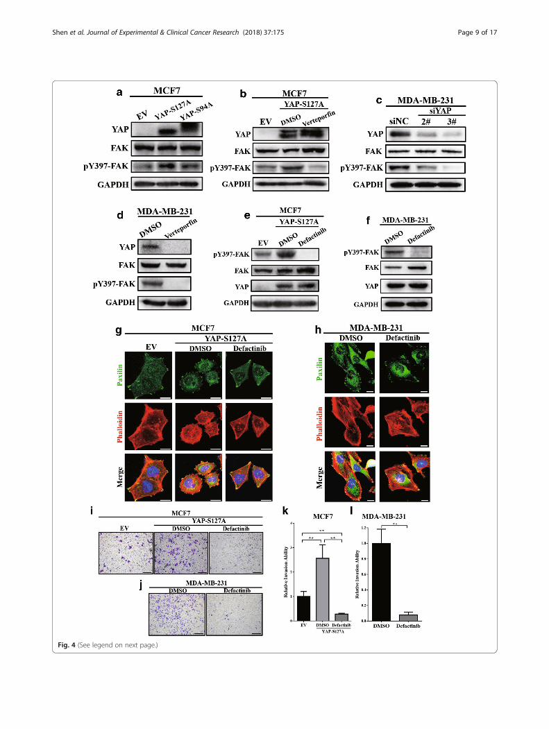
Fig. 4 (See legend on next page.)
Shen et al. Journal of Experimental & Clinical Cancer Research (2018) 37:175 Page 9 of 17
potential, respectively [37]; therefore, these two cell lineswere selected for further study. MCF7 was stably transfectedwith pcDNA3.1-YAP plasmid to overexpress YAP protein(Fig. 2a), and a collection of siRNAs was used to knockdownendogenous YAP expression in MDA-MB-231 cells (Fig. 2b).As shown in Fig. 2b, siYAP-#2 and siYAP-#3 demonstrated arelatively high knockdown efficiency, thus these two siRNAswere used in this research. The transwell assay revealed thatthe expression level of YAP was positively correlated withcell migration and invasion ability, both in MCF7 (Fig. 2c, d)and MDA-MB-231 (Fig. 2e, f) cells.Due to the important role of cell adhesion in cancer inva-
sion and metastasis [3], we next validated whether YAPcould regulate the cell adhesion ability. The overexpressionof YAP could significantly induce MCF7 cell adhesion togelatin (Fig. 2g, h), while knockdown of YAP inMDA-MB-231 cells significantly inhibited cell adhesion abil-ity (Fig. 2i, j). Interestingly, through immunofluorescence weobserved that the number of focal adhesions, an importantsub-cellular structure that mediates the regulatory effects ofa cell to ECM adhesion [38], was strongly associated withoverexpression of YAP in MCF7 (Fig. 2k, m) and was sig-nificantly decreased by YAP knockdown in MDA-MB-231cells (Fig. 2l, n). These results indicated that YAP could in-crease cell migration, invasion and adhesion abilities and in-duce focal adhesion formation in breast cancer cell lines.
YAP-TEAD interaction was essential for tumour cellinvasiveness and focal adhesion formationTo investigate the regulation mechanism of YAP in cell inva-siveness and focal adhesion formation, two pcDNA3.1-YAPmutant plasmids, FLAG-tagged-YAP-S127A (constitutivenuclei-accommodation mutant) and GFP-tagged-YAP-S94A(TEAD-binding domain mutant) were selected and stablytransfected into MCF7 cells (Fig. 3a). Compared to theYAP-S94A mutant, ectopic expression of YAP-S127A inMCF7 could significantly increase the cell adhesion ability(Fig. 3b, c) and promoted cell migration and invasion (Fig.3d-f). In addition, through immunofluorescence we found
that expression of the YAP-S127A mutant, rather thanYAP1-S94A, significantly increased focal adhesions in MCF7(Fig. 3g). Thus, nucleus accommodation and TEAD-bindingdomain are required for the YAP-induced cell invasivenessand focal adhesion formation.To further validate the essential role of the YAP-TEAD
interaction in tumour invasiveness and focal adhesion, ver-teporfin (MCE, Cat. # HY-B0146), a small molecular inhibi-tor of the YAP-TEAD interaction [39], was employed.Verteporfin could significantly reverse YAP-S127A-inducedcell adhesion (Fig. 3h, i) and invasion (Fig. 3j, k) in MCF7cells. Furthermore, after treating with verteporfin,MCF7-YAP-S127A presented a reduction of focal adhesions(Fig. 3l). Additionally, verteporfin could also significantlyinhibited cell adhesion (Fig. 3m, n), invasion (Fig. 3o, p) andfocal adhesions (Fig. 3q) in MDA-MB-231 cells. Togetherthese data suggest that the YAP-TEAD interaction was es-sential for tumour cell invasiveness and focal adhesion for-mation in breast cancer cell lines.
YAP-TEAD promoted focal adhesion formation in breastcancer cell lines by inducing FAK phosphorylationFocal adhesion kinase (FAK, also known as PTK2) is amajor component of focal adhesions, and the phosphoryl-ation of FAK at Tyr397 has been demonstrated to be amain step in the assembly of focal adhesion complexes [8,9]. In our study, we observed that the overexpression ofYAP-S127A, rather than YAP-S94A, significantly inducedFAK phosphorylation at Tyr397 in MCF7 (Fig. 4a). Apply-ing verteporfin could significantly reverse FAK phosphor-ylation in MCF7 cells (Fig. 4b). Similarly, eitherknockdown of YAP expression or treatment with vertepor-fin significantly inhibited FAK Tyr397 phosphorylation inMDA-MB-231 cells (Fig. 4c, d). Interestingly, verteporfinalso significantly decreased endogenous YAP protein levelsin MDA-MB-231 (Fig. 4d). This may have been caused bythe increasing proteasomal degradation of YAP due to thedisruption of the YAP-TEAD complex; however, the spe-cific mechanism requires further investigation.
(See figure on previous page.)Fig. 4 YAP-TEAD promoted focal adhesion formation in breast cancer cell lines through inducing FAK phosphorylation. (a) Western blot revealedthat compared with the YAP-S94A mutant, overexpression of the YAP-S127A mutant could promote FAK Y397 phosphorylation. (b) Verteporfinreversed YAP-S127A-induced FAK phosphorylation in MCF7 cells. MCF7-YAP-S127A was treated with verteporfin at a dose of 10 μM for 24 hbefore the Western blot assay was performed. DMSO was used as a negative control. (c) Knockdown of endogenous YAP expression inhibitedFAK Y397 phosphorylation in MDA-MB-231 cells. (d) Verteporfin inhibited FAK phosphorylation in MDA-MB-231 cells. Cells were treated withverteporfin at a dose of 10 μM for 24 h before Western blot assays were performed. DMSO was used as a negative control. (e, f) Western blotverified the inhibition of FAK Y397 phosphorylation via defactinib in MCF7-YAP-S127A (e) and MDA-MB-231 (f) cells. Cells were exposed todefactinib at a dose of 10 μM for 8 h before Western blot assays were performed. DMSO was used as a negative control. (g, h) Treatment withdefactinib (10 μM) for 8 h decreased focal adhesions, both in MCF7-YAP-S127A (g) and MDA-MB-231 (h). DMSO was used as a negative control.Paxilin (green), F-actin (stained with phalloidin, red). Nuclei (blue). Scale bar: 20 μm. (i) Transwell invasion assays showed that exposure todefactinib (10 μM) could significantly reverse YAP-S127A-induced cell invasion in MCF7 cells. DMSO was used as a negative control. Theexperiment was performed in triplicate. Scale bar: 100 μm. (j) Exposure to defactinib (10 μM) significantly decreased cell invasion ability inMDA-MD-231 cells. DMSO was used as a negative control. The experiment was performed in triplicate. Scale bar: 100 μm. (k) Quantification of therelative invasion ability in (i). ** p < 0.01 by ANOVA test. (l) Quantification of the relative invasion ability in (j). ** p < 0.01 by Student’s t-test
Shen et al. Journal of Experimental & Clinical Cancer Research (2018) 37:175 Page 10 of 17
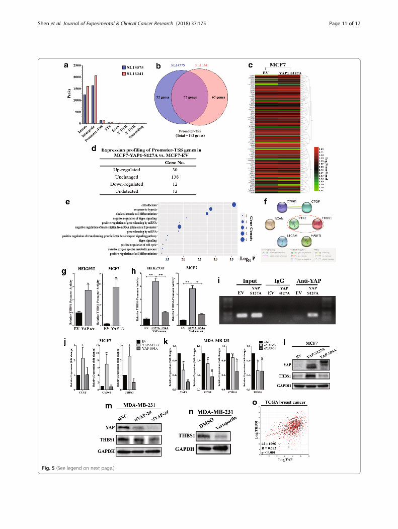
Fig. 5 (See legend on next page.)
Shen et al. Journal of Experimental & Clinical Cancer Research (2018) 37:175 Page 11 of 17
Next, to determine the role of FAK phosphorylation inYAP-induced FA formation, a novel FAK inhibitor, defacti-nib (MedChemExpress, Cat. # HY-12289), was used to in-hibit FAK Tyr397 phosphorylation in both MCF7-S127A(Fig. 4e) and MDA-MB-231 (Fig. 4f). As shown in Fig. 4gand Fig. 4h, after treating with defactinib, the number offocal adhesions was significantly decreased inMCF7-YAP-S127A and MDA-MB-231 cells. In addition,defactinib could also inhibit cell invasion in bothMCF7-YAP-S127A (Fig. 4i, k) and MDA-MB-231 (Fig. 4j,l). This evidence revealed that FAK-Tyr397 phosphorylationwas essential for YAP-TEAD regulated FA formation.
YAP-TEAD transcriptionally promoted expression of FAKupstream regulatory factor, thrombospondin 1 (THBS1)To determine how YAP-TEAD regulates FAK phosphoryl-ation, a collection of TEAD4 ChIP-sequence data (two repli-cations, SL14575 and SL16341) in MCF7 cells was firstobtained from the ENCODE database (GSM1010860) andanalysed via the ChIP-Seek tool (Fig. 5a). Peaks located inthe promoter-transcription start site (TSS) region wereexacted and annotated. As shown in Fig. 5b, a total of 192genes whose promoter was potentially combined withTEAD4 were identified. Next, gene expression profiling wasperformed to confirm the mRNA expression levels of these192 genes in MCF7 cells overexpressing the YAP-S127A mu-tant (Fig. 5c). Among them, 30 genes were upregulated inthe YAP-S127A mutant (fold change greater than 2) (Fig.5d). To further identify the potential upstream genes of FAK
which could be transcriptionally activated by YAP-TEAD,gene ontology (GO) enrichment analysis of these 30 upregu-lated genes was performed. As shown in Fig. 5e, “cell adhe-sion” was represented as the first GO enrichment categoryand included 6 genes (THBS1, HABP2, L1CAM, BCAM,CYR61 and CTGF). Interestingly, the “cell adhesion” cat-egory also ranked 6th in the GO enrichment analysis of allupregulated genes (1416 genes) that were identified in theYAP-S127A mutant in MCF7 cells (Additional file 4 TableS3). Through the String program, THBS1 appeared to be thepotential upstream regulatory factor of FAK (Fig. 5f).Thrombospondin 1 (THBS1) has been widely reported to
be an activator of FAK Tyr397 phosphorylation [26–28].According to the ChIP-sequence data from the ENCODEdatabase, TEAD4 can bind to the promoter region ofTHBS1 in MCF7 cells (Additional file 5: Fig. S2). Tocharacterize the transcriptional regulation of THBS1 byYAP, a luciferase-based reporter containing the promoterregion of THBS1 (TSS: -2000 ~ + 50 bp) was constructedand co-transfected with pcDNA3.1-YAP and Renilla plas-mids into HEK293T and MCF7 cells. The dual luciferasereporter assay revealed that YAP overexpression could sig-nificantly increase THBS1 promoter activity (Fig. 5g). Sub-sequently, YAP-S127A and YAP-S94A mutants wereco-transfected, and the results showed that compared withthe YAP1-S94A mutant, YAP1-S127A could significantlypromote THBS1 promoter activities in both HEK293T andMCF7 (Fig. 5h). Chromatin immunoprecipitation showedthat overexpression of the YAP-S127A mutant led to an
(See figure on previous page.)Fig. 5 YAP-TEAD transcriptionally promoted expression of FAK upstream regulatory factor, thrombospondin 1 (THBS1). (a) Analysis of TEAD ChIP-sequence data of MCF7 cells from the ENCODE database (GSM1010860) via the ChIP-seek tool. SL14575 and SL16341 were two bio-replications ofthe ChIP-sequence data. (b) Peaks in promoter-TSS category from (a) were exacted and annotated. A total of 192 genes whose promoter waspotentially combined with TEAD4 were identified. (c) Gene expression profiling was performed in MCF7 cells overexpressing the YAP-S127Amutant compared with empty vector. Expression values of the 192 genes from (b) were exacted and presented in a heat map. (d) The 192 geneswere divided into four categories according to the expression fold change (FC) in MCF7-YAP-S127A vs. MCF7-EV cells. Upregulated: genes thatwere upregulated by the YAP-S127A mutant with a FC≥ 2; Unchanged: genes with an expression fold change between the two groups of lessthan two; Downregulated: genes were downregulated by the YAP-S127A mutant with a FC ≥ 2; Undetected: genes that were not detected bythe expression profiling. (e) Gene ontology analysis (biological processes) was performed for the 30 upregulated genes from (d). “Cell adhesion”was the first enrichment category and contained 6 genes. (f) Using the STRING program to analyse potential interactions between FAK (alsoknown as PTK) and the 6 upregulated genes that were included in the “cell adhesion” category (THBS1, HABP2, L1CAM, BCAM, CYR61 and CTGF).THBS1 appeared to be highly correlated to FAK (confidence score: 0.849). (g) Dual luciferase reporter assays showed that THBS1 promoter activitycould be significantly enhanced by YAP, both in HEK293T and MCF7 cells. *p < 0.05 by Student’s t-test. The experiments were performed intriplicate. (h) Dual luciferase reporter assays showed that compared to the YAP-S94A mutant, YAP-S127A could significantly increase THBS1promoter activity in HEK293T and MCF7 cells. *p < 0.05 and **p < 0.01 by ANOVA. The experiments were performed in triplicate. (i) Throughoverexpressing the YAP-S127A mutant, the combination of YAP protein and THBS1 promoter was significantly increased in MCF7 cells. Chromatinand proteins were cross-linked, and mouse monoclonal anti-YAP antibodies were used for pulldown. The promoter of THBS1 was amplified andverified via agarose gel electrophoresis. Mouse IgG was used as a negative control. (j) Quantitative real-time PCR showed mRNA levels of YAPtarget genes (CTGF, CYR61) and THBS1 in MCF7-EV, MCF7-YAP-S127A and MCF7-S94A cells. The YAP-S127A mutant could significantly induceTHBS1 and YAP target gene expression. GAPDH was used as an internal control. *p < 0.05 by ANOVA. The experiments were performed intriplicate. (k) Knockdown of endogenous YAP significantly downregulated THBS1, CTGF and CYR61 expression in MDA-MB-231 cells. GAPDH wasused as an internal control. *p < 0.05 by ANOVA. The experiments were performed in triplicate. (l) Western blot showed that compared with theYAP-S94A mutant, overexpression of the YAP-S127A mutant significantly induced THBS1 expression. (m) Knockdown of endogenous YAPexpression significantly decreased THBS1 protein levels in MDA-MB-231 cells. (n) Verteporfin could inhibit THBS1 expression in MDA-MB-231 cells.Cells were exposed to verteporfin (10 μM) for 24 h before the Western blot assay was performed. (o) THBS1 expression was positively associatedwith YAP in clinical breast cancer specimens (R = 0.382, p < 0.01). Gene correlation analysis was based on the TCGA breast invasive carcinomadataset and was analysed via the R2: Genomics Analysis and Visualization Platform. The degrees of freedom (df) was 1095
Shen et al. Journal of Experimental & Clinical Cancer Research (2018) 37:175 Page 12 of 17

Fig. 6 (See legend on next page.)
Shen et al. Journal of Experimental & Clinical Cancer Research (2018) 37:175 Page 13 of 17
increased binding between YAP and the THBS1 promoterin MCF7 cells (Fig. 5i). Next, quantitative real-time PCRwas performed to validate whether YAP could regulateTHBS1 mRNA expression. Compared to YAP-S94A,overexpression of the YAP-S127A mutant significantly in-creased YAP target genes (CTGF and CYR61) [35] andTHBS1 mRNA levels in MCF7 cells (Fig. 5j). Meanwhile,knockdown of endogenous YAP significantly inhibited bothYAP target genes (CTGF and CYR61) and THBS1 expres-sion in MDA-MB-231 cells (Fig. 5k). We further verifiedthat the YAP-S127A mutant could upregulate THBS1 pro-tein levels in MCF7 cells via Western blot assay (Fig. 5l).Furthermore, either knockdown of YAP expression or dis-rupting the YAP-TEAD complex with verteporfin could sig-nificantly inhibit THBS1 protein expression inMDA-MB-231 cells (Fig. 5m, n). Collectively, these datademonstrated that THBS1 was the target gene of YAP andcould be transcriptionally activated by the YAP-TEADcomplex. Finally, to validate the expression correlation be-tween YAP and THBS1 in clinical breast cancer specimens,the TCGA database (breast invasive carcinoma dataset) wasused and analysed via the R2: Genomics Analysis andVisualization Platform (http://r2.amc.nl). As shown inFig. 5o, THBS1 expression was positively associated withYAP expression in breast cancer (R = 0.382, p < 0.001).
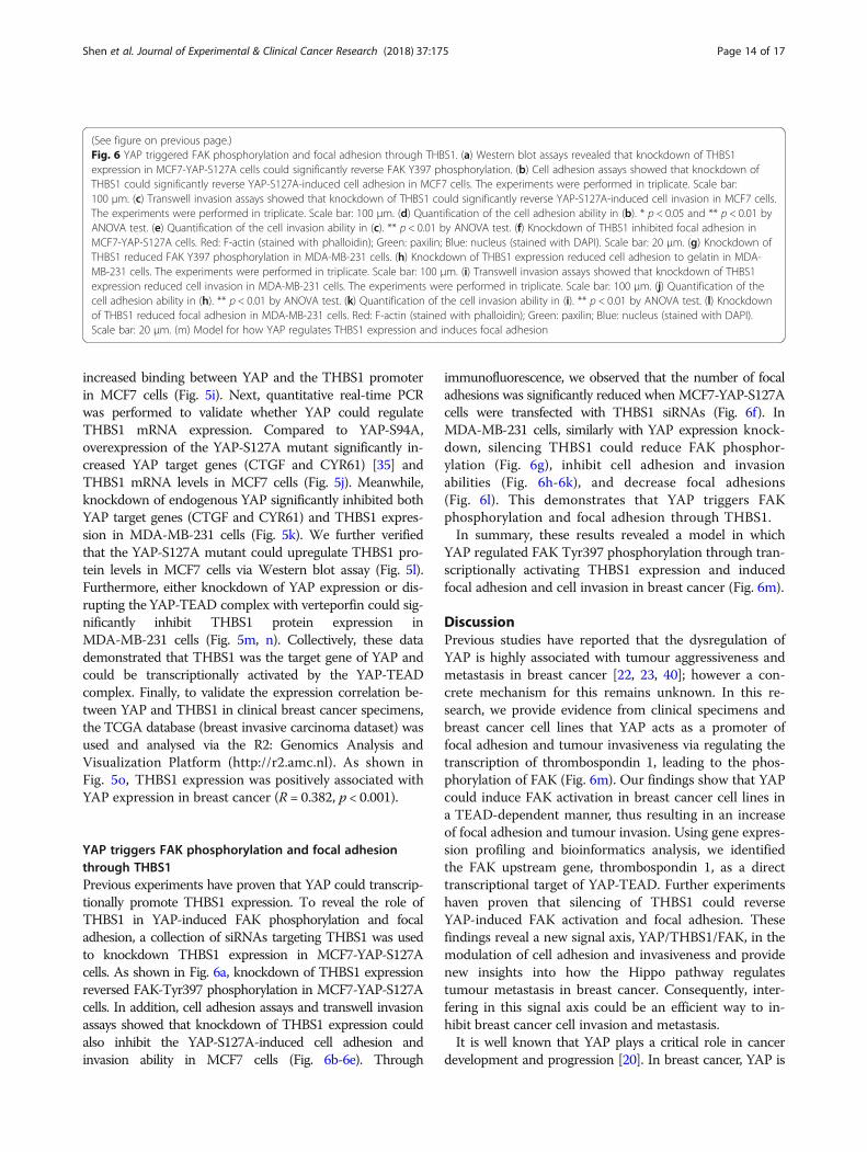
YAP triggers FAK phosphorylation and focal adhesionthrough THBS1Previous experiments have proven that YAP could transcrip-tionally promote THBS1 expression. To reveal the role ofTHBS1 in YAP-induced FAK phosphorylation and focaladhesion, a collection of siRNAs targeting THBS1 was usedto knockdown THBS1 expression in MCF7-YAP-S127Acells. As shown in Fig. 6a, knockdown of THBS1 expressionreversed FAK-Tyr397 phosphorylation in MCF7-YAP-S127Acells. In addition, cell adhesion assays and transwell invasionassays showed that knockdown of THBS1 expression couldalso inhibit the YAP-S127A-induced cell adhesion andinvasion ability in MCF7 cells (Fig. 6b-6e). Through
immunofluorescence, we observed that the number of focaladhesions was significantly reduced when MCF7-YAP-S127Acells were transfected with THBS1 siRNAs (Fig. 6f). InMDA-MB-231 cells, similarly with YAP expression knock-down, silencing THBS1 could reduce FAK phosphor-ylation (Fig. 6g), inhibit cell adhesion and invasionabilities (Fig. 6h-6k), and decrease focal adhesions(Fig. 6l). This demonstrates that YAP triggers FAKphosphorylation and focal adhesion through THBS1.In summary, these results revealed a model in which
YAP regulated FAK Tyr397 phosphorylation through tran-scriptionally activating THBS1 expression and inducedfocal adhesion and cell invasion in breast cancer (Fig. 6m).
DiscussionPrevious studies have reported that the dysregulation ofYAP is highly associated with tumour aggressiveness andmetastasis in breast cancer [22, 23, 40]; however a con-crete mechanism for this remains unknown. In this re-search, we provide evidence from clinical specimens andbreast cancer cell lines that YAP acts as a promoter offocal adhesion and tumour invasiveness via regulating thetranscription of thrombospondin 1, leading to the phos-phorylation of FAK (Fig. 6m). Our findings show that YAPcould induce FAK activation in breast cancer cell lines ina TEAD-dependent manner, thus resulting in an increaseof focal adhesion and tumour invasion. Using gene expres-sion profiling and bioinformatics analysis, we identifiedthe FAK upstream gene, thrombospondin 1, as a directtranscriptional target of YAP-TEAD. Further experimentshaven proven that silencing of THBS1 could reverseYAP-induced FAK activation and focal adhesion. Thesefindings reveal a new signal axis, YAP/THBS1/FAK, in themodulation of cell adhesion and invasiveness and providenew insights into how the Hippo pathway regulatestumour metastasis in breast cancer. Consequently, inter-fering in this signal axis could be an efficient way to in-hibit breast cancer cell invasion and metastasis.It is well known that YAP plays a critical role in cancer
development and progression [20]. In breast cancer, YAP is
(See figure on previous page.)Fig. 6 YAP triggered FAK phosphorylation and focal adhesion through THBS1. (a) Western blot assays revealed that knockdown of THBS1expression in MCF7-YAP-S127A cells could significantly reverse FAK Y397 phosphorylation. (b) Cell adhesion assays showed that knockdown ofTHBS1 could significantly reverse YAP-S127A-induced cell adhesion in MCF7 cells. The experiments were performed in triplicate. Scale bar:100 μm. (c) Transwell invasion assays showed that knockdown of THBS1 could significantly reverse YAP-S127A-induced cell invasion in MCF7 cells.The experiments were performed in triplicate. Scale bar: 100 μm. (d) Quantification of the cell adhesion ability in (b). * p < 0.05 and ** p < 0.01 byANOVA test. (e) Quantification of the cell invasion ability in (c). ** p < 0.01 by ANOVA test. (f) Knockdown of THBS1 inhibited focal adhesion inMCF7-YAP-S127A cells. Red: F-actin (stained with phalloidin); Green: paxilin; Blue: nucleus (stained with DAPI). Scale bar: 20 μm. (g) Knockdown ofTHBS1 reduced FAK Y397 phosphorylation in MDA-MB-231 cells. (h) Knockdown of THBS1 expression reduced cell adhesion to gelatin in MDA-MB-231 cells. The experiments were performed in triplicate. Scale bar: 100 μm. (i) Transwell invasion assays showed that knockdown of THBS1expression reduced cell invasion in MDA-MB-231 cells. The experiments were performed in triplicate. Scale bar: 100 μm. (j) Quantification of thecell adhesion ability in (h). ** p < 0.01 by ANOVA test. (k) Quantification of the cell invasion ability in (i). ** p < 0.01 by ANOVA test. (l) Knockdownof THBS1 reduced focal adhesion in MDA-MB-231 cells. Red: F-actin (stained with phalloidin); Green: paxilin; Blue: nucleus (stained with DAPI).Scale bar: 20 μm. (m) Model for how YAP regulates THBS1 expression and induces focal adhesion
Shen et al. Journal of Experimental & Clinical Cancer Research (2018) 37:175 Page 14 of 17
often reported as an oncogene, and its hyper-activationoften leads to various tumour-promoting effects [41, 42].Previous studies have reported the association betweenYAP and breast cancer cell aggressiveness [22]. Further-more, the overexpression of YAP has been shown to be atrigger of epithelial–mesenchymal transition [43] and actindynamics [24]. As the main effector of the Hippo pathway,activation of YAP is controlled by Hippo signalling [19, 20].Hippo signalling is an evolutionarily conserved regulator oftissue growth and cell fate and is mainly regulated by theactin cytoskeleton and cellular tension [44, 45]. Recentstudies have revealed numerous upstream signalling mech-anisms involved in the Hippo pathway, including cell polar-ity, mechanotransduction and G protein-coupled receptorsignalling [45]. As a key component of cell-ECM crosstalk,focal adhesions have been demonstrated to play an import-ant role in cellular mechanotransduction [6] and act as alink between integrin and Hippo signalling [45] in tumours.In the conventional viewpoint, FAK activation and focal ad-hesion appear to be an upstream signalling mechanism thattriggers Hippo-off and induces YAP activation signalling[46]. However, a recent study has unveiled that YAP coulddirectly control the RhoA GTPase pathway to induce FAassembly in AD-MSC and CAL51 cells [47]. Therefore, it isreasonable that YAP could promote tumour metastasis inan FA-dependent manner.In our current study, we observed that YAP induces focal
adhesions in breast cancer cells. Through gene expressionscreening and bioinformatics analysis, we discovered a newsignalling axis, YAP/THBS1/FAK, in Hippo-mediated celladhesion and invasion. THBS1 was the first member to beidentified in the thrombospondins family and is a mainplayer in the tumour microenvironment [48]. THBS1 wasdemonstrated decades ago to be a cell adhesion protein[49]. Thereafter, numerous studies have proven that THBS1regulates cell adhesion in different cell types, regardless ofspecies [48]. Previous studies have shown that THBS1could modulate FAK phosphorylation to regulate focal ad-hesion dynamics [26, 27, 50, 51]. In addition, increased ex-pression of THBS1 has also been reported to be associatedwith tumour invasiveness and metastasis [52–54]. Due toits essential role in tumour progression, THBS1 representsa perspective therapeutic target in cancer treatment. How-ever, very little is known about its upstream regulation. Ourcurrent results reveal a novel mechanism where YAP in-duces FAK phosphorylation via activating THBS1 transcrip-tion in a TEAD-dependent manner. These findings reveal anew crosstalk mechanism between the Hippo pathway andTHBS1-FAK signalling and provide a new interpretation ofYAP-regulated tumour aggressiveness in breast cancer.Numerous reports have shown that the FAK gene is
amplified in a large fraction of breast cancer specimens;meanwhile, increased FAK expression and activity fre-quently correlates with metastatic disease and poor
prognosis (reviewed in [15]). FAK has been demonstratedto play an important role in the progression of tumour ag-gressiveness; thus, it has been selected as a potential targetfor cancer therapeutics [18]. Several FAK inhibitors(GSK2256098, VS-4718, VS-6062, defactinib, andBI853520) have been entered into clinical trials, and somehave achieved promising clinical activities in patients withselected solid cancers [18, 55]. Although inhibition of FAKhas shown effectiveness in the control of cancer, little isknown regarding the predictive response biomarkers ofFAK-targeting agents. In our research, we have shown acorrelation between Hippo signalling and FAK activation;therefore, YAP overexpression may act as a potential bio-marker of FAK inhibitor treatment in breast cancer.In summary, with this study, we have proven that YAP
acts as an upstream regulator in focal adhesion dynamicsand have discovered a YAP/THBS1/FAK signallingmechanism in the regulation of cell invasiveness and ad-hesion in breast cancer. These findings reveal a new roleof Hippo signalling in focal adhesion in breast cancerand provide exciting opportunities for future studies.
ConclusionsIn this study, we offer evidences that YAP promotes focaladhesion and tumour invasiveness in breast cancer. More-over, we unveil a new signal axis, YAP/THBS1/FAK, in themodulation of cell adhesion and invasiveness, and providesnew insights into the crosstalk between Hippo signallingand focal adhesion (Fig. 6m). These findings reveal a newrole of Hippo signalling in focal adhesion in breast cancerand provide exciting opportunities for future studies.
Additional files
Additional file 1: Table S1. The sequences of siRNAs used in thisresearch. (DOC 32 kb)
Additional file 2: Table S2. Primer sequences used in this research.(DOC 38 kb)
Additional file 3: Figure S1. Gene set enrichment analysis (GSEA) ofpurified tumour cells from 14 primary breast tumour tissues and 6metastatic lymph nodes from the GEO database (GSE30480). C6: oncogenicgene sets were used in this analysis. ES: enrichment score; NES: normalizedenrichment score; NOM-p: normalized p-value; FDR-q: false discovery rate q-value; FWER-p: family-wise error rate p-value. (JPG 17399 kb)
Additional file 4: Table S3. Gene ontology enrichment (biologicalprocesses) of all upregulated genes (1416 genes with fold change greaterthan 1.5) affected by the YAP-S127A mutant in MCF7 cells. “Cell adhesion”was the 6th enrichment category and contained 30 genes. Gene categor-ies were ranked by –Log10P value. The categories with p > 0.01 wereomitted from this table. (DOC 82 kb)
Additional file 5: Figure S2. The binding sequence of TEAD4 to theTHBS1 gene. SL14575 and SL16341 were two bio-replications of theTEAD4 ChIP-sequence data from the ENCODE database. Sequence datawere mapped to NCBI GRCh37 (hg19) according to the protocol and ana-lysed via the ChIP-seek tool. The TEAD4 binding site was calculated asthe aggregate of the TEAD4 binding peaks from the two bio-replicates.TSS: transcription start site. (JPG 986 kb)
Shen et al. Journal of Experimental & Clinical Cancer Research (2018) 37:175 Page 15 of 17
AbbreviationsATCC: American Type Culture Collection; ChIP: Chromatinimmunoprecipitation; CTGF: Connective tissue growth factor;CYR61: Cysteine-rich angiogenic inducer 61; DAPI: 4′,6-diamidino-2-phenylindole; DAVID: The Database for Annotation, Visualization andIntegrated Discovery; DMEM: Dulbecco’s modified Eagle medium;DMSO: Dimethyl sulfoxide; ECM: Extracellular matrix; ENCODE: Encyclopaediaof DNA Elements; FA: Focal adhesion; FAK: Focal adhesion kinase (alsoknown as PTK2, protein tyrosine kinase 2); GAPDH: Glyceraldehyde-3-phosphate dehydrogenase; GFP: Green fluorescent protein; GSEA: Gene setenrichment analysis; IHC: Immunohistochemistry; L15: Leibovitz’s L-15medium; qRT-PCR: Quantitative real-time PCR; SDS-PAGE: Sodium dodecylsulfate polyacrylamide gel electrophoresis; TCGA: The Cancer Genome Atlas;TEAD: TEA domain family member; THBS1: Thrombospondin 1;TSS: Transcription start site; YAP: Yes-associated protein
AcknowledgementsThe authors thank the experimental medicine centre of Tongji Hospital forproviding experimental support and the Department of Pathology of TongjiHospital for immunohistochemistry analysis. We apologize to the colleagueswhose work was not cited due to space constraints.
FundingThis work was funded by the National Natural Science Foundation of China(81572861, 81773053).
Availability of data and materialsAll data can be provided upon request.
Author contributionsConceived and designed the experiments: JS, DX and JG Performed theexperiments: JS, BC and ZZ Analysed the data: JS, YW, CM, and LLContributed reagents/materials/analysis tools: XL and DT Wrote the paper: JS.All authors’ read and approved the final manuscript.
Ethics approval and consent to participateNot applicable.
Consent for publicationNot applicable.
Competing interestsThe authors declare that they have no competing interests.
Publisher’s NoteSpringer Nature remains neutral with regard to jurisdictional claims inpublished maps and institutional affiliations.
Author details1Molecular Medicine Center, Tongji Hospital, Tongji Medical College,Huazhong University of Science and Technology, 1095 Jiefang Av., Wuhan,Hubei 430030, People’s Republic of China. 2Department of Surgery, TongjiHospital, Tongji Medical College, Huazhong University of Science andTechnology, 1095 Jiefang Av., Wuhan, Hubei 430030, People’s Republic ofChina.
Received: 21 June 2018 Accepted: 20 July 2018
References1. Siegel RL, Miller KD, Jemal A. Cancer statistics, 2018. CA Cancer J Clin. 2018;
68(1):7–30.2. Weigelt B, Peterse JL, Van’t veer LJ. Breast cancer metastasis: markers and
models. Nat Rev Cancer. 2005;5(8):591–602.3. Gkretsi V, Stylianopoulos T. Cell adhesion and matrix stiffness: coordinating
Cancer cell invasion and metastasis. Front Oncol. 2018;8:145.4. He X, Lee B, Jiang Y. Cell-ECM interactions in tumour invasion. Adv Exp Med
Biol. 2016;936:73–91.5. Carragher NO, Frame MC. Focal adhesion and actin dynamics: a place
where kinases and proteases meet to promote invasion. Trends Cell Biol.2004;14(5):241–9.
6. Burridge K. Focal adhesions: a personal perspective on a half century ofprogress. FEBS J. 2017;284(20):3355–61.
7. Paluch EK, Aspalter IM, Sixt M. Focal adhesion-independent cell migration.Annu Rev Cell Dev Biol. 2016;32:469–90.
8. Kirchner J, Kam Z, Tzur G, Bershadsky AD, Geiger B. Live-cell monitoring oftyrosine phosphorylation in focal adhesions following microtubuledisruption. J Cell Sci. 2003;116(Pt 6):975–86.
9. Miyamoto S, Akiyama SK, Yamada KM. Synergistic roles for receptoroccupancy and aggregation in integrin transmembrane function. Science.1995;267(5199):883–5.
10. Webb DJ, Donais K, Whitmore LA, Thomas SM, Turner CE, Parsons JT, et al.FAK-Src signalling through paxillin, ERK and MLCK regulates adhesiondisassembly. Nat Cell Biol. 2004;6(2):154–61.
11. McLean GW, Carragher NO, Avizienyte E, Evans J, Brunton VG, Frame MC.The role of focal-adhesion kinase in cancer - a new therapeutic opportunity.Nat Rev Cancer. 2005;5(7):505–15.
12. Owens LV, Xu L, Craven RJ, Dent GA, Weiner TM, Kornberg L, et al.Overexpression of the focal adhesion kinase (p125FAK) in invasive humantumours. Cancer Res. 1995;55(13):2752–5.
13. Cance WG, Harris JE, Iacocca MV, Roche E, Yang X, Chang J, et al.Immunohistochemical analyses of focal adhesion kinase expression inbenign and malignant human breast and colon tissues: correlation withpreinvasive and invasive phenotypes. Clin Cancer Res. 2000;6(6):2417–23.
14. Schlaepfer DD, Mitra SK, Ilic D. Control of motile and invasive cellphenotypes by focal adhesion kinase. Biochim Biophys Acta. 2004;1692(2–3):77–102.
15. Luo M, Guan JL. Focal adhesion kinase: a prominent determinant in breastcancer initiation, progression and metastasis. Cancer Lett. 2010;289(2):127–39.
16. van Nimwegen MJ, Verkoeijen S, van Buren L, Burg D, van de Water B.Requirement for focal adhesion kinase in the early phase of mammaryadenocarcinoma lung metastasis formation. Cancer Res. 2005;65(11):4698–706.
17. Provenzano PP, Inman DR, Eliceiri KW, Beggs HE, Keely PJ. Mammaryepithelial-specific disruption of focal adhesion kinase retards tumourformation and metastasis in a transgenic mouse model of human breastcancer. Am J Pathol. 2008;173(5):1551–65.
18. Lee BY, Timpson P, Horvath LG, Daly RJ. FAK signalling in human cancer asa target for therapeutics. Pharmacol Ther. 2015;146:132–49.
19. Pfleger CM. The hippo pathway: a master regulatory network important indevelopment and dysregulated in disease. Curr Top Dev Biol. 2017;123:181–228.
20. Zanconato F, Cordenonsi M, Piccolo S. YAP/TAZ at the roots of Cancer.Cancer Cell. 2016;29(6):783–803.
21. Ji XY, Zhong G, Zhao B. Molecular mechanisms of the mammalian hipposignalling pathway. Yi Chuan. 2017;39(7):546–67.
22. Pegoraro S, Ros G, Ciani Y, Sgarra R, Piazza S, Manfioletti G. A novel HMGA1-CCNE2-YAP axis regulates breast cancer aggressiveness. Oncotarget. 2015;6(22):19087–101.
23. Kim HM, Jung WH, Koo JS. Expression of yes-associated protein (YAP) inmetastatic breast cancer. Int J Clin Exp Pathol. 2015;8(9):11248–57.
24. Qiao Y, Chen J, Lim YB, Finch-Edmondson ML, Seshachalam VP, Qin L, et al.YAP regulates actin dynamics through ARHGAP29 and promotes metastasis.Cell Rep. 2017;19(8):1495–502.
25. Wang Z, Wu Y, Wang H, Zhang Y, Mei L, Fang X, et al. Interplay ofmevalonate and hippo pathways regulates RHAMM transcription via YAP tomodulate breast cancer cell motility. Proc Natl Acad Sci U S A 2014;111(1):E89–E98.
26. Hu C, Wen J, Gong L, Chen X, Wang J, Hu F, et al. Thrombospondin-1promotes cell migration, invasion and lung metastasis of osteosarcomathrough FAK dependent pathway. Oncotarget. 2017;8(44):75881–92.
27. Gahtan V, Wang XJ, Ikeda M, Willis AI, Tuszynski GP, Sumpio BE.Thrombospondin-1 induces activation of focal adhesion kinase in vascularsmooth muscle cells. J Vasc Surg. 1999;29(6):1031–6.
28. Wang XJ, Maier K, Fuse S, Willis AI, Olson E, Nesselroth S, et al.Thrombospondin-1-induced migration is functionally dependent upon focaladhesion kinase. Vasc Endovasc Surg. 2008;42(3):256–62.
29. Flamini MI, Gauna GV, Sottile ML, Nadin BS, Sanchez AM, Vargas-Roig LM.Retinoic acid reduces migration of human breast cancer cells: role ofretinoic acid receptor beta. J Cell Mol Med. 2014;18(6):1113–23.
30. Yu H, Simons DL, Segall I, Carcamo-Cavazos V, Schwartz EJ, Yan N, et al.PRC2/EED-EZH2 complex is up-regulated in breast cancer lymph nodemetastasis compared to primary tumour and correlates with tumourproliferation in situ. PLoS One. 2012;7(12):e51239.
Shen et al. Journal of Experimental & Clinical Cancer Research (2018) 37:175 Page 16 of 17

31. Subramanian A, Tamayo P, Mootha VK, Mukherjee S, Ebert BL, Gillette MA,et al. Gene set enrichment analysis: a knowledge-based approach forinterpreting genome-wide expression profiles. Proc Natl Acad Sci U S A.2005;102(43):15545–50.
32. Aguirre-Gamboa R, Gomez-Rueda H, Martinez-Ledesma E, Martinez-Torteya A, Chacolla-Huaringa R, Rodriguez-Barrientos A, et al.SurvExpress: an online biomarker validation tool and database forcancer gene expression data using survival analysis. PLoS One. 2013;8(9):e74250.
33. Chen TW, Li HP, Lee CC, Gan RC, Huang PJ, Wu TH, et al. ChIPseek, a web-based analysis tool for ChIP data. BMC Genomics. 2014;15:539.
34. Szklarczyk D, Franceschini A, Wyder S, Forslund K, Heller D, Huerta-Cepas J, et al. STRING v10: protein-protein interaction networks,integrated over the tree of life. Nucleic Acids Res. 2015;43(Databaseissue):D447–52.
35. Moroishi T, Hansen CG, Guan KL. The emerging roles of YAP and TAZ incancer. Nat Rev Cancer. 2015;15(2):73–9.
36. Zhou Y, Huang T, Cheng AS, Yu J, Kang W, To KF. The TEAD Family and ItsOncogenic Role in Promoting Tumorigenesis. Int J Mol Sci. 2016;17(1).https://doi.org/10.3390/ijms17010138.
37. Yang H, Wang B, Wang T, Xu L, He C, Wen H, et al. Toll-like receptor 4prompts human breast cancer cells invasiveness via lipopolysaccharidestimulation and is overexpressed in patients with lymph node metastasis.PLoS One. 2014;9(10):e109980.
38. Chen CS, Alonso JL, Ostuni E, Whitesides GM, Ingber DE. Cell shapeprovides global control of focal adhesion assembly. Biochem Biophys ResCommun. 2003;307(2):355–61.
39. Liu-Chittenden Y, Huang B, Shim JS, Chen Q, Lee SJ, Anders RA, et al.Genetic and pharmacological disruption of the TEAD-YAP complexsuppresses the oncogenic activity of YAP. Genes Dev. 2012;26(12):1300–5.
40. Wang T, Mao B, Cheng C, Zou Z, Gao J, Yang Y, et al. YAP promotes breastcancer metastasis by repressing growth differentiation factor-15. BiochimBiophys Acta. 2018;1864(5 Pt A):1744–53.
41. Maugeri-Sacca M, Barba M, Pizzuti L, Vici P, Di Lauro L, Dattilo R, et al. Thehippo transducers TAZ and YAP in breast cancer: oncogenic activities andclinical implications. Expert Rev Mol Med. 2015;17:e14.
42. Overholtzer M, Zhang J, Smolen GA, Muir B, Li W, Sgroi DC, et al.Transforming properties of YAP, a candidate oncogene on the chromosome11q22 amplicon. Proc Natl Acad Sci U S A. 2006;103(33):12405–10.
43. Liu Y, He K, Hu Y, Guo X, Wang D, Shi W, et al. YAP modulates TGF-beta1-induced simultaneous apoptosis and EMT through upregulation of the EGFreceptor. Sci Rep. 2017;7:45523.
44. Harvey KF, Zhang X, Thomas DM. The hippo pathway and human cancer.Nat Rev Cancer. 2013;13(4):246–57.
45. Yu FX, Guan KL. The hippo pathway: regulators and regulations. Genes Dev.2013;27(4):355–71.
46. Kim NG, Gumbiner BM. Adhesion to fibronectin regulates hippo signallingvia the FAK-Src-PI3K pathway. J Cell Biol. 2015;210(3):503–15.
47. Nardone G, Oliver-De La Cruz J, Vrbsky J, Martini C, Pribyl J, Skladal P, et al.YAP regulates cell mechanics by controlling focal adhesion assembly. NatCommun. 2017;8:15321.
48. Huang T, Sun L, Yuan X, Qiu H. Thrombospondin-1 is a multifaceted playerin tumour progression. Oncotarget. 2017;8(48):84546–58.
49. Tuszynski GP, Rothman V, Murphy A, Siegler K, Smith L, Smith S, et al.Thrombospondin promotes cell-substratum adhesion. Science. 1987;236(4808):1570–3.
50. Lymn JS, Rao SJ, Clunn GF, Gallagher KL, O'Neil C, Thompson NT, et al.Phosphatidylinositol 3-kinase and focal adhesion kinase are early signalsin the growth factor-like responses to thrombospondin-1 seen inhuman vascular smooth muscle. Arterioscler Thromb Vasc Biol. 1999;19(9):2133–40.
51. Orr AW, Pallero MA, Xiong WC, Murphy-Ullrich JE. Thrombospondin inducesRhoA inactivation through FAK-dependent signalling to stimulate focaladhesion disassembly. J Biol Chem. 2004;279(47):48983–92.
52. Pal SK, Nguyen CT, Morita KI, Miki Y, Kayamori K, Yamaguchi A, et al. THBS1is induced by TGFB1 in the cancer stroma and promotes invasion of oralsquamous cell carcinoma. J Oral Pathol Med. 2016;45(10):730–9.
53. Borsotti P, Ghilardi C, Ostano P, Silini A, Dossi R, Pinessi D, et al.Thrombospondin-1 is part of a slug-independent motility and metastaticprogram in cutaneous melanoma, in association with VEGFR-1 and FGF-2.Pigment Cell Melanoma Res. 2015;28(1):73–81.
54. Horiguchi H, Yamagata S, Rong Qian Z, Kagawa S, Sakashita N.Thrombospondin-1 is highly expressed in desmoplastic components ofinvasive ductal carcinoma of the breast and associated with lymphnode metastasis. J Med Investig. 2013;60(1–2):91–6.
55. Roy-Luzarraga M, Hodivala-Dilke K. Molecular pathways: endothelial cellFAK-A target for Cancer treatment. Clin Cancer Res. 2016;22(15):3718–24.
Shen et al. Journal of Experimental & Clinical Cancer Research (2018) 37:175 Page 17 of 17