absence of mycobacterium avium subspecies · pdf fileabsence of mycobacterium avium subspecies...
TRANSCRIPT
JUNE 10, 2003
Clinical Medicine & ResearchVolume 1, Number 3: 217-226
©2003 Clinical Medicine & Research www.mfldclin.edu/clinmedres
Original Research
Absence of Mycobacterium avium subspecies paratuberculosisComponents from Crohn’s Disease Intestinal Biopsy Tissues
*Jay L.E. Ellingson, PhD, Department of Veterinary Pathology, College of Veterinary Medicine, Iowa State University, Ames, Iowa and the Center forVeterinary Biologics Laboratory, Veterinary Services, Animal and Plant Health Inspection Service, United States Department of Agriculture, Ames, Iowa
John C. Cheville, MD, Mayo Clinic, Rochester, Minnesota†Dominique Brees, DVM, PhD, DACVP, Department of Veterinary Pathology, College of Veterinary Medicine, Iowa State University, Ames, Iowa
Janice M. Miller, DVM, PhD, Zoonotic Diseases Research Unit, National Animal Disease Center, Agricultural Research Service, United States Department of Agriculture, Ames, Iowa
Norman F. Cheville, DVM, PhD, DrHc, Department of Veterinary Pathology, College of Veterinary Medicine, Iowa State University, Ames, Iowa*Current affiliation Marshfield Clinic Laboratories-Food Safety Services, Marshfield Clinic, Marshfield, Wisconsin
†Current affiliation Pfizer, Inc., Groton, Connecticut
ABSTRACTBACKGROUND
Crohn’s disease is a chronic human intestinal inflammatory disorder for which an etiologic agent has not been identified.Johne’s disease is a similar chronic enteric granulomatous disease of ruminant species and has been used as a model ofCrohn’s disease. Johne’s disease has been proven to be caused by Mycobacterium avium subspecies paratuberculosis(M. avium ss paratuberculosis). It has been proposed that M. avium ss paratuberculosis may also cause Crohn’s disease. This is of particular concern because the organism may be spread to humans through inadequately pasteurized dairy products.
OBJECTIVE
We sought to determine whether M. avium ss paratuberculosis could be detected using identical techniques in paraffin-embedded tissue samples of bovine Johne’s disease and human Crohn’s, ulcerative colitis and diverticular diseases. Sampleswere obtained for analysis from national tissue banks.
DESIGN
Cross-species and cross-disease sample comparisons by multiple detection techniques.METHODS
Histology, immunocytochemistry and polymerase chain reaction (PCR) were utilized to test and compare the presence of M. avium ss paratuberculosis components. Insertion sequence IS900, present in multiple copies and found only in M. avium ss paratuberculosis, was utilized in both PCR and immunocytochemical analyses.
RESULTS
The IS900 sequence was demonstrable in all samples of confirmed positive Johne’s disease tissue. The sequence was not identified in the 35 Crohn’s, 36 ulcerative colitis, and 21 diverticular disease samples.
CONCLUSION
M. avium ss paratuberculosis was not associated with the lesions in these Crohn’s disease samples, using these methods.
RECEIVED: REVISED AND ACCEPTED: JULY 1, 2003
REPRINT REQUESTS: KEYWORDS:Jay L.E. Ellingson, PhDMarshfield Clinic LaboratoriesFood Safety ServicesMarshfield Clinic1000 North Oak AvenueMarshfield, WI 54449Telephone: 715-389-5958Fax: 715-389-7599 Email: [email protected]
Crohn’s disease; Johne’s disease; Inflammatory bowel disease;Mycobacterium avium subspecies paratuberculosis; IS900;Polymerase chain reaction
GRANT SUPPORT:USDA grant 1999-038181.
217
July 2003 Body.qxd 7/14/03 8:53 AM Page 217
INTRODUCTION
Crohn’s disease is a chronic inflammatory disorder of thehuman intestine characterized by protein losing enteropathy,general malabsorption and steatorrhea. Lesions that begin asmucosal erosions and neutrophil infiltrates within crypts andcrypt abscesses, progress to transmural lymphogranulomatousenteritis with lymphocyte populations being predominantlyTH1 cells.1-3 In approximately 50% of the cases, lymphoidaggregates contain noncaseating granulomas composed ofepithelioid histiocytes and multinucleate giant cells of theLanghan’s type. Ulcers both discrete and serpiginous, arehallmarks of Crohn’s lesions.4 Neutrophils and eosinophilsare prominent in many lesions, as are plasmacytes bearingIgG, IgM, IgA or IgD. These inflammatory processes underlie the characteristic cobblestone appearance of themucosal surface.
The cause and some critical aspects in the pathogenesis ofhuman Crohn’s disease are not known. Suggestions that theetiological agent involves mycobacteria5,6 have been basedon the detection of mycobacterial species in intestinal biopsytissue from Crohn’s disease patients including: Mycobacteriumavium-intracellulare, Mycobacterium cheloni, Mycobacteriumfortuitum, Mycobacterium kansasii, Mycobacterium aviumsubspecies paratuberculosis (M. avium ss paratuberculosis).5
Like human Crohn’s disease, Johne’s disease of cattle is achronic, progressive lymphogranulomatous process withpreference for the terminal ileum and ileocecal valve. UnlikeCrohn’s disease, acid-fast intracellular bacilli are always present in lesions, there is no ulceration and the causal bacterium, M. avium ss paratuberculosis, can be isolated.Early in the disease process there is marked edema and lymphangitis at the edge of the expanding enteric lesion.Enteric lesions in Johne’s disease are infiltrated with M. avium ss paratuberculosis infected macrophages. Lesionsspread and grow by accumulation, reinfection and expansionof macro-phage populations in the intestinal wall. M. aviumss paratuberculosis disseminate from infected to non-infected macro-phages, allowing the bacterium to avoidhumoral and cellular mechanisms of immunity. Transmissionof M. avium ss paratuberculosis among cattle is by ingestionfrom milk or feces and by placental infection. Chronic infiltration of macrophages in the lamina propria throughoutthe lower intestine results in villous atrophy and malabsorption.Although infection is often restricted to the intestine, bacteria can spread via monocytes from the primary site ofinfection in the intestine to liver, spleen, kidney, uterus andmammaries through hematogenous or lymphatic routes.
Due to the clinical symptoms of Crohn’s disease closelymimicking those found in animals with Johne’s disease, itwas proposed almost 90 years ago that the two diseasesshared the same etiology.7,8 Specifically, M. avium ssparatuberculosis continues to be suspected as the causativeagent of both disorders. However, reviews of Johne’s diseaseand its connection to human Crohn’s have not establishedany connection.6,9-12
There is no sound epidemiological evidence that links exposure to M. avium ss paratuberculosis to an increasedincidence of Crohn’s disease, even though cows with clinicalparatuberculosis do shed viable organisms in their milk atlow levels (50 CFU/50 ml milk).13 In Great Britain it hasbeen demonstrated that M. avium ss paratuberculosis DNAis present in milk samples obtained from retail markets.14
Viable M. avium ss paratuberculosis have been cultured fromthe pasteurized retail milk samples. These results suggestthat current pasteurization techniques may not be adequateto kill M. avium ss paratuberculosis in raw milk, or thatmilk may become contaminated post pasteurization. Becauseof the potential for spreading M. avium ss paratuberculosisthrough widely consumed dairy products, it has become crucial to definitively establish whether or not this organismis actually involved in the etiology of Crohn’s diseases.
In recent serologic studies, up to 83% of Crohn’s patientsshowed evidence of serum antibodies to M. avium ss paratuberculosis.15-20 Recently it has been demonstratedthat lactating mothers with Crohn’s disease shed M. avium ssparatuberculosis in their breast milk.21 However, unlike thecase for Johne’s disease, reports are mixed as to the presenceor absence of M. avium ss paratuberculosis at the site of theintestinal lesions.
Studies that used genetic primers in polymerase chain reaction (PCR) analyses to detect the organism in Crohn’spatient tissues have yielded the most provocative data.22-28
The most widely used primers are for insertion sequence900 (IS900), a genetic element proven to be quite specificfor M. avium ss paratuberculosis.29-32 In those studies, from13% to 100% of Crohn’s patients tested positive for M. avium ss paratuberculosis by IS900 DNA/PCR.33-44
Other studies, however, have been unable to demonstrate M. avium ss paratuberculosis DNA in Crohn’s disease tissue.5,11,12,45-50 One potential problem with these results isthat some primers designed from IS900 can cross-react withclosely related IS901 and IS902.51-55
It is therefore crucial to use primers that are very sensitiveand specific to the M. avium ss paratuberculosis organism.In this study we used the specific IS900 primers in PCRanalyses to search for evidence of M. avium ss paratubercu-losis-infection in formalin-fixed, paraffin-embedded tissuesections from bovine Johne’s disease and human Crohn’sdisease, ulcerative colitis and diverticular disease. For comparison the more widely used immunohistochemistrywas applied. This was compared to the PCR analyses of aDNA insertion sequence that allows for specific identificationof DNA from M. avium ss paratuberculosis. The IS900sequence was demonstrable in all samples of confirmed positives of Johne’s disease tissue. The sequence was notidentified in the 35 Crohn’s, 36 ulcerative colitis, and 21diverticular disease samples. The evidence does not supportthe involvement of M. avium ss paratuberculosis in the etiology of Crohn’s disease.
218 CM&R 2003 : 1 (July) Ellingson et al.
July 2003 Body.qxd 7/14/03 8:53 AM Page 218
METHODS
Tissue samplesA retrospective sample from a large individual collection ofhuman Crohn’s, ulcerative colitis and diverticular disease,was obtained from the Department of Pathology at MayoClinic. Additionally, samples of confirmed bovine Johne’sdisease from the United States Department of Agriculturewere obtained and examined. Tissue sections were formalin-fixed, paraffin-embedded and processed by standard procedures via standard operating protocols from both theDepartment of Pathology, Mayo Clinic and the NationalVeterinary Services Laboratory, Animal and Plant HealthInspection Service, United States Department ofAgriculture.56,57 Samples were screened for colonic lesionsthat showed clear evidence of granulomas. These sampleswere then analyzed by immunohistochemistry and transmis-sion electron microscopy for cytologic characteristics ofaffected macrophages, and lymphoid and vascular cells atthe margins of the progressive granulomatous lesions.
ImmunohistochemistryAn immunohistochemical technique to specifically detect M. avium ss paratuberculosis has been developed to detectantigens of M. avium ss paratuberculosis in formalin-fixed,paraffin-embedded tissue sections.58 The probe detected M. avium ss paratuberculosis in infected bovine tissues. Tissuesections from animals infected with M. avium ss aviumwere negative. Thus, differentiation between M. avium ssparatuberculosis and M. avium ss avium in formalin-fixed,paraffin-embedded bovine tissues was possible.58
Identification of mycobacteriaProcedures used to cut paraffin-embedded tissue sectionsand prepare crude DNA extracts have been previouslydescribed.56,57 Ten microliter aliquots of each crude extractwere tested with each set of PCR primers (table 1). Samplesthat failed to react with any primer were retested using purified DNA. The DNA purification procedure was modifiedslightly from the original.47,56 The 10 µl aliquots were usedfor all PCR amplifications.
PCREach sample was examined with a PCR primer set derivedfrom IS900, as previously described.56 IS900 characteristi-cally is present in multiple copies and is considered specificfor M. avium ss paratuberculosis.56,62,71
All amplification reactions were conducted with a “Hot Start”procedure that is based on heat-activated polymerase, as previously described. The concentration of reactants (50 µlfinal volume) was as follows: 20 pM primers, 1.25 U DNA
polymerase, 0.2 mM nucleotides, and 2.5 mM Mg++. Eachset of test samples included a negative control tube in whichwater was substituted for a test sample. A complete mastermixwas prepared for the IS900 primer set. The 40 µL aliquotswere introduced into the reaction tubes which were held at -70°C until use. Positive control DNA (10 ng) was obtainedfrom bacterial culture extracts and from sections cut fromparaffin-embedded tissue of confirmed Johne’s disease.
Amplification conditions for the IS900 primers were as follows:10 minutes at 94°C, then 1 minute at 94°C, 15 seconds at 65°C, and 2 minutes at 72°C for 50 cycles, and a final 10 minute extension at 72°C.
RESULTS
Histology and immunohistochemistryMacrophages and multinucleate giant cells in granulomas in Crohn’s biopsies did not contain bacilli; there was no evidence of other bacteria, fungi or viruses in tissues stainedusing acid-fast Stain (auramine-rhodamine) 59 and gramstains. There was no ultrastructural evidence of intracellularbacterial replication; e.g., cells lacked bacteriophorous vacuoles, well-developed Golgi complexes and vacuolartransport systems. In three cases of Crohn’s disease, acid-fast material was present, but by electron microscopy thismaterial was shown to be cellular debris, not bacilli. Noacid-fast bacilli were detected within enteric lesions of tissues from ulcerative colitis or from diverticular disease.
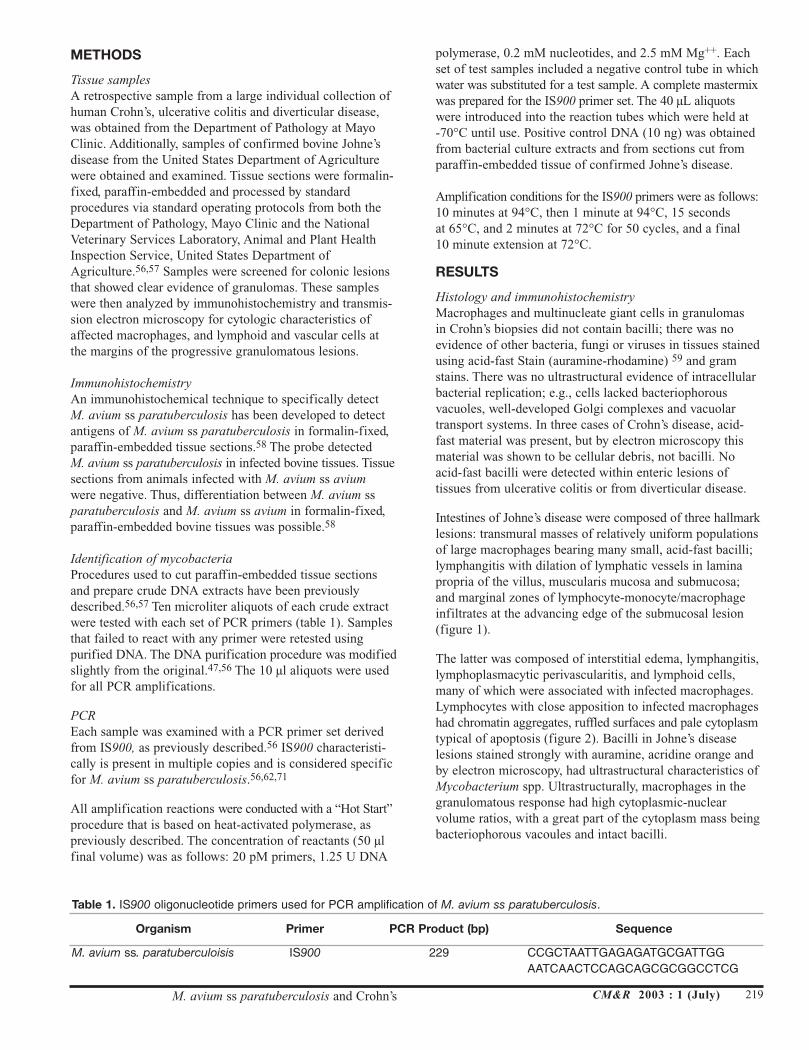
Intestines of Johne’s disease were composed of three hallmarklesions: transmural masses of relatively uniform populationsof large macrophages bearing many small, acid-fast bacilli;lymphangitis with dilation of lymphatic vessels in laminapropria of the villus, muscularis mucosa and submucosa;and marginal zones of lymphocyte-monocyte/macrophageinfiltrates at the advancing edge of the submucosal lesion(figure 1).
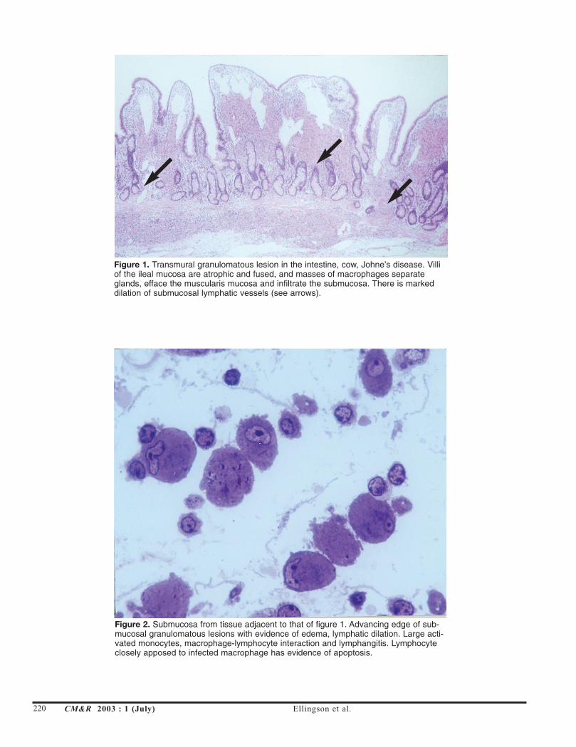
The latter was composed of interstitial edema, lymphangitis,lymphoplasmacytic perivascularitis, and lymphoid cells,many of which were associated with infected macrophages.Lymphocytes with close apposition to infected macrophageshad chromatin aggregates, ruffled surfaces and pale cytoplasmtypical of apoptosis (figure 2). Bacilli in Johne’s diseaselesions stained strongly with auramine, acridine orange andby electron microscopy, had ultrastructural characteristics ofMycobacterium spp. Ultrastructurally, macrophages in thegranulomatous response had high cytoplasmic-nuclear volume ratios, with a great part of the cytoplasm mass beingbacteriophorous vacoules and intact bacilli.
M. avium ss paratuberculosis and Crohn’s CM&R 2003 : 1 (July) 219
Table 1. IS900 oligonucleotide primers used for PCR amplification of M. avium ss paratuberculosis.
Organism Primer PCR Product (bp) Sequence
M. avium ss. paratuberculoisis IS900 229 CCGCTAATTGAGAGATGCGATTGGAATCAACTCCAGCAGCGCGGCCTCG
July 2003 Body.qxd 7/14/03 8:53 AM Page 219
220 CM&R 2003 : 1 (July) Ellingson et al.
Figure 1. Transmural granulomatous lesion in the intestine, cow, Johne’s disease. Villiof the ileal mucosa are atrophic and fused, and masses of macrophages separateglands, efface the muscularis mucosa and infiltrate the submucosa. There is markeddilation of submucosal lymphatic vessels (see arrows).
Figure 2. Submucosa from tissue adjacent to that of figure 1. Advancing edge of sub-mucosal granulomatous lesions with evidence of edema, lymphatic dilation. Large acti-vated monocytes, macrophage-lymphocyte interaction and lymphangitis. Lymphocyteclosely apposed to infected macrophage has evidence of apoptosis.
July 2003 Body.qxd 7/14/03 8:53 AM Page 220
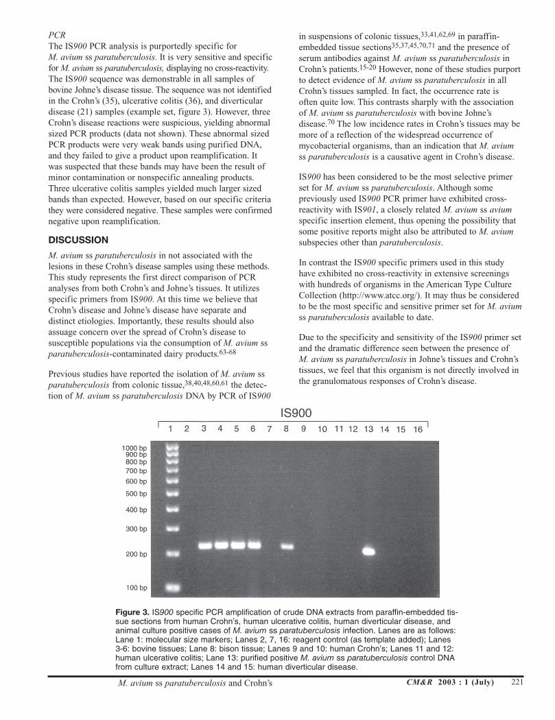
PCRThe IS900 PCR analysis is purportedly specific for M. avium ss paratuberculosis. It is very sensitive and specificfor M. avium ss paratuberculosis, displaying no cross-reactivity.The IS900 sequence was demonstrable in all samples ofbovine Johne’s disease tissue. The sequence was not identifiedin the Crohn’s (35), ulcerative colitis (36), and diverticulardisease (21) samples (example set, figure 3). However, threeCrohn’s disease reactions were suspicious, yielding abnormalsized PCR products (data not shown). These abnormal sizedPCR products were very weak bands using purified DNA,and they failed to give a product upon reamplification. Itwas suspected that these bands may have been the result ofminor contamination or nonspecific annealing products.Three ulcerative colitis samples yielded much larger sizedbands than expected. However, based on our specific criteriathey were considered negative. These samples were confirmednegative upon reamplification.
DISCUSSION
M. avium ss paratuberculosis in not associated with thelesions in these Crohn’s disease samples using these methods.This study represents the first direct comparison of PCRanalyses from both Crohn’s and Johne’s tissues. It utilizesspecific primers from IS900. At this time we believe thatCrohn’s disease and Johne’s disease have separate and distinct etiologies. Importantly, these results should alsoassuage concern over the spread of Crohn’s disease to susceptible populations via the consumption of M. avium ssparatuberculosis-contaminated dairy products.63-68
Previous studies have reported the isolation of M. avium ssparatuberculosis from colonic tissue,38,40,48,60,61 the detec-tion of M. avium ss paratuberculosis DNA by PCR of IS900
in suspensions of colonic tissues,33,41,62,69 in paraffin-embedded tissue sections35,37,45,70,71 and the presence ofserum antibodies against M. avium ss paratuberculosis inCrohn’s patients.15-20 However, none of these studies purportto detect evidence of M. avium ss paratuberculosis in allCrohn’s tissues sampled. In fact, the occurrence rate is often quite low. This contrasts sharply with the associationof M. avium ss paratuberculosis with bovine Johne’sdisease.70 The low incidence rates in Crohn’s tissues may bemore of a reflection of the widespread occurrence ofmycobacterial organisms, than an indication that M. aviumss paratuberculosis is a causative agent in Crohn’s disease.
IS900 has been considered to be the most selective primerset for M. avium ss paratuberculosis. Although some previously used IS900 PCR primer have exhibited cross-reactivity with IS901, a closely related M. avium ss aviumspecific insertion element, thus opening the possibility thatsome positive reports might also be attributed to M. aviumsubspecies other than paratuberculosis.
In contrast the IS900 specific primers used in this studyhave exhibited no cross-reactivity in extensive screeningswith hundreds of organisms in the American Type CultureCollection (http://www.atcc.org/). It may thus be consideredto be the most specific and sensitive primer set for M. aviumss paratuberculosis available to date.
Due to the specificity and sensitivity of the IS900 primer setand the dramatic difference seen between the presence of M. avium ss paratuberculosis in Johne’s tissues and Crohn’stissues, we feel that this organism is not directly involved inthe granulomatous responses of Crohn’s disease.
M. avium ss paratuberculosis and Crohn’s CM&R 2003 : 1 (July) 221
Figure 3. IS900 specific PCR amplification of crude DNA extracts from paraffin-embedded tis-sue sections from human Crohn’s, human ulcerative colitis, human diverticular disease, andanimal culture positive cases of M. avium ss paratuberculosis infection. Lanes are as follows:Lane 1: molecular size markers; Lanes 2, 7, 16: reagent control (as template added); Lanes3-6: bovine tissues; Lane 8: bison tissue; Lanes 9 and 10: human Crohn’s; Lanes 11 and 12:human ulcerative colitis; Lane 13: purified positive M. avium ss paratuberculosis control DNAfrom culture extract; Lanes 14 and 15: human diverticular disease.
July 2003 Body.qxd 7/14/03 8:53 AM Page 221
Other evidence lends credence to this conclusion. Prior tothis, there has been no clinical evidence that unequivocallyshows an increased incidence of Crohn’s disease in farmersor agricultural workers associated with dairy herds with ahigh incidence of Johne’s disease. Nor has eating organs ortissues from infected animals been documented to causeinfection of humans with M. avium ss paratuberculosis.Subclinically infected animals harbor the organisms in thelower small intestine and the regional lymph nodes, makingthem an unlikely source of infection for consumers. However,M. avium ss paratuberculosis can be widely disseminated inthe latter stages of clinical disease, with viable organismspresent in the liver, spleen, kidneys, lungs, pancreas, thymusgland, uterus and epicardium.72 Consumption of theseorgans by humans might transmit the organism, even thoughno infection occurs. No published information is available to date on whether M. avium ss paratuberculosis can be cultured from muscle tissue of infected cows.
The most worrisome avenue of potential spread of M. aviumss paratuberculosis to humans is thought to be through theconsumption of inadequately pasteurized dairy products.Standard temperatures and times for heat pasteurization ofmilk, especially either via standard holder methods (63.5°Cfor 30 minutes) or high-temperature, short-time methods(71.7°C for 15 seconds), have been shown to be insufficientto kill all M. avium ss paratuberculosis in milk.73,74 Studiesof raw milk inoculated with live M. avium ss paratuberculo-sis (104, 106 CFU/ml), have demonstrated that it is possibleto effectively kill all the bacteria using laboratory-scale pasteurizers (72°C for 15 seconds).43 A group in Australiahas further corroborated this data in a study using a small-scale commercial pasteurizing unit.75 These studies indicatethe transmission of viable M. avium ss paratuberculosisfrom animals to humans via adequately pasteurized dairyproducts can be made even less likely, thus minimizing anyremaining potential concern that it may act as a zoonoticagent in Crohn’s disease.
Additionally, M. avium ss paratuberculosis has been reportedto cause Johne’s disease only in a variety of ungulate species.No non-ruminant animals have been found to be affected.Aside from the dramatic differences between ruminant andnon-ruminant gastrointestinal physiologies, an explanationfor this difference has not yet been forwarded.
Finally, human Crohn’s disease may prove to be an immunity-mediated susceptibility to general bacterial infections in thegastrointestinal mucosa. A gene associated with susceptibility to Crohn’s disease has now been identified. CARD15/NOD2situated within the IBD1 region of chromosome 16q12,76-79
is only expressed in monocytes and macrophages. CARD15encodes a protein composed of two caspase recruitmentdomains, a nucleotide-binding domain and leucine-richrepeats.80,81 The caspase recruitment domains enable a protein to induce apoptosis and the NF-κB signaling pathways.CARD15 has been proposed to be an intracellular receptorfor bacterial components in monocytes where it is mainly
expressed.80 Leucine-rich repeats are involved in the interaction with the lipopolysaccharides of infecting bacteria.82 Three main mutations of CARD15 have beenfound in patients with Crohn’s disease, but not in patientswith ulcerative colitis. All occur in the leucine-rich repeatsdomain or its vicinity.76,77,83 Thus these mutations may alterthe recognition of the bacterial lipopolysaccharides. Otherrare variants also occur.81,83
However, this is not yet the complete mechanism. Crohn’smay be stratified into ileal, colonic and perianal fistular. Ithas been independently demonstrated by two groups that the CARD15/NOD2 mutations are seen only in the casesrestricted to ileal Crohn’s disease.84-86 It has been speculatedthat gut bacteria occur at much higher concentrations in thecolon than in the ileum. Therefore additional mechanisms tocombat higher levels of bacteria have evolved in the colon,whereas the ileum may be more dependent on the functionof NOD2.84 Other mutations are actively being sought, especially in the interleukin and major histocompatibilitycomplex regions.87-89
M. avium ss paratuberculosis is resilient in the environmentand is relatively ubiquitous. It is likely that M. avium ssparatuberculosis may commonly or transiently exist in theintestinal tract of the normal population.16,27 In light of thenew genetic findings, it seems likely that patients withCrohn’s disease have an altered immune reaction against M. avium ss paratuberculosis and a variety of other intestinalmicroorganisms. Some patient populations may be exposedto M. avium ss paratuberculosis, while others are not, and inthe latter group some other offending organism triggers theaberrant immune response in the intestinal wall. Our patientpopulation showed no evidence of M. avium ss paratubercu-losis infection. However, this study accurately confirmed analready established association between M. avium ss paratu-berculosis and Johne’s disease lesions in ruminants, no etiology of Crohn’s disease was determined.
ACKNOWLEDGMENTS
The authors thank Marshfield Clinic Research Foundationfor its support through the assistance of Alice Stargardt andGraig Eldred in the preparation of this manuscript.
222 CM&R 2003 : 1 (July) Ellingson et al.
July 2003 Body.qxd 7/14/03 8:53 AM Page 222

REFERENCES1. Desreumaux P, Brandt E, Gambiez L, Emilie D, Geboes K,
Klein O, Ectors N, Cortot A, Capron M, Colombel JF.Distinct cytokine patterns in early and chronic ileallesions of Crohn’s disease. Gastroenterology1997;113:118-126.
2. Fuss IJ, Neurath M, Boirivant M, Klein JS, de la Motte C,Strong SA, Fiocchi C, Strober W. Disparate CD4+ laminapropria (LP) lymphokine secretion profiles in inflamma-tory bowel disease. Crohn’s disease LP cell manifestincreased secretion of IFN-gamma, whereas ulcerativecolitis LP cells manifest increased secretion of IL-5. JImmunol 1996;157:1261-1270.
3. Parronchi P, Romagnani P, Annunziato F, Sampognaro S,Becchio A, Giannarini L, Maggi E, Pupilli C, Tonelli F,Romagnani S. Type 1 T-helper cell predominance andinterleukin-12 expression in the gut of patients withCrohn’s disease. Am J Pathol 1997;150:823-832.
4. Hamilton SR, Morson BC. Crohn’s disease. Part 1:Pathology. In: Haubrich WS, Schaffner F, Berk JE,Bockus HL (Eds). Bockus gastroenterology, Volume 2.Philadelphia, W.B. Saunders, 1995, pp 1398-1409.
5. Chiodini RJ. Crohn’s disease and the mycobacterioses: areview and comparison of two disease entities. ClinMicrobiol Rev 1989;2:90-117.
6. Thompson DE. The role of mycobacteria in Crohn’s disease.J Med Microbiol 1994, 41:74-94.
7. Dalziel TK. Chronic intestinal enteritis. Br Med J1913;ii:1068-1070.
8. Crohn BB, Ginzburg L, Oppenheimer GD. Regional ileitis; apathologic and clinical entity. JAMA 1932;99:1323-1329.
9. Engstrand L. Mycobacterium paratuberculosis and Crohn’sdisease. Scand J Infect Dis Suppl 1995;98:27-29.
10. Erasmus DL, Victor TC, Van Eeden PJ, Falck V, Van HeldenP. Mycobacterium paratuberculosis and Crohn’s disease.Gut 1995;36:942.
11. Quirke P. Antagonist. Mycobacterium avium subspeciesparatuberculosis is a cause of Crohn’s disease. Gut2001;49:757-760.
12. Van Kruiningen HJ. Lack of support for a common etiologyin Johne’s disease of animals and Crohn’s disease inhumans. Inflamm Bowel Dis 1999;5:183-191.
13. Sweeney RW, Whitlock RH, Rosenberger AE.Mycobacterium paratuberculosis cultured from milk andsupramammary lymph nodes of infected asymptomaticcows. J Clin Microbiol 1992;30:166-171.
14. Millar D, Ford J, Sanderson J, Withey S, Tizard M, Doran T,Hermon-Taylor J. IS900 PCR to detect Mycobacteriumparatuberculosis in retail supplies of whole pasteurizedcows’ milk in England and Wales. Appl EnvironMicrobiol 1996;62:3446-3452.
15. Elsaghier A, Prantera C, Moreno C, Ivanyi J. Antibodies toMycobacterium paratuberculosis-specific protein antigensin Crohn’s disease. Clin Exp Immunol 1992;90:503-508.
16. Suenaga K, Yokoyama Y, Nishimori I, Sano S, Morita M,Okazaki K, Onishi S. Serum antibodies to Mycobacteriumparatuberculosis in patients with Crohn’s disease. Dig DisSci 1999;44:1202-1207.
17. Naser S, Shafran I, El-Zaatari F. Mycobacterium aviumsubsp. paratuberculosis in Crohn’s disease is serologicallypositive. Clin Diagn Lab Immunol 1999;6:282.
18. Naser SA, Hulten K, Shafran I, Graham DY, El-Zaatari FA.Specific seroreactivity of Crohn’s disease patients againstp35 and p36 antigens of M. avium subsp.paratuberculosis. Vet Microbiol 2000;77:497-504.
19. Olsen I, Wiker HG, Johnson E, Langeggen H, Reitan LJ.Elevated antibody responses in patients with Crohn’s dis-ease against a 14-kDa secreted protein purified fromMycobacterium avium subsp. paratuberculosis. Scand JImmunol 2001;53:198-203.
20. El-Zaatari FA, Naser SA, Hulten K, Burch P, Graham DY.Characterization of Mycobacterium paratuberculosis p36antigen and its seroreactivities in Crohn’s disease. CurrMicrobiol 1999;39:115-119.
21. Naser SA, Schwartz D, Shafran I. Isolation ofMycobacterium avium subsp. paratuberculosis frombreast milk of Crohn’s disease patients. Am JGastroenterol 2000;95:1094-1095.
22. El-Zaatari FA, Osato MS, Graham DY. Etiology of Crohn’sdisease: the role of Mycobacterium avium paratuberculo-sis. Trends Mol Med 2001;7:247-252.
23. Hermon-Taylor J, Bull TJ, Sheridan JM, Cheng J, StellakisML, Sumar N. Causation of Crohn’s disease byMycobacterium avium subspecies paratuberculosis. Can JGastroenterol 2000;14:521-539.
24. Hermon-Taylor J. Protagonist. Mycobacterium avium sub-species paratuberculosis is a cause of Crohn’s disease.Gut 2001;49:755-756.
25. Hulten K, El-Zimaity HM, Karttunen TJ, Almashhrawi A,Schwartz MR, Graham DY, El-Zaatari FA. Detection ofMycobacterium avium subspecies paratuberculosis inCrohn’s diseased tissues by in situ hybridization. Am JGastroenterol 2001;96:1529-1535.
26. Sechi LA, Mura M, Tanda F, Lissia A, Solinas A, Fadda G,Zanetti S, Manuela M, Francesco T, Amelia L, AntonelloS, Giovanni F, Stefania Z. Identification ofMycobacterium avium subsp. paratuberculosis in biopsyspecimens from patients with Crohn’s disease identifiedby in situ hybridization. J Clin Microbiol 2001;39:4514-4517.
27. Chamberlin W, Graham DY, Hulten K, El-Zimaity HM,Schwartz MR, Naser S, Shafran I, El-Zaatari FA. Reviewarticle: Mycobacterium avium subsp. paratuberculosis asone cause of Crohn’s disease. Aliment Pharmacol Ther2001;15:337-346.
28. Ikonomopoulos JA, Gorgoulis VG, Kastrinakis NG,Zacharatos PV, Kokotas SN, Evangelou K, Kotsinas AG,Tsakris AG, Manolis EN, Kittas CN. Sensitive differentialdetection of genetically related mycobacterial pathogensin archival material. Am J Clin Pathol 2000;114:940-950.
29. Hulten K, Karttunen TJ, El-Zimaity HM, Naser SA,Almashhrawi A, Graham DY, El-Zaatari FA. In situhybridization method for studies of cell wall deficient M.paratuberculosis in tissue samples. Vet Microbiol2000;77:513-518.
30. Hulten K, Karttunen TJ, El-Zimaity HM, Naser SA, CollinsMT, Graham DY, El-Zaatari FA. Identification of cell walldeficient forms of M. avium subsp. paratuberculosis inparaffin embedded tissues from animals with Johne’s dis-ease by in situ hybridization. J Microbiol Methods2000;42:185-195.
M. avium ss paratuberculosis and Crohn’s CM&R 2003 : 1 (July) 223
July 2003 Body.qxd 7/14/03 8:53 AM Page 223
31. Schwartz D, Shafran I, Romero C, Piromalli C, BiggerstaffJ, Naser N, Chamberlin W, Naser SA. Use of short-termculture for identification of Mycobacterium avium subsp.paratuberculosis in tissue from Crohn’s disease patients.Clin Microbiol Infect 2000;6:303-307.
32. Tiveljung A, Soderholm JD, Olaison G, Jonasson J,Monstein HJ. Presence of eubacteria in biopsies fromCrohn’s disease inflammatory lesions as determined by16S rRNA gene-based PCR. J Med Microbiol1999;48:263-268.
33. Dell’Isola B, Poyart C, Goulet O, Mougenot JF, Sadoun-Journo E, Brousse N, Schmitz J, Ricour C, Berche P.Detection of Mycobacterium paratuberculosis by poly-merase chain reaction in children with Crohn’s disease. JInfect Dis 1994;169:449-451.
34. Dubos RJ, Middlebrook G. The effect of wetting agents onthe growth of tubercle bacilli. J Exp Med 1948;88:81-89.
35. Fidler HM, Thurrell W, Johnson NM, Rook GA, McFaddenJJ. Specific detection of Mycobacterium paratuberculosisDNA associated with granulomatous tissue in Crohn’s dis-ease. Gut 1994;35:506-510.
36. Hermon-Taylor J. Mycobacterium paratuberculosis as achronic enteric pathogen in humans. In: Chiodini RJ,Collins MT, Bassey EOE (Eds). Proceedings of the FourthInternational Colloquium on Paratuberculosis, 1995, pp388-394.
37. Lisby G, Andersen J, Engbaek K, Binder V. Mycobacteriumparatuberculosis in intestinal tissue from patients withCrohn’s disease demonstrated by a nested primer poly-merase chain reaction. Scand J Gastroenterol1994;29:923-929.
38. McFadden J, Collins J, Beaman B, Arthur M, Gitnick G.Mycobacteria in Crohn’s disease: DNA probes identifythe wood pigeon strain of Mycobacterium avium andMycobacterium paratuberculosis from human tissue. JClin Microbiol 1992;30:3070-3073.
39. Mishina D, Katsel P, Brown ST, Gilberts EC, Greenstein RJ.On the etiology of Crohn’s disease. Proc Natl Acad SciUSA 1996;93:9816-9820.
40. Moss MT, Sanderson JD, Tizard ML, Hermon-Taylor J, el-Zaatari FA, Markesich DC, Graham DY. Polymerase chainreaction detection of Mycobacterium paratuberculosis andMycobacterium avium subsp silvaticum in long term cul-tures from Crohn’s disease and control tissues. Gut1992;33:1209-1213.
41. Murray A, Oliaro J, Schlup MM, Chadwick VS.Mycobacterium paratuberculosis and inflammatory boweldisease: frequency distribution in serial colonoscopicbiopsies using the polymerase chain reaction. Microbios1995;83:217-228.
42. Schaefer WB, Lewis CW Jr. Effect of oleic acid on growthand cell structure of mycobacteria. J Bacteriol1965;90:1438-1447.
43. Stabel JR, Steadham E, Bolin CA. Heat inactivation ofMycobacterium paratuberculosis in raw milk: are currentpasteurization conditions effective? Appl EnvironMicrobiol 1997;63:4975-4977.
44. Suenaga K, Yokoyama Y, Okazaki K, Yamamoto Y.Mycobacteria in the intestine of Japanese patients withinflammatory bowel disease. Am J Gastroenterol1995;90:76-80.
45. Frank TS, Cook SM. Analysis of paraffin sections ofCrohn’s disease for Mycobacterium paratuberculosisusing polymerase chain reaction. Mod Pathol 1996;9:32-35.
46. Rosenberg WM, Bell JI, Jewell DP. Mycobacterium paratu-berculosis DNA cannot be detected in Crohn’s disease tis-sues. Gastroenterology 1991;100:A611.
47. Rowbotham DS, Mapstone NP, Trejdosiewicz LK, HowdlePD, Quirke P. Mycobacterium paratuberculosis DNA notdetected in Crohn’s disease tissue by fluorescent poly-merase chain reaction. Gut 1995;37:660-667.
48. Sanderson JD, Moss MT, Tizard ML, Hermon-Taylor J.Mycobacterium paratuberculosis DNA in Crohn’s diseasetissue. Gut 1992;33:890-896.
49. Wu SW, Pao CC, Chan J, Yen TS. Lack of mycobacterialDNA in Crohn’s disease tissue. Lancet 1991;337:174-175.
50. Kanazawa K, Haga Y, Funakoshi O, Nakajima H, MunakataA, Yoshida Y. Absence of Mycobacterium paratuberculo-sis DNA in intestinal tissues from Crohn’s disease bynested polymerase chain reaction. J Gastroenterol1999;34:200-206.
51. Ahrens P, Giese SB, Klausen J, Inglis NF. Two markers,IS901-IS902 and p40, identified by PCR and by usingmonoclonal antibodies in Mycobacterium avium strains. JClin Microbiol 1995;33:1049-1053.
52. Klausen J, Giese SB, Fuursted K, Ahrens P. Distribution ofserotypes, IS901 and a 40 kDa protein in Mycobacteriumavium complex strains isolated from man and animals inDenmark. APMIS 1997;105:277-282.
53. Kunze ZM, Portaels F, McFadden JJ. Biologically distinctsubtypes of Mycobacterium avium differ in possession ofinsertion sequence IS901. J Clin Microbiol 1992;30:2366-2372.
54. Kunze ZM, Wall S, Appelberg R, Silva MT, Portaels F,McFadden JJ. IS901, a new member of a widespread classof atypical insertion sequences, is associated withpathogenicity in Mycobacterium avium. Mol Microbiol1991;5:2265-2272.
55. Nishimori K, Eguchi M, Nakaoka Y, Onodera Y, Ito T,Tanaka K. Distribution of IS901 in strains ofMycobacterium avium complex from swine by usingIS901-detecting primers that discriminate between M.avium and Mycobacterium intracellulare. J. ClinMicrobiol 1995;33:2102-2106.
56. Miller JM, Jenny AL, Ellingson JL. Polymerase chain reac-tion identification of Mycobacterium avium in formalin-fixed, paraffin-embedded animal tissues. J Vet DiagnInvest 1999;11:436-440.
57. Miller J, Jenny A, Rhyan J, Saari D, Suarez D. Detection ofMycobacterium bovis in formalin-fixed, paraffin-embed-ded tissues of cattle and elk by PCR amplification of anIS6110 sequence specific for Mycobacterium tuberculosiscomplex organisms. J Vet Diagn Invest 1997;9:244-249.
58. Brees DJ, Reimer SB, Cheville NF, Florance A, Thoen CO.Immunohistochemical detection of Mycobacteriumparatuberculosis in formalin-fixed, paraffin-embeddedbovine tissue sections. J Vet Diagn Invest 2000;12:60-63.
59. Mote RF, Muhm RL, Gigstad DC. A staining method usingacridine orange and auramine O for fungi and mycobacte-ria in bovine tissue. Stain Technol 1975;50:5-9.
224 CM&R 2003 : 1 (July) Ellingson et al.
July 2003 Body.qxd 7/14/03 8:53 AM Page 224

60. Chiodini RJ, Kruiningen HJ, Thayer WR, Coutu JA.Spheroplastic phase of mycobacteria isolated frompatients with Crohn’s disease. J Clin Microbiol1986;24:357-363.
61. McFadden JJ, Butcher PD, Chiodini R, Hermon-Taylor J.Crohn’s disease-isolated mycobacteria are identical toMycobacterium paratuberculosis, as determined by DNAprobes that distinguish between mycobacterial species. JClin Microbiol 1987;25:796-801.
62. Wards BJ, Collins DM, de Lisle GW. Detection ofMycobacterium bovis in tissues by polymerase chain reac-tion. Vet Microbiol 1995;43:227-240.
63. Odumeru J, Gao A, Chen S, Raymond M, Mutharia L. Useof the bead beater for preparation of Mycobacteriumparatuberculosis template DNA in milk. Can J Vet Res2001;65:201-205.
64. Rowan NJ, MacGregor SJ, Anderson JG, Cameron D, FarishO. Inactivation of Mycobacterium paratuberculosis bypulsed electric fields. Appl Environ Microbiol2001;67:2833-2836.
65. Stabel JR. Johne’s disease and milk: do consumers need toworry? J Dairy Sci 2000;83:1659-1663.
66. Bakker D, Willemsen PT, van Zijderveld FG.Paratuberculosis recognized as a problem at last: a review.Vet Q 2000;22:200-204.
67. Hermon-Taylor J, Bull T. Crohn’s disease caused byMycobacterium avium subspecies paratuberculosis: apublic health tragedy whose resolution is long overdue. JMed Microbiol 2002;51:3-6.
68. Harris JE, Lammerding AM. Crohn’s disease andMycobacterium avium subsp. paratuberculosis: currentissues. J Food Prot 2001;64:2103-2110.
69. Collins MT, Lisby G, Moser C, Chicks D, Christensen S,Reichelderfer M, Hoiby N, Harms BA, Thomsen OO,Skibsted U, Binder V. Results of multiple diagnostic testsfor Mycobacterium avium subsp. paratuberculosis inpatients with inflammatory bowel disease and in controls.J Clin Microbiol 2000;38:4373-4381.
70. Vary PH, Andersen PR, Green E, Hermon-Taylor J,McFadden JJ. Use of highly specific DNA probes and thepolymerase chain reaction to detect Mycobacteriumparatuberculosis in Johne’s disease. J Clin Microbiol1990;28:933-937.
71. Richter E, Schluter C, Duchrow M, Hahn M, Rusch-GerdesS, Galle J, Flad HD, Gerdes J. An improved method forthe species-specific assessment of mycobacteria in rou-tinely formalin-fixed and paraffin-embedded tissues. JPathol 1995;175:85-92.
72. Hines SA, Buergelt CD, Wilson JH, Bliss EL. DisseminatedMycobacterium paratuberculosis infection in a cow. J AmVet Med Assoc 1987;190:681-683.
73. Chiodini RJ, Hermon-Taylor J. The thermal resistance ofMycobacterium paratuberculosis in raw milk under condi-tions simulating pasteurization. J Vet Diagn Invest1993;5:629-631.
74.Grant IR, Ball HJ, Neill SD, Rowe MT. Inactivation ofMycobacterium paratuberculosis in cows’ milk at pasteur-ization temperatures. Appl Environ Microbiol1996;62:631-636.
75. Hope AF, Tulk PA, Condron RJ. Commercial pasteurizationof Mycobacterium paratuberculosis in whole milk. In:Chiodini RJ, Hines II ME, Collins MT (Eds). Proceedingsof the Fifth International Colloquium on Paratuberculosis,1997.
76. Ogura Y, Bonen DK, Inohara N, Nicolae DL, Chen FF,Ramos R, Britton H, Moran T, Karaliuskas R, Duerr RH,Achkar JP, Brant SR, Bayless TM, Kirschner BS, HanauerSB, Nunez G, Cho JH. A frameshift mutation in NOD2associated with susceptibility to Crohn’s disease. Nature2001;411:603-606.
77. Hugot JP, Chamaillard M, Zouali H, Lesage S, Cezard JP,Belaiche J, Almer S, Tysk C, O’Morain CA, Gassull M,Binder V, Finkel Y, Cortot A, Modigliani R, Laurent-PuigP, Gower-Rousseau C, Macry J, Colombel JF, SahbatouM, Thomas G. Association of NOD2 leucine-rich repeatvariants with susceptibility to Crohn’s disease. Nature2001;411:599-603.
78. McGovern DP, Van Heel DA, Ahmad T, Jewell DP. NOD2(CARD15), the first susceptibility gene for Crohn’s dis-ease. Gut 2001;49:752-754.
79. Judge T, Lichtenstein GR. The NOD2 gene and Crohn’s dis-ease: another triumph for molecular genetics.Gastroenterology 2002;122:826-828.
80. Ogura Y, Inohara N, Benito A, Chen FF, Yamaoka S, NunezG. NOD2, a Nod1/Apaf-1 family member that is restrictedto monocytes and activates NF-kappaB. J Biol Chem2001;276:4812-4818.
81. Lesage S, Zouali H, Cezard JP, Colombel JF, Belaiche J,Almer S, Tysk C, O’Morain C, Gassull M, Binder V,Finkel Y, Modigliani R, Gower-Rousseau C, Macry J,Merlin F, Chamaillard M, Jannot AS, Thomas G, HugotJP; EPWG-IBD Group; EPIMAD Group; GETAIDGroup. Card15/NOD2 mutational analysis and genotype-phenotype correlation in 612 patients with inflammatorybowel disease. Am J Hum Genet 2002;70:845-857.
82. Inohara N, Ogura Y, Chen FF, Muto A, Nunez G. HumanNod1 confers responsiveness to bacterial lipopolysaccha-rides. J Biol Chem 2001;276:2551-2554.
83. Hampe J, Cuthbert A, Croucher PJ, Mirza MM, MascherettiS, Fisher S, Frenzel H, King K, Hasselmeyer A,MacPherson AJ, Bridger S, van Deventer S, Forbes A,Nikolaus S, Lennard-Jones JE, Foelsch UR, Krawczak M,Lewis C, Schreiber S, Mathew CG. Association betweeninsertion mutation in NOD2 gene and Crohn’s disease inGerman and British populations. Lancet 2001;357:1925-1928.
84. Cuthbert AP, Fisher SA, Mirza MM, King K, Hampe J,Croucher PJ, Mascheretti S, Sanderson J, Forbes A,Mansfield J, Schreiber S, Lewis CM, Mathew CG. Thecontribution of NOD2 gene mutations to the risk and siteof disease in inflammatory bowel disease.Gastroenterology 2002;122:867-874.
M. avium ss paratuberculosis and Crohn’s CM&R 2003 : 1 (July) 225
July 2003 Body.qxd 7/14/03 8:53 AM Page 225
85. Ahmad T, Armuzzi A, Bunce M, Mulcahy-Hawes K,Marshall SE, Orchard TR, Crawshaw J, Large O, de SilvaA, Cook JT, Barnardo M, Cullen S, Welsh KI, Jewell DP.The molecular classification of the clinical manifestationsof Crohn’s disease. Gastroenterology 2002;122:854-866.
86. Sachar DB. Genomics and phenomics in Crohn’s disease.Gastroenterology 2002;122:1161-1162.
87. Meresse B, Rutgeerts P, Malchow H, Dubucquoi S,Dessaint JP, Cohard M, Colombel JF, Desreumaux P. Lowileal interleukin 10 concentrations are predictive of endo-scopic recurrence in patients with Crohn’s disease. Gut2002;50:25-28.
88. Schreiber S. Monocytes or T cells in Crohn’s disease: doesIL-16 allow both to play at that game? Gut 2001;49:747-748.
89. Glas J, Martin K, Brunnler G, Kopp R, Folwaczny C, WeissEH, Albert ED. MICA, MICB and C1_4_1 polymorphismin Crohn’s disease and ulcerative colitis. Tissue Antigens2001;58:243-249.
226 CM&R 2003 : 1 (July) Ellingson et al.
July 2003 Body.qxd 7/14/03 8:53 AM Page 226