abdominal and gastrointestinal imaging 1529 …...abdominal and gastrointestinal imaging 1529...
TRANSCRIPT

Note: This copy is for your personal non-commercial use only. To order presentation-ready copies for distribution to your colleagues or clients, contact us at www.rsna.org/rsnarights.
1529ABDOMINAL AND GASTROINTESTINAL IMAGING
Venkata S. Katabathina, MD • Christine O. Menias, MD • Alampady K. P. Shanbhogue, MD • Jaishree Jagirdar, MD • Raj Mohan Paspulati, MD Srinivasa R. Prasad, MD
Hepatocellular adenomas are benign liver neoplasms with specific but varied histopathologic findings and tumor biology. The results from recent studies of the pathologic and genetic basis of hepatocellular ad-enomas provide important insights into the pathogenesis and molecular changes, as well as the putative oncologic pathways used by diverse adenoma subtypes. On the basis of the genetic and pathologic features, hepatocellular adenomas are categorized into three distinct subtypes: (a) inflammatory hepatocellular adenomas, (b) hepatocyte nuclear factor 1 a–mutated hepatocellular adenomas, and (c) b-catenin–mu-tated hepatocellular adenomas. Different subtypes show variable clinical behavior, imaging findings, and natural history, and thus the options for treatment and surveillance may vary. Cross-sectional imaging plays an important role in the diagnosis, subtype characterization, identification of complications, and surveillance of hepatocellular adenomas. New schemas for genotype-phenotype classification of hepatic adenomas, as well as management triage of patients with specific subtypes of adeno-mas, are being proposed in an attempt to improve clinical outcomes.©RSNA, 2011 • radiographics.rsna.org
Genetics and Imaging of Hepatocellular Adeno-mas: 2011 Update1
CME FEATURE
See www.rsna .org/education /rg_cme.html
LEARNING OBJECTIVES FOR TEST 2
After completing this journal-based CME activity, participants
will be able to:
■ Describe a current update of genotype-phenotype classifica-tion of hepatocellu-lar adenomas.
■ Explain the cor-relation of MR im-aging findings with genotype-phenotype tumor features.
■ Discuss manage-ment implications of the current clas-sification of the sub-types of hepatocel-lular adenomas.
Abbreviations: H-E = hematoxylin-eosin, HNF-1a = hepatocyte nuclear factor 1 alpha, JAK = Janus kinase, MODY = maturity-onset diabetes of the young, STAT = signal transducer and activator of transcription
RadioGraphics 2011; 31:1529–1543 • Published online 10.1148/rg.316115527 • Content Codes: 1From the Departments of Radiology (V.S.K., A.K.P.S.) and Pathology (J.J.), University of Texas Health Science Center at San Antonio, San Antonio, Tex; Mallinckrodt Institute of Radiology, Washington University School of Medicine, St Louis, Mo (C.O.M.); Case Western Reserve University, Cleve-land, Ohio (R.M.P.); and University of Texas M. D. Anderson Cancer Center, 1400 Pressler St, Houston, TX 77030 (S.R.P.). Presented as an education exhibit at the 2010 RSNA Annual Meeting. Received February 22, 2011; revision requested March 28 and received June 17; accepted June 22. For this journal-based CME activity, the authors, editor, and reviewers have no relevant relationships to disclose. Address correspondence to S.R.P. (e-mail: [email protected]).
©RSNA, 2011
INVITED COMMENTARY
See discussion on this article by Ros
and Goodman (pp 1543–1545).

1530 October Special Issue 2011 radiographics.rsna.org
IntroductionHepatocellular adenomas are rare benign hepatic tumors that commonly occur in women who have been receiving oral contraceptives for more than 2 years (1). The results of recent studies indicate that hepatocellular adenoma is not a single disease but a heterogeneous group of tumors character-ized by specific genetic and pathologic abnormali-ties and tumor biology. Accordingly, hepatocellular adenomas are currently categorized into three dis-tinct genetic and pathologic subtypes: (a) inflam-matory hepatocellular adenomas, (b) hepatocyte nuclear factor 1 alpha (HNF-1a)–mutated hepa-tocellular adenomas, and (c) b-catenin–mutated hepatocellular adenomas (2). Hepatocellular ad-enomas without any known genetic abnormalities are categorized in the “unclassified” subtype. Geno-type-phenotype classification of hepatocellular adenomas was proposed after the completion of a large multicenter study of the molecular markers of hepatocellular adenoma and their correlation with pathologic findings (3,4). Although this clas-sification is new and is not widely accepted, it has definitive diagnostic and management implica-tions. Recently, Bioulac-Sage et al (5) also con-cluded that hepatocellular adenoma is a complex entity and that management needs to be adapted to the phenotype classification. It is important to understand the different subtypes of hepatocellular adenoma as separate entities because of the no-ticeable differences in the imaging findings and the management strategies. Although image-guided biopsy or surgical resection with histopathologic and immunohistochemical analysis is necessary for complete characterization of hepatocellular adeno-mas, imaging plays an important role in diagnosis, subtype characterization, identification of compli-cations, and surveillance.
The purpose of this article is to present the genetic abnormalities, oncogenesis, and imaging characteristics of specific subtypes of hepatocel-lular adenoma and to discuss their management implications. First, the characterization of the subtypes of hepatic adenoma is discussed, along with the role of magnetic resonance (MR) imag-ing. Then the following categories are presented:
inflammatory hepatocellular adenoma, HNF-1a–mutated hepatocellular adenoma, b-catenin–mutated hepatocellular adenoma, unclassified hepatocellular adenoma, and hepatic adenomato-sis. Finally, the genotype-phenotype classification and management implications are discussed.
Characterization of Subtypes: Role of MR Imaging
Ultrasonography (US), multidetector computed tomography (CT), and MR imaging constitute commonly used imaging modalities for the eval-uation of hepatocellular adenomas. Among these modalities, MR imaging is the imaging modality of choice for subtype characterization of hepato-cellular adenomas. Currently, to our knowledge, no large studies have been performed to evaluate the role of multidetector CT and nonenhanced US for this purpose. Contrast material–enhanced US may show certain imaging findings that can help in subtype categorization of hepatocellular adenomas (6).
At MR imaging, the imaging features of hepa-tocellular adenomas vary on the basis of the histo-pathologic findings and associated complications. Lewin et al (7) concluded that three MR imaging patterns in patients with hepatic adenomatosis are associated with three pathologic forms, which include steatotic, peliotic, and mixed types. In a study with results that support the genotype-phe-notype classification of hepatocellular adenomas proposed by Bioulac-Sage et al (2), Laumonier et al (8) analyzed the MR imaging findings of different subtypes of hepatocellular adenomas for correlation between the MR imaging features and pathologic findings. In the findings from this study, Laumonier et al (8) concluded that HNF-1a–mutated hepatocellular adenomas and inflam-matory hepatocellular adenomas were associated with specific MR imaging patterns that were related to diffuse fat distribution and to sinusoidal dilatation, respectively. Although biopsy with his-topathologic confirmation is needed to establish the specific subtype, certain imaging findings may be able to help in the subtype characterization of hepatocellular adenomas, the prediction of com-plications, and the guidance of patient manage-ment (5). In the results of a recent study, Ronot

RG • Volume 31 Number 6 Katabathina et al 1531
et al (9) concluded that MR imaging and biopsy analysis are two efficient methods for subtyping hepatocellular adenomas, and the association of these two methods increases the diagnostic confi-dence. The MR imaging appearances of different subtypes of hepatocellular adenoma are summa-rized in the Table.
Inflammatory Hepatocellular Adenoma
Inflammatory hepatocellular adenoma is the most common subtype and accounts for about 40%–50% of all hepatocellular adenomas. Inflammatory hepatocellular adenomas include liver tumors pre-viously referred to as “telangiectatic focal nodular hyperplasia” or “telangiectatic adenomas” (5,10). Inflammatory hepatocellular adenomas occur most frequently in young women with a history of oral contraceptive usage and in obese patients (10). Patients with inflammatory hepatocellular adenomas may present with signs of chronic ane-mia and/or “systemic inflammatory syndrome,” characterized by fever, leukocytosis, and elevated serum levels of C-reactive protein (11). Abnormal results of liver function tests, including increases in the serum levels of transaminases, alkaline phos-phatase, and g-glutamyl transferase, may be found, especially in patients with intratumoral bleeding or multiple adenomas (12).
Sustained activation of the Janus kinase (JAK)–signal transducer and activator of transcription (STAT) pathway (JAK-STAT pathway), with resultant hepatocellular proliferation, is the pro-posed pathogenesis in the development of inflam-matory hepatocellular adenomas (13). About 60% of inflammatory hepatocellular adenomas result from somatic gain-of-function mutations of the interleukin-6 signal transducer gene (IL6ST) (13). The IL6ST gene is located at chromosome 5q11 and encodes for glycoprotein 130 (13,14). Gain-of-function mutations in glycoprotein 130 constitutively trigger glycoprotein 130 dimeriza-tion and JAK–STAT-3 activation without inter-leukin-6 binding, which leads to the development of inflammatory hepatocellular adenomas. The remaining 40% of inflammatory hepatocellular adenomas show overexpression of wild-type gly-coprotein 130, which results in STAT-3 activation by an unidentified mechanism (Fig 1) (15). In ad-dition, the chemokine CCL20 (C-C motif ligand 20) is also overexpressed in these tumors, which results in the recruitment of polymorphous inflam-matory cells within the tumors (15,16). Although the exact pathogenesis of marked tumoral peliosis is unknown, the condition has been attributed to suppression of the transthyretin gene (TTR), the
MR Imaging Characteristics of Different Subtypes of Hepatocellular Adenoma
Subtype T1-weighted
Gradient-Echo MR ImagesT2-weighted MR Images
Gadolinium-enhanced T1-weighted MR Images
Inflammatory hepatocellular adenoma
Isointense or mildly hyperin-tense, without signal drop-off with use of chemical shift sequence
Diffusely hyper- intense
Intense enhancement during arterial phase that persists in the portal venous and delayed phases
HNF-1a–mutated hepatocellular adenoma
Hyper- or isointense, with diffuse signal drop-off with use of chemical shift sequence
Isointense to slightly hyper- intense
Moderate enhancement in the arterial phase, with no persistent enhance-ment in the portal venous and delayed phases
b-Catenin–mutated hepatocellular adenoma*
… … …
*No specific MR imaging patterns; may mimic hepatocellular carcinoma on MR images (strong enhancement during arterial phase, with portal venous washout).

1532 October Special Issue 2011 radiographics.rsna.org
Figure 2. Histopathologic findings of an inflammatory hepatocellular adenoma in a 27-year-old woman. (a) Cut sec-tion of the gross specimen shows scattered areas of frank hemorrhage (arrows). (b) Low-power photomicrograph (hematoxylin-eosin [H-E] stain; original magnification, ×20) shows multiple hemorrhagic areas (arrows). (c) High-power photomicrograph (H-E stain; original magnification, ×100) shows dilated sinusoidal spaces (arrowheads).
insulinlike growth factor gene (IGF1), and the hu-man albumin gene (ALB) in some inflammatory hepatocellular adenomas (15).
At gross pathologic examination, inflamma-tory hepatocellular adenomas are heterogeneous in appearance, with areas of congestion and frank hemorrhage (Fig 2a) (3). At histopatho-logic examination, inflammatory hepatocellular adenomas show intense polymorphous inflam-matory infiltrates, marked sinusoidal dilatation or congestion, and thick-walled arteries (Fig 2b,
2c) (17). Tumor cells show immunoreactivity to acute phase inflammatory markers such as serum amyloid A and C-reactive protein (3).
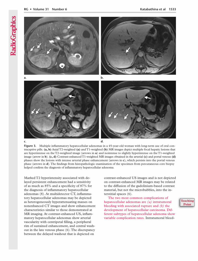
At MR imaging, inflammatory hepatocel-lular adenomas are diffusely hyperintense on T2-weighted images, with a higher signal inten-sity in the periphery of the lesion, correlating with dilated sinusoids. On T1-weighted images, inflammatory hepatocellular adenomas are iso-intense or mildly hyperintense, with minimal or no signal drop-off with chemical shift sequences (7,8). After administration of gadolinium-based contrast material, inflammatory hepatocellular adenomas usually show intense enhancement during the arterial phase, which persists in the portal venous and delayed phases (Fig 3) (7,8).
Figure 1. Flowchart illus-trates the pathogenesis of inflammatory hepatocellular adenomas (IHCAs). ALB = albumin, CCL20 = C-C motif ligand 20, gp-130 = glyco-protein 130, HCA = hepato-cellular adenoma, IGF1 = insulinlike growth factor, IL-6 = interleukin-6, IL6ST = inter-leukin-6 signal transducer, JAK = Janus kinase, STAT = signal transducer and activator of transcription, TTR = trans- thyretin.
RG • Volume 31 Number 6 Katabathina et al 1533
Figure 3. Multiple inflammatory hepatocellular adenomas in a 45-year-old woman with long-term use of oral con-traceptive pills. (a, b) Axial T2-weighted (a) and T1-weighted (b) MR images depict multiple focal hepatic lesions that are hyperintense on the T2-weighted image (arrows in a) and isointense to slightly hyperintense on the T1-weighted image (arrow in b). (c, d) Contrast-enhanced T1-weighted MR images obtained in the arterial (c) and portal venous (d) phases show the lesions with intense arterial phase enhancement (arrows in c), which persists into the portal venous phase (arrows in d). The findings from histopathologic examination of the specimen from percutaneous core biopsy helped confirm the diagnosis of inflammatory hepatocellular adenoma.
Marked T2 hyperintensity associated with de-layed persistent enhancement had a sensitivity of as much as 85% and a specificity of 87% for the diagnosis of inflammatory hepatocellular adenomas (8). At multidetector CT, inflamma-tory hepatocellular adenomas may be depicted as heterogeneously hyperattenuating masses on nonenhanced CT images and show enhancement characteristics similar to those demonstrated at MR imaging. At contrast-enhanced US, inflam-matory hepatocellular adenomas show arterial vascularity with centripetal filling, a peripheral rim of sustained enhancement, and central wash-out in the late venous phase (6). The discrepancy between the delayed washout that is depicted on
contrast-enhanced US images and is not depicted on contrast-enhanced MR images may be related to the diffusion of the gadolinium-based contrast material, but not the microbubbles, into the in-terstitial spaces (6).
The two most common complications of hepatocellular adenomas are (a) intratumoral bleeding with associated rupture and (b) the development of hepatocellular carcinoma. Dif-ferent subtypes of hepatocellular adenoma show variable complication rates. Intratumoral bleed-
1534 October Special Issue 2011 radiographics.rsna.org
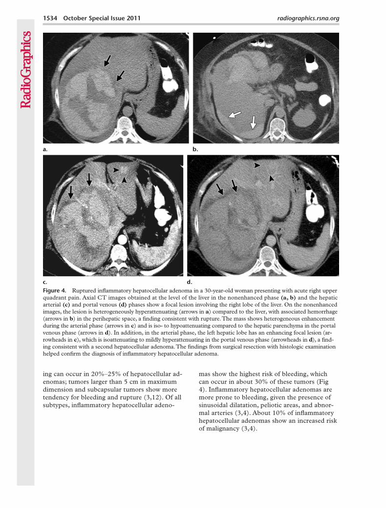
ing can occur in 20%–25% of hepatocellular ad-enomas; tumors larger than 5 cm in maximum dimension and subcapsular tumors show more tendency for bleeding and rupture (3,12). Of all subtypes, inflammatory hepatocellular adeno-
mas show the highest risk of bleeding, which can occur in about 30% of these tumors (Fig 4). Inflammatory hepatocellular adenomas are more prone to bleeding, given the presence of sinusoidal dilatation, peliotic areas, and abnor-mal arteries (3,4). About 10% of inflammatory hepatocellular adenomas show an increased risk of malignancy (3,4).
Figure 4. Ruptured inflammatory hepatocellular adenoma in a 30-year-old woman presenting with acute right upper quadrant pain. Axial CT images obtained at the level of the liver in the nonenhanced phase (a, b) and the hepatic arterial (c) and portal venous (d) phases show a focal lesion involving the right lobe of the liver. On the nonenhanced images, the lesion is heterogeneously hyperattenuating (arrows in a) compared to the liver, with associated hemorrhage (arrows in b) in the perihepatic space, a finding consistent with rupture. The mass shows heterogeneous enhancement during the arterial phase (arrows in c) and is iso- to hypoattenuating compared to the hepatic parenchyma in the portal venous phase (arrows in d). In addition, in the arterial phase, the left hepatic lobe has an enhancing focal lesion (ar-rowheads in c), which is isoattenuating to mildly hyperattenuating in the portal venous phase (arrowheads in d), a find-ing consistent with a second hepatocellular adenoma. The findings from surgical resection with histologic examination helped confirm the diagnosis of inflammatory hepatocellular adenoma.

RG • Volume 31 Number 6 Katabathina et al 1535
HNF-1a–mutated Hepatocellular Adenoma
HNF-1a–mutated hepatocellular adenomas are the second most common type of hepatocellular adenoma and constitute about 30%–35% of all hepatocellular adenomas. Biallelic inactivat-ing mutations of the HNF-1a gene (HNF1A) are responsible for the development of HNF-1a–mutated hepatocellular adenomas, and some cases have an association with (a) maturity-onset diabetes of the young (MODY), type 3, and (b) familial hepatic adenomatosis (4,5,18). HNF-1a–mutated hepatocellular adenomas de-velop exclusively in female patients, with more than 90% of patients having a history of oral contraceptive use, and the tumors are multiple in about 50% of patients. Many HNF-1a–mu-tated hepatocellular adenomas are asymptomatic and are depicted incidentally during imaging studies performed for other reasons, such as trauma or nonspecific abdominal pain.
HNF-1a–mutated hepatocellular adenomas result from biallelic inactivating mutations of the HNF1A gene (also known as transcription fac-tor-1 gene) (19). The HNF1A gene is a tumor
suppressor gene located at chromosome 12q24, and the gene encodes the HNF-1a protein, a transcription factor involved in hepatocyte dif-ferentiation (10). In about 90% of cases, both mutations are of somatic origin; in the remain-ing 10% of cases, HNF-1a–mutated hepatocel-lular adenomas show one germline mutation and one somatic mutation (3,19). Patients with germline mutations have a predisposition to MODY type 3 and to familial hepatic adenoma-tosis (20,21). Estrogens in oral contraceptives may act as endogenous genotoxic agents, and it has been proposed that estrogens are respon-sible for somatic mutations in HNF-1a–mu-tated hepatocellular adenomas (19,22). The final outcome of all of these mutations is the production of nonfunctioning HNF-1a protein, which promotes lipogenesis and hepatocellular proliferation (Fig 5) (23). In addition, abnormal HNF-1a protein causes silencing of liver fatty acid–binding protein, resulting in impaired fatty acid trafficking in hepatocytes, which leads to intracellular fat deposition (23).
Figure 5. Flowchart shows pathogenesis of HNF-1a–mu-tated hepatocellular adeno-mas. HCA = hepatocellular adenoma, L-FABP = liver fatty acid–binding protein, OCP = oral contraceptive pill.

1536 October Special Issue 2011 radiographics.rsna.org
Figure 6. Photomicrograph (H-E stain; original mag-nification, ×40) of an HNF-1a–mutated hepatocellular adenoma in a 39-year-old woman shows fat deposition (arrows) within the hepatocytes.
Finally, all of these changes lead to the de-velopment of hepatocellular adenomas with diffuse intratumoral steatosis. At histologic examination, HNF-1a–mutated hepatocellular adenomas are characterized by excessive lipid accumulation in the tumor hepatocytes (Fig 6) (18). No portal tract elements or cytologic and/or nuclear abnormalities are identified within the tumor cells. Lack of liver fatty acid–binding protein is a characteristic feature of HNF-1a–mutated hepatocellular adenomas at immuno-chemical analysis (18).
At MR imaging, HNF-1a–mutated hepato-cellular adenomas are predominantly hyper- or isointense on T1-weighted images, with diffuse signal drop-off with use of a chemical shift se-quence because of intracellular steatosis (7,8). In addition, the background liver also shows diffuse fatty infiltration because HNF-1a–mu-tated hepatocellular adenomas are associated with MODY type 3 and hepatic steatosis (24). HNF-1a–mutated hepatocellular adenomas are isointense to slightly hyperintense on T2-weighted images. Moderate enhancement in the arterial phase is identified after administration of gadolinium-based contrast material, with no persistent enhancement in the portal venous and delayed phases (Fig 7) (8). The sensitivity and specificity of homogeneous signal drop-off on chemical shift images for the diagnosis of HNF-1a–mutated hepatocellular adenomas were as high as 86% and 100%, respectively (8). The intra- and intercellular lipid of the hepatocel-lular adenomas may uncommonly manifest as macroscopic fat deposits that can be identified at multidetector CT, and such lipid is diagnostic of HNF-1a–mutated hepatocellular adenomas (25). MR imaging is more sensitive and specific for the identification of intratumoral fat. Ap-proximately 7% of hepatocellular adenomas may show fat when imaged with CT, whereas about 35%–77% of hepatocellular adenomas demon-strate fat at chemical shift MR imaging (26,27). At contrast-enhanced US, HNF-1a–mutated hepatocellular adenomas show isovascularity to moderately increased vascularity, with mixed filling in the arterial phase after contrast mate-rial administration, and are isoechoic in the por-tal venous and delayed phases (6).
Among all hepatocellular adenomas, the HNF-1a–mutated hepatocellular adenomas are the least aggressive subtype: Tumors less than 5 cm in maximum dimension show minimal risk of bleeding and subsequent rupture and carry minimal or no risk for the development of malignancy.
b-Catenin–mutated Hepatocellular Adenoma
b-Catenin–mutated hepatocellular adenomas constitute about 10%–15% of all hepatocellular adenomas and are due to activating mutations of the b-catenin gene (18). b-Catenin–mutated hepatocellular adenomas occur more frequently in men and are associated with male hormone administration, glycogen storage disease, and fa-milial adenomatosis polyposis (10).
b-Catenin–mutated hepatocellular adenomas develop because of sustained activation of b-catenin protein, resulting in uncontrolled hepato-cyte proliferation (28). b-Catenin is encoded by the catenin b 1 gene (CTNNB1), which is located at chromosome 3p21. b-Catenin is an impor-tant downstream effector of the Wnt/b-catenin pathway, which has a major role in liver embryo-genesis, cell adhesion, growth, zonation, and regeneration. In the steady state, in the absence of a Wnt signal, b-catenin is phosphorylated and degraded by a cytoplasmic destruction complex formed by the adenomatous polyposis coli gene (APC) and the Axin family of genes in associa-

RG • Volume 31 Number 6 Katabathina et al 1537
tion with glycogen synthase kinase (29). Mutated b-catenin protein, abnormal activation of Wnt protein, or mutations of the APC gene cause increased cytoplasmic availability of b-catenin, which causes sustained activation of its targeted genes, resulting in uncontrolled hepatocyte pro-
Figure 7. HNF-1a–mutated (steatotic) hepatocellular adenoma in a 29-year-old woman with a history of ele- vated liver enzyme levels. (a–c) Axial T2-weighted (a) and T1-weighted in-phase (b) and out-of-phase (c) MR images depict a focal hepatic lesion that is mildly hyper-intense on the T2-weighted image (arrows in a) and isointense to the liver on the T1-weighted in-phase image (arrows in b), with focal areas of signal loss on the out-of- phase image (arrows in c). (d, e) Contrast-enhanced T1-weighted MR images obtained in the arterial (d) and portal venous (e) phases show that the lesion has mild en-hancement in the arterial phase (arrows in d), which does not persist into the portal venous phase (arrows in e).
liferation and the development of b-catenin–mu-tated hepatocellular adenomas (Fig 8). At his-topathologic examination, the hepatocytes of b-catenin–mutated hepatocellular adenomas show cytologic abnormalities, such as a high nuclear-cytoplasmic ratio, nuclear atypia, and formation

1538 October Special Issue 2011 radiographics.rsna.org
Figure 8. Flowcharts relating to b-catenin–mutated hepatocellular adenoma ( bHCA). (a) Cyto-plasmic destruction of b-catenin in the steady state with absence of Wnt binding and with normal APC, b-catenin, and Axin genes. (b) Pathogenesis of b-catenin–mutated hepatocellular adenomas and hepatocellular carcinomas (HCCs) via the Wnt/b-catenin pathway, secondary to mutations of the APC, b-catenin, and Axin genes and abnormal binding of Wnt protein. APC = adenomatous polyposis coli, GSK = glycogen synthase kinase, M- = mutated gene.
of acini, and are difficult to distinguish from the hepatocytes of well-differentiated hepatocellular carcinomas (10). The glutamate-ammonia ligase gene (GLUL), a b-catenin–targeted gene that encodes glutamine synthase, is overexpressed in b-catenin–mutated hepatocellular adenomas, resulting in strong and diffuse positivity to gluta-mine synthase at immunohistochemical analysis (2). In addition, heterogeneous aberrant nuclear and cytoplasmic staining of b-catenin is also identified (2,10).
No specific MR imaging patterns have yet been proposed to identify b-catenin–mutated hepato-cellular adenomas, and on T1- and T2-weighted images, these tumors may show homogeneous or heterogeneous hyperintense signal intensity, depending on the presence of hemorrhage and/or necrosis. b-Catenin–mutated hepatocellular adenomas commonly demonstrate strong arte-rial enhancement that may or may not persist on the portal venous and delayed phases, and these tumors may mimic hepatocellular carcinomas at imaging (8). No specific US or multidetector CT findings for b-catenin–mutated hepatocellular ad-enomas have been reported in the literature.
Overall, the risk of hepatocellular carcinoma development in hepatocellular adenomas is about 5%–10% (30). The important risk factors for malignant transformation of hepatocellular adenomas are male sex, concomitant glycogen storage disease, anabolic steroid usage, the b-catenin–mutated subtype, and tumors larger than 5 cm in maximum dimension (24,30,31). Of all hepatocellular adenomas, b-catenin–mutated hepatocellular adenomas carry the highest risk of malignancy and are interpreted as borderline lesions between hepatocellular adenoma and hepatocellular carcinoma (Fig 9) (10). The Wnt/b-catenin pathway involved in the pathogenesis of b-catenin–mutated hep-atocellular adenomas is also a major player in the development of hepatocellular carcinoma, with about 35% of hepatocellular carcinomas showing b-catenin mutations (32,33). This find-ing explains the high incidence of cancer in b-catenin–mutated hepatocellular adenomas compared with the incidence in other subtypes. In a recent study, Farges et al (31) concluded that metabolic syndrome is an emerging condi-tion associated with malignant transformation of hepatocellular adenoma in men. In the study, these investigators also showed that the presence of malignancy with hepatocellular adenoma is

RG • Volume 31 Number 6 Katabathina et al 1539
10 times more frequent in men than in women, and management should primarily be based on the sex of the patient (31). Hepatocellular car-cinomas may develop either as a macroscopic nodule larger than 1 cm in maximum dimension or as multiple microscopic foci. Although b-catenin–mutated hepatocellular adenomas also have a risk of bleeding, the exact incidence of this complication is not known.
Unclassified Hepatocellular Adenoma
Approximately 10% of all hepatocellular adeno-mas are without specific genetic and/or pathologic abnormalities. This subset of tumors is grouped under the rubric of the unclassified subtype (10).
No specific MR imaging patterns have yet been proposed to identify unclassified hepatocel-lular adenomas. These hepatocellular adenomas are poorly understood; detailed studies will be required to evaluate the oncogenesis, clinical fea-tures, and pathologic and radiologic findings of this rare subtype of hepatocellular adenoma.
Hepatic AdenomatosisTo our knowledge, hepatic adenomatosis was first described by Flejou et al (34) in 1985 and is defined as the presence of multiple adenomas (arbitrarily >10) involving both lobes of the liver, without any history of steroid therapy or glycogen
Figure 9. Hepatocellular adenoma with malignant change in a 40-year-old man. (a) Coronal T2-weighted MR image shows an isointense exophytic focal lesion (arrows), 7 cm in maxi-mum dimension, arising from hepatic segment VI, with focal hyperintense areas within the lesion. (b, c) Axial T1-weighted contrast-enhanced MR images obtained in the arterial (b) and portal venous (c) phases show intense arterial enhancement in the arterial phase (arrow in b), with washout in the portal ve-nous phase (arrow in c). The findings from surgical resection and histopathologic examination showed a well-differentiated hepatocellular carcinoma arising from a hepatocellular ad-enoma with b-catenin mutations.

1540 October Special Issue 2011 radiographics.rsna.org
Figure 10. Hepatic adenomatosis in a 38-year-old woman. Coronal T2-weighted (a) and axial contrast-en-hanced T1-weighted (b) MR images depict multiple T2 hyperintense lesions (arrows in a) throughout the liver, which show intense enhancement in the arterial phase (arrowheads in b), a finding consistent with multiple inflammatory hepatocellular adenomas.
storage disease (35). Hepatic adenomatosis com-monly occurs in women during the 4th and 5th decades of life (35,36). Although the exact etiology of hepatic adenomatosis is still unclear, congeni-tal or acquired hepatic vascular abnormalities, mutations of the HNF1A gene, and nonalcoholic fatty liver disease have been proposed as potential causes for the development of hepatic adenomato-sis (35,37,38). In addition, patients with familial hepatic adenomatosis who have germline muta-tions of the HNF1A gene tend to develop MODY type 3. Hepatocellular adenomas in patients with hepatic adenomatosis may be of the inflammatory, HNF-1a–mutated, or b-catenin–mutated sub-types, and their imaging appearances may vary ac-cordingly (Fig 10) (7). In contradistinction to pre-vious beliefs, hepatic adenomatosis per se does not have any increased risk of complications; tumor size and subtype determine the risks of malignancy and bleeding (36). Management of hepatocellular adenomas in patients with hepatic adenomatosis is similar to management of other hepatocellular adenomas. In addition, diabetes mellitus and mu-tations of the HNF1A gene should be sought in patients with hepatic adenomatosis, and liver im-aging is recommended in their relatives (20).
Genotype-Phenotype Classifi- cation: Management Implications
The classification of hepatocellular adenomas into different subtypes on the basis of genetic and pathologic characteristics allows a better un-derstanding of the natural history and prognoses of these rare tumors, so that management and follow-up options may be modified accordingly. On the basis of the recommendations by Bioulac-Sage et al (5), the management strategies for dif-ferent hepatocellular adenoma subtypes in asymp- tomatic and symptomatic patients have been summarized in a chart (Fig 11).
Incidental depiction in an asymptomatic patient is one of the common clinical presentations of hep- atocellular adenoma. For this group of patients, the first step in management is subtype classifica-tion, on the basis of the cross-sectional imaging findings, into one of two groups: (a) hepatocellular adenomas with diffuse steatosis or (b) heteroge-neous hepatocellular adenomas without steato-sis (5). As discussed previously, MR imaging is preferred to CT for identification of intracellular microscopic fat and for better characterization of the hepatocellular adenoma. A thorough clinical examination with determination of the medica-tion history is recommended to help identify any
known cause, such as usage of oral contraceptives, barbiturates, or steroids. If such usage is found, withholding the medication and repeating the im-aging studies in 3–6 months are suggested.
Stable and regressing hepatocellular adenomas may be monitored with imaging studies without any therapeutic interventions. Hepatocellular ad-enomas that are growing despite discontinuation of the usage of potentially causative medications need further evaluation on the basis of the sex of the patient and the size of the tumor (3). Genetic counseling with regard to a family history of hep-atocellular adenomas, hepatic adenomatosis, and MODY type 3 may be performed in patients with suspected steatotic hepatocellular adenomas mea-suring less than 5 cm in maximum dimension (5).

RG • Volume 31 Number 6 Katabathina et al 1541
In patients with heterogeneous small (<5-cm) hepatocellular adenomas, percutaneous biopsy and histopathologic review are warranted to iden-tify b-catenin mutations. Hepatocellular adenomas larger than 5 cm in maximum dimension, in male patients, and in patients with glycogen storage disease should be surgically resected (5,12,39,40). Surgically resected hepatocellular adenomas and those subjected to biopsy should undergo detailed histopathologic review to characterize them on the basis of the genotype-phenotype classification. Hep-atocellular adenomas with b-catenin mutations, inflammatory features with b-catenin mutations, and hepatocellular adenomas mimicking hepato-cellular carcinomas at histopathologic evaluation are to be considered as hepatocellular carcinomas that require treatment and surveillance accord-ingly (5). Female patients with inflammatory hep-atocellular adenomas, HNF-1a–mutated hepato-cellular adenomas, and unclassified tumors require treatment of diabetes mellitus and clinical and MR imaging follow-up until menopause (5). To our knowledge, there are no definite guidelines to rec-ommend an optimal interval for follow-up exami-nations; annual imaging follow-up has been rec-ommended by some investigators for both solitary hepatocellular adenoma and hepatic adenomatosis (41). In addition to surgery, other definitive treat-ment options include radiofrequency ablation and hepatic artery embolization. Radiofrequency abla-tion is indicated for (a) tumors smaller than 4 cm in maximum dimension, (b) patients who are not surgical candidates, and (c) those who prefer to avoid surgery after discussion and full understand-ing of available treatment options (40,42). Tumors
that grow to larger than 5 cm in maximum dimen-sion during follow-up need definitive treatment.
The management of patients with sympto-matic hepatocellular adenomas depends on the duration and type of symptoms. Patients with ruptured hepatocellular adenomas who present in hemodynamically unstable condition and patients with hepatocellular adenomas larger than 5 cm in maximum dimension require immediate treat-ment with either hepatic artery embolization or surgery (43,44). Conservative treatment may be considered in hemodynamically stable patients, even though they present with rupture (44). Pa-tients with continued symptoms, with growing hepatocellular adenomas, or with tumors larger than 5 cm in maximum dimension need defini-tive treatment. Further management is guided by the specific subtype of hepatocellular adenoma on the basis of the genotype-phenotype classifica-tion, as discussed for asymptomatic patients (5).
ConclusionsHepatocellular adenomas are a diverse group of benign neoplasms that are characterized by spe-cific genetic mutations, molecular abnormalities, histopathologic features, imaging findings, tumor biology, and natural history. The current geno-type-phenotype classification of hepatocellular adenomas identifies three distinct subtypes (in-flammatory, HNF-1a–mutated, and b-catenin–mutated hepatocellular adenomas). Novel in-sights into the pathogenesis of adenomas permit
Figure 11. Flowchart illustrates management strategies for different subtypes of hepatocel-lular adenoma (HCA). GSD = glycogen storage disease, RFA = radiofre-quency ablation.

1542 October Special Issue 2011 radiographics.rsna.org
better understanding of the tumor subtypes and their biologic behaviors. Hepatocellular adeno-mas with b-catenin mutations frequently undergo malignant change, inflammatory hepatocellular adenomas commonly bleed, and steatotic hepa-tocellular adenomas typically portend a favorable prognosis.
Contrast-enhanced MR imaging is the imag-ing modality of choice for the differentiation of the subtypes of hepatocellular adenoma. Inflam-matory hepatocellular adenomas are diffusely hyperintense on T2-weighted images and have isointense or mildly hyperintense signal inten-sity on T1-weighted images, with minimal or no signal drop-off with the use of chemical shift sequences. After contrast material administra-tion, inflammatory hepatocellular adenomas show intense enhancement during the arterial phase that persists in the portal venous and delayed phases. HNF-1a–mutated hepatocel-lular adenomas are hyper- or isointense on T1-weighted images, with diffuse signal drop-off with use of the chemical shift sequence, and are isointense to slightly hyperintense on T2-weighted images. Moderate enhancement is depicted in the arterial phase, with an absence of persistent enhancement in the portal venous and delayed phases. No specific MR imaging patterns have been proposed to identify b-catenin–mutated and unclassified hepatocellular adenomas. A knowledge of the biologic diversity of hepatocellular adenomas and a familiarity with the clinical and imaging findings of the subtypes of hepatocellular adenoma and their associated complications permit optimal patient management.
References 1. Edmondson HA, Henderson B, Benton B. Liver-cell
adenomas associated with use of oral contraceptives. N Engl J Med 1976;294(9):470–472.
2. Bioulac-Sage P, Rebouissou S, Thomas C, et al. Hepatocellular adenoma subtype classification us-ing molecular markers and immunohistochemistry. Hepatology 2007;46(3):740–748.
3. Bioulac-Sage P, Balabaud C, Zucman-Rossi J. Sub-type classification of hepatocellular adenoma. Dig Surg 2010;27(1):39–45.
4. Zucman-Rossi J, Jeannot E, Nhieu JT, et al. Genotype-phenotype correlation in hepatocellular adenoma: new classification and relationship with HCC. Hepatology 2006;43(3):515–524.
5. Bioulac-Sage P, Laumonier H, Couchy G, et al. Hep-atocellular adenoma management and phenotypic classification: the Bordeaux experience. Hepatology 2009;50(2):481–489.
6. Laumonier H, Cailliez H, Balabaud C, Bioulac-Sage P, Zucman-Rossi J, Trillaud H. Contrast-enhanced ultrasonography to identify the two major subtypes of hepatocellular adenoma. J Hepatol 2010;52 (suppl 1):S223.
7. Lewin M, Handra-Luca A, Arrivé L, et al. Liver adenomatosis: classification of MR imaging features and comparison with pathologic findings. Radiology 2006;241(2):433–440.
8. Laumonier H, Bioulac-Sage P, Laurent C, Zucman-Rossi J, Balabaud C, Trillaud H. Hepatocellular adenomas: magnetic resonance imaging features as a function of molecular pathological classification. Hepatology 2008;48(3):808–818.
9. Ronot M, Bahrami S, Calderaro J, et al. Hepatocel-lular adenomas: accuracy of magnetic resonance imaging and liver biopsy in subtype classification. Hepatology 2011;53(4):1182–1191.
10. Bioulac-Sage P, Laumonier H, Laurent C, Zucman-Rossi J, Balabaud C. Hepatocellular adenoma: what is new in 2008. Hepatol Int 2008;2(3):316–321.
11. Paradis V, Champault A, Ronot M, et al. Telangiec-tatic adenoma: an entity associated with increased body mass index and inflammation. Hepatology 2007;46(1):140–146.
12. Dokmak S, Paradis V, Vilgrain V, et al. A single-cen-ter surgical experience of 122 patients with single and multiple hepatocellular adenomas. Gastroenter-ology 2009;137(5):1698–1705.
13. Rebouissou S, Amessou M, Couchy G, et al. Fre-quent in-frame somatic deletions activate gp130 in inflammatory hepatocellular tumours. Nature 2009; 457(7226):200–204.
14. Rodriguez C, Grosgeorge J, Nguyen VC, Gaudray P, Theillet C. Human gp130 transducer chain gene (IL6ST) is localized to chromosome band 5q11 and possesses a pseudogene on chromosome band 17p11. Cytogenet Cell Genet 1995;70(1-2):64–67.
15. Spannbauer MM, Trautwein C. Frequent in-frame somatic deletions activate gp130 in inflammatory hepatocellular tumors. Hepatology 2009;49(4): 1387–1389.
16. Schutyser E, Struyf S, Van Damme J. The CC che-mokine CCL20 and its receptor CCR6. Cytokine Growth Factor Rev 2003;14(5):409–426.
17. Bioulac-Sage P, Rebouissou S, Sa Cunha A, et al. Clinical, morphologic, and molecular features de-fining so-called telangiectatic focal nodular hyper-plasias of the liver. Gastroenterology 2005;128(5): 1211–1218.
18. Bioulac-Sage P, Blanc JF, Rebouissou S, Balabaud C, Zucman-Rossi J. Genotype phenotype classifica-tion of hepatocellular adenoma. World J Gastroen-terol 2007;13(19):2649–2654.
19. Bluteau O, Jeannot E, Bioulac-Sage P, et al. Bi-allelic inactivation of TCF1 in hepatic adenomas. Nat Genet 2002;32(2):312–315.
20. Bacq Y, Jacquemin E, Balabaud C, et al. Familial liver adenomatosis associated with hepatocyte nuclear factor 1alpha inactivation. Gastroenterology 2003;125(5):1470–1475.
21. Ellard S. Hepatocyte nuclear factor 1 alpha (HNF-1 alpha) mutations in maturity-onset diabetes of the young. Hum Mutat 2000;16(5):377–385.
22. Cavalieri E, Frenkel K, Liehr JG, Rogan E, Roy D. Estrogens as endogenous genotoxic agents: DNA adducts and mutations. J Natl Cancer Inst Monogr 2000;2000(27):75–93.

RG • Volume 31 Number 6 Katabathina et al 1543
23. Rebouissou S, Imbeaud S, Balabaud C, et al. HNF1alpha inactivation promotes lipogenesis in human hepatocellular adenoma independently of SREBP-1 and carbohydrate-response element-binding protein (ChREBP) activation. J Biol Chem 2007;282(19):14437–14446.
24. Bioulac-Sage P, Laumonier H, Sa Cunha A, Bala-baud C. Hepatocellular adenomas. Liver Int 2009; 29(1):142.
25. Prasad SR, Wang H, Rosas H, et al. Fat-containing lesions of the liver: radiologic-pathologic correlation. RadioGraphics 2005;25(2):321–331.
26. Grazioli L, Federle MP, Brancatelli G, Ichikawa T, Olivetti L, Blachar A. Hepatic adenomas: imaging and pathologic findings. RadioGraphics 2001;21(4): 877–892; discussion 892–894.
27. Ichikawa T, Federle MP, Grazioli L, Nalesnik M. Hepatocellular adenoma: multiphasic CT and histo-pathologic findings in 25 patients. Radiology 2000; 214(3):861–868.
28. Chen YW, Jeng YM, Yeh SH, Chen PJ. P53 gene and Wnt signaling in benign neoplasms: beta-catenin mutations in hepatic adenoma but not in focal nodular hyperplasia. Hepatology 2002;36(4 pt 1): 927–935.
29. Thompson MD, Monga SP. WNT/beta-catenin signaling in liver health and disease. Hepatology 2007;45(5):1298–1305.
30. Farges O, Dokmak S. Malignant transformation of liver adenoma: an analysis of the literature. Dig Surg 2010;27(1):32–38.
31. Farges O, Ferreira N, Dokmak S, Belghiti J, Bedossa P, Paradis V. Changing trends in malignant trans-formation of hepatocellular adenoma. Gut 2011;60 (1):85–89.
32. Takigawa Y, Brown AM. Wnt signaling in liver can-cer. Curr Drug Targets 2008;9(11):1013–1024.
33. Buendia MA. Genetic alterations in hepatoblas-toma and hepatocellular carcinoma: common and distinctive aspects. Med Pediatr Oncol 2002;39(5): 530–535.
34. Flejou JF, Barge J, Menu Y, et al. Liver adenomato-sis: an entity distinct from liver adenoma? Gastro-enterology 1985;89(5):1132–1138.
35. Veteläinen R, Erdogan D, de Graaf W, et al. Liver adenomatosis: re-evaluation of aetiology and man-agement. Liver Int 2008;28(4):499–508.
36. Greaves WO, Bhattacharya B. Hepatic adenomatosis. Arch Pathol Lab Med 2008;132(12):1951–1955.
37. Grazioli L, Federle MP, Ichikawa T, Balzano E, Nalesnik M, Madariaga J. Liver adenomatosis: clini-cal, histopathologic, and imaging findings in 15 pa-tients. Radiology 2000;216(2):395–402.
38. Brunt EM, Wolverson MK, Di Bisceglie AM. Be-nign hepatocellular tumors (adenomatosis) in non-alcoholic steatohepatitis: a case report. Semin Liver Dis 2005;25(2):230–236.
39. Rhim H, Lim HK, Kim YS, Choi D, Lee WJ. Ra-diofrequency ablation of hepatic tumors: lessons learned from 3000 procedures. J Gastroenterol Hepatol 2008;23(10):1492–1500.
40. van der Sluis FJ, Bosch JL, Terkivatan T, de Man RA, Ijzermans JN, Hunink MG. Hepatocellular adenoma: cost-effectiveness of different treatment strategies. Radiology 2009;252(3):737–746.
41. Ribeiro A, Burgart LJ, Nagorney DM, Gores GJ. Management of liver adenomatosis: results with a conservative surgical approach. Liver Transpl Surg 1998;4(5):388–398.
42. Atwell TD, Brandhagen DJ, Charboneau JW, Nagorney DM, Callstrom MR, Farrell MA. Suc-cessful treatment of hepatocellular adenoma with percutaneous radiofrequency ablation. AJR Am J Roentgenol 2005;184(3):828–831.
43. Huurman VA, Stoot JH, van der Linden E, Terpstra OT, Schaapherder AF. Necrosis of a large hepatic tumor after hemorrhage and subsequent selective arterial embolization. World J Gastroenterol 2006;12 (37):6059–6061.
44. Erdogan D, Busch OR, van Delden OM, Ten Kate FJ, Gouma DJ, van Gulik TM. Management of spontaneous haemorrhage and rupture of hepatocel-lular adenomas: a single centre experience. Liver Int 2006;26(4):433–438.
This journal-based CME activity has been approved for AMA PRA Category 1 CreditTM. See www.rsna.org/education/rg_cme.html.

Teaching Points October Special Issue 2011
Genetics and Imaging of Hepatocellular Adenomas: 2011 UpdateVenkata S. Katabathina, MD • Christine O. Menias, MD • Alampady K. P. • Shanbhogue, MD • Jaishree Jagir-dar, MD • Raj Mohan Paspulati, MD Srinivasa R. Prasad, MD
RadioGraphics 2011; 31:1529–1543 • Published online 10.1148/rg.316115527 • Content Codes:
Page 1530Accordingly, hepatocellular adenomas are currently categorized into three distinct genetic and pathologic subtypes: (a) inflammatory hepatocellular adenomas, (b) hepatocyte nuclear factor 1 alpha (HNF-1a)–mu-tated hepatocellular adenomas, and (c) b-catenin–mutated hepatocellular adenomas (2).
Page 1532 (Figure on page 1533)At MR imaging, inflammatory hepatocellular adenomas are diffusely hyperintense on T2-weighted im-ages, with a higher signal intensity in the periphery of the lesion, correlating with dilated sinusoids. On T1-weighted images, inflammatory hepatocellular adenomas are iso-intense or mildly hyperintense, with minimal or no signal drop-off with chemical shift sequences (7,8). After administration of gadolinium-based contrast material, inflammatory hepatocellular adenomas usually show intense enhancement dur-ing the arterial phase, which persists in the portal venous and delayed phases (Fig 3) (7,8).
Page 1533The two most common complications of hepatocellular adenomas are (a) intratumoral bleeding with associated rupture and (b) the development of hepatocellular carcinoma. Different subtypes of hepato-cellular adenoma show variable complication rates.
Page 1536At MR imaging, HNF-1a–mutated hepatocellular adenomas are predominantly hyper- or isointense on T1-weighted images, with diffuse signal drop-off with use of a chemical shift sequence because of intracellular steatosis (7,8).
Page 1541Hepatocellular adenomas larger than 5 cm in maximum dimension, in male patients, and in patients with glycogen storage disease should be surgically resected (5,12,39,40). Surgically resected hepatocellular ad-enomas and those subjected to biopsy should undergo detailed histopathologic review to characterize them on the basis of the genotype-phenotype classification.