a.a
TRANSCRIPT

Aggregatibacteractinomycetemcomitans
Presented by:Dr. Yogender Singh

CONTENTS
INTRODUCTION
HISTORICAL PERSPECTIVE
TAXONOMIC CLASSIFICATION
CULTURAL CHARACTERISTICS
BIOCHEMICAL CHARACTERISTICS
FACTORS INFLUENCING THE GROWTH AND VIABILITY
VIRULENCE FACTORS
DIAGNOSTIC METHODS
PRO-ATHEROGENETIC PROPERTIES
ROLE IN AGGRESSIVE PERIODONTITIS
CONCLUSION
REFERENCES

The human fetus inside the uterus is sterile
Within hours after birth, the sterile oral cavity becomes colonized by low numbers of mainly facultative and aerobic bacteria.
2nd day-anaerobic bacteria develops.
Within 2 weeks-mature microbiota established in the gut of new born.
After 2yrs, the entire human microbial flora is formed by a complex collection of 1014
microorganisms with more than 400 different types of bacteria.

Criteria for identification of pathogens
Koch’s postulates
Applicability of Koch postulates in periodontal disease
In periodontitis 3 main problem existed –
1.Inability to culture all the organism
2.Difficulties in defining and culturing sites of active disease
3.Lack of animal model system
Sigmund socransky, a researcher at forsyth dental center,boston, proposed criteria by which periopathogens can be identified.

Socransky’s postulates
Must be associated with the disease evident by organism must be found in relatively high numbers in proximity to the periodontal lesion;
Must be eliminated or decreased in sites that demonstrate clinical resolution of the disease with treatment.
Must demonstrate a host response, in the form of an alteration in the cellular or humoral immune response.
Must be capable of causing disease in experimental animal models
.
Must demonstrate virulence factors responsible for enabling the microorganism to cause destruction of periodontal tissues.

Bacterial characteristics based on Oxygen Environment….
Aerobes - require oxygen for growth
Anaerobic - do not require oxygen
Facultative anaerobic - use oxygen when it is present
Obligate Anaerobic - Cannot survive in presence of oxygen
Aerotolerant anaerobes - do not use oxygen but can tolerate oxidizing environments
Shift from health to Periodontal disease:
In the process…..
Gram positive to Gram negative
Aerobic to Anaerobic
Facultative to Obligatory
Fermentive to Proteolytic
Non-motile to motile

INTRODUCTION
More than 500 cultivable bacterial species have been isolated from the gingival crevices of human beings.
But the actual species count may be thousands, since a wide proportion of bacteria remains uncultivable or unidentified.
The sub gingival bacterial flora consists of facultatively anaerobic gram positive species in the healthy oral cavity, but in gingivitis the proportion of gram negative bacteria increases.
In periodontitis , only 10 to 30 species, mainly gram negative anaerobic bacteria, are putative pathogens.
Mainly three species
Actinobacillus actinomycetemcomitans ,
Porphyromonas gingivalis and
Bacteroides forsythus
Are presently considered as primary etiological agents in periodontitis (WWP 1996 ).

Additional putative periodontal pathogens include:
Prevotella intermedia,
Prevotella nigrescens,
Campylobacter rectus ,
Fusobacterium nucleatum ,
Peptostreptococcus micros and
Spirochetes.
(AAP 1992,Haffajee & Socransky1994,WWP1996 )

HISTORICAL PERSPECTIVE A. actinomycetemcomitans was first isolated and
identified in 1912 by Klinger.
It was so named because of its consistent association with Actinomyces israelii in actinomycotic infections. (1934 Klaber )
The genus name Actinobacillus refers to
actin- star shaped
bacillus- rod shaped
actinomycetum comitans- referring its close association with actinomyces israelii in actinomycotic lesions

In 1951 – Home put forward that A.a could cause disease in humans
In 1959 Heinerish suggested that it a part of normal flora.
In 1976 , Newman, Socransky and Slots – related A.a to Juvenile periodontitis.
In 1979 , Tsai et al, discovered A.a leukotoxin.

Page et al 1991,Perry et al 1996,Kaplan et al 2001- described Six serotypes, a–f.
In 2004 , Roe et al – identified the complete A.a genome
In 2006- Phylogenetic similarity of A. actinomycetemcomitans and Haemophilus aphrophilus, H. paraphrophilus, and H. segnis, suggesting the new genus Aggregatibacter.

TAXONOMIC CLASSIFICATION
Aggregatibacter a member of family Pasteurellaceae, has been recognized as one of the key pathogens in periodontitis.
Was first isolated from human cervicofacial actinomycosis together with Actinomyces and was thus described as Bacterium actinomycetemcomitans by Klinger in 1912.
Later Lieske in 1921 reclassified as Bacterium comitans.

Due to their phylogeny and their typical phenotypic characteristics –
The autoaggregation, the species Actinobacillus a, Haemophilus aphrophilus , Haemophilus paraaphrophilus and Haemophilus segnis were recently
reclassified to a novel genus Aggregatibacter
(Norskov – Lauritsen and Kilan; 2006)

CULTURAL CHARACTERISTICS
Small non motile
Gram negative coccobacillus
Capnophilic
Facultatively anaerobic,
Grows well in 5% CO2 in air or anaerobically (24-48 hrs)

SELECTIVE MEDIUM
Tryptic soy–serum–bacitracin–vancomycin agar (contains 10% horse serum, 75 mg ⁄ l bacitracin and 5 mg ⁄ l vancomycin)
The presence of these antibiotics suppresses the growth of gram-positive bacteria.
Colonies incubated for 4–7 days on a serum-containing medium will develop star-like structures centrally.
This rough star-like morphology may be lost on subculturing, producing smooth isolates that are less adherent to the agar surface.

Star-like structures centrally present
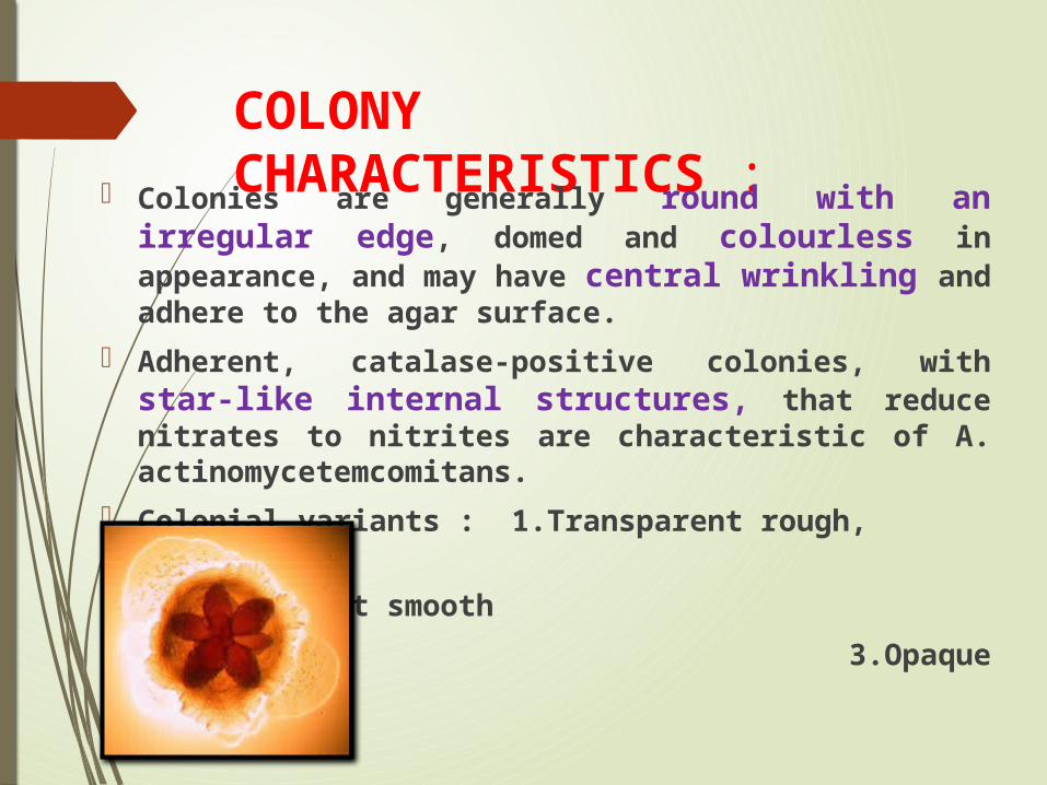
COLONY CHARACTERISTICS : Colonies are generally round with an
irregular edge, domed and colourless in appearance, and may have central wrinkling and adhere to the agar surface.
Adherent, catalase-positive colonies, with star-like internal structures, that reduce nitrates to nitrites are characteristic of A. actinomycetemcomitans.
Colonial variants : 1.Transparent rough,
2.transparent smooth
3.Opaque smooth

BIOCHEMICAL CHARECTERISTICS
Lack of growth on Mac Conkey and other enteric agars
Catalase production : + ve Nitrate reduction : + ve Oxidase negative : + ve Urease : - ve Indole production : - ve Glucose , Fructose , Mannose : strong
fermentation Acid production from maltose , mannitol
and xylose varies Non – haemolytic
FACTORS INFLUENCING THE GROWTH AND VIABILITY OF A.
ACTINOMYCETEMCOMITANS Growth rate Increases Yeast extract , L- cysteine , Thiamine,
Steroids
Optimal pH 7-8 in a medium containing 0.5-1.0 % Nacl.
A rapid reduction of A.actinomycetemcomitans a viability occurred following suspension in distilled water .
The presence of detergent Triton X – 100 at conc. above 2% ( u/v) also decreased the viability of A. a within 10 min.

VIRULENCE FACTORS
Virulence is defined as the relative capacity of a microbe to cause disease .(Slots, 1999)
Poulin and Combes (1999) defined the concept of virulence in terms of the “virulence factors”, which are molecules or components from a microbe that harm the host.
Leukotoxin
Endotoxin
Collagenase
Bacteriocin
Cytolethal distending toxin
Adhesins
Neutrophil inhibitor
Fc binding proteins

A. actinomycetemcomitans is one of the few oral bacteria capable of colonizing buccal mucosa as well as dental plaque.
The establishment of A.actinomycetemcomitans in the human oral cavity is dependent on:
Initial colonisation
Interacting microflora
Various host factors.

Aa COLONISATION FACTORS
Pili or fimbriae Capsule Interactions with other bacteria Vesicles
FIMBRIAE (PILI)
Thin surface appendages that serve as adhesion determinants for initial attachment of A. actinomycetemcomitans to oral surface.
BACTERIAL INTERACTIONS
Streptococcus sanguis , Actinomyces naeslundi and Streptococcus uberis produced factors that were inhibitory to the growth of A. actinimycetemcomitans.
Hillman and co – workers ( 1982, 1985, 1987 )
The hydrogen peroxide formation by the “ beneficial species “, either directly or via a host peroxidase system inhibits the growth of pathogen.

Stevens et al. ( 1987 ) and Hammond et al ( 1987 )
Demonstrated the reverse antagonism.
A. a specifically inhibit the growth of S. sanguis, S. uberis, and A. naeslundii genospecies 2 ( but not other species ) by the production of a bacteriocin.
This mutual antagonism is highly specific and its outcome may strongly influence whether a subject or a site will exhibit disease due to A. actinomycetemcomitans.

VESICLES
A.a elaborates numerous vesicles or “ blebs” on the cell surfaces.
Plays important role in initial colonisation.

PLASMIDS AND BACTERIOPHAGES
These may alter:
1.The physiological properties of a microorganism ,
2.Contribute to virulence ,
3.Modify taxonomic status , and
4.Spread biological properties among different strains, species , and genera.

VIRULENCE FACTORS
Leukotoxin
It was found in 1979 , Tsai et al.
It is a 116kDa protein and a member of the RTX( repeats in toxin ) family whose cellular receptor is the beta 2 – integrin , LFA-1, thus accounting for its selective effect on leukocytes.
The gene operon that produces A.a leukotoxin is designated as lkt. (lkt A-C)

The lktA protein is the active toxin.
Acylated, by lktC and an acyl carrier protein, to be become biologically active.
The other two proteins (lktB and lktD ) are responsible for transport and secretion of lktA .
The differences in leukotoxin expression may be due to transcriptional regulation.
Since there is a direct correlation between the level of leukotoxin and the amount of lkt RNA in a given strain.

The RTX leukotoxins are secreted except lkt A
Which is entirely cell associated ; either bound to cell surface/ – associated nucleic acids /or within membranous vesicles .
Or
lkt A bud from the bacterium’s surface and makes bacterium itself toxic to target cells.

A.a leukotoxin specifically kills leucocyte function associated antigen bearing cells such as:
Polymorphonuclear leucocytes and macrophages.
Whereas other types of cells e.g.:
Epithelial and endothelial cells,
Fibroblasts,
Erythrocytes and platelets are resistant to lysis.

The leukotoxin mediated killing is extremely rapid ( a matter of minutes )
Is caused by formation of pores in the cell membrane of target cells.
Cell lysis might be induced by the rapid formation of highly conducive ion channels that lead to:
Membrane depolarization ,
Loss of intracellular K+ ,
Osmotic swelling and subsequent cell death .

A. actinomycetemcomitans produces higher amounts of leukotoxin in anaerobic conditions than in aerobic conditions.
Which is due to the presence of a 35 – bp oxygen response element that represents leukotoxin production in aerobic conditions.

SUPERANTIGENS
Superantigens are generally bacterial proteins that activate T cells bearing specific beta T- cell receptors.
Activation lead to apoptosis of T cells and thus superantigens can be considered as immunosuppresants.

CYTOLETHAL DISTENDING TOXIN
CDT is a cell cycle – modulatory protein with immunosuppressive function .
This toxin is the product of a three gene operon ( cdtA, cdtB, cdtC ) which is found in a range of bacteria including E coli , Shigella spp., Campylobacter spp., Helicobacter spp.

The mechanism of action of this toxin is believed to be due to nuclease activity of cdtB .
cdtA and cdtC facilitate the entry of cdtB into host cells.
The cdtB enters into the nucleus and degrade chromosomal DNA,
Inducing cell cycle arrest in G2/M phase via specific checkpoint kinases .

Fc BINDING PROTEINS
The role of Fc receptors found on the bacterial surfaces and those released in soluble form during growth is not well established.
However, in vitro studies suggest that these molecules may function to inhibit complement activation.

CHEMOTACTIC INHIBITOR
Neutrophils are recruited to infected areas by following a concentration gradient of chemotactic signals.
Disruption or inhibition of neutrophil chemotaxis is advantageous for the infecting organism.
A. actinomycetemcomitans has been shown to be capable of inhibiting chemotaxis.

IMMUNOSUPPRESSIVE FACTOR A. a produces a protein capable of
inhibiting DNA, RNA, and protein synthesis in human T cells activated by mitogens or antigens.
The purified protein inhibits IgG and IgM production by human B cells and affects immunoglobulin production by interfering with the early stage of cell activation.
This factor also affects both B lymphocytes and T- regulatory cells.

LIPOPOLYSACCHARIDE
LPS is toxic to human NK cells
LPS induces the production of cytokines such as IL-6,8,1B and TNF-a from host cells, thus promoting inflammatory reaction.
Induces bone resorption in vitro and in vivo, which may enhance progression of periodontitis.
Stimulates production of MMPs and plasminogen activator which contribute to destruction of periodontal connective tissue.
A. a LPS induces foam cell formation and cholesteryl ester accumulation in murine macrophages which suggests that it also has pro-atherogenic activity.

CYTOKINE INDUCER PROTEINS SECRETED BY
BACTERIUMCell stress protein, chaperonin 60 :
Potent bone degrading molecule.
Normally intracellular, appears to be secreted by this bacterium and stimulates bone resorption by acting as an osteoclast “ growth factor”

HEAT SHOCK PROTEINS
Several HSPs are present in A.a including GroEL- like ( HSP-60) and DnaK like ( HSP -70) proteins.
Protein homologous to GroEL-like HSP found in the surface associated material of A. actinomycetemcomitans has osteolytic activity by murine bone resorption assay.
Purified native GroEl – like HSP from A.a promotes epithelial cell proliferation at lower HSP concentrations, but has a toxic effect on epithelial cells at higher HSP concentrations.

ACTINOMYCETEMCOMITIN: A NEW BACTERIOCIN PRODUCED BY A. a
Lima FL et al 2008 isolated a bacteriocin named as actinomycetemcomitin from A.actinomycetemcomitans P ( 7-20) strain that is active against Peptostreptococcus anaerobius ATCC 27337.
Actinomycetemcomitin was produced during exponential and stationary growth phases and its amount decreased until it disappeared during the decline growth phase.

DIAGNOSTIC METHODSCommercial diagnostic kits:
1.Evalusite ( Kodak)
This is a number of enzyme linked immunosorbant assays (ELISA ) using antibodies to detect antigens for A.actinomycetemcomitans.
The reactions are carried out in a simple chair side reaction kit.
Subgingival plaque samples are reacted with the antibodies and detection substrate in a multilevel reaction dish.

2. Omnigene (Omni Gene, Inc ) and BTD
( Biotechnica Diagnostics , Inc)
These are DNA probe systems for a number of subgingival bacteria.
A paper point sample of subgingival plaque is placed in a container provided and mailed off to the company for assay.

SELECTIVE MEDIUM FOR ISOLATION OF A.a
Socransky et al 1981 developed a selective medium , malachite green bacitracin agar , for the isolation of A.a.
The medium consists of Trypticase soy agar 40 gm/l , bacitracin 128μg/ml , malachite green 8μg/ml and 5% defibrinated sheep blood.
The medium, when incubated in an atmosphere of air plus 10% CO2 for 5 days, permitted greater than 80% recovery of pure cultures of A.actinomycetemcomitans when compared with a non selective media.

Slots et al 1982 developed TSBV ( tryptic soy- serum – bacitracin – vancomycin ) agar.
TSBV agar contained ( per litre ) 40 g of tryptic soy agar , 1 g of yeast extract , 100 ml of horse serum , 75 mg of bacitracin and 5 mg of vancomycin.
The TSBV medium suppressed most oral species and permitted significantly higher recovery of A.actinomycetemcomitans than non selective blood agar medium.

Martijn van Steenbergen T J et al 1986
Compared the two selective media Malachite green bacitracin agar ( MGB agar ) and tryptic soy serum bacitracin vancomycin ( TSBV ) agar .
The highest recovery was found on TSBV agar plates cultured in air - 5% CO2 both for plaque samples from periodontal pockets and for pure cultures.
Tsuzukibashi O et al 2008 developed a novel selective medium designated as AASM .
It was prepared by adding 200μg/ml of vancomycin and 10μg/ml of bacitracin to AAGM , which contains dextrose , sodium bicarbonate , trypticase soy , yeast extract and agar.
They showed that all serotypes ( a-f ) of A.actinomycetemcomitans strains grow well and the average growth recovery of A.actinomycetemcomitans on AASM medium was 94.4% .

PHENOTYPIC TYPING METHODS
Biotyping: Serological typing:
Six A. a serotypes have been designated (a
through f).
The serological specificity is defined by six structurally and antigenically distinct O-polysaccharide components of their respective lipopolysaccharide molecules (Page et al., 1991; Perry et al., 1996a, b; Kaplan et al., 2001).
However 3% to 8% of A. actinomycetemcomitans isolates remain non serotypeable.

Most periodontal patients with A. a infections harbor only one serotype.
Multiple serotypes are found in less than 10% of the subjects.
The serotype distribution of A. a in oral cavities of periodontal patients .
serotype a in 25%
serotype b in 29 %
serotype c in 23 to 26 %
serotype d in 3 to 4 %
serotype e in 6 to 10 % and
non serotypeable isolates in 3 to 5 % of subjects.

The serotype specificity is located in the lipopolysaccharide ( LPS ) O – antigen .
The serotypes a, b, c, e and f contain repeated disaccharide or trisaccharide units in their O- antigens ;
Serotype d has tetrasaccharide repeating units.

MOLECULAR TYPING METHODS:
Restriction enzyme analysis ( REA )
Restriction fragment length polymorphism (RFLP)
Ribotyping: Pulse field gel electrophoresis (PFGE)
Arbitrarily primed (AP)-PCR
Amplified fragment length polymorphism (AFLP)

PRO- ATHEROGENETIC PROPERTIES OF A. a
An association between cardiovascular and periodontal disease may be due to lipopolysaccharide (LPS) promoted release of inflammatory mediators, adverse alterations in lipoprotein profile and an imbalance in cholesterol homeostasis.
Laura Lakio et al in 2006 studied the proatherogenic properties of LPS preparations from serotypes b and d strains on macrophages ( RAW 264.7) .

A. actinomycetemcomitans LPS preparations induced a time dependant release of TNF – α and IL-1β .
LPS induced foam cell formation and cholesterol ester accumulation from native low density lipoprotein.
A.ACTINOMYCETEMCOMITANS AND AGGRESSIVE PERIODONTITIS
Localised Aggressive periodontitis
This species was first recognized as a possible periodontal pathogen by its increased frequency of detection and high numbers in lesions of localized juve periodontitis
(Newman et al. 1976, Slots 1976, Newman & Socransky 1977, Slots et al. 1980, Mandell & Socransky 1981, Zambon et al 1983, Chung et al 1989 )

When compared with numbers in plaque samples from other clinical conditions including periodontitis, gingivitis and health.
Majority of subjects with Localised Juvenile Periodontitis demonstrated elevated serum antibody response to this species and that there was local synthesis of antibody to this species.
When subjects with LJP were treated successfully, the species was eliminated or lowered in level, while treatment failures were associated with failure to lower the numbers of the species in treated sites.
The species induced disease in experimental animals.

A.a IN AILING OR FAILING DENTAL IMPLANTS AND IN PERIODONTAL REGENERATION.
A. a was detected in failing root – formed dental implants
P. gingivalis was related to implant failure.
A. a and P.g can attach to barrier membranes and P.g can penetrate porous barrier membranes from one side to the other side.
Both organism have been implicated in failing regenerative periodontal therapy.
TRANSMISSION OF AAVERTICAL TRANSMISSION
This occurs between the parents and children, and the parent is source of infection.
Especially the children from the parents with adult periodontitis are susceptible to get transmitted by A a.
This can be due to poor oral health, family members share habits deficient in dental care, living habits. (Slots et al ,1996)
HORIZONTAL TRANSMISSION
This may occur between siblings or between spouses. the siblings may transmit them to each other or The possibility remains that the bacterium +ve spouse may act as a source of reinfection for the treated spouse. (Slots et al ,1996)

PREVENTION AND CONTROL OF PERIODONTITIS CAUSED BY Aa
Alter subgingival environment
1. Reduction in probing depth
2. Mechanical removal or disruption of subgingival plaque biofilm
3. Application of oxygenating and redox agents: The dyes can raise the redox potential of an ecosystem.The dye most commonly used is methylene blue.
4. Use of antimicrobials
Replacement therapy
1. Pre-eruptive colonization
2. Competitive replacement

Replacement therapy:-Phenomenon by which one member of the ecosystem can inhibit the growth of another is termed as bacterial interference.
Use of antagonistic organism to control pathogens and prevent disease is termed replacement therapy.
1. Pre-eruptive colonization: ecological niches within the Plaque are filled by a harmless or potentially beneficial organism before the undesirable strain has had the opportunity to colonize
2. Competitive displacement: here, a more competitive strain would displace a pre-existing organism from plaque In health; it has been shown that H2O2 producing strains of S.sanguis inhibit the growth of Aa, whereas the converse is true for plaque from sites with LAP.

EFFECT OF PERIODONTAL TREATMENT ON SUBGINGIVAL Aa:-
Aa may survive in untreated periodontal lesions for a considerable period of time.
Slots and Rosling, 1983, showed that SRP alone was unable to remove Aa from LJP lesions.
Failure of non-surgical therapy to effectively control Aa from gingival sites may stem from the ability of organisms to invade gingival tissue and thereby evade the effect of mechanical debridement and periodontal healing.
Periodontal surgery often fails to control effectively subgingival Aa
Modified widman may suppress Aa to below detectable levels in about 50% of LAP lesions.
Resistive periodontal surgery is more effective than access flap surgery in combating subgingival Aa.
This may be due to the excision of Aa infected gingival tissue and to pocket depth reduction to levels permitting adequate cleaning by tooth brushing, flossing or other oral hygiene measures.
Systemic antibiotics have the potential to eradicate Aa residing in the periodontal pockets and gingival tissues.
Slots et al, 1983, combined the use of tetracycline with SRP or with periodontal flap surgery and found that the levels of Aa was markedly suppressed in LJP lesions

Compared to systemic antibiotic therapy, topical periodontal drug therapy substantially reduces adverse patient reactions and antimicrobial resistance.
However, topical subgingival application of antimicrobials have largely been unsuccessful in eradicating subgingival Aa.

CONCLUSION A. actinomycetemcomitans is a bacterium with an
array of diverse potential virulence characteristics, including multiple immune evasion mechanisms and novel mechanisms for binding to host matrices and invading host cells, any one of which may play a crucial role in the local tissue pathology of LAP.
Our understanding of this organism still lags behind that of enteric pathogens, largely because methods for genetic manipulation have only just become available and the genome sequence, while almost complete, still awaits annotation.
With the availability of such methodology and genome information we should begin to see rapid advances in understanding how A. actinomycetemcomitans produces such profound, but local, pathology and shed light on its ability to induce systemic pathology such as the recent report of glomerulonephritis caused by this bacterium.
REFERENCES
CARRANZA, 9 TH,10TH,11TH EDITION
PATHOGENESIS OF PERIODONTAL DISEASES, PERIO 2000,vol14 1997 (9-11)
ACTINOBACILLUS ACTINOMYCETUMCOMITANS AND PORPHYROMONAS
GINGIVALIS, PERIO 2000,vol 20 1999 (136-167)
IMMUNOREGULATION, PERIO-2000, vol 35 2004 (9-13)
Moore WEC , Moore LVH The bacteria of periodontal diseases. Periodontol 2000 1994 ; 5 :66-67
Socransky SS , Haffajee AD, Smith GLF, Dzink JL Difficulties encountered in the search for the etiologic agents for destructive periodontal diseases. J Clin Periodontol 1987; 14 : 588-593.
Van Palenstein Helderman WH Microbial etiology of periodontal disease. J Clin Periodontol 1981 ; 8 : 261 – 280.
Socransky SS , Haffajee AD Evidence of bacterial etiology: a historical perspective. Periodontol 2000 1994 ; 5: 7-25.