a virus in american blackcurrant (ribes americanum) with
TRANSCRIPT
viruses
Article
A Virus in American Blackcurrant (Ribes americanum)with Distinct Genome Features ReshapesClassification in the Tymovirales
Thanuja Thekke-Veetil 1, Thien Ho 1, Joseph D. Postman 2, Robert R. Martin 3 ID andIoannis E. Tzanetakis 1,* ID
1 Department of Plant Pathology, Division of Agriculture, University of Arkansas System, Fayetteville,AR 72701, USA; [email protected] (T.T.-V.); [email protected] (T.H.)
2 National Clonal Germplasm Repository, United States Department of Agriculture, Corvallis, OR 97333, USA;[email protected]
3 Horticultural Crops Research Unit, United States Department of Agriculture, Corvallis, OR 97331, USA;[email protected]
* Correspondence: [email protected]; Tel.: +1-479-575-3180; Fax: +1-479-575-7601
Received: 21 April 2018; Accepted: 26 July 2018; Published: 3 August 2018�����������������
Abstract: A novel virus with distinct genome features was discovered by high throughput sequencingin a symptomatic blackcurrant plant. The virus, tentatively named Ribes americanum virus A(RAVA), has distinct genome organization and molecular features bridging genera in the orderTymovirales. The genome consists of 7106 nucleotides excluding the poly(A) tail. Five open readingframes were identified, with the first encoding a putative viral replicase with methyl transferase(MTR), AlkB, helicase, and RNA dependent RNA polymerase (RdRp) domains. The genomeorganization downstream of the replicase resembles that of members of the order Tymovirales withan unconventional triple gene block (TGB) movement protein arrangement with none of the otherfour putative proteins exhibiting significant homology to viral proteins. Phylogenetic analysis usingreplicase conserved motifs loosely placed RAVA within the Betaflexiviridae. Data strongly suggest thatRAVA is a novel virus that should be classified as a species in a new genus in the Betaflexiviridae or anew family within the order Tymovirales.
Keywords: Betaflexiviridae; blackcurrant; Ribes americanum virus A; characterization; detection
1. Introduction
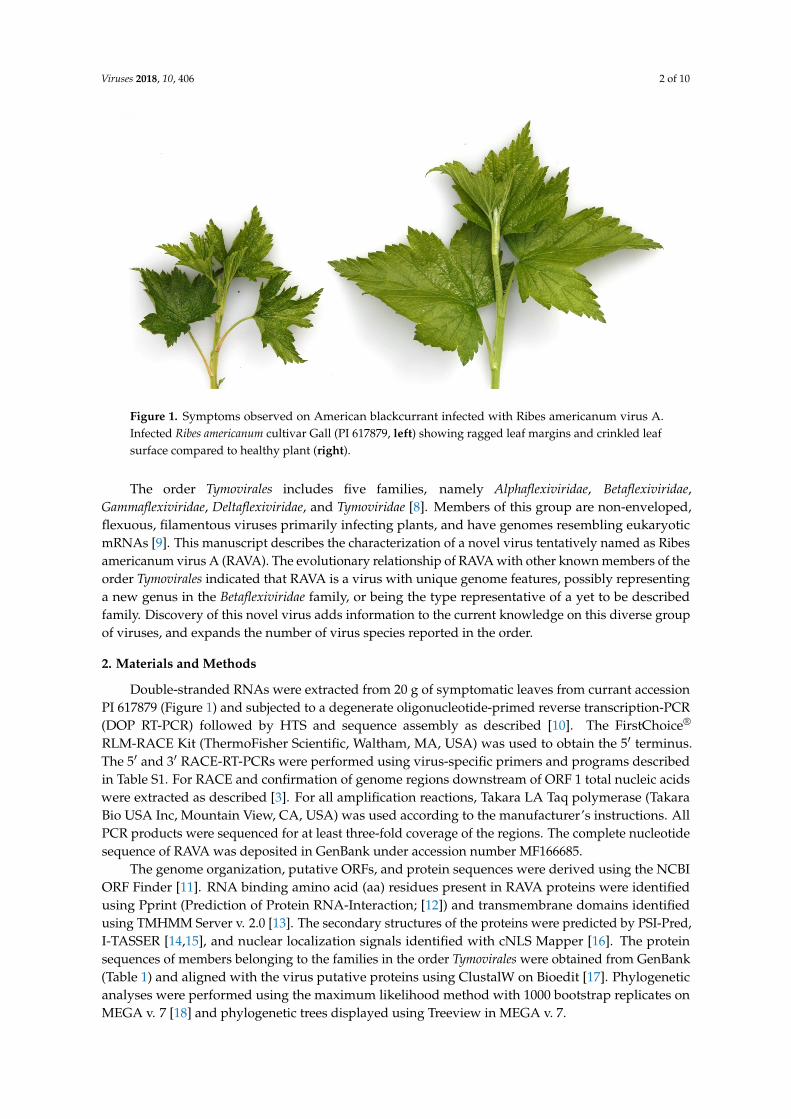
Technological developments in high throughput sequencing (HTS) have resulted in the rapiddiscovery and characterization of several novel plant viruses [1–4]. The USDA National ClonalGermplasm Repository (NCGR) maintains and distributes various accessions of specialty fruit andnut crop species from around the world [5]. This includes currant (Ribes spp.), a berry known forits potential health effects [6]. This study was initiated when an American blackcurrant maintainedin the USDA-NCGR, Corvallis, Oregon (Ribes americanum; PI 617879) showed virus-like symptoms(Figure 1). The plant was infected with two viruses [3]: A waikavirus [7] and an undescribed virus inthe order Tymovirales.
Viruses 2018, 10, 406; doi:10.3390/v10080406 www.mdpi.com/journal/viruses
Viruses 2018, 10, 406 2 of 10Viruses 2018, 10, x FOR PEER REVIEW 2 of 10
Figure 1. Symptoms observed on American blackcurrant infected with Ribes americanum virus A. Infected Ribes americanum cultivar Gall (PI 617879, left) showing ragged leaf margins and crinkled leaf surface compared to healthy plant (right).
The order Tymovirales includes five families, namely Alphaflexiviridae, Betaflexiviridae, Gammaflexiviridae, Deltaflexiviridae, and Tymoviridae [8]. Members of this group are non-enveloped, flexuous, filamentous viruses primarily infecting plants, and have genomes resembling eukaryotic mRNAs [9]. This manuscript describes the characterization of a novel virus tentatively named as Ribes americanum virus A (RAVA). The evolutionary relationship of RAVA with other known members of the order Tymovirales indicated that RAVA is a virus with unique genome features, possibly representing a new genus in the Betaflexiviridae family, or being the type representative of a yet to be described family. Discovery of this novel virus adds information to the current knowledge on this diverse group of viruses, and expands the number of virus species reported in the order.
2. Materials and Methods
Double-stranded RNAs were extracted from 20 g of symptomatic leaves from currant accession PI 617879 (Figure 1) and subjected to a degenerate oligonucleotide-primed reverse transcription-PCR (DOP RT-PCR) followed by HTS and sequence assembly as described [10]. The FirstChoice® RLM-RACE Kit (ThermoFisher Scientific, Waltham, MA, USA) was used to obtain the 5′ terminus. The 5′ and 3′ RACE-RT-PCRs were performed using virus-specific primers and programs described in Table S1. For RACE and confirmation of genome regions downstream of ORF 1 total nucleic acids were extracted as described [3]. For all amplification reactions, Takara LA Taq polymerase (Takara Bio USA Inc, Mountain View, CA, USA) was used according to the manufacturer’s instructions. All PCR products were sequenced for at least three-fold coverage of the regions. The complete nucleotide sequence of RAVA was deposited in GenBank under accession number MF166685.
The genome organization, putative ORFs, and protein sequences were derived using the NCBI ORF Finder [11]. RNA binding amino acid (aa) residues present in RAVA proteins were identified using Pprint (Prediction of Protein RNA-Interaction; [12]) and transmembrane domains identified using TMHMM Server v. 2.0 [13]. The secondary structures of the proteins were predicted by PSI-Pred, I-TASSER [14,15], and nuclear localization signals identified with cNLS Mapper [16]. The protein sequences of members belonging to the families in the order Tymovirales were obtained from GenBank (Table 1) and aligned with the virus putative proteins using ClustalW on Bioedit [17]. Phylogenetic analyses were performed using the maximum likelihood method with 1000 bootstrap replicates on MEGA v. 7 [18] and phylogenetic trees displayed using Treeview in MEGA v. 7.
Figure 1. Symptoms observed on American blackcurrant infected with Ribes americanum virus A.Infected Ribes americanum cultivar Gall (PI 617879, left) showing ragged leaf margins and crinkled leafsurface compared to healthy plant (right).
The order Tymovirales includes five families, namely Alphaflexiviridae, Betaflexiviridae,Gammaflexiviridae, Deltaflexiviridae, and Tymoviridae [8]. Members of this group are non-enveloped,flexuous, filamentous viruses primarily infecting plants, and have genomes resembling eukaryoticmRNAs [9]. This manuscript describes the characterization of a novel virus tentatively named as Ribesamericanum virus A (RAVA). The evolutionary relationship of RAVA with other known members of theorder Tymovirales indicated that RAVA is a virus with unique genome features, possibly representinga new genus in the Betaflexiviridae family, or being the type representative of a yet to be describedfamily. Discovery of this novel virus adds information to the current knowledge on this diverse groupof viruses, and expands the number of virus species reported in the order.
2. Materials and Methods
Double-stranded RNAs were extracted from 20 g of symptomatic leaves from currant accessionPI 617879 (Figure 1) and subjected to a degenerate oligonucleotide-primed reverse transcription-PCR(DOP RT-PCR) followed by HTS and sequence assembly as described [10]. The FirstChoice®
RLM-RACE Kit (ThermoFisher Scientific, Waltham, MA, USA) was used to obtain the 5′ terminus.The 5′ and 3′ RACE-RT-PCRs were performed using virus-specific primers and programs describedin Table S1. For RACE and confirmation of genome regions downstream of ORF 1 total nucleic acidswere extracted as described [3]. For all amplification reactions, Takara LA Taq polymerase (TakaraBio USA Inc, Mountain View, CA, USA) was used according to the manufacturer’s instructions. AllPCR products were sequenced for at least three-fold coverage of the regions. The complete nucleotidesequence of RAVA was deposited in GenBank under accession number MF166685.
The genome organization, putative ORFs, and protein sequences were derived using the NCBIORF Finder [11]. RNA binding amino acid (aa) residues present in RAVA proteins were identifiedusing Pprint (Prediction of Protein RNA-Interaction; [12]) and transmembrane domains identifiedusing TMHMM Server v. 2.0 [13]. The secondary structures of the proteins were predicted by PSI-Pred,I-TASSER [14,15], and nuclear localization signals identified with cNLS Mapper [16]. The proteinsequences of members belonging to the families in the order Tymovirales were obtained from GenBank(Table 1) and aligned with the virus putative proteins using ClustalW on Bioedit [17]. Phylogeneticanalyses were performed using the maximum likelihood method with 1000 bootstrap replicates onMEGA v. 7 [18] and phylogenetic trees displayed using Treeview in MEGA v. 7.
Viruses 2018, 10, 406 3 of 10
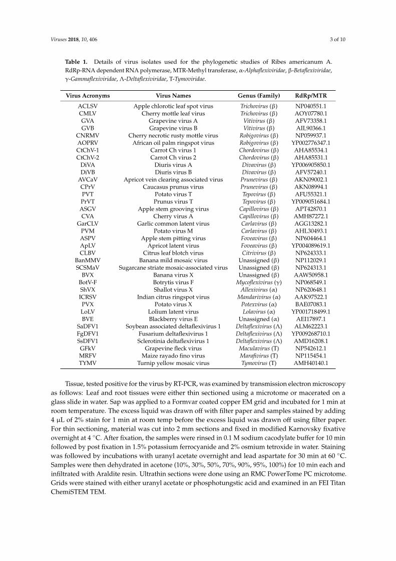
Table 1. Details of virus isolates used for the phylogenetic studies of Ribes americanum A.RdRp-RNA dependent RNA polymerase, MTR-Methyl transferase, α-Alphaflexiviridae, β-Betaflexiviridae,γ-Gammaflexiviridae, Λ-Deltaflexiviridae, T-Tymoviridae.
Virus Acronyms Virus Names Genus (Family) RdRp/MTR
ACLSV Apple chlorotic leaf spot virus Trichovirus (β) NP040551.1CMLV Cherry mottle leaf virus Trichovirus (β) AOY07780.1GVA Grapevine virus A Vitivirus (β) AFV73358.1GVB Grapevine virus B Vitivirus (β) AIL90366.1
CNRMV Cherry necrotic rusty mottle virus Robigovirus (β) NP059937.1AOPRV African oil palm ringspot virus Robigovirus (β) YP002776347.1CtChV-1 Carrot Ch virus 1 Chordovirus (β) AHA85534.1CtChV-2 Carrot Ch virus 2 Chordovirus (β) AHA85531.1
DiVA Diuris virus A Divavirus (β) YP006905850.1DiVB Diuris virus B Divavirus (β) AFV57240.1
AVCaV Apricot vein clearing associated virus Prunevirus (β) AKN09002.1CPrV Caucasus prunus virus Prunevirus (β) AKN08994.1PVT Potato virus T Tepovirus (β) AFU55321.1PrVT Prunus virus T Tepovirus (β) YP009051684.1ASGV Apple stem grooving virus Capillovirus (β) APT42870.1CVA Cherry virus A Capillovirus (β) AMH87272.1
GarCLV Garlic common latent virus Carlavirus (β) AGG13282.1PVM Potato virus M Carlavirus (β) AHL30493.1ASPV Apple stem pitting virus Foveavirus (β) NP604464.1ApLV Apricot latent virus Foveavirus (β) YP004089619.1CLBV Citrus leaf blotch virus Citrivirus (β) NP624333.1
BanMMV Banana mild mosaic virus Unassigned (β) NP112029.1SCSMaV Sugarcane striate mosaic-associated virus Unassigned (β) NP624313.1
BVX Banana virus X Unassigned (β) AAW50958.1BotV-F Botrytis virus F Mycoflexivirus (γ) NP068549.1ShVX Shallot virus X Allexivirus (α) NP620648.1ICRSV Indian citrus ringspot virus Mandarivirus (α) AAK97522.1PVX Potato virus X Potexvirus (α) BAE07083.1LoLV Lolium latent virus Lolavirus (α) YP001718499.1BVE Blackberry virus E Unassigned (α) AEI17897.1
SaDFV1 Soybean associated deltaflexivirus 1 Deltaflexivirus (Λ) ALM62223.1FgDFV1 Fusarium deltaflexivirus 1 Deltaflexivirus (Λ) YP009268710.1SsDFV1 Sclerotinia deltaflexivirus 1 Deltaflexivirus (Λ) AMD16208.1GFkV Grapevine fleck virus Maculavirus (T) NP542612.1MRFV Maize rayado fino virus Marafivirus (T) NP115454.1TYMV Turnip yellow mosaic virus Tymovirus (T) AMH40140.1
Tissue, tested positive for the virus by RT-PCR, was examined by transmission electron microscopyas follows: Leaf and root tissues were either thin sectioned using a microtome or macerated on aglass slide in water. Sap was applied to a Formvar coated copper EM grid and incubated for 1 min atroom temperature. The excess liquid was drawn off with filter paper and samples stained by adding4 µL of 2% stain for 1 min at room temp before the excess liquid was drawn off using filter paper.For thin sectioning, material was cut into 2 mm sections and fixed in modified Karnovsky fixativeovernight at 4 ◦C. After fixation, the samples were rinsed in 0.1 M sodium cacodylate buffer for 10 minfollowed by post fixation in 1.5% potassium ferrocyanide and 2% osmium tetroxide in water. Stainingwas followed by incubations with uranyl acetate overnight and lead aspartate for 30 min at 60 ◦C.Samples were then dehydrated in acetone (10%, 30%, 50%, 70%, 90%, 95%, 100%) for 10 min each andinfiltrated with Araldite resin. Ultrathin sections were done using an RMC PowerTome PC microtome.Grids were stained with either uranyl acetate or phosphotungstic acid and examined in an FEI TitanChemiSTEM TEM.
Viruses 2018, 10, 406 4 of 10
3. Results
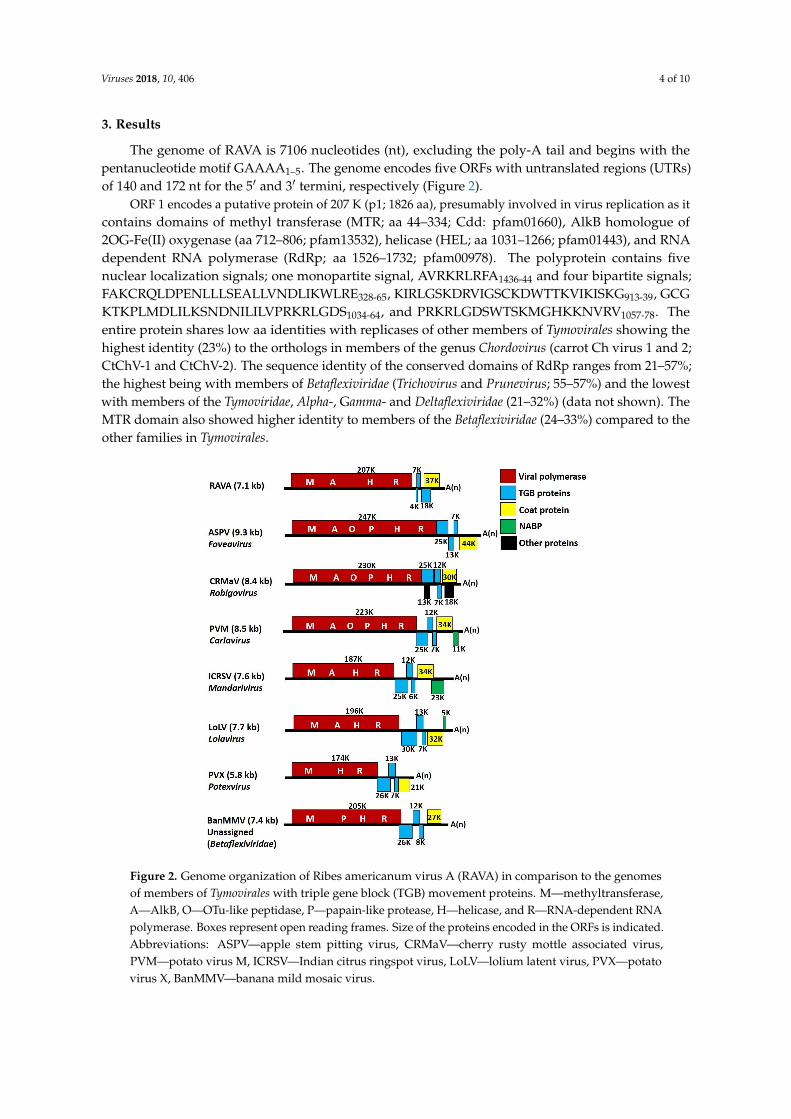
The genome of RAVA is 7106 nucleotides (nt), excluding the poly-A tail and begins with thepentanucleotide motif GAAAA1–5. The genome encodes five ORFs with untranslated regions (UTRs)of 140 and 172 nt for the 5′ and 3′ termini, respectively (Figure 2).
ORF 1 encodes a putative protein of 207 K (p1; 1826 aa), presumably involved in virus replication as itcontains domains of methyl transferase (MTR; aa 44–334; Cdd: pfam01660), AlkB homologue of2OG-Fe(II) oxygenase (aa 712–806; pfam13532), helicase (HEL; aa 1031–1266; pfam01443), and RNAdependent RNA polymerase (RdRp; aa 1526–1732; pfam00978). The polyprotein contains fivenuclear localization signals; one monopartite signal, AVRKRLRFA1436-44 and four bipartite signals;FAKCRQLDPENLLLSEALLVNDLIKWLRE328-65, KIRLGSKDRVIGSCKDWTTKVIKISKG913-39, GCGKTKPLMDLILKSNDNILILVPRKRLGDS1034-64, and PRKRLGDSWTSKMGHKKNVRV1057-78. Theentire protein shares low aa identities with replicases of other members of Tymovirales showing thehighest identity (23%) to the orthologs in members of the genus Chordovirus (carrot Ch virus 1 and 2;CtChV-1 and CtChV-2). The sequence identity of the conserved domains of RdRp ranges from 21–57%;the highest being with members of Betaflexiviridae (Trichovirus and Prunevirus; 55–57%) and the lowestwith members of the Tymoviridae, Alpha-, Gamma- and Deltaflexiviridae (21–32%) (data not shown). TheMTR domain also showed higher identity to members of the Betaflexiviridae (24–33%) compared to theother families in Tymovirales.
Viruses 2018, 10, x FOR PEER REVIEW 4 of 10
2% osmium tetroxide in water. Staining was followed by incubations with uranyl acetate overnight and lead aspartate for 30 min at 60 °C. Samples were then dehydrated in acetone (10%, 30%, 50%, 70%, 90%, 95%, 100%) for 10 min each and infiltrated with Araldite resin. Ultrathin sections were done using an RMC PowerTome PC microtome. Grids were stained with either uranyl acetate or phosphotungstic acid and examined in an FEI Titan ChemiSTEM TEM.
3. Results
The genome of RAVA is 7106 nucleotides (nt), excluding the poly-A tail and begins with the pentanucleotide motif GAAAA1–5. The genome encodes five ORFs with untranslated regions (UTRs) of 140 and 172 nt for the 5′ and 3′ termini, respectively (Figure 2).
ORF 1 encodes a putative protein of 207 K (p1; 1826 aa), presumably involved in virus replication as it contains domains of methyl transferase (MTR; aa 44–334; Cdd: pfam01660), AlkB homologue of 2OG-Fe(II) oxygenase (aa 712–806; pfam13532), helicase (HEL; aa 1031–1266; pfam01443), and RNA dependent RNA polymerase (RdRp; aa 1526–1732; pfam00978). The polyprotein contains five nuclear localization signals; one monopartite signal, AVRKRLRFA1436-44 and four bipartite signals; FAKCRQLDPENLLLSEALLVNDLIKWLRE328-65, KIRLGSKDRVIGSCKDWTTKVIKISKG913-39, GCGKTKPLMDLILKSNDNILILVPRKRLGDS1034-64, and PRKRLGDSWTSKMGHKKNVRV1057-78. The entire protein shares low aa identities with replicases of other members of Tymovirales showing the highest identity (23%) to the orthologs in members of the genus Chordovirus (carrot Ch virus 1 and 2; CtChV-1 and CtChV-2). The sequence identity of the conserved domains of RdRp ranges from 21–57%; the highest being with members of Betaflexiviridae (Trichovirus and Prunevirus; 55–57%) and the lowest with members of the Tymoviridae, Alpha-, Gamma- and Deltaflexiviridae (21–32%) (data not shown). The MTR domain also showed higher identity to members of the Betaflexiviridae (24–33%) compared to the other families in Tymovirales.
Figure 2. Genome organization of Ribes americanum virus A (RAVA) in comparison to the genomes of members of Tymovirales with triple gene block (TGB) movement proteins. M—methyltransferase, A—AlkB, O—OTu-like peptidase, P—papain-like protease, H—helicase, and R—RNA-dependent RNA polymerase. Boxes represent open reading frames. Size of the proteins encoded in the ORFs is indicated. Abbreviations: ASPV—apple stem pitting virus, CRMaV—cherry rusty mottle associated virus, PVM—potato virus M, ICRSV—Indian citrus ringspot virus, LoLV—lolium latent virus, PVX—potato virus X, BanMMV—banana mild mosaic virus.
Figure 2. Genome organization of Ribes americanum virus A (RAVA) in comparison to the genomesof members of Tymovirales with triple gene block (TGB) movement proteins. M—methyltransferase,A—AlkB, O—OTu-like peptidase, P—papain-like protease, H—helicase, and R—RNA-dependent RNApolymerase. Boxes represent open reading frames. Size of the proteins encoded in the ORFs is indicated.Abbreviations: ASPV—apple stem pitting virus, CRMaV—cherry rusty mottle associated virus,PVM—potato virus M, ICRSV—Indian citrus ringspot virus, LoLV—lolium latent virus, PVX—potatovirus X, BanMMV—banana mild mosaic virus.
Viruses 2018, 10, 406 5 of 10
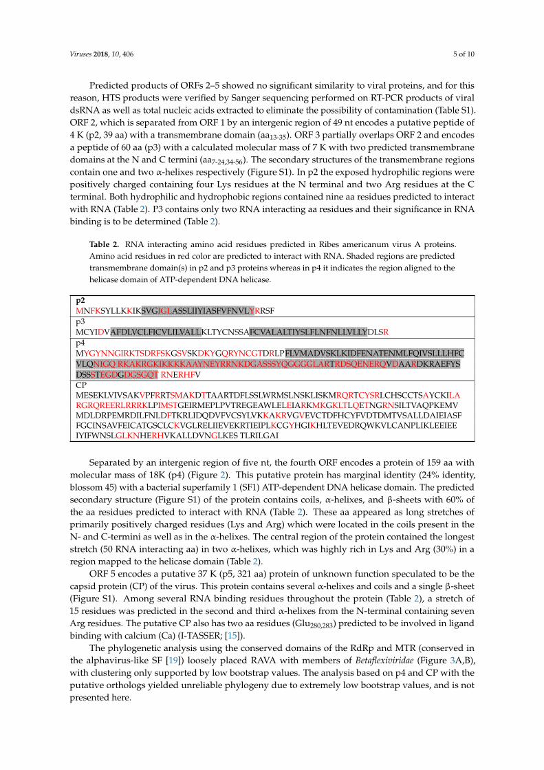
Predicted products of ORFs 2–5 showed no significant similarity to viral proteins, and for thisreason, HTS products were verified by Sanger sequencing performed on RT-PCR products of viraldsRNA as well as total nucleic acids extracted to eliminate the possibility of contamination (Table S1).ORF 2, which is separated from ORF 1 by an intergenic region of 49 nt encodes a putative peptide of4 K (p2, 39 aa) with a transmembrane domain (aa13-35). ORF 3 partially overlaps ORF 2 and encodesa peptide of 60 aa (p3) with a calculated molecular mass of 7 K with two predicted transmembranedomains at the N and C termini (aa7-24,34-56). The secondary structures of the transmembrane regionscontain one and two α-helixes respectively (Figure S1). In p2 the exposed hydrophilic regions werepositively charged containing four Lys residues at the N terminal and two Arg residues at the Cterminal. Both hydrophilic and hydrophobic regions contained nine aa residues predicted to interactwith RNA (Table 2). P3 contains only two RNA interacting aa residues and their significance in RNAbinding is to be determined (Table 2).
Table 2. RNA interacting amino acid residues predicted in Ribes americanum virus A proteins.Amino acid residues in red color are predicted to interact with RNA. Shaded regions are predictedtransmembrane domain(s) in p2 and p3 proteins whereas in p4 it indicates the region aligned to thehelicase domain of ATP-dependent DNA helicase.
p2MNFKSYLLKKIKSVGIGLASSLIIYIASFVFNVLYRRSFp3MCYIDVAFDLVCLFICVLILVALLKLTYCNSSAFCVALALTIYSLFLNFNLLVLLYDLSRp4MYGYNNGIRKTSDRFSKGSVSKDKYGQRYNCGTDRLPFLVMADVSKLKIDFENATENMLFQIVSLLLHFCVLQNIGQ RKAKRGKIKKKKAAYNEYRRNKDGASSSYQGGGGLARTRDSQENERQVDAARDKRAEFYSDSSSTEGDGDGSGQT RNERHFVCPMESEKLVIVSAKVPFRRTSMAKDTTAARTDFLSSLWRMSLNSKLISKMRQRTCYSRLCHSCCTSAYCKILARGRQREERLRRRKLPIMSTGEIRMEPLPVTREGEAWLELEIARKMKGKLTLQETNGRNSILTVAQPKEMVMDLDRPEMRDILFNLDFTKRLIDQDVFVCSYLVKKAKRVGVEVCTDFHCYFVDTDMTVSALLDAIEIASFFGCINSAVFEICATGSCLCKVGLRELIIEVEKRTIEIPLKCGYHGIKHLTEVEDRQWKVLCANPLIKLEEIEEIYIFWNSLGLKNHERHVKALLDVNGLKES TLRILGAI
Separated by an intergenic region of five nt, the fourth ORF encodes a protein of 159 aa withmolecular mass of 18K (p4) (Figure 2). This putative protein has marginal identity (24% identity,blossom 45) with a bacterial superfamily 1 (SF1) ATP-dependent DNA helicase domain. The predictedsecondary structure (Figure S1) of the protein contains coils, α-helixes, and β-sheets with 60% ofthe aa residues predicted to interact with RNA (Table 2). These aa appeared as long stretches ofprimarily positively charged residues (Lys and Arg) which were located in the coils present in theN- and C-termini as well as in the α-helixes. The central region of the protein contained the longeststretch (50 RNA interacting aa) in two α-helixes, which was highly rich in Lys and Arg (30%) in aregion mapped to the helicase domain (Table 2).
ORF 5 encodes a putative 37 K (p5, 321 aa) protein of unknown function speculated to be thecapsid protein (CP) of the virus. This protein contains several α-helixes and coils and a single β-sheet(Figure S1). Among several RNA binding residues throughout the protein (Table 2), a stretch of15 residues was predicted in the second and third α-helixes from the N-terminal containing sevenArg residues. The putative CP also has two aa residues (Glu280,283) predicted to be involved in ligandbinding with calcium (Ca) (I-TASSER; [15]).
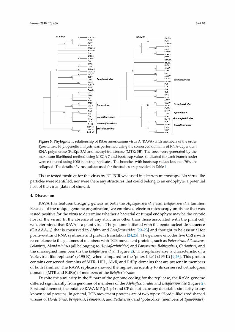
The phylogenetic analysis using the conserved domains of the RdRp and MTR (conserved inthe alphavirus-like SF [19]) loosely placed RAVA with members of Betaflexiviridae (Figure 3A,B),with clustering only supported by low bootstrap values. The analysis based on p4 and CP with theputative orthologs yielded unreliable phylogeny due to extremely low bootstrap values, and is notpresented here.
Viruses 2018, 10, 406 6 of 10
Viruses 2018, 10, x FOR PEER REVIEW 6 of 10
clustering only supported by low bootstrap values. The analysis based on p4 and CP with the putative orthologs yielded unreliable phylogeny due to extremely low bootstrap values, and is not presented here.
Figure 3. Phylogenetic relationship of Ribes americanum virus A (RAVA) with members of the order Tymovirales. Phylogenetic analysis was performed using the conserved domains of RNA-dependent RNA polymerase (RdRp; 3A) and methyl transferase (MTR; 3B). The trees were generated by the maximum likelihood method using MEGA 7 and bootstrap values (indicated for each branch node) were estimated using 1000 bootstrap replicates. The branches with bootstrap values less than 70% are collapsed. The details of virus isolates used for the studies are provided in Table 1.
Tissue tested positive for the virus by RT-PCR was used in electron microscopy. No virus-like particles were identified, nor were there any structures that could belong to an endophyte, a potential host of the virus (data not shown).
4. Discussion
RAVA has features bridging genera in both the Alphaflexiviridae and Betaflexiviridae families. Because of the unique genome organization, we employed electron microscopy on tissue that was tested positive for the virus to determine whether a bacterial or fungal endophyte may be the cryptic host of the virus. In the absence of any structures other than those associated with the plant cell, we determined that RAVA is a plant virus. The genome initiated with the pentanucleotide sequence (GAAAA1-5) that is conserved in Alpha- and Betaflexiviridae [20–23] and thought to be essential for positive-strand RNA synthesis and protein translation [24,25]. The genome encodes five ORFs with resemblance to the genomes of members with TGB movement proteins, such as Potexvirus, Allexivirus, Lolavirus, Mandarivirus (all belonging to Alphaflexiviridae) and Foveavirus, Robigovirus, Carlavirus, and the unassigned members (in the Betaflexiviridae) (Figure 2). The replicase size is characteristic of a ‘carlavirus-like replicase’ (>195 K), when compared to the ‘potex-like’ (<195 K) [9,26]. This protein contains conserved domains of MTR, HEL, AlkB, and RdRp domains that are present in members of both families. The RAVA replicase showed the highest aa identity to its conserved orthologous domains (MTR and RdRp) of members of the Betaflexiviridae.
Despite the similarity in the 5′ part of the genome coding for the replicase, the RAVA genome differed significantly from genomes of members of the Alphaflexiviridae and Betaflexiviridae (Figure 2). First and foremost, the putative RAVA MP (p2–p4) and CP do not share any detectable similarity to
Figure 3. Phylogenetic relationship of Ribes americanum virus A (RAVA) with members of the orderTymovirales. Phylogenetic analysis was performed using the conserved domains of RNA-dependentRNA polymerase (RdRp; 3A) and methyl transferase (MTR; 3B). The trees were generated by themaximum likelihood method using MEGA 7 and bootstrap values (indicated for each branch node)were estimated using 1000 bootstrap replicates. The branches with bootstrap values less than 70% arecollapsed. The details of virus isolates used for the studies are provided in Table 1.
Tissue tested positive for the virus by RT-PCR was used in electron microscopy. No virus-likeparticles were identified, nor were there any structures that could belong to an endophyte, a potentialhost of the virus (data not shown).
4. Discussion
RAVA has features bridging genera in both the Alphaflexiviridae and Betaflexiviridae families.Because of the unique genome organization, we employed electron microscopy on tissue that wastested positive for the virus to determine whether a bacterial or fungal endophyte may be the cryptichost of the virus. In the absence of any structures other than those associated with the plant cell,we determined that RAVA is a plant virus. The genome initiated with the pentanucleotide sequence(GAAAA1-5) that is conserved in Alpha- and Betaflexiviridae [20–23] and thought to be essential forpositive-strand RNA synthesis and protein translation [24,25]. The genome encodes five ORFs withresemblance to the genomes of members with TGB movement proteins, such as Potexvirus, Allexivirus,Lolavirus, Mandarivirus (all belonging to Alphaflexiviridae) and Foveavirus, Robigovirus, Carlavirus, andthe unassigned members (in the Betaflexiviridae) (Figure 2). The replicase size is characteristic of a‘carlavirus-like replicase’ (>195 K), when compared to the ‘potex-like’ (<195 K) [9,26]. This proteincontains conserved domains of MTR, HEL, AlkB, and RdRp domains that are present in membersof both families. The RAVA replicase showed the highest aa identity to its conserved orthologousdomains (MTR and RdRp) of members of the Betaflexiviridae.
Despite the similarity in the 5′ part of the genome coding for the replicase, the RAVA genomediffered significantly from genomes of members of the Alphaflexiviridae and Betaflexiviridae (Figure 2).First and foremost, the putative RAVA MP (p2–p4) and CP do not share any detectable similarity to anyknown viral proteins. In general, TGB movement proteins are of two types: ‘Hordei-like’ (rod shapedviruses of Hordeivirus, Benyvirus, Pomovirus, and Pecluvirus), and ‘potex-like’ (members of Tymovirales),
Viruses 2018, 10, 406 7 of 10
and are usually overlapping [27], with the exception of TGBps reported in the genus Robigovirus andan unassigned member, sugarcane striate mosaic-associated virus, in which only two, TGBp 2 andTGBp 3, are overlapping.
The organization of TGB ORFs is highly conserved among plant viruses [27,28]. Potex-like TGBproteins occur in the order of a bigger sized protein, TGBp 1 (~25 K) followed by smaller proteins,TGBp 2 and TGBp 3, (~12 K and ~7 K respectively; Figure 2) in which TGBp 2 has two and TGBp 3 hassingle transmembrane domains bordered by charged residues. TGBp 2 and TGBp 3 are membraneproteins and localize on endomembranes and cell walls [26,27]. TGBp 1 of these viruses usuallyharbors an NTP binding helicase domain of SFI and has ATPase, RNA binding, and RNA helicaseactivity, and is believed to increase the size exclusion limit of plasmodesmata [26]. Based on proteinsize and characteristics, RAVA seems to have potex-like TGB proteins, however the arrangement ofthe putative TGB ORFs (ORFs 2–4) is reversed. The RNA binding site of potexviral TGBp1 conservedpositively charged residue (Arg), which is essential for interaction with RNA [27], as with the caseof RAVA p4 protein (Table 2). The size of these three proteins was significantly different from othermembers (Figure 2). Although there was no significant sequence identity with its putative orthologs,the presence of transmembrane domains in p2 and p3 proteins and the NTP binding helicase domainin p4 makes us hypothesize that these proteins constitute the TGB movement proteins of RAVA.
ORFs downstream of the viral replicase are thought to be translated from 3′-terminal subgenomicRNAs in which TGB proteins get translated through leaky scanning mechanisms in all familiesof the order other than Tymoviridae [27]. Sequence context around the initiation codon affects thetranslation efficiency of mRNAs. The consensus optimal context for translation initiation in mammalsis GCC(A/G)CCAUGG (the initiation codon in bold; [29]). In some cases, viruses also follow thisrule [30]. The Kozak context of translation initiation was observed in RAVA ORF 5, although itsinitiation codon is 15 nt downstream of the overlapping ORF 4. This is expected if the protein isexpressed, given that the ribosomes first identify the start codon of p4, and the CP initiation contextneeds to be optimal if any ribosomes are to bypass that of p4.
The last ORF of most Alpha- and Betaflexiviridae members is the CP (Figure 2). However, the lastORF of RAVA lacked the domains conserved in the CP of flexuous viruses [31]. The predicted RNAbinding region of the protein is rich in Arg. Arg-rich RNA binding motif in the CP is known to beinvolved in genome binding and subsequent packaging [32]. The protein also has aa residues thoughtto be involved in Ca binding. Calcium plays an important role in the assembly and disassemblyand/or replication processes in viruses [33–35]. The size of the protein is similar to the range shown bymembers that have TGB MP (28–45 K; Figure 2). The position in the genome, predicted size, Arg-richRNA binding region, and presence of Ca binding sites suggests that the protein is the RAVA CP.
The genomes of the members of Tymovirales are highly diverse with respect to the number andorganization of genes, indicative of the major role of recombination in the evolution of this virus group.There is significant diversity in the 3′ region downstream of viral replicase (30 K/TGB MP, CP, andNABP) suggestive of possible recombination events in which the ancestral viruses had a common5′ part while acquiring the 3′ genes from different origins, which further diversified over time [36].Several studies have also described evidence of recombination in the members of this group [37–41].Martelli et al. [26] suggested coevolution of MP and CP in this virus group. RAVA genome followsthis theory in which the replicase, although with low bootstrap values, groups with the Betaflexiviridae,whereas the remaining proteins are phylogenetically distant. Changes in phylogenetic relationshipsfor different regions of the genome of a virus is an indication of recombination [42]. RAVA polymeraseis similar to the members of the Trivirinae subfamily, however the rest of the genome is Quinvirinae-like(Carlavirus, Foveavirus, Robigovirus; Figure 2) in which the TGBp-like proteins’ organization is reversed,suggesting the role of recombination between these two subfamilies in the evolution of this novel virus.
Viruses 2018, 10, 406 8 of 10
5. Conclusions
RAVA is a unique, previously undescribed virus of tymo-like lineage which cannot be assigned toany of the currently recognized virus taxa. With regard to its molecular features such as the numberof genes encoded in the genome, organization, and the presence of conserved pentanucleotide at 5′
termini, RAVA resembles members of the order Tymovirales. Phylogenetic analysis based on conservedRdRp domain, the principal determinant in the evolutionary framework of positive-strand RNAviruses, and MTR domain, clearly qualify RAVA as a putative species in the Betaflexiviridae family.However, differences in the genome downstream of polymerase (organization of TGBp-like proteinsand lack of conserved domains of ortholog proteins) and the distant phylogenetic relationship withother members distinguishes it from both existing genera and unassigned members of the family,suggesting a tentative classification. Therefore, we propose that RAVA represents a new, monotypicgenus in the family Betaflexiviridae or a family (given the low replicase aa identities with existingviruses) for which the names Ravavirus/Ravaviridae are proposed.
Supplementary Materials: The following are available online at http://www.mdpi.com/1999-4915/10/8/406/s1,Figure S1: Predicted secondary structures (PSI-Pred; [14]) of the Ribes americanum virus A proteins. Table S1:Primers and PCR conditions used for the amplification of the Ribes americanum virus A genome.
Author Contributions: T.T.-V., T.H., and I.E.T. conceived and designed the experiments; T.T.-V. and T.H.performed the experiments; T.T.-V. and T.H. analyzed the data; T.T.-V. and I.E.T. wrote the paper; J.D.P. and R.R.M.did the electron microscopy work. All authors approved the final version of the manuscript.
Funding: This research was funded by USDA awards 14-8130-0392-CA and Hatch project 1002361.
Conflicts of Interest: The authors declare no conflict of interest. The funding sponsors had no role in the designof the study; in the collection, analyses, or interpretation of data; in the writing of the manuscript, and in thedecision to publish the results.
References
1. Barba, M.; Czosnek, H.; Hadidi, A. Historical perspective, development and applications of next-generationsequencing in plant virology. Viruses 2014, 6, 106–136. [CrossRef] [PubMed]
2. Di Bello, P.L.; Laney, A.G.; Druciarek, T.; Ho, T.; Gergerich, R.C.; Keller, K.E.; Martin, R.R.; Tzanetakis, I.E.A novel emaravirus is associated with redbud yellow ringspot disease. Virus Res. 2016, 222, 41–47. [CrossRef][PubMed]
3. Ho, T.; Tzanetakis, I.E. Developing a virus detection and discovery pipeline using next generation sequencing.Virology 2014, 471–473, 54–60. [CrossRef] [PubMed]
4. Massart, S.; Olmos, A.; Jijakli, H.; Candresse, T. Current impact and future directions of high throughputsequencing in plant virus diagnostics. Virus Res. 2014, 188, 90–96. [CrossRef] [PubMed]
5. Postman, J.; Hummer, K.; Stover, E.; Krueger, R.; Forsline, P.; Grauke, L.J.; Zee, F.; Ayala-Silva, T.; Irish, B.Fruit and nut genebanks in the US national plant germplasm system. HortScience 2006, 41, 1188–1194.
6. Terry, L. Health-Promoting Properties of Fruit and Vegetables; CABI: Wallingford, UK, 2014; p. 432.7. Thekke-Veetil, T.; Ho, T.; Postman, J.D.; Tzanetakis, I.E. Molecular characterization of a new member of the
genus Waikavirus. Phytopathology 2017, 107, 102.8. ICTV. Virus Taxonomy 2017 Release, Order Tymovirales. Available online: https://talk.ictvonline.org/
taxonomy/ (accessed on 31 July 2018).9. Adams, M.J.; Antoniw, J.F.; Bar-Joseph, M.; Brunt, A.A.; Candresse, T.; Foster, G.D.; Martelli, G.P.; Milne, R.G.;
Fauquet, C.M. The new plant virus family Flexiviridae and assessment of molecular criteria for speciesdemarcation. Arch. Virol. 2004, 149, 1045–1060. [CrossRef] [PubMed]
10. Ho, T.; Martin, R.R.; Tzanetakis, I.E. Next-generation sequencing of elite berry germplasm and data analysisusing a bioinformatics pipeline for virus detection and discovery. In Plant Pathology: Techniques and Protocols(Methods in Molecular Biology); Lacomme, C., Ed.; Springer: New York, NY, USA, 2015; pp. 301–313.
11. Wheeler, D.L.; Church, D.M.; Federhen, S.; Lash, A.E.; Madden, T.L.; Pontius, J.U.; Schuler, G.D.;Schriml, L.M.; Sequeira, E.; Tatusova, T.A.; et al. Database resources of the National Center for Biotechnology.Nucleic Acids Res. 2003, 31, 28–33. [CrossRef] [PubMed]
Viruses 2018, 10, 406 9 of 10
12. Kumar, M.; Gromiha, M.M.; Raghava, G.P.S. Prediction of RNA binding sites in a protein using SVM andPSSM profile. Proteins 2008, 71, 189–194. [CrossRef] [PubMed]
13. Krogh, A.; Larsson, B.; von Heijne, G.; Sonnhammer, E.L.L. Predicting transmembrane protein topologywith a hidden Markov model: Application to complete genomes. J. Mol. Biol. 2001, 305, 567–580. [CrossRef][PubMed]
14. Jones, D.T. Protein secondary structure prediction based on position-specific scoring matrices. J. Mol. Biol.1999, 292, 195–202. [CrossRef] [PubMed]
15. Yang, J.; Yan, R.; Roy, A.; Xu, D.; Poisson, J.; Zhang, Y. The I-TASSER Suite: Protein structure and functionprediction. Nat. Methods 2015, 12, 7–8. [CrossRef] [PubMed]
16. Kosugi, S.; Hasebe, M.; Tomita, M.; Yanagawa, H. Systematic identification of yeast cell cycle-dependentnucleocytoplasmic shuttling proteins by prediction of composite motifs. Proc. Natl. Acad. Sci. USA 2009, 106,10171–10176. [CrossRef] [PubMed]
17. Hall, T.A. BioEdit: A user-friendly biological sequence alignment editor and analysis program for Windows95/98/NT. Nucleic Acids Symp. Ser. 1999, 41, 95–98.
18. Kumar, S.; Stecher, G.; Tamura, K. MEGA7: Molecular evolutionary genetics analysis version 7.0 for biggerdatasets. Mol. Biol. Evol. 2016, 33, 1870–1874. [CrossRef] [PubMed]
19. Koonin, E.V.; Dolja, V.V. Evolution and taxonomy of positive-strand RNA viruses: Implications ofcomparative analysis of amino acid sequences. Crit. Rev. Biochem. Mol. Biol. 1993, 28, 375–430. [CrossRef][PubMed]
20. Li, R.; Mock, R. Characterization of a flowering cherry strain of cherry necrotic rusty mottle virus. Arch.Virol. 2008, 153, 973–978. [CrossRef] [PubMed]
21. Sabanadzovic, S.; Abou Ghanem-Sabanadzovic, N.; Tzanetakis, I.E. Blackberry virus E: An unusual flexivirus.Arch. Virol. 2011, 156, 1665–1669. [CrossRef] [PubMed]
22. Villamor, D.V.; Druffel, K.L.; Eastwell, K.C. Complete nucleotide sequence of a virus associated with rustymottle disease of sweet cherry (Prunus avium). Arch. Virol. 2013, 158, 1805–1810. [CrossRef] [PubMed]
23. Zhang, Y.P.; Kirkpatrick, B.C.; Smart, C.D.; Uyemoto, J.K. cDNA cloning and molecular characterization ofcherry green ring mottle virus. J. Gen. Virol. 1998, 79, 2275–2281. [CrossRef] [PubMed]
24. Verchot-Lubicz, J.; Ye, C.M.; Bamunusinghe, D. Molecular biology of potexviruses: Recent advances. J. Gen.Virol. 2007, 88, 1643–1655. [CrossRef] [PubMed]
25. Wong, S.M.; Mahtani, P.H.; Lee, K.C.; Yu, H.H.; Tan, Y.; Neo, K.K.; Chan, Y.; Wu, M.; Chng, C.G. Cymbidiummosaic potexvirus RNA: Complete nucleotide sequence and phylogenetic analysis. Arch. Virol. 1997, 142,383–391. [CrossRef] [PubMed]
26. Martelli, G.P.; Adams, M.J.; Kreuze, J.F.; Dolja, V.V. Family Flexiviridae: A case study in virion and genomeplasticity. Annu. Rev. Phytopathol. 2007, 45, 73–100. [CrossRef] [PubMed]
27. Morozov, S.Y.; Solovyev, A.G. Triple gene block: Modular design of a multifunctional machine for plantvirus movement. J. Gen. Virol. 2003, 84, 1351–1366. [CrossRef] [PubMed]
28. Rupasov, V.V.; Morozov, S.Y.; Kanyuka, K.V.; Zavriev, S.K. Partial nucleotide sequence of potato virus MRNA shows similarities to potexviruses in gene arrangement and the encoded amino acid sequences. J. Gen.Virol. 1989, 70, 1861–1869. [CrossRef] [PubMed]
29. Kozak, M. Point mutations define a sequence flanking the AUG initiator codon that modulates translationby eukaryotic ribosomes. Cell 1986, 44, 283–292. [CrossRef]
30. Sánchez-Puig, J.M.; Blasco, R. AUG context and mRNA translation in vaccinia virus. Span. J. Agric. Res. 2008,6, 73–80. [CrossRef]
31. Dolja, V.V.; Boyko, V.P.; Agranovsky, A.A.; Koonin, E.V. Phylogeny of capsid proteins of rod-shaped andfilamentous RNA plant viruses: Two families with distinct patterns of sequence and probably structureconservation. Virology 1991, 184, 79–86. [CrossRef]
32. Choi, Y.G.; Rao, A.L.N. Molecular studies on bromovirus capsid protein: VII. Selective packaging of BMVRNA4 by specific N-terminal arginine residues. Virology 2000, 275, 207–217. [CrossRef] [PubMed]
33. Abdel-Meguid, S.; Yamane, S.T.; Fukuyama, K.; Rossmann, M.G. The location of calcium ions in southernbean mosaic virus. Virology 1981, 114, 81–85. [CrossRef]
34. Durham, A.C.; Hendry, D.A. Cation binding by tobacco mosaic virus. Virology 1977, 77, 510–519. [CrossRef]35. Gajardo, R.; Vende, P.; Poncet, D.; Cohen, J. Two proline residues are essential in the calcium-binding activity
of rotavirus VP7 outer capsid protein. J. Virol. 1997, 71, 2211–2216. [PubMed]
Viruses 2018, 10, 406 10 of 10
36. Marais, A.; Faure, C.; Mustafayev, E.; Candresse, T. Characterization of new isolates of apricot veinclearing-associated virus and of a new Prunus-Infecting virus: Evidence for recombination as a driving forcein Betaflexiviridae evolution. PLoS ONE 2015, 10, e0129469. [CrossRef] [PubMed]
37. Alabi, O.J.; Al Rwahnih, M.; Mekuria, T.A.; Naidu, R.A. Genetic diversity of grapevine virus A in Washingtonand California vineyards. Phytopathology 2014, 104, 548–560. [CrossRef] [PubMed]
38. Singh, L.; Hallan, V.; Martin, D.P.; Ram, R.; Zaidi, A.A. Genomic sequence analysis of four newchrysanthemum virus B isolates: Evidence of RNA recombination. Arch. Virol. 2012, 157, 531–537. [CrossRef][PubMed]
39. Villamor, D.E.; Eastwell, K.C. Viruses associated with rusty mottle and twisted leaf diseases of sweet cherryare distinct species. Phytopathology 2013, 103, 1287–1295. [CrossRef] [PubMed]
40. Yoon, J.Y.; Joa, J.H.; Choi, K.S.; Do, K.S.; Lim, H.C.; Chung, B.M. Genetic diversity of a natural population ofapple stem pitting virus isolated from apple in Korea. Plant Pathol. J. 2014, 30, 195–199. [CrossRef] [PubMed]
41. Zanardo, L.G.; Silva, F.N.; Lima, A.T.M.; Milanesi, D.F.; Castilho-Urquiza, G.P.; Almeida, A.M.R. Molecularvariability of cowpea mild mottle virus infecting soybean in Brazil. Arch. Virol. 2014, 159, 727–737. [CrossRef][PubMed]
42. Salminen, M. Detecting recombination in viral sequences. In The Phylogenetic Handbook: A Practical Approachto DNA and Protein Phylogeny; Salemi, M., Vandamme, A.M., Eds.; University Press: Cambridge, UK, 2003;pp. 348–377.
© 2018 by the authors. Licensee MDPI, Basel, Switzerland. This article is an open accessarticle distributed under the terms and conditions of the Creative Commons Attribution(CC BY) license (http://creativecommons.org/licenses/by/4.0/).