a subsequent-memory effect in dorsolateral prefrontal cortex
TRANSCRIPT

Cognitive Brain Research 16 (2003) 162–166www.elsevier.com/ locate/cogbrainres
Research report
A subsequent-memory effect in dorsolateral prefrontal cortexa , b*Bart Rypma , Mark D’Esposito
aDepartment of Psychology, Rutgers University, Smith Hall, 101 Warren Street, Newark, NJ 07102,USAbHelen Wills Neuroscience Institute and Department of Psychology, University of California, Berkeley, CA, USA
Accepted 26 September 2002
Abstract
The importance of brain regions for long-term memory encoding has been examined by comparison of encoding-related neural activityon trials in which successful recollection subsequently occurred to the encoding-related activity on trials in which successful recollectiondid not occur. We applied similar analyses to event-related functional magnetic resonance imaging (fMRI) data to explore the relativeroles of dorsolateral and ventrolateral prefrontal cortex (PFC) regions during specific components of a working-memory (WM)maintenance task. The results of this study indicated that increases in dorsolateral PFC activity during encoding was related to subsequentretrieval-success. These results lend support to the hypothesis that ventrolateral PFC mediates a limited-capacity WM buffer that supportsrehearsal maintenance functions while dorsolateral PFC mediates WM organization functions that accommodate the capacity limits ofWM maintenance. 2002 Published by Elsevier Science B.V.
Theme: Neural basis of behaviour
Topic: Learning and memory: systems and functions
Keywords: Working memory; Prefrontal cortex; Functional magnetic resonance imaging; Subsequent-memory effect
1 . Introduction cesses that mediate performance under low memory de-mand conditions and those that mediate performance under
Working memory (WM), the cognitive system that high memory demand conditions [24]. Specifically, re-allows individuals to maintain information over brief time search on the capacity limits of WM storage suggest thatintervals, may be divided into separate processes such as low memory load storage is mediated by a short-termthose required for brief retention of information, and those buffer of limited capacity (perhaps 41 /21 items, seerequired for allocating attention and coordinating infor- Cowan [7]), whereas higher memory loads require addi-mation that is being temporarily maintained [4] Evidence tional processing to compress, or ‘chunk’ the informationhas accumulated to support the notion that PFC may be for efficient storage and retrieval, so as to accommodateorganized to support different WM processes. Rypma et al. the capacity limits of the short-term maintenance buffer[25] and Rypma and D’Esposito [22,23] for instance, [18,29,24].observed that, under low memory load conditions (two to Neuroimaging results showing differential activationthree letters), activation in frontal regions was limited to patterns under low and high memory demands, andleft ventrolateral PFC (BAs 44, 45 and 47). Additional behavioral results suggesting dissociable memory pro-activation of dorsolateral PFC (BAs 9 and 46) was cesses under such memory-demands suggest a ‘memoryobserved, only during encoding, under high memory organization hypothesis’ of PFC function in WM. Thisdemand conditions (six letters). hypothesis states that ventrolateral PFC mediates the
These findings are important because they parallel limited capacity short-term buffer of WM while dorsolater-observed behavioral dissociations between cognitive pro- al PFC plays a role in the strategic organization, or
‘chunking,’ of information [7,12,18] that permit storage oflarge amounts of information [22,24].*Corresponding author. Tel.:11-510-643-4416.
E-mail address: [email protected](B. Rypma). The role of cortical regions in memory encoding have
0926-6410/02/$ – see front matter 2002 Published by Elsevier Science B.V.doi:10.1016/S0926-6410(02)00247-1
163B. Rypma, M. D’ Esposito / Cognitive Brain Research 16 (2003) 162–166
been explored in long-term episodic memory paradigms by acquisition to allow tissue to reach steady-state mag-comparing activation during encoding on trials in which netization.the subjects later successfully retrieved the information toactivation on trials in which later retrieval was unsuccess- 2 .3. Behavioral taskful [19,10,5,17]. These studies have yielded an importantphenomenon, a ‘subsequent-memory effect,’ that has aided To start each trial, letter strings, ranging in length fromin understanding the necessary neural network for success- 1 to 8, were presented simultaneously in pseudo-randomful memory encoding. The ‘subsequent memory effect’ order for 4 s followed by a 12-s unfilled delay. A probemay be defined as significantly greater activation of a letter then appeared for 2 s during which the subjectparticular brain region during memory encoding for trials pressed a button with their right thumb if the probe itemin which later retrieval was successful compared to that in was part of the memory set or with their left thumb if thetrials in which later retrieval was unsuccessful. probe item was not part of the memory set. Following
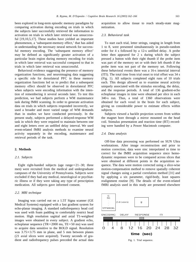
Behavioral evidence suggesting the presence of memory these behavioral events there was a 16-s intertrial intervalorganization functions, and neuroimaging data suggesting (ITI). The total time from trial onset to trial offset was 34 sa specific role for dorsolateral PFC in these memory (Fig. 1). All subjects completed eight runs of 10 trialsorganization functions led us to predict that a subsequent each. This design allowed us to examine neural activitymemory effect should be observed in dorsolateral PFC uniquely associated with the stimulus encoding, the delay,when subjects were encoding information with the inten- and the response periods. A total of 136 gradient-echotion of remembering it several seconds later. To test this echoplanar images in time were obtained per slice in eachprediction, we had subjects perform a WM maintenance 340-s run. Thus, a total of 1360 observations weretask during fMRI scanning. In order to generate activation obtained for each voxel in the brain for each subject,data on trials in which subjects responded incorrectly, we giving us considerable power to estimate effects withinused a broader and more varied range of WM demands subjects.than in studies we have conducted previously. In the Subjects viewed a backlit projection screen from withinpresent study, subjects performed a delayed-response WM the magnet bore through a mirror mounted on the headtask in which they were required to maintain between one coil. Stimulus presentation and reaction time (RT) record-and eight letters over an unfilled delay interval. We used ing were handled by a Power Macintosh computer.event-related fMRI analysis methods to examine neuralactivity separately in the encoding, maintenance and 2 .4. Data analysisretrieval periods of the task.
Off-line data processing was performed on SUN Ultraworkstations. After image reconstruction and prior to
2 . Methods motion correction, data were sinc interpolated in time tocorrect for the fMRI acquisition sequence since hemo-
2 .1. Subjects dynamic responses were to be compared across slices thatwere obtained at different points in the acquisition se-
Eight right-handed subjects (age range521–30; three quence. The data were motion corrected using a slice-wisemen) were recruited from the medical and undergraduate motion-compensation method to remove spatially coherentcampuses of the University of Pennsylvania. Subjects were signal changes using a partial correlation method [31] andexcluded if they had any medical, neurological or psychiat- by applying a six parameter, rigid-body, least squaresric illness or if they were taking any type of prescription realignment routine [9]. The details of the event-relatedmedication. All subjects gave informed consent. fMRI analysis used in this study are presented elsewhere
2 .2. MRI technique
Imaging was carried out on a 1.5T Signa scanner (GEMedical Systems) equipped with a fast gradient system forecho-planar imaging. A standard radiofrequency head coilwas used with foam padding to comfortably restrict headmotion. High resolution sagittal and axial T1-weightedimages were obtained in every subject. A gradient echo,echoplanar sequence (TR52000 ms, TE550 ms) was usedto acquire data sensitive to the BOLD signal. Resolutionwas 3.7533.75 mm in plane, and 5 mm between planes(21 axial slices were acquired). Twenty seconds of gra-dient and radiofrequency pulses preceded the actual data Fig. 1. Trial sequence.

B. Rypma, M. D’ Esposito / Cognitive Brain Research 16 (2003) 162–166164
[31]. Briefly, fMRI signal changes that occurred during each ROI and averaged separately for dorsolateral andparticular temporal periods of the behavioral trials were ventrolateral PFC ROIs for each subject. Random-effectsmodeled with covariates comprised of shifted, BOLD tests were then used to compare activation (as measured byhemodynamic response functions (HRFs), the fMRI re- ROI-wise parameter estimates) for each task period insponse resulting from a brief pulse of neural activity. correct and incorrect trials. Note that our procedure doesChanges in BOLD signal associated with the encoding, not entail creation of statistical maps and therefore doesdelay, and response periods of the behavioral task were not depend on thresholds for determination of neuralassessed with covariates that modeled the expected BOLD activity. Thus, we assessed cortical activation in eachsignal response in the event of an increase in neural subject’s dorsolateral and ventrolateral PFC in the encod-activity (relative to the ITI) occurring in each of the task ing, delay and response periods in each of the eightperiods. memory load conditions (i.e. one to eight letters) using the
Our rationale for deriving an HRF is explained else- parameter estimates (non-thresholded) for the covariateswhere [1]. An HRF was derived from primary sen- that modeled each task-period, in each memory-loadsorimotor cortex in each subject in the following manner. condition. Testing of a priori hypotheses involved plannedBefore performing the WM task described above, each comparisons of the activation differences between correctsubject performed a simple RT task in which a central and incorrect trials in each task period.white fixation cross changed briefly (500 ms) to a circleevery 16 s cueing subjects to make a bilateral button press.Twenty such events occurred during the 320-s scan (1603 . Resultsimages). All scanning parameters were identical to thoseused for the WM experiment. 3 .1. Behavioral performance
Because fMRI data are temporally autocorrelated underthe null hypothesis [31], the data were analyzed using the RT increased with increasing memory-load,F(7,49)5modified general linear model for serially correlated error 24.1, P,0.0001, MSe59069.6. The relationship betweenterms [30]. A time-domain representation of the expected RT and memory load was well-described by a linearly
21/ f power structure and a filter that removes frequencies increasing function (slope50.97, r 50.96), which wasabove 0.244 was placed within the K matrix. This filter significant, t(7)513.3, P,0.0001, typical of performancewas also applied to the fMRI time series to remove on such tasks [26]. Performance accuracy was high acrossartifacts at the Nyquist frequency (0.25 Hz). Low fre- memory-load conditions (91.8%) but decreased with in-quency (sine and cosine) confounds up to 0.025 Hz andcreasing memory loadF(7, 49)57.6, P,0.0001, MSe5trial-effect covariates were included in our model, to 67.3. This relationship was well-described by a linearly
2account for frequency components and mean signal decreasing function (slope520.87, r 50.75), which waschange, respectively, that were associated with each trial. significant, t(7)524.26, P,0.003. These results did not
Relationships with each task period and the ITI were differ with the presence or absence of the probe in theassessed by contrasts (yieldingt-statistics with|1195 df) memory set (i.e. whether the correct response was ‘yes’ orinvolving the parameter estimates that corresponded to‘no’; ts,1).covariates that modeled each task period. Three covariatesmodeled each 4-s of the delay period. Given estimates of
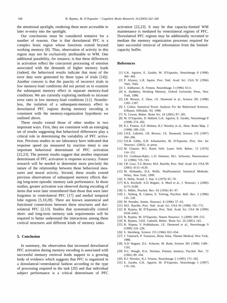
3 .2. FMRI signalthe temporal smoothness of the hemodynamic response,the covariate modeling the first 4-s of the delay periodwould be contaminated by hemodynamic activity from the 3 .2.1. Correct vs. incorrect trialsencoding period. Thus, only the second 4-s interval To assess our prediction of increased neural activity in(designated as delay period 1) and third 4-s interval of the dorsolateral PFC during encoding of correct trials, we useddelay period (designated as delay period 2) are considered planned comparisons of average fMRI signal values forin the analyses. Delay periods 1 and 2 were analyzed trials in which subjects responded correctly to those inseparately. To examine activity in specific regions of PFC, which subjects responded incorrectly in each task period,dorsolateral PFC regions-of-interest (ROIs) were drawn to in dorsolateral and ventrolateral PFC. Fig. 2 shows theinclude middle and superior frontal gyri (corresponding to mean parameter estimates from dorsolateral and ventrola-BAs 9 and 46) according to the Talairach and Tournoux teral PFC in each task period (averaged across memory[27] atlas on standard T1 axial slices in the axial plane. A loads) for correct and incorrect trials. In dorsolateral PFC,similar procedure was used to draw ventrolateral PFC during the encoding period, reliable differences wereROIs to include inferior frontal gyrus corresponding to observed between parameter estimates from correct trialsBAs 44, 45 and 47. These ROIs were then normalized to and those from incorrect trials, using Bonferroni-correctedeach subject’s T1 axial images using a 12-parameter affine ([16]) parametrict-tests, t(7)52.63, P,0.03, and non-transformation [9]. with a non-linear deformation routine parametric paired sign tests (P50.008; [15]). No other[3]. Parameter estimates were derived from each voxel of effects were significant in dorsolateral PFC and no signifi-

165B. Rypma, M. D’ Esposito / Cognitive Brain Research 16 (2003) 162–166
incorrect trials, is qualitatively distinct from any other taskperiod or prefrontal region.
The meaning of the negative activation we observed on‘incorrect’ trials is difficult to interpret for at least tworeasons. First, whereas the positive activation on ‘correct’trials (similar to RTs on correct trials) could reasonably beinterpreted as reflecting a fairly circumscribed sequence ofcognitive processes that are well studied and well under-stood, the negative activation on incorrect trials may resultfrom a number of possible processes including attentionallapses or pursuit of nonoptimal strategies. Whatever psy-chological process negative activations may reflect, theseresults suggest that a considerable number of dorsolateralPFC neurons simultaneously reduce their activation (com-pared to baseline) during the encoding of to-be-remem-bered information on some trials [21]. This processappears to have a sufficiently deleterious effect on ‘down-stream’ memory processes to impede successful retrievalof information.
The finding, that positive dorsolateral PFC encodingactivation is associated with retrieval success and negativedorsolateral PFC encoding activation is associated withretrieval failure suggests support for the hypothesis thatdorsolateral PFC is critical for mediation of workingmemory processes [25], specifically, for initial acquisitionof to-be-remembered information [22]. Rypma et al. [25]observed ventrolateral PFC activation during low (threeletters) and high (six letters) memory demand conditions.Additional dorsolateral PFC activity was observed only inthe high memory-demand conditions. Rypma and D’E-
Fig. 2. Mean fMRI signal (parameter estimates) from dorsolateral and sposito [22], using event-related fMRI, isolated this de-ventrolateral PFC during encoding, delay and retrieval periods for trials in mand-differential activity to the encoding period. Based onwhich subjects responded correctly (white bars) and those in which
these observations, Rypma and his colleagues have arguedsubjects responded incorrectly (hatched bars).that dorsolateral PFC plays a critical role in encodingprocesses that permit efficient memory maintenance and
cant effects were observed in ventrolateral PFC in both retrieval. These results suggest further support for theparametric and nonparametric tests. notion that capacity-limited WM maintenance processes
may be mediated by ventrolateral PFC whereas strategicorganization processes may be mediated by dorsolateralPFC [22–24].
4 . Discussion How might such strategic organization processes facili-tate subsequent retrieval of stored information? A number
In this study, we observed greater encoding-related of theoretical frameworks have been proposed that couldneural activity in trials in which subjects responded provide an answer to this question. In Baddeley’s WMcorrectly than in trials in which subjects responded incor- model [23], maintenance rehearsal is a process of perpetualrectly on a WM maintenance task. Significant activation reactivation of items currently stored in the maintenancedifferences between correct and incorrect trials did not buffer. In our view, strategic organization acts as a data-occur in ventrolateral PFC but did occur in dorsolateral compression program to permit supracapacity informationPFC. The relatively small number of participants in this maintenance within this buffer and availability to retrievalstudy could raise concerns about statistical power in the mechanisms. Another framework, proposed by Cowan [7],present study. Two points are worth noting in this regard. emphasizes the role of attention in information mainte-First, the single largest effect size (1.14; [6]) was observed nance. In this view, items in long-term memory may bein dorsolateral PFC during encoding and this effect was activated based on the context of information that ismore than twice that in any other task period or in currently within the focus of a capacity-limited attentionalventrolateral PFC. Second, the reliable pattern, of positive spotlight. Chunking of information then, serves to activateactivation for correct trials and negative activation for the long-term memory representations of items currently in
B. Rypma, M. D’ Esposito / Cognitive Brain Research 16 (2003) 162–166166
the attentional spotlight, rendering them more accessible to activation [22,23]. It may be that capacity-limited WMlater re-entry into the spotlight. maintenance is mediated by ventrolateral regions of PFC.
Our conclusions must be considered tentative for a Dorsolateral PFC regions may be additionally recruited tonumber of reasons. One is that dorsolateral PFC is a mediate the memory organization processes required forcomplex brain region whose functions extend beyond later successful retrieval of information from the limited-working memory [8]. Thus, observation of activity in this capacity buffer.region may not be exclusively attributable to WM. Oneadditional possibility, for instance, is that these differencesin activation reflect the concurrent processing of emotion R eferencesassociated with the demands of higher memory loads(indeed, the behavioral results indicate that most of the [1] G.K. Aguirre, E. Zarahn, M. D’Esposito, NeuroImage 8 (1998)
360–369.error data were generated by these types of trials [14]).[2] P. Alvarez, L.R. Squire, Proc. Natl. Acad. Sci. USA 91 (1994)Another concern is that the paucity of incorrect trials in
7041–7045.low memory-load conditions did not permit us to examine [3] J. Ashburner, K. Friston, NeuroImage 3 (1996) S111.the subsequent memory effect in separate memory-load [4] A. Baddeley, Working Memory, Oxford University Press, Newconditions. We are currently exploring methods to increase York, 1986.
[5] J.B. Brewer, Z. Zhao, J.E. Desmond et al., Science 281 (1998)error rates in low memory-load conditions [11]. Nonethe-1185–1187.less, the isolation of a subsequent-memory effect to
[6] J. Cohen, Statistical Power Analysis For the Behavioral Sciences,dorsolateral PFC regions during memory encoding is Erlbaum, Hillsdale, NJ, 1998.consistent with the memory-organization hypothesis we [7] N. Cowan, Behav. Brain Sci. 24 (2001) 87–185.outlined above. [8] M. D’Esposito, D. Ballard, G.K. Aguirre, E. Zarahn, NeuroImage 8
(1998) 274–282.These results extend those of other studies in two[9] K.J. Friston, A.P. Holmes, K.J. Worsley et al., Hum. Brain Map. 2important ways. First, they are consistent with an emerging
(1998) 189–210.set of results suggesting that behavioral differences play a[10] J.D.E. Gabrieli, J.B. Brewer, J.E. Desmond, Science 276 (1997)critical role in determining the variability of PFC activa- 264–266.tion. Previous studies in our laboratory have indicated that [11] S.E.B. Gibbs, E.H. Schumacher, M. D’Esposito, Proc. Soc. for
Neurosci. (2002), in press.response speed (as measured by reaction time) is one[12] M. Glanzer, M.J. Razel, Verb. Learn. Verb. Behav. 13 (1974)important behavioral determinant of PFC activation
114–131.[22,23]. The present results suggest that another important[13] P.S. Goldman-Rakic, L.D. Selemon, M.L. Schwartz, Neurosciencedeterminant of PFC activation is response accuracy. Future 12 (1984) 719–743.research will be needed to determine more precisely the[14] J.R. Gray, T.S. Braver, M.E. Raichle, Proc. Natl. Acad. Sci. USA 99
(2002) 4115–4120.nature of the relationship between these behavioral mea-[15] M. Hollander, D.A. Wolfe, NonParametric Statistical Methods,sures and neural activity. Second, these results extend
Wiley, New York, 1999.previous observations of subsequent memory effects dur-[16] S. Holm, Scand. J. Stat. 6 (1979) 65–70.ing long-term episodic memory task performance. In those [17] B.A. Kirchoff, A.D. Wagner, A. Maril et al., J. Neurosci. 1 (2000)studies, greater activation was observed during encoding of 6175–6180.
[18] G. Miller, Psychol. Rev. 63 (1956) 81–97.items that were later remembered than those that were later[19] L. Nyberg, R. Cabeza, E. Tulving, Psychon. Bull. Rev. 3 (1996)forgotten in ventrolateral PFC [17] and medial temporal
135–138.lobe regions [5,10,28]. There are known anatomical and [20] M. Petrides, Semin. Neurosci. 8 (1996) 57–63.functional connections between these structures and dor-[21] M.E. Raichle, Proc. Natl. Acad. Sci. USA 95 (1998) 765–772.solateral PFC [2,13]. Studies that systematically control [22] B. Rypma, M. D’Esposito, Proc. Natl. Acad. Sci. USA 96 (1999)
6558–6563.short- and long-term memory task requirements will be[23] B. Rypma, M. D’Esposito, Nature Neurosci. 3 (2000) 509–515.required to better understand the interactions among these[24] B. Rypma, J.D.E. Gabrieli, Behav. Brain Sci. 24 (2001) 143.
cortical structures and different kinds of memory tasks. [25] B. Rypma, V. Prabhakaran, J.E. Desmond et al., NeuroImage 9(1999) 216–226.
[26] S. Sternberg, Science 153 (1966) 652–654.[27] J. Talairach, P. Tournoux, Brain Atlas, Thieme Medical, New York,5 . Conclusion
1988.[28] A.D. Wagner, D.L. Schacter, M. Rotte, Science 281 (1998) 1188–
In summary, the observation that increased dorsolateral 1191.PFC activation during memory encoding is associated with [29] N.C. Waugh, D.A. Norman, Primary memory, Psychol. Rev. 72successful memory retrieval lends support to a growing (1965) 89–104.
[30] K.J. Worsley, K.J. Friston, NeuroImage 2 (1995) 173–182.body of evidence which suggests that PFC is organized in[31] E. Zarahn, G.K. Aguirre, M. D’Esposito, NeuroImage 5 (1997)a dorsolateral /ventrolateral fashion according to the type
179–195.of processing required in the task [20] and that individualsubject performance is a critical determinant of PFC