a study on histology of fetal liver - semantic scholar · a study on histology of fetal liver...
TRANSCRIPT
26National Journal of Clinical Anatomy
A study on histology of fetal liver1Jaiswal A, 2Sinha DN, 3Singh AK1Assistant Professor, 2Professor & HOD, 3ProfessorDepartment of Anatomy, Govt. Medical College, Haldwani, Nainital, Uttarakhand
Abstract
Background and aims : The liver during fetogenesisdoes not follow classical lobular pattern. Normal histologyof the fetal liver at various stages of development wasstudied to get insight into the morphology of fetal liverand special function it performs in fetal life. Method:Dissection of 29 normal human fetuses was done andhistological findings of liver were noted with respect tothe age of fetus. The histology of fetal liver was studiedusing H & E stain and important features of fetal liverwere studied. Result: Fetal liver histology is differentfrom adult liver. Unlike adult liver, fetal liver showshepatocytes arranged in sheets predominantly with cord-like pattern at places. The sinusoids are dilated and filledwith hemopoietic cells, which could be appreciated atdifferent stages of fetal development. The Kupffer cellswere also noticed in fetal liver. Conclusion: The present
study will be helpful in understanding the normal histologyof fetal liver and add to the existing knowledge regardingdevelopment of fetal liver.
Key words: fetal hepatocytes, hematopoiesis,acinus, portal, sinusoid
Introduction
Fetal liver is one of most primitive embryonic organsdeveloped from endodermal evagination of foregut andseptum transversum mesenchyme1. The fetal
hepatocytes were formed from endodermal cells ofhepatic diverticulum. The portal veins develop from
vitelline veins. The hepatoblast adjacent to mesenchymedifferentiate in to future biliary tubular structures whichmature from the hilum to outward, begin around the 11th
week of gestation and continue past birth2,3. The fetalliver is also center of hematopoiesis in fetus4.
The aim of the present study is to find out thehistological structure of fetal liver and to contribute toavailable literature in understanding the liver histologyduring gestational period including liver architecture,hepatocyte, Kupffer cells, sinusoid and hemopoeticnature of the liver during the fetal life.
Materials and methods
This prospective study was conducted at theDepartment of Anatomy, Government Medical College,Haldwani. The fetuses were obtained from Obstetricsand Gynecology Department, Dr Sushila Tiwari Hospitalover a period of two years (period commencing from2011 to 2013), with due regards on ethical ground. Thefetuses were preserved in formalin.
The crown rump lengths (CRLs) of fetuses wererecorded and histological features were studied inrelation to CRL of the fetuses. In the fetus, the liverwas observed for any gross and congenital anomalies.Its weight and size were recorded before embalming.The liver was removed by dissecting inferior vena cavaand excising ligaments of the liver, and after that itsweight was recorded. The liver tissue was taken forhistological processing. Five µm-thick fetal liver sectionswere stained with hematoxylin-eosin stain and werestudied by light microscopy.
Observations and results
Observations on the light microscopic structure ofliver during fetal development : The hepatic lobularpattern was not defined up to CRL of 8.5 cm. The liverarchitecture showed ill-defined lobular pattern of fetalhepatocytes from CRL of 10.4 cm with a well-definedpattern noticed only in late gestation with CRL of 28 cm(Table 1). The fetal hepatocytes were appreciated from
Original article
Vol. - 4 (1) Pg. 26- 29 (2015)
27National Journal of Clinical Anatomy
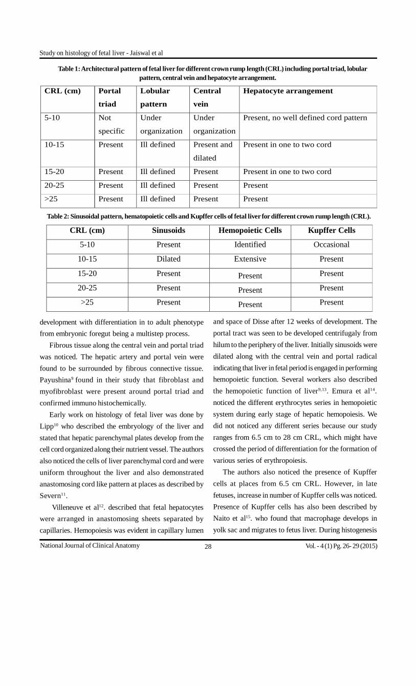
Fig. 3. Photomicrograph of liver of foetus21cm CRL showsmigration of connective tissue from capsular region towards portaltriad, X100.
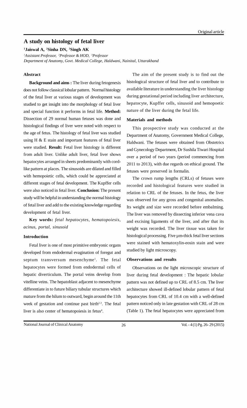
Fig. 1. Photomicrograph of liver 12cmCRL shows central veinand opening of the sinusoid, central vein , liver cells andlongitudinally placed portal traid bile ductile surrounded byfibrous connective tissue ,X100,H& E.
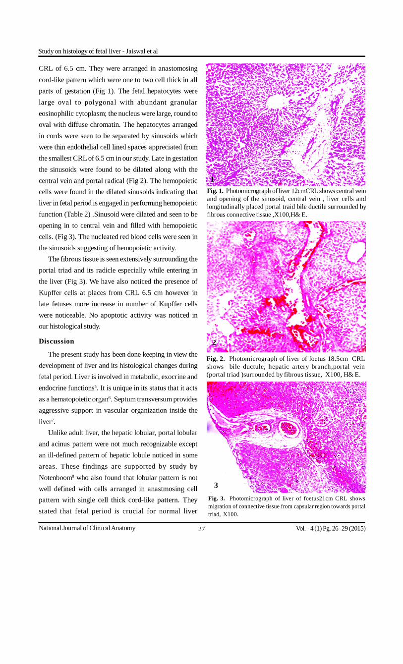
Fig. 2. Photomicrograph of liver of foetus 18.5cm CRLshows bile ductule, hepatic artery branch,portal vein(portal triad )surrounded by fibrous tissue, X100, H& E.
CRL of 6.5 cm. They were arranged in anastomosingcord-like pattern which were one to two cell thick in allparts of gestation (Fig 1). The fetal hepatocytes werelarge oval to polygonal with abundant granulareosinophilic cytoplasm; the nucleus were large, round tooval with diffuse chromatin. The hepatocytes arrangedin cords were seen to be separated by sinusoids whichwere thin endothelial cell lined spaces appreciated fromthe smallest CRL of 6.5 cm in our study. Late in gestationthe sinusoids were found to be dilated along with thecentral vein and portal radical (Fig 2). The hemopoieticcells were found in the dilated sinusoids indicating thatliver in fetal period is engaged in performing hemopoieticfunction (Table 2) .Sinusoid were dilated and seen to beopening in to central vein and filled with hemopoieticcells. (Fig 3). The nucleated red blood cells were seen inthe sinusoids suggesting of hemopoietic activity.
The fibrous tissue is seen extensively surrounding theportal triad and its radicle especially while entering inthe liver (Fig 3). We have also noticed the presence ofKupffer cells at places from CRL 6.5 cm however inlate fetuses more increase in number of Kupffer cellswere noticeable. No apoptotic activity was noticed inour histological study.
Discussion
The present study has been done keeping in view thedevelopment of liver and its histological changes duringfetal period. Liver is involved in metabolic, exocrine andendocrine functions5. It is unique in its status that it actsas a hematopoietic organ6. Septum transversum providesaggressive support in vascular organization inside theliver7.
Unlike adult liver, the hepatic lobular, portal lobularand acinus pattern were not much recognizable exceptan ill-defined pattern of hepatic lobule noticed in someareas. These findings are supported by study byNotenboom8 who also found that lobular pattern is notwell defined with cells arranged in anastmosing cellpattern with single cell thick cord-like pattern. Theystated that fetal period is crucial for normal liver
1
2
3
Study on histology of fetal liver - Jaiswal et al
Vol. - 4 (1) Pg. 26- 29 (2015)
28National Journal of Clinical Anatomy
development with differentiation in to adult phenotypefrom embryonic foregut being a multistep process.
Fibrous tissue along the central vein and portal triadwas noticed. The hepatic artery and portal vein werefound to be surrounded by fibrous connective tissue.Payushina9 found in their study that fibroblast andmyofibroblast were present around portal triad andconfirmed immuno histochemically.
Early work on histology of fetal liver was done byLipp10 who described the embryology of the liver andstated that hepatic parenchymal plates develop from thecell cord organized along their nutrient vessel. The authorsalso noticed the cells of liver parenchymal cord and wereuniform throughout the liver and also demonstratedanastomosing cord like pattern at places as described bySevern11.
Villeneuve et al12. described that fetal hepatocyteswere arranged in anastomosing sheets separated bycapillaries. Hemopoiesis was evident in capillary lumen
Table 1: Architectural pattern of fetal liver for different crown rump length (CRL) including portal triad, lobularpattern, central vein and hepatocyte arrangement.
Table 2: Sinusoidal pattern, hematopoietic cells and Kupffer cells of fetal liver for different crown rump length (CRL).
and space of Disse after 12 weeks of development. Theportal tract was seen to be developed centrifugaly fromhilum to the periphery of the liver. Initially sinusoids weredilated along with the central vein and portal radicalindicating that liver in fetal period is engaged in performinghemopoietic function. Several workers also describedthe hemopoietic function of liver9,13. Emura et al14.noticed the different erythrocytes series in hemopoieticsystem during early stage of hepatic hemopoiesis. Wedid not noticed any different series because our studyranges from 6.5 cm to 28 cm CRL, which might havecrossed the period of differentiation for the formation ofvarious series of erythropoiesis.
The authors also noticed the presence of Kupffercells at places from 6.5 cm CRL. However, in latefetuses, increase in number of Kupffer cells was noticed.Presence of Kupffer cells has also been described byNaito et al15. who found that macrophage develops inyolk sac and migrates to fetus liver. During histogenesis
Study on histology of fetal liver - Jaiswal et al
Vol. - 4 (1) Pg. 26- 29 (2015)
CRL (cm)
Portal
triad
Lobular
pattern
Central
vein
Hepatocyte arrangement
5-10 Not
specific
Under
organization
Under
organization
Present, no well defined cord pattern
10-15 Present Ill defined Present and
dilated
Present in one to two cord
15-20 Present Ill defined Present Present in one to two cord
20-25 Present Ill defined Present Present
>25 Present Ill defined Present Present
CRL (cm) Sinusoids Hemopoietic Cells Kupffer Cells
5-10 Present Identified Occasional
10-15 Dilated Extensive Present
15-20 Present Present Present
20-25 Present Present Present
>25 Present Present Present
29National Journal of Clinical Anatomy
the role of apoptosis has been also described indevelopment of liver16. In our study we did not noticethe activity of apoptosis because our field of study waslimited from CRL 6.5 cm to 28 cm of the human fetuses.The role of apoptosis plays is seen in early stages offetal development.
Conclusion
The histology of liver was studied in 29 human fetusesof either sex from CRL 6.5 cm to 28 cm, in other words2.5 month to 5.6 month. Hepatic lobule, portal lobule andacinus pattern were not much evident except ill-definedpattern at places. The liver during fetogenesis does notfollow classical lobular pattern. Fetal liver is primarilydesigned for hemopoiesis there for shows prominenterythropoietic activity. The vessels (portal vein, centralvein and sinusoid) were extremely dilated; perhaps theywere in way of organizational changes to achieve theadult morphological feature.
References
1. Baruah P, Choudhury PR. Tongue-like elongation ofthe left lobe of liver. OA Case Reports. 2013;2(17):161.
2. Crawford JM. Development of the intrahepatic biliarytree. Semin Liver Dis. 2002;22(3):213-26.
3. Hisami Ando. Embryology of the biliary tract. DigSurg. 2010;27:87-9.
4. Aurelie, Brunet de la Grange P, Burlen-Defranoux O, etal. Immature haemopoitic stem cells undergo maturationin the fetal liver. Development. 2012;139(19):3521-30.
5. Zorn AM. Liver development. 2008 Oct 31. In: StemBook[Internet]. Cambridge (MA): Harvard Stem CellInstitute. Available from: http://www.ncbi.nlm.nih.gov/books/NBK27068/ [Accessed 2008]
6. Slayton WB, Juul SE, Calhoun DA, Li Y, Braylan RC,Christensen RD. Hematopoiesis in the liver andmarrow of human fetuses at 5 to 16 weekspostconception: quant itative assessment ofmacrophage and neutrophil populations. Pediatr Res.1998;43(6):774-82.
7. Couvelard A, Scoazec JY, Dauge MC, Bringuier AF,Potet F, Feldmann G. Structural and functionaldifferentiation of sinusoidal endothelial cells during
liver organogenesis in humans. Blood. 1996; 87(11):4568-80.
8. Notenboom RG, de Boer PA, Moorman AF, LamersWH. The establishment of the hepatic architecture isa prerequisite for the development of a lobular patternof gene expression. Development. 1996:122;321-32.
9. Payushina OV. Hematopoietic Microenvironment inthe Fetal Liver: Roles of Different Cell Populations.ISRN Cell Biology, vol. 2012, Article ID 979480, 7 pages,2012. doi:10.5402/2012/979480.
10. LippW, Zeitschr J. Mikro-Anat. Forsch. 1952;59:161.11. Severn CB. A morphological study of the development
of the human liver. II. Establishment of liverparenchyma, extrahepatic ducts and associated venouschannels. Am J Anat. 1972;133:85-107.
12. Villeneuve J, Pelluard-Nehme F, Combe C, Carles D,Chaponnier C, Ripoche J, et al. Immunohistochemicalstudy of the phenotypic change of the mesenchymalcells during portal tract maturation in normal andfibrous (ductal plate malformation) fetal liver. CompHepatol. 2009;8:5.
13. Miranda RN, Omurtag K, Castellani WJ, CasasLE, Quintanilla NM, Kaabipour E. Myelopoiesisin the l iver of st i l lborns with evidence ofintrauterine infection. Arch Pathol Lab Med.2006;130(12):1786-91.
14. Emura I, Sekiya M, Ohnishi Y. Two types of immatureerythrocytic series in the human fetal liver. ArchHistol Jpn. 1983;46(5):631-43.
15. Naito M, Hasegawa G, Takahash K. Development,differentiation, and maturation of Kupffer cells.Microsc Res Tech. 1997;39:350-64.
16. TeardaT, NakanumaY, Ohta G. Glandular elementaround int rahepatic bi le duct in man; theirmorphology and distribution in normal livers.Liver. 1987;7:1-8.
Address for communication:Dr. Anamika Jaiswal
B-19, Mahesh Park, Modinagar, Ghaziabad - 201 201.Uttar Pradesh.
Mobile : 09897633426e-mail ID : [email protected]
Study on histology of fetal liver - Jaiswal et al
Vol. - 4 (1) Pg. 26- 29 (2015)
