liver anatomy and histology - massachusetts institute of technology
TRANSCRIPT
Liver Anatomy and Histology
Comparative Pathology Laboratory Division of Comparative Medicine
Arlin Rogers
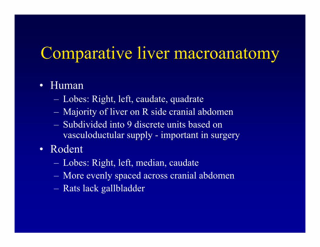
Comparative liver macroanatomy
• Human – Lobes: Right, left, caudate, quadrate – Majority of liver on R side cranial abdomen – Subdivided into 9 discrete units based on
vasculoductular supply - important in surgery
• Rodent – Lobes: Right, left, median, caudate – More evenly spaced across cranial abdomen – Rats lack gallbladder
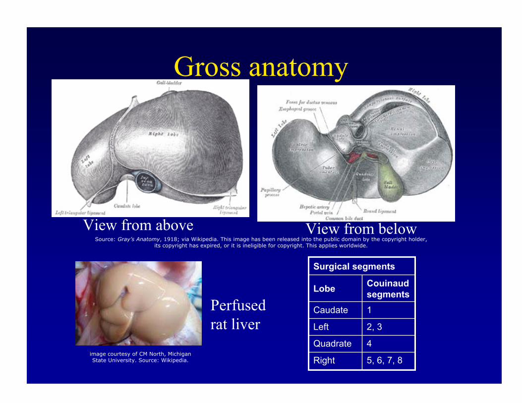
Gross anatomy
View from above View from below
Perfused rat liver
5, 6, 7, 8Right
4Quadrate
2, 3Left
1Caudate
Couinaud segmentsLobe
i
Surgical segments
Source: Gray’s Anatomy, 1918; via Wikipedia. This image has been released into the public domain by the copyright holder, its copyright has expired, or it is inelig ble for copyright. This applies worldwide.
image courtesy of CM North, Michigan State University. Source: Wikipedia.
Several diagrams of liver structure removed for copyright reasons. (Vertical and horiztonal section, anterior and interior surfaces, and a detail cutaway showing interior ducts.)
Mouse liver lobes
Source: Figures 2 and 3 in Harada, T., et al. "Liver and Gallbladder." Chapter 7 in Pathology of the Mouse. Edited by Robert Maronpot. Vienna, IL: Cache River Press, 1999. ISBN: 188989902X.
Images removed for copyright reasons.
Liver functional unit classification schemes
• Lobule first described by Weppler, 1665
• Functional anatomy still not fully known!• Three main models:
– Classical lobule – Rappaport’s acinus model – Matsumoto’s primary lobule

Classical lobule
• Central vein & peripheral portal triads • Roughly hexagonal outline • Blood flows from portal triads to central vein• Species differences
– Pigs have well outlined lobules due to Ç portal fibrous connective tissue (many anatomy studies done with this species as a result)
– Humans and nonhuman primates have discernible lobules
– Mice have poorly visualized lobules
Two photos removed for copyright reasons.Fig. 10-74 and 10-75 from unknown source.
Lobule--reticulin stain
Figure removed for copyright reasons.Source: Figure 1.33 in [MacSween].MacSween, R., et al. Pathology of the Liver, 4th ed. Philadelphia, PA: Elsevier, 2002
Lobular division
Figure removed for copyright reasons. Source: Figure 1.4 in [MacSween].
Hepatic acinus
• Rappaport, 1950’s
• Portal tracts are headwater, elliptical zones spread to terminal hepatic (central) vein – Zone 1: Periportal, high enzyme & O2
– Zone 2: Intermediate – Zone 3: Perivenular, low O2, most susceptible to
hypoxic injury
• Simple & complex acini in berry-like bunches
Classical lobule vs. acini
Figure removed for copyright reasons. Source: Figure 1.7 in [MacSween].
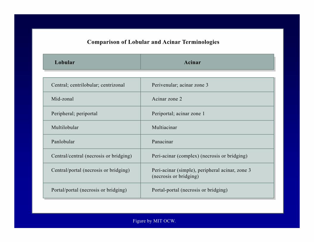
Lobular
Central; centrilobular; centrizonal
Mid-zonal
Peripheral; periportal
Multilobular
Central/central (necrosis or bridging)
Central/portal (necrosis or bridging)
Portal/portal (necrosis or bridging)
Acinar
Perivenular; acinar zone 3
Acinar zone 2
Periportal; acinar zone 1
Multiacinar
Peri-acinar (complex) (necrosis or bridging)
PanacinarPanlobular
Peri-acinar (simple), peripheral acinar, zone 3 (necrosis or bridging)
Portal-portal (necrosis or bridging)
Comparison of Lobular and Acinar Terminologies
Figure by MIT OCW.
Matsumoto’s Primary Lobule
• Similar to classical lobule, but incorporates current knowledge of vascular supply
• “Sickle zone” of complex periportal branching
• Otherwise a hybrid of classical & acinar models
Figure removed for copyright reasons. Source: Figure 1.9 in [MacSween].
Primary vs. classical lobule: vascular network models
Matsumoto Primary Lobule Figures removed for copyright reasons.
Source: Figures 1.10-1.12 in [MacSween].
Classical Lobule
adult liver
Figure removed for copyright reasons.
Arteriovenous connections in
Source: Figure 1.6 in [MacSween].
Hepatic functional unit
• Kidney-Nephron (glomerulus to collecting duct)
• Liver-Not as well defined– Choleon: hepatocytes drained by a single canal
of Herring – Hepaton: hepatocytes served by a single
vascular twig
– Choleohepaton: hybrid of above
Hepatic microcirculatory subunit
Figure removed for copyright reasons. Source: Figure 1.14 in [MacSween].
Zonal heterogeneity
• Hepatocytes in different zones of lobule have different morphology, gene expression, and function – O2 concentration – Matrix proteoglycans
– Bile concentration – Endothelial & Kuffer cell adhesion molecules
and cytokines

Figure removed for copyright reasons. Source: Figure 1.5 in [MacSween].
Biliary Tree
• Canaliculi • Canals of Herring • Bile ductules • Intrahepatic bile ducts
• Extrahepatic bile ducts
• Gallbladder • Common bile duct
Canaliculi
• Interhepatocytic channels for bile flow
• 1-2 um diameter • Bounded by zonula occludentes and zonulae
and maculae adherentes • Microfilaments in microvilli provide
motility • Bile flows opposite direction of blood
Figures removed for copyright reasons.Source: Figure 1.23, 1.24 (Canal of Herring), 1.39 (Portal Triad) and 1.26 in [MacSween].
Gallbladder
• Sac for bile storage
• Empties into common bile duct under hormonal stimulation (e.g. cholecystikinin)
• Absent in rat and horse
Source: Figure 9 in Harada, T., et al. "Liver and Gallbladder." Chapter 7 in Pathology of the Mouse. Edited by Robert Maronpot.
Vienna, IL: Cache River Press, 1999. ISBN: 188989902X.
Image removed for copyright reasons.

Hepatic sinusoids
• ~10 um diameter • Fenestrated endothelium• Space of Disse between endothelium and
hepatocyte membrane - exchange of molecules – small frequent pores periportally “sieve plate”– large pores centrilobularly – pore size affected by hormones and alcohol

Figure removed for copyright reasons. Source: Figure 1.31 in [MacSween].

Resident Cells of the Liver• Hepatocytes • Biliary epithelium • Endothelial
– Blood vessels – Sinusoids – Lymphatics
• Hepatic stellate (Ito) cells • Kupffer cells (resident MØ) • Liver-associated lymphocytes • Nerves & connective tissue cells
Hepatocytes
• 30-40 um, polyhedral • Polarized
– Apical - canalicular region – Lateral - adjacent to canaliculi– Basolateral - facing sinusoids
• Endocrine and exocrine function• Round open nuclei with dispersed and aggregated
chromatin and prominent nucleoli • Polyploidy is common, increases with age

Granular vesicle (secretory granule)
Golgi apparatus
Centriole
Granular endoplasmic
reticulum
Mitochondrion
Basement membrane
Nuclear pore
Schematic Drawing of the General Organization of a Cell
Nuclear envelope
Figure by MIT OCW.

Figure removed for copyright reasons.Source: Figure 1.1 in [Cheville] Cheville, N.F. Ultrastructural Pathology: An introduction to interpretation. Ames, IA: Blackwell Publishing, 1994. ISBN: 0813823986.

Hepatocyte organelles: nucleus
• Large, 5--10% of cell volume • Double-layered membrane with many pores• Polyploidy (humans)
– Birth: Nearly 100% diploid – 8 years: 90% diploid – 15 years: <85% diploid
• Cytoplasm doubles in volume for each nucleus, maintaining steady ratio of genetic material to cell size
• Mitoses infrequent in adults

Figure removed for copyright reasons. Source: Figure 1.18 in [MacSween].
Hepatocyte organelles: ER
• Cisternal network of membrane lined tubes• In general rough ER (ribosomes bound) more
concentrated at periphery, smooth ER centrally merging into perinuclear Golgi – Parallels protein processing
• Zonal variation – Centrilobular hepatocytes 2X SER vs periportal
• Important site of enzymatic activity – SER significantly expands following toxic exposures
• ER can be isolated in vitro by density gradient centrifugation = “microsomes”
Figure removed for copyright reasons. Source: Figure 1.7 from unknown source.
Hepatocyte organelles: Golgi
• Up to 50 interconnected Golgi zones • Stacks of curved membrane-bound sacs
– convex (outer) surface = cis – concave (inner) surface = trans
• Raw proteins arrive from SER on cis face• Processed glyco- & lipoproteins bleb from
trans face for trafficking to cellular site of function or secretion into blood or bile

Hepatocyte organelles: lysosomes
• Landfills of the cell• Primary lysosomes contain hydrolytic enzymes
(acid phosphatase, etc) • Fuse with phagocytic vesicles to form 2o
lysosomes or effete cell products to form autophagic vacuoles
• Enzymes degrade contents• Subunits recycled, excreted (e.g. into bile), or
retained for long periods (sometimes with pathologic consequences)

Figure removed for copyright reasons. Source: Figure 1.36 in [Cheville].
Hepatocyte organelles: peroxisomes (microbodies)
• Ovoid membrane-bound granules 0.2-1 um• Fatty acid beta-oxidases • Upregulated by peroxisome proliferator-activated
receptor -alpha (PPARa) • Peroxisome proliferator compounds (PPC)
– Phthalate plasticizers, hypolipidemic fibrate drugs (e.g. clofibrate), Wy 14,643
• Rodents are exquisitely sensitive to PPC (tumors)• Humans and other species (e.g. guinea pig)
resistant

Figure removed for copyright reasons. Source: Figure 1.19 in [MacSween].
Hepatocyte organelles: mitochondria
• “Powerhouse of cell” • Outer membrane with lipid transport proteins• Inner folded membrane (cristae) with oxidative
enzymes • Main function to produce ATP• Also important in apoptosis (mitochondrial
permeability transition, MPT, cytochrome c, Bcl family)
• Highly mobile organelles • Comprise 20% of hepatocyte volume
Figure removed for copyright reasons. Source: Figure 1.22 in [Cheville].
• Cytoplasm (hyaloplasm) •
• Liposomes •
Other hepatocyte contents
Glycogen – alpha-rosettes – beta-particles
Free ribosomes • Other
Glycogen in the non-Atkins liver
Figure removed for copyright reasons. Source: Figure 1.35 in [MacSween].
Glycogen - fasting vs. fed mice
Source: Figures 5 and 6 in Harada, T., et al. "Liver and Gallbladder." Chapter 7 in Pathology of the Mouse. Edited by Robert Maronpot. Vienna, IL: Cache River Press, 1999. ISBN: 188989902X.
Images removed for copyright reasons.
Cell debris
Figure removed for copyright reasons. Source: Figure 1. 36 in [MacSween].
Hepatocyte organelles: cytoskeleton
• Microfilaments (6 nm) – Fibrous (F) actin polymerized fibrils – Globular (G) actin monomers
• Intermediate filaments (cytokeratins; 8-10 nm)– Hepatocyte Ck 8 & 18 – Biliary epithelium add Ck 7 & 19
• Microtubules (20 nm) – Polymers of alpha & beta tubulin – Part of mitotic spindle apparatus & cyotplasmic
centrioles – Scaffold for rigidity and directed organelle trafficking
Junctional complexes
• Desmosome (macula adherens)– button-like connector between cytokeratin filaments of
adjacent cellls
• Intermediate junction (zonula adherens)– continous belt-like junction below tight jxns – Ca2+ dependent adherins incl beta catenin
• Tight junction (zonula occludens) – sealing belt separates canaliculus from intercellular
space
Figure removed for copyright reasons. Source: Figure 1-22, 1-23 in unknown source.
Canalicular membrane
• Specialized region of hepatocyte membrane• Bile canaliculus bounded by apposing hepatocyte
hemicanaliculi • Finger-like projectios (microvilli)
– actin/myosin etc for bile propulsion • Canalicular-rich enzymes
– gamma glutamyl transpeptidase (GGT) – alkaline phosphatase (ALP) – backflow into blood during cholestasis (bile sludging)

Figure removed for copyright reasons. Source: Figure 1.16 in [MacSween].
Bile secretory apparatus-Uptake
• Conjugated bilirubin uptake from blood via high affinity receptors – Sodium taurocholate cotransporting polypeptide
(NTPC) – Organic anion and cation transporting proteins (Oat1 &
2, Oct1 & 2) • Most bile taken up by periportal hepatocytes
(decreasing bile gradient in sinusoids approaching central vein)
• Hepatocyte receptor for unconjugated bilirubin unknown

Bile secretory apparatus-Vesicle transport
• Endocytosis of ligand-bound receptors (larger than clathrin-coated pits)
• Endosome contents acidified (releases ligand)• Most endosome contents (80%) returned to cell
membrane • ~18% directed to lysosomes• ~2% secreted into bile • Vesicular trafficking along microtubules accounts
for 95% of blood-to-bile transport
Bile secretory apparatus-excretion
• Bile salt export pump (BSEP)– Other names: canalicular bile salt transporter (CSBT),
sister of P glycoprotein (SPGP) – rate-limiting step of bile outflow– absent in progressive familial intrahepatic cholestasis
type 2 (PFIC 2) – important in drug excretion
• Other known or putative bile export molecules– Multidrug-resistance associated protein 2 (MRP2) – Multidrug resistance proteins 1 & 3 (MDR1,3)

Biliary epithelium
• Cuboidal or columnar w/microvilli • Actively secrete substances and resorb bile
– cholehepatic shunt pathway • Peribiliary glands (human)
– intramural mucous glands communicate w/lumen – extramural mixed seromucous glands form
tubuloalveoli that connect to lumen via ducts • Duct epithelium secretes IgA and IgM• Stimulated by secretin and inhibited by
somatostatin
Bile ductule
Figure removed for copyright reasons. Source: Figure 1.25 in [MacSween].
Endothelial cells
• Specialized for compartment & function
• Display unique cell adhesion molecules & cytokine production based on location
• Sinusoidal endothelium fenestrated – frequent small pores periportally (sieve plate)– infrequent large pores centrally (fixation artifact?) – no basement membrane; hepatocytes bathed in filtered
plasma
Figures removed for copyright reasons. Source: Figure 1.27, 1.28 in [MacSween].
Hepatic stellate (Ito) cells
• In space of Disse; inapparent w/stndrd stains
• Long thin cellular projections • Important in lipid and vitamin A storage • 4 main functions:
– extracellular matrix proteins – pericytic - microvascular tone – Vitamin A and retinyl storage – cytokines (HGF-alpha in response to IGF-2)

Figure removed for copyright reasons. Source: Figure 1.29 in [MacSween].
Kupffer cells
• Resident macrophages line sinusoids • Clearance of gut derived toxins (e.g. LPS)• Secrete cytokines
– IL-1 – IL-6 – TNF-alpha
• Express class II MHC- antigen presentation• At least partially derived from bone marrow
monocytes

Figure removed for copyright reasons. Source: Figure 1.30 in [MacSween].
Liver-associated lymphocytes
• Normal human liver has 1x10¹⁰ lymphoid cells • Predominantly in portal regions but also scattered
• Predominant classes – NKT cells – gamma-delta T cells (most g/d T cells of any organ)– CD8+ T cells
• Immune function of liver comparable to that of GI tract
Lymphatics
• Liver is single largest source of lymph– 15-20% overall volume – 25-50% thoracic duct flow
• Very high protein content • ~80% lymphoid cells & 20% MØ • Terminal twigs of lymphatic channels accompany
hepatic arterioles – dilated w/visible cells in some disease states
• Continuous with subcapsular lymphatic sinus• Drains fluid collected in space of Disse

Nerve supply
• Liver is richly innervated although difficult to see neural structures histologically
• Nerve fibers demonstrated by immunohistochemistry
• Sympathetic and parasympathetic signals affect vascular tone and metabolism
• Successful transplant engraftment, however, suggests neural control is dispensible to overall function
Hepatic embryonic development
• Human embryos: Hepatic bud @ 3 wk • Endoderm - pouch off of future duodenum
• Cues from mesenchyma guide hepatic differentiation – Septum transversum
– Coelomic lining
• Hepatic diverticulum invades stroma, induces sinusoidal plexus formation from vitelline veins

Figure removed for copyright reasons. Source: Figure 1.1 in [MacSween].
Development (cont’d)
• Epithelial bud breaks into anastomosing parallel sheets around sinusoids
• Caudal bud forms cystic duct & gallbladder• Growing liver fills available space,
displacing stomach & duodenum from septum transversum
• Hepatic stalk migrates with duodenum to form extrahepatic bile ducts
Figure removed for copyright reasons. Source: Figure 1.2 in [MacSween].
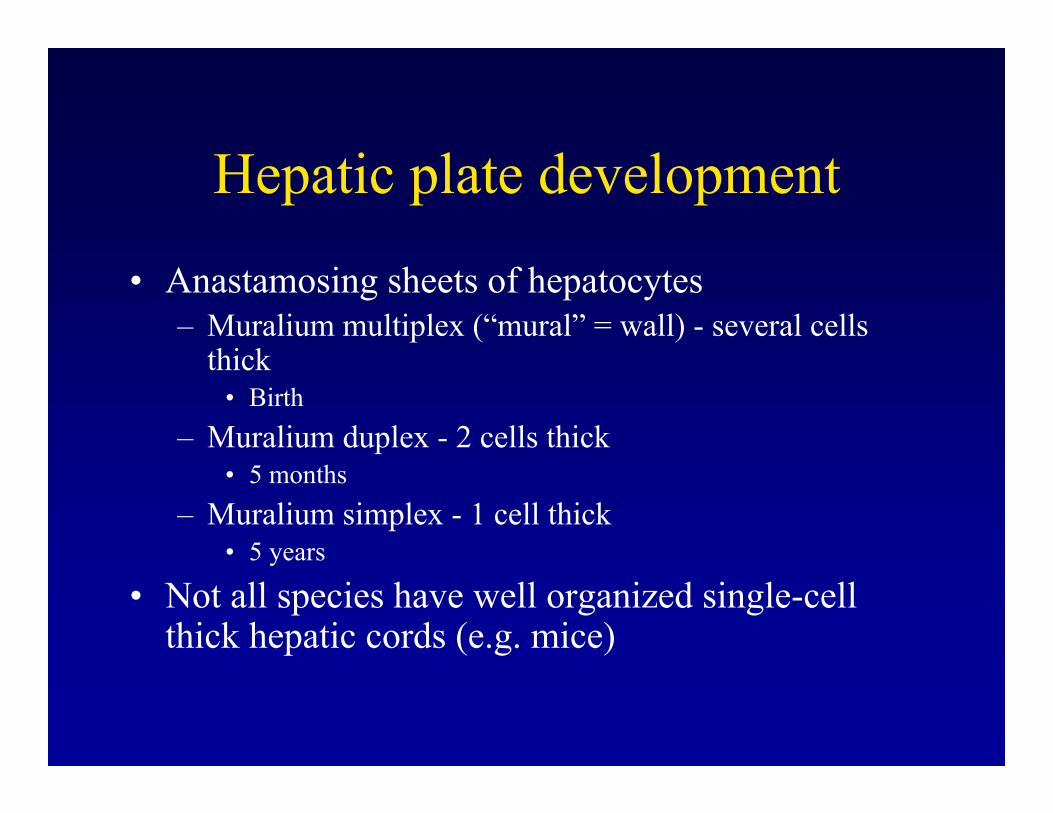
Hepatic plate development
• Anastamosing sheets of hepatocytes– Muralium multiplex (“mural” = wall) - several cells
thick • Birth
– Muralium duplex - 2 cells thick• 5 months
– Muralium simplex - 1 cell thick• 5 years
• Not all species have well organized single-cell thick hepatic cords (e.g. mice)
Figure removed for copyright reasons. Source: Figure 1.34 in [MacSween].
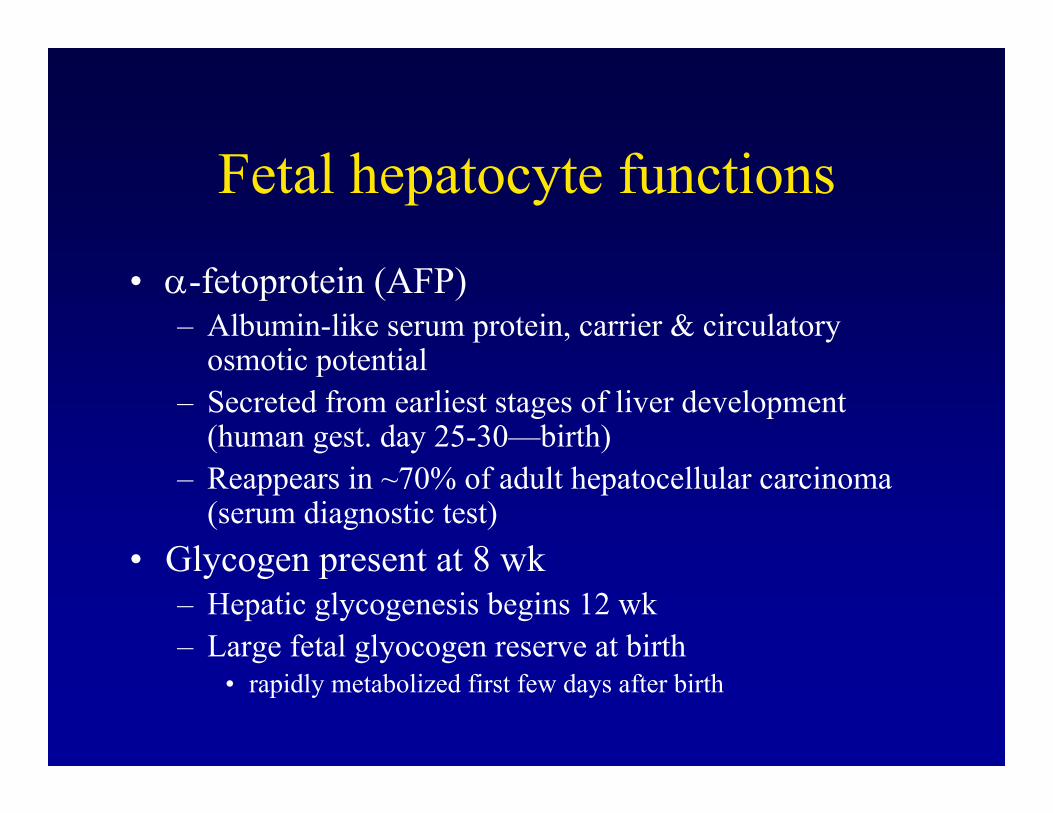
Fetal hepatocyte functions
• α-fetoprotein (AFP)– Albumin-like serum protein, carrier & circulatory
osmotic potential – Secreted from earliest stages of liver development
(human gest. day 25-30—birth) – Reappears in ~70% of adult hepatocellular carcinoma
(serum diagnostic test) • Glycogen present at 8 wk
– Hepatic glycogenesis begins 12 wk– Large fetal glyocogen reserve at birth
• rapidly metabolized first few days after birth

Fetal hepatocyte functions (cont’d)
• Hemosiderin (iron) deposits early in development – Increased during hepatic hematopoiesis (12--18
wks gestation), then decrease – Mostly periportal; also site of copper storage
• Sinusoidal endothelial, Kupffer & stellate cells appear @ 10-12 wk
Biliary functional development
• Bile acid synthesis begins 5--9 wk
• Secretion 12 wk • However, canalicular excretion & transport
remains immature until 4--6 wk postpartum • Bile excretion across placenta is important
for fetus
Figure removed for copyright reasons.
Developing ductal plate Cytokeratin IHC
Source: Figure 1.3 in [MacSween].
Summary
• Liver main functions– Catabolism – Metabolism – Detoxification – Bile production
• Several ways to classify liver based on histology, physiology & vascular supply
• Species differences in anatomy and physiology• Knowledge of liver functional anatomy informs
hypotheses about disease pathogenesis
Sources
Kumar, Vinay, Abul K. Abbas, and Nelson Fausto, eds. Robbins and Cotran Pathologic Basis of Disease. 7th ed. Philadelphia, PA: Elsevier Saunders, 2005.
MacSween, R., et al. Pathology of the Liver, 4th ed. Philadelphia, PA: Elsevier, 2002.
Maronpot, R., editor. Pathology of the Mouse. Vienna, IL: Cache River Press, 1999.
Cheville N.F. Ultrastructural Pathology: An introduction to interpretation. Ames, IA: Blackwell Publishing, 1994.