a study of the distance between the free edges of medial
TRANSCRIPT

Contents lists available at BioMedSciDirect Publications
Journal homepage: www.biomedscidirect.com
International Journal of Biological & Medical Research
Int J Biol Med Res. 2012; 3(2): 1520-1524
A study of the distance between the free edges of Medial Pterygoid Plates in the Skulls of Central Indian Population Manmohan patel*
A R T I C L E I N F O A B S T R A C T
Keywords:
Original Article
Medial Pterygoid platesNasopharynxSkulls
1. Introduction
Objective: To stabilize the craniometric data for central Indian population and to find out any
sexual dimorphism in distance between the free edges of medial Pterygoid plates in the
nasopharyngeal region. Method: The present study was conducted on three hundred fourteen
dry skulls of central India region. The dry skulls taken for study are first examined to determine
the sex of the skull then various measurements of the distance between the free edges of medial
Pterygoid plates were taken and compared to find out any sexual dimorphism. The maximum
transverse distance between free edges of medial pterygoid plates has been measured at three
sites-Site-I : At the uppermost part where the pterygoid plates meet the body of sphenoid bone,Site-II : At middle of pterygoid plates,Site-III : At the level of posterior free margin of hard palate.Results: At the site I, the average distance between free edges of medial pterygoid plates for
male cases and female cases were 27.84(2.44) mm and 26.93(2.45) mm respectively. At the site
II, the average distance between free edges of medial pterygoid plates for male cases and
female cases were 29.32(2.86) mm and 28.25(2.57) mm respectively. At site III, the average
distance between free edges of medial pterygoid plates for male cases and female cases were
0.44((2.50) mm and 29.54(2.77) mm respectively. For all the three sites, the average values for
the male cases were significantly higher as compared with the female cases (P 0.05).Conclusion: This study shows that the distance between the free edges of medial Pterygoid
plates in the skulls of central Indian population is slightly lower than that mentioned in other
studies and the distance between the free edges of medial Pterygoid plates were found
significantly higher in the male skulls than female skulls.
Pharynx is a 12-14 cm long musculomembranous tube shaped
like an inverted cone; it extends from the cranial base to the lower
border of cricoid cartilage (at the level of sixth cervical vertebra)
where it becomes continuous with the Oesophagus.
The pharynx is subdivided into Nasopharynx, Oropharynx and
Laryngopharynx. This subdivision of the pharynx is used in most
modern textbooks of Anatomy, and the Nasopharynx is described as
the upper portion of the pharynx lying above the soft palate and
behind the posterior nares, which allows the free respiratory
passage between the nasal cavity and lower airway. Nasopharynx
is an open Cuboidal chamber that lies beneath base of skull at the
posterior aspect of nasal fossa. The Nasopharynx measures 4.0-5.5
cm transversely, 2.5-3.5 cm antero-posteriorly and roughly 4.0 cm
in height [1].
The nasal and oral part of the pharynx communicates through
the pharyngeal isthmus, which lies between the posterior border
of the soft palate and the posterior pharyngeal wall. Elevation of
the soft palate and constriction of the palatopharyngeal isthmus
occurs during swallowing
BioMedSciDirectPublications
Copyright 2010 BioMedSciDirect Publications IJBMR - All rights reserved.ISSN: 0976:6685.c
International Journal ofBIOLOGICAL AND MEDICAL RESEARCH
www.biomedscidirect.comInt J Biol Med ResVolume 3, Issue 1, Jan 2012
*Assistant professor, Deptt. Of Anatomy, Gandhi Medical college, Bhopal, India,
* Corresponding Author : Dr. Manmohan patel
Deptt. Of Anatomy, Gandhi Medical college, Bhopal, India,E-mail: [email protected]
Copyright 2010 BioMedSciDirect Publications. All rights reserved.c

1521
The walls of the Nasopharynx are rigid except for the soft
palate therefore the cavity of the Nasopharynx is never obliterated
unlike the cavity of the Oropharynx and Laryngopharynx [2]. The
Nasopharynx has a roof, a posterior wall, two lateral walls and a
floor.
The roof and posterior wall form a continuous slope that leads
down from the nasal septum to the Oropharynx. It is bounded
above by the mucosa overlying the posterior part of the body of
sphenoid and further backs the basilar part of the occipital bone as
for as pharyngeal tubercle, further down the mucosa overlies the
Pharyngobasilar fascia and the superior constrictor and behind
these the anterior Arch of the Atlas.
The Nasopharyngeal tonsil lies in the midline in the roof and
posterior wall. The superior constrictor and fascia completes the
posterior wall [3].
It is a non-expansible sheet of fascia. Pharyngobasilar fascia is
thick above where muscular fibers are absent and starting at the
Pharyngeal tubercle of basioccipital bone passes laterally anterior
to the longus capitis muscle. It is firmly attached to the basilar
occipital and petrous temporal bones medial to the carotid canal,
curving under the auditory tube and forwards to the sharp
p o s t e r i o r b o r d e r o f m e d i a l p t e r y g o i d p l a t e a n d
pterygomandibular raphe. As it descents it diminishes in thickness
but is strengthened posteriorly by a fibrous band attached to the
occipital's pharyngeal tubercle and descending as the median
raphe of constrictors. This provides firmness to the
nasopharyngeal wall to help it in keeping its patency [2-4].
From above anatomical knowledge we know that the
Nasopharynx has bony relations in the roof and posterior wall by
body of sphenoid and occipital bone and also to the atlas and axis
vertebra. On anterior side the posterior nares are bounded both
side by the medial pterygoid plates. Only the lateral wall in its
posterior part is muscular (superior constrictor muscle).
So if there are any deformities in the normal development of
skull in this region it leads to variation in the size of the
Nasopharynx.
As we know that Nasopharynx is a communication between
nose and rest of the respiratory apparatus, any variation in the size
of the Nasopharynx leads to disturbed respiratory function.
Syndrome related to sleep disorder called, as 'sleep apnea
syndrome' in which there is difficulty in breathing during the sleep.
Restricted bony pharynx is one of the causes of sleep apnea
syndrome [5]. The knowledge that sleeps apnea commonly results
from upper airway obstruction rises the questions How, Where
and why the airway is obstructed. The answers to these questions
may continue to provide the basis for the management of patients
with obstructive sleep apneas (OSA) through the development of
therapies designed to eliminate the airway closer. Understanding
the airway occlusion in patients of sleep apnea will be facilitated by
first considering the normal Anatomy and Physiology of the upper
air way [6].
Instrumentation of Nasopharynx like nasopharyngoscopy and
nasogastric intubation will also need the normal structure and
dimension of the region to be known for meticulous manipulation
of instruments as well as for their better designing.
The cases of nasopharyngeal carcinoma are common and the
knowledge of anatomy of Nasopharynx and its relations is useful in
interpretation of images for investigative purpose and
understanding the course of spread of carcinoma.
While going through the available literature on Nasopharynx it
was found that data regarding its measurements are scanty.
Therefore the current study was proposed. It was decided that
various dimensions determining the size of Nasopharynx be
measured at the base of skull and the variations and sexual
dimorphism be noted if any.
OBJECTIVE: To stabilize the craniometric data for central
Indian population and to find out any sexual dimorphism in the
distance between the free edges of medial Pterygoid plates in the
nasopharyngeal region.
The present study was conducted in the department of
anatomy at NSCB Medical College Jabalpur India, from August
2005 to July 2007. Total 314 dry skulls of central India region were
taken for this study. The skulls were selected only after confirming
that there was intact base of skull especially in the anterior cranial
fosse & middle cranial fosse region. The skulls taken for study are
of adult to old age groups where development in the
Nasopharyngeal region has completed.
The dry skulls taken for this study was first examined for
determine the sex of the skull. With the help of various traits, we
first determined the probable sex of the skull. When the skull
shows more points towards male, it was marked male and the
same criteria taken for female skull.
The instruments used in present study for the measurement
size of are a metallic scale (having calibrations of 0.5 millimeter)
and a spreading caliper. The spreading caliper is spreaded between
the two bony points to be measured and fixed. The fixed caliper is
then matched with the calibrations on the scale and the reading is
taken. Each reading was repeated twice to rule out any manual
error.
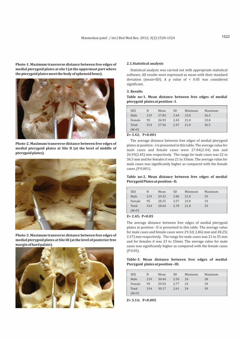
The parameter measured in this study is maximum transverse
distance between free edges of medial pterygoid plates. This has
been measured at three sites-
Site-I : At the uppermost part where the pterygoid plates meet the
body of sphenoid bone,
Site-II : At middle of pterygoid plates,
Site-III : At the level of posterior free margin of hard palate.
2. Material and Methods
Manmohan patel / Int J Biol Med Res. 2012; 3(2):1520-1524
SEX
Male
Female
Total
(M+F)
SEX
Male
Female
Total
(M+F)
SEX
Male
Female
Total
(M+F)
N
219
95
314
N
219
95
314
N
219
95
314
Mean
27.84
26.93
27.56
Mean
29.32
28.25
28.64
Mean
30.44
29.54
30.17
SD
2.44
2.45
2.47
SD
2.86
2.57
2.78
SD
2.50
2.77
2.61
Minimum
23.0
21.0
21.0
Minimum
21.0
23.0
21.0
Minimum
24
24
24
Maximum
36.5
33.0
36.5
Maximum
35
33
35
Maximum
38
39
39
Table no-1. Mean distance between free edges of medial
pterygoid plates at position –I.
Table no-2. Mean distance between free edges of medial
Pterygoid Plates at position –II.
Table-3. Mean distance between free edges of medial
Pterygoid plates at position –III.
Z= 3.14; P<0.005
Z= 2.45; P<0.05
Z= 3.42; P<0.001
2.1.Statistical analysis
3. Results
Photo-1. Maximum transverse distance between free edges of
medial pterygoid plates at site I (at the uppermost part where
the pterygoid plates meet the body of sphenoid bone).
Photo-2. Maximum transverse distance between free edges of
medial pterygoid plates at Site II (at the level of middle of
pterygoid plates).
Photo-3. Maximum transverse distance between free edges of
medial pterygoid plates at Site III (at the level of posterior free
margin of hard palate).
Statistical analysis was carried out with appropriate statistical
software. All results were expressed as mean with their standard
deviation (mean=SD). A p value of < 0.05 was considered
significant.
The average distance between free edges of medial pterygoid
plates at position –I is presented in this table. The average value for
male cases and female cases were 27.84(2.44) mm and
26.93(2.45) mm respectively. The range for male cases was 22 to
36.5 mm and for females it was 21 to 33mm. The average value for
male cases was significantly higher as compared with the female
cases. (P 0.001).
The average distance between free edges of medial pterygoid
plates at position –II is presented in this table. The average value
for male cases and female cases were 29.32( 2.86) mm and 28.25(
2.57) mm respectively. The range for male cases was 21 to 35 mm
and for females it was 23 to 33mm. The average value for male
cases was significantly higher as compared with the female cases
(P 0.05).
1522Manmohan patel / Int J Biol Med Res. 2012; 3(2):1520-1524

4. Discussion
4. Discussion
6.References
The average distance between free edges of medial pterygoid
plates at position–III is presented in this table. The average value
for male cases and female cases were 30.44(( 2.50) mm and 29.54(
2.77) mm respectively. The range for male cases was 24 to 38mm
and for female it was 24 to 39mm. The average value for male cases
was significantly higher as compared with the female cases (P
0.005).
Nasopharynx has always been an area of special interest for
workers of various disciplines. Its situation at the base of the skull
has attracted attention during craniometry. Craniometrical
measurements have been done and attempts have been made to
stabilize a proper relationship between size of the mandible and
the Nasopharynx [7].
Being associated with important vital functions like respiration,
Nasopharynx has attracted attention of otorhinolaryngologists.
Young children with difficulty in breathing are found to have
enlarged lymphoid tissue in the posterior pharyngeal wall. The
calculation of Adenoid –Nasopharyngeal ratio has been suggested
for assessment of adenoidal hypertrophy, as adenoidectomy will
not be effective in cases of stenosis of bony Nasopharynx [8, 9]. A
another study demonstrated that the upper airway was smaller in
children with 'Sleep Apnoea Syndrome' in comparison with
control subjects whereas the volume of tongue and mandible were
same in both cases [10]. Transverse measurements of
Nasopharynx are done to estimate the width of operating field in
cases planned for Transsphenoidal Hypophysectomy [11].
Cephalometric studies have been used for many years for
evaluation of growth and development of face. In recent past
various sleep disorders like snoring, sleep apnoeas and upper
airway diseases has drown the attention of scientists for study of
measurement of Nasopharynx. Radiological and Imaging
techniques have been used for measurement of nasopharyngeal
dimensions.
The present study was conducted on three hundred fourteen dry
skulls of central India region. Various measurements of the
Nasopharyngeal region were taken. The purpose of the study is to
stabilize data for central India population and to find out if there is
any sexual dimorphism.
A measurement for transverse diameter of Nasopharynx is the
distance between the free edges of medial Pterygoid plates which
was measured at three places in our study –
Site- I. Where the Pterygoid plates meet the Sphenoid bone -
27.564± 2.47mm
Site- II . At the Middle of the Pterygoid plates -
28.64±2.78mm
S i t e - I I I . A t t h e l e v e l o f h a r d p a l a t e -
30.17±2.61mm
The various patterns are observed in the distance between the
medial Pterygoid plates as we go downwards from position I to
position III.
In 68.79 % Medial Pterygoid plate deviate from each other.
In 17.2 % cases the Medial Pterygoid plates are parallel,
In 06.05% cases the Medial Pterygoid plates are narrowest in the
middle,
In 05.73 % cases the Medial Pterygoid plates moves closer as we
go down from position I –III.
And in 2.23 % cases the Medial Pterygoid plates are broadest in
the middle. These findings are compared with that of Kobayashi H,
Kato I and Terabayashi T [12].
Kobayashi H, Kato I and Terabayashi T.[12] also measured the
distance between the right and left Medial Pterygoid plates in
twenty-seven normal adults from antero-posteriorly Tomograms.
They observed the mean distance between Medial Pterygoid plates
about 32mm. They related this value for the calculation of actual
operating field, which was about 1cm less than the distance
between the Medial Pterygoid plates. The space was calculated for
Sublabial transeptal transsphenoidal Hypophysectomy in Asiatic
type of skull. They did not mention that at which site, they
measured the distance between the Medial Pterygoid plates.
We measured the distance between the free edges of Medial
Pterygoid plates at three sites. All values of our measurements are
lower than as mentioned by Kobayashi H, Kato I and Terabayashi T.
This study shows that the distance between the free edges of
medial Pterygoid plates in the skulls of central Indian population is
slightly lower than that mentioned in other studies and the
distance between the free edges of medial Pterygoid plates were
found significantly higher in male skulls than female skulls in
central Indian population.
1523
Mean distance between medial Pterygoid plates
Kobayashi H, Kato I and Terabayashi T
In present study
Site I - 27.56± 2.47mm
Site II. - 28.64±2.78mm
Site III. - 30.17±2.61mm
[1] Frank G. Ondrey, Simon K. Wright. Neoplasm of Nasopharynx. 2003.
[2] Ballenger's Otorhinolaryngology 16th edition .Published by B. C. Decker Hamilton, Ontario pages 1392-1407.
[3] Simon A Hickey, Allan Thexton, John D. Langdon, Patrica Collins and Caroline Wigley. 'Pharynx' Chapter 35 Gray's Anatomy 39th Edition 2005 Published by ELSEVIER Churchill Livingstone pages 619-631.
[4] Frank G. Ondrey, Simon K. Wright. Neoplasm of Nasopharynx. Ballenger's Otorhinolaryngology 16th edition .Published by BC.Decker Hamilton, Ontario pages,2003; 1392-1407.
[5] Last's Anatomy Regional and Applied 11th edition 2006. Published by ELSEVIER Churchill Livingstone , London Page-398.
[5] Mangat K, Chaudhary G, Salahudeen S, Chandra SV, Pahor AL. MRI in assessment of Adenoids in Children.' International Congress series.2003; 1240 :1437-1441.
[6] Raanan Arens, Joseph M., Mc Donough. Andrew T. Costarino, Soroosh Mahboubi, Catherinee Tyag Kier, Greg Maislin, Richard. J. Schwab and Allan. I. Pack. 'Magnetic Resonance Imaging of Upper airway structures of Children with Obstructive Sleep Apnoea Syndrome.' American Journal of Respiratory and Critical care Medicine.2001; 164 :698-703.
Manmohan patel / Int J Biol Med Res. 2012; 3(2):1520-1524
1524
[7] Samuel T. Kuna, and John E. Remmers. 'Neural and Anatomic factors Related to upper airway occlusion during Sleep'. Medical clinics of North America vol. 69, 6 November 1985 pages 1221-42.
[8] Hillowala RA, Trent RB. The mandible –a measurement of the Nasopharynx Human Evolution vol. 7 N-1 (35-42) 1992.
[9] Mutsuhisa Fujioka, Lionel W. Young and Bertram R. Girdany. Radiographic Evaluation of Adenoid size in Children: Adenoid Nasopharyngeal Ratio.' A J R – 133; 401-404 September 1979.
[10] Mangat K, Chaudhary G, Salahudeen S, Chandra SV, Pahor AL. MRI in assessment of Adenoids in Children.' International Congress series.2003; 1240 :1437-1441.
[11] Raanan Arens, Joseph M., Mc Donough. Andrew T. Costarino, Soroosh Mahboubi, Catherinee Tyag Kier, Greg Maislin, Richard. J. Schwab and Allan. I. Pack. 'Magnetic Resonance Imaging of Upper airway structures of Children with Obstructive Sleep Apnoea Syndrome.' American Journal of Respiratory and Critical care Medicine.2001; 164 :698-703.
[12] Kabayashi H, Kato J, Terebayashu T. Anthropometric relevance on sublabial transpalatal transsphenoidal Hypophysectomy in the Asiatic type of skull.' ORL J Otorhinilaryngol Realt Spec. 1988; 50(5): 340-4.
[13] Kabayashi H, Kato J, Terebayashu T. Anthropometric relevance on sublabial transpalatal transsphenoidal Hypophysectomy in the Asiatic type of skull. ORL J Otorhinilaryngol Realt Spec. 1988; 50(5): 340-344.
Copyright 2010 BioMedSciDirect Publications IJBMR - All rights reserved.
ISSN: 0976:6685.c
Manmohan patel / Int J Biol Med Res. 2012; 3(2):1520-1524