a study of corneal thickness, shape and collagen
TRANSCRIPT

Experimental Eye Research 84 (2007) 423e434www.elsevier.com/locate/yexer
A study of corneal thickness, shape and collagen organisation inkeratoconus using videokeratography and
X-ray scattering techniques
Sally Hayes a, Craig Boote a, Stephen J. Tuft b, Andrew J. Quantock a, Keith M. Meek a,*
a Structural Biophysics Research Group, School of Optometry and Vision Sciences, Cardiff University, Redwood Building, King Edward VII Avenue,
Cardiff, CF10 3NB, United Kingdomb Moorfields Eye Hospital, 162 City Road, London, EC1V 2PD, United Kingdom
Received 20 July 2006; accepted in revised form 18 October 2006
Available online 18 December 2006
Abstract
In keratoconus, the cornea becomes progressively ectactic resulting in severe visual impairment. Here, we use a combination of videokeratog-raphy and synchrotron X-ray diffraction to investigate the relationship between corneal shape and thickness, and the distribution and predom-inant orientation of stromal fibrillar collagen in five keratoconus corneas. In all but the least advanced case, the thinning and ectasia measured invivo using corneal videokeratography was accompanied by corresponding changes in the relative distribution and orientation of stromal collagenin the excised corneal buttons. Although the most severe case of keratoconus possessed the most pronounced stromal collagen alterations, andonly a minor disruption to stromal collagen arrangement was seen in the least advanced case, a variability in the extent of stromal collagenalteration was seen between these clinical extremes. The observed abnormalities in collagen distribution and orientation are consistent witha mechanism of keratoconus progression that involves inter-fibrillar or inter-lamellar slippage causing a redistribution of tissue within the cornea.� 2006 Elsevier Ltd. All rights reserved.
Keywords: cornea; keratoconus; X-ray scattering; videokeratography
1. Introduction
As the cornea is responsible for over two-thirds of the eye’srefractive power, any acquired variations in its shape are po-tentially detrimental to vision. The stroma, which forms thebulk of the cornea, is comprised primarily of water, collagens,proteoglycans and keratocytes. At normal hydration the uni-formly small diameter of stromal collagen fibrils and the
* Corresponding author. Structural Biophysics Research Group, School of
Optometry and Vision Sciences, Cardiff University, Redwood Building,
King Edward VII Avenue, POB 905, Cardiff, CF10 3NB, United Kingdom.
Tel.: þ44 02920 876317; fax: þ44 02920 874859.
E-mail addresses: [email protected] (S. Hayes), [email protected] (C.
Boote), [email protected] (S.J. Tuft), [email protected] (A.J. Quantock),
[email protected] (K.M. Meek).
0014-4835/$ - see front matter � 2006 Elsevier Ltd. All rights reserved.
doi:10.1016/j.exer.2006.10.014
regular separation distance between them is believed to beregulated by proteoglycans (Borcherding et al., 1975; Chakra-varti et al., 1998; Kao and Liu, 2003; Quantock et al., 2001).Collagen fibrils lie parallel to each other within stacked layers(lamellae) which are themselves interspersed with thin, flatkeratocytes. Although most lamellae lie parallel to the cornealsurface (Komai and Ushiki, 1991), lamellar interweaving isa common feature of the anterior (Radner et al., 1998a) andmid-stroma (Radner and Mallinger, 2002). The specific fibril-lar arrangement is believed to provide the cornea with itsshape, transparency and strength (Benedek, 1971; Daxer andFratzl, 1997; Komai and Ushiki, 1991; Maurice, 1957; Meeket al., 2003; Muller et al., 2001; Radner and Mallinger,2002; Smolek and McCarey, 1990). As a result, abnormalitiesin the structural organisation of stromal collagen have beenimplicated in keratoconus, a pathology characterised by
424 S. Hayes et al. / Experimental Eye Research 84 (2007) 423e434
a progressive thinning and ectasia of the stroma resulting ina cone-shaped cornea and severe irregular astigmatism (Klint-worth, 1994).
Despite much research into the pathogenesis of keratoco-nus, the precise mechanism by which affected corneasprogressively thin and steepen remains unknown. In particular,it is unclear if the stromal thinning associated with the diseaseis caused by an enzyme-mediated loss of collagen (Fini et al.,1992; Kenney et al., 1989, 1994; Kenney and Brown, 2003;Rehany et al., 1982; Sawaguchi et al., 1990, 1994, 1989;Teng, 1963; Zhou et al., 1998), a disruption to fibrillogenesisdue to the presence of defective keratocytes (Brookes et al.,2003; Kim et al., 1999; Sherwin et al., 2002; Somodi et al.,1996) or abnormal proteoglycans (Funderburgh et al., 1989),or whether it is due to slippage between lamellae resultingin a redistribution of collagen away from the apex of thecone (Polack, 1976).
In the normal human cornea, more collagen fibrils lie inthe superioreinferior and nasaletemporal directions than inany other meridian (Aghamohamadzadeh et al., 2004; Daxerand Fratzl, 1997; Meek et al., 1987). The exact purpose ofthis arrangement is unknown, but it may be related to theposition at which the four extra-ocular rectus muscles insertinto the anterior sclera, thereby providing increased tensilestrength to prevent corneal distortion during eye movement(Daxer and Fratzl, 1997). Previous studies have shown that,in corneas with keratoconus, the normal orientation offibrillar collagen is disturbed in the scarred apical region(Daxer and Fratzl, 1997; Radner et al., 1998b) and lamellarinterweaving is greatly reduced (Radner et al., 1998b).More recently, it was further shown that the progressiveand symmetrical increase of collagen mass from the centreto the edge of the cornea is also disturbed in keratoconus(Meek et al., 2005). These factors likely contribute to thediffering biomechanical properties of normal andkeratoconus corneas (Andreassen et al., 1980; Nash et al.,1982).
X-ray scattering is a specialised technique that has beenused to gain structural information about the constituent col-lagen in the corneal stroma. The wide-angle equatorial scat-tering pattern that is produced from the lateral packing ofmolecules within the stromal collagen fibrils can be used todetermine the intermolecular spacing within the fibrils aswell as the arrangement and distribution of fibrillar collagenin the intact cornea. Since the collagen molecules are ar-ranged roughly parallel to each other and to the fibril axis,the orientation of the molecules derived from an X-ray scatterimage reasonably represents the direction of the collagen fi-brils at that position in the cornea. The intensity of X-rayscatter gives quantitative information regarding the relativenumbers of fibrils lying in a certain direction, as well asthe relative tissue mass at the point through which thebeam passes.
Using a combination of videokeratography and X-rayscattering, the current study investigates for the first time therelationship between keratoconus corneal shape and thickness,and abnormalities in stromal collagen arrangement.
2. Materials and methods
2.1. Tissue
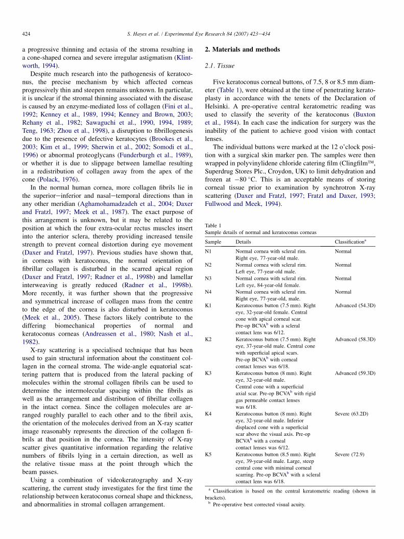
Five keratoconus corneal buttons, of 7.5, 8 or 8.5 mm diam-eter (Table 1), were obtained at the time of penetrating kerato-plasty in accordance with the tenets of the Declaration ofHelsinki. A pre-operative central keratometric reading wasused to classify the severity of the keratoconus (Buxtonet al., 1984). In each case the indication for surgery was theinability of the patient to achieve good vision with contactlenses.
The individual buttons were marked at the 12 o’clock posi-tion with a surgical skin marker pen. The samples were thenwrapped in polyvinylidene chloride catering film (Clingfilm�,Superdrug Stores Plc., Croydon, UK) to limit dehydration andfrozen at �80 �C. This is an acceptable means of storingcorneal tissue prior to examination by synchrotron X-rayscattering (Daxer and Fratzl, 1997; Fratzl and Daxer, 1993;Fullwood and Meek, 1994).
Table 1
Sample details of normal and keratoconus corneas
Sample Details Classificationa
N1 Normal cornea with scleral rim.
Right eye, 77-year-old male.
Normal
N2 Normal cornea with scleral rim.
Left eye, 77-year-old male.
Normal
N3 Normal cornea with scleral rim.
Left eye, 84-year-old female.
Normal
N4 Normal cornea with scleral rim.
Right eye, 77-year-old, male.
Normal
K1 Keratoconus button (7.5 mm). Right
eye, 32-year-old female. Central
cone with apical corneal scar.
Pre-op BCVAb with a scleral
contact lens was 6/12.
Advanced (54.3D)
K2 Keratoconus button (7.5 mm). Right
eye, 37-year-old male. Central cone
with superficial apical scars.
Pre-op BCVAb with corneal
contact lenses was 6/18.
Advanced (58.3D)
K3 Keratoconus button (8 mm). Right
eye, 32-year-old male.
Central cone with a superficial
axial scar. Pre-op BCVAb with rigid
gas permeable contact lenses
was 6/18.
Advanced (59.3D)
K4 Keratoconus button (8 mm). Right
eye, 32-year-old male. Inferior
displaced cone with a superficial
scar above the visual axis. Pre-op
BCVAb with a corneal
contact lenses was 6/12.
Severe (63.2D)
K5 Keratoconus button (8.5 mm). Right
eye, 39-year-old male. Large, steep
central cone with minimal corneal
scarring. Pre-op BCVAb with a scleral
contact lens was 6/18.
Severe (72.9)
a Classification is based on the central keratometric reading (shown in
brackets).b Pre-operative best corrected visual acuity.

425S. Hayes et al. / Experimental Eye Research 84 (2007) 423e434
The normal right and left corneas with scleral rim (from thesame donor) were provided by Manchester Eye Hospital, UK.They were obtained within 3 h of death and a suture wasinserted in the superior aspect of the sclera to mark the orien-tation. Two other normal corneas (N3 and N4) of known ori-entation were obtained from the Bristol Eye Bank, UK(Table 1). The samples were wrapped in polyvinylidene chlo-ride catering film, frozen in liquid nitrogen cooled isopentaneand stored at �80 �C.
2.2. Videokeratography
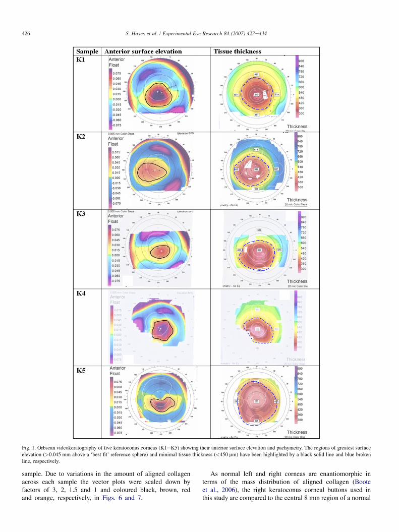
Prior to surgery, a videokeratography image of all fivekeratoconus corneas was recorded using the Orbscan II� im-aging system (Bausch and Lomb, London, UK) to producetopography maps of anterior surface elevation and cornealthickness. The regions of greatest anterior surface elevation(>0.045 mm above a ‘best fit’ reference sphere) and maximaltissue thinning (defined as a pachymetry reading of<450 mm) are highlighted by a black solid line and a bluebroken line, respectively, in Fig. 1. For the purposes of thisstudy the combined regions of greatest anterior surface eleva-tion and maximal tissue thinning will be referred to as the‘apical region’.
2.3. X-ray scattering e data collection
All X-ray scattering data were collected on Station 14.1 atthe UK Synchrotron Radiation Source (Daresbury Laboratory,Warrington, UK), using an X-ray beam focused to 200�200 mm at the specimen with a wavelength of 0.1488 nm.The samples were wrapped tightly in polyvinylidene chloridecatering film to prevent tissue dehydration during data collec-tion and placed in their correct orientation in a sealed sampleholder enclosed between two sheets of Mylar (DuPont TeijinFilms�, London, UK). The sample holder was then carefullysecured onto a computer-operated translation stage (NewportSpectra-Physics Ltd., Newbury, UK) with the anterior surfaceof the cornea facing the X-ray beam. A series of wide-angle X-ray scattering images resulting from a beam exposure ofbetween 45 and 75 s were collected over the entire sampleat regular intervals of 0.4e0.5 mm in the case of the normalcorneas, and 0.25 mm for each of the keratoconus buttons.The exposure time remained constant for individual samplesbut varied between samples due to differences in the beamintensity at the time of data collection. The resulting X-rayscattering images were recorded on a Quantum 4R CCDdetector (ADSC, Poway, CA) placed 15 cm behind the cornea.The X-ray scattering patterns were calibrated using the0.304 nm X-ray reflection from powdered calcite.
2.4. X-ray scattering e data analysis
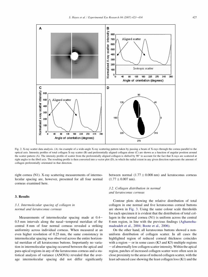
The angular distribution of scatter intensity around thecollagen intermolecular reflection (Fig. 2A) was measuredto form a 0e360� distribution pattern using Optimus 6.5
(Media Cybernetics, Wokingham, UK) image analysis soft-ware and Excel (Microsoft, UK) spreadsheets (Fig. 2B). Fur-ther details of this analysis procedure are provided byNewton and Meek (1998). To take into account the variationsin beam intensity and exposure time between samples, theintensity profile for each image was normalised against theaverage X-ray intensity at the time of data collection (re-corded by an ion chamber) multiplied by the beam exposuretime. The distance from the centre of the scattering pattern tothe intermolecular reflection was measured and calibratedagainst the known spacing of calcite to obtain the intermo-lecular Bragg spacing of collagen. Based on the assumptionthat collagen molecules are packed in a pseudo-hexagonallattice, the Bragg spacing was multiplied by a factor of1.11 to give the mean intermolecular centre-to-centre spacing(Klug and Alexander, 1974).
The total area under the intensity profile (i.e. the total X-rayscatter) is proportional to the total amount of fibrillar collagenin the path of the X-ray beam e the more collagen, the greaterthe intensity of X-ray scatter (Newton and Meek, 1998). Thetotal X-ray scatter is comprised of two components: the scatterarising from collagen fibrils that lie equally in all directionswithin the plane of the cornea (isotropic scatter) and the scatterfrom collagen fibrils that adopt a preferred orientation (alignedscatter) (Fig. 2B). Removal of the isotropic scatter leaves onlythe scatter from preferentially aligned lamellae, which will bereferred to throughout as ‘aligned collagen’ (Fig. 2C).
The area under each intensity profile (Fig. 2B, C) wassummed to produce a numeric value of scatter intensity forboth total collagen and aligned collagen. The ratio of the scat-ter intensity from aligned collagen as a proportion of the totalcollagen scatter intensity was defined as the ‘index of orienta-tion’. In this scheme a value of 1 indicates that all of the col-lagen present is preferentially aligned and a value of 0 meansthat the collagen is distributed equally in all directions withinthe plane of the cornea. By plotting each calculated intensityvalue/index on a grid relating to corneal position, contourmaps showing the distribution of total collagen scatter (andhence relative fibrillar mass) and the indices of orientationwere produced. By presenting the distribution of aligned col-lagen as an index of orientation, the reduced stromal thicknessin keratoconus corneas is taken into account and so the distri-bution of aligned collagen can be directly compared betweennormal and keratoconus corneas.
In order to show the preferred orientation of alignedcollagen within a sample, each profile of aligned collagenscatter was shifted by 90� to account for the fact that X-rays are scattered at right angles to the orientation of thecollagen molecules. The resulting scatter intensity profilewas then transformed into polar co-ordinates to producevector plots (Fig. 2D) (Connon and Meek, 2003). Thedistance from the origin of a vector plot to the edge, atany given angle, represents the intensity of scatter fromaligned collagen lying in a particular direction. The individ-ual vector plots were compiled onto a grid relating tocorneal position to form a ‘vector plot map’, showing thepreferred orientation of aligned collagen over the entire
426 S. Hayes et al. / Experimental Eye Research 84 (2007) 423e434
Fig. 1. Orbscan videokeratography of five keratoconus corneas (K1eK5) showing their anterior surface elevation and pachymetry. The regions of greatest surface
elevation (>0.045 mm above a ‘best fit’ reference sphere) and minimal tissue thickness (<450 mm) have been highlighted by a black solid line and blue broken
line, respectively.
sample. Due to variations in the amount of aligned collagenacross each sample the vector plots were scaled down byfactors of 3, 2, 1.5 and 1 and coloured black, brown, redand orange, respectively, in Figs. 6 and 7.
As normal left and right corneas are enantiomorphic interms of the mass distribution of aligned collagen (Booteet al., 2006), the right keratoconus corneal buttons used inthis study are compared to the central 8 mm region of a normal
427S. Hayes et al. / Experimental Eye Research 84 (2007) 423e434
Fig. 2. X-ray scatter data analysis. (A) An example of a wide-angle X-ray scattering pattern taken by passing a beam of X-rays through the cornea parallel to the
optical axis. Intensity profiles of total collagen X-ray scatter (B) and preferentially aligned collagen alone (C) are shown as a function of angular position around
the scatter pattern (A). The intensity profile of scatter from the preferentially aligned collagen is shifted by 90� to account for the fact that X-rays are scattered at
right angles to the fibril axis. The resulting profile is then converted into a vector plot (D), in which the radial extent in any given direction represents the amount of
collagen preferentially orientated in that direction.
right cornea (N1). X-ray scattering measurements of intermo-lecular spacing are, however, presented for all four normalcorneas examined here.
3. Results
3.1. Intermolecular spacing of collagen innormal and keratoconus corneas
Measurements of intermolecular spacing made at 0.4e0.5 mm intervals along the nasaletemporal meridian of thecentral 8 mm of four normal corneas revealed a strikinguniformity across individual corneas. When measured at aneven higher resolution of 0.25 mm, the same consistency inintermolecular spacing was observed across the entire horizon-tal meridian of all keratoconus buttons. Importantly no varia-tion in intermolecular spacing occurred between the apical andpara-apical regions in any of the keratoconus corneas and a sta-tistical analysis of variance (ANOVA) revealed that the aver-age intermolecular spacing did not differ significantly
between normal (1.77� 0.008 nm) and keratoconus corneas(1.77� 0.007 nm).
3.2. Collagen distribution in normaland keratoconus corneas
Contour plots showing the relative distribution of totalcollagen in one normal and five keratoconus corneal buttonsare shown in Fig. 3. Using the same colour scale thresholdsfor each specimen it is evident that the distribution of total col-lagen in the normal cornea (N1) is uniform across the central8 mm region, in line with the previous findings (Aghamoha-madzadeh et al., 2004; Boote et al., 2006).
On the other hand, all keratoconus buttons showed a non-uniform distribution of collagen scatter. In all cases thehighlighted region of reduced corneal thickness coincideswith a region e or in some cases (K3 and K5) multiple regionse of abnormally low collagen scatter intensity. Within the apicalregion, patches of increased collagen scatter were often seen inclose proximity to the areas of reduced collagen scatter, with theleast advanced case showing the least collagen loss (K1) and the

428 S. Hayes et al. / Experimental Eye Research 84 (2007) 423e434
Fig. 3. Total collagen distribution. Contour maps showing the relative distribution of total collagen in a normal cornea with scleral rim (N1a) and in the central
7.5e8.5 mm of the same cornea and five keratoconus corneas (N1b, K1eK5). Green line in N1a denotes central 8 mm region - shown enlarged as N1b. Regions of
greatest anterior surface elevation (black solid line) and tissue thinning (blue broken line) are highlighted.
most severe keratoconus (K5) associated with the most exten-sive loss. There was, however, variability in collagen distribu-tion between these clinical extremes, so it is likely thata severe phenotype must be reached before the link between dis-ease severity and collagen mass redistribution is manifest.
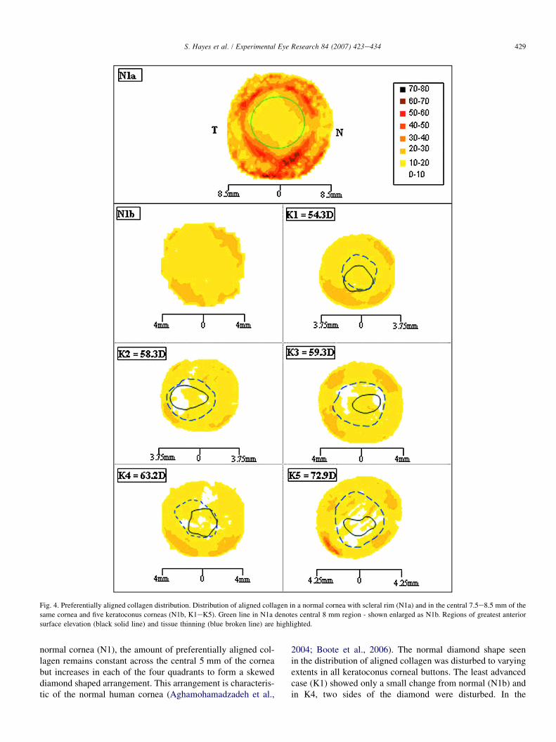
3.3. Preferentially aligned collagen distribution innormal and keratoconus corneas
Fig. 4 shows the distribution of aligned collagen scatter inthe normal and keratoconus corneas depicted in Fig. 3. In the
429S. Hayes et al. / Experimental Eye Research 84 (2007) 423e434
Fig. 4. Preferentially aligned collagen distribution. Distribution of aligned collagen in a normal cornea with scleral rim (N1a) and in the central 7.5e8.5 mm of the
same cornea and five keratoconus corneas (N1b, K1eK5). Green line in N1a denotes central 8 mm region - shown enlarged as N1b. Regions of greatest anterior
surface elevation (black solid line) and tissue thinning (blue broken line) are highlighted.
normal cornea (N1), the amount of preferentially aligned col-lagen remains constant across the central 5 mm of the corneabut increases in each of the four quadrants to form a skeweddiamond shaped arrangement. This arrangement is characteris-tic of the normal human cornea (Aghamohamadzadeh et al.,
2004; Boote et al., 2006). The normal diamond shape seenin the distribution of aligned collagen was disturbed to varyingextents in all keratoconus corneal buttons. The least advancedcase (K1) showed only a small change from normal (N1b) andin K4, two sides of the diamond were disturbed. In the

430 S. Hayes et al. / Experimental Eye Research 84 (2007) 423e434
Fig. 5. Index of orientation in a normal cornea with scleral rim (N1a) and in the central 7.5e8.5 mm of the same cornea and five keratoconus corneas (N1b, K1e
K5). Green line in N1a denotes central 8 mm region - shown enlarged as N1b. Regions of greatest anterior surface elevation (black solid line) and tissue thinning
(blue broken line) are highlighted.
remaining three keratoconus buttons (K2, K3 and K5), all fourquadrants showed abnormalities in the distribution of alignedcollagen scatter.
With the exception of K1, a reduction in aligned collagenscatter was apparent in the apical region of each keratoconuscornea. Although a general trend indicated a relationship
between the severity of the disease and the reduction inaligned collagen scatter (with the least advanced keratoconusshowing no change in aligned collagen scatter in the centralcornea and the most severe case (K5) displaying a large regionof reduced aligned collagen scatter), marked variations wereobserved between specimens. For example, in keratoconus

431S. Hayes et al. / Experimental Eye Research 84 (2007) 423e434
cornea K4 the region of reduced aligned collagen scatter ex-tended beyond the apical region, and in another cornea (K3)two distinct regions of reduced aligned collagen scatter wereseen close to the edge of the apical region.
When the distribution of aligned collagen was plotted as theindex of orientation (Fig. 5), it was found that in the normalcornea (N1a) the index of orientation increased in all fourquadrants of its periphery, with the greatest increase occurringin the superiorenasal and inferioretemporal quadrants.Within the central 8 mm region, this increase in the index oforientation can still be clearly seen in the superiorenasaland inferioretemporal quadrants of the normal cornea(N1b). In three out of five of the keratoconus buttons exam-ined (K2, K4 and K5) the index of orientation failed toincrease in one or more of the quadrants, and an abnormal in-crease in the index of orientation was observed close to theapex of the cone. In all but the least advanced case of kerato-conus, patches of abnormally low index of orientation (visibleas white areas in Fig. 5) were observed both inside and outsidethe apical region.
3.4. Collagen orientation in normaland keratoconus corneas
In the central 6 mm of the normal cornea (N1) the preferredalignment of collagen fibrils is in the superioreinferior and na-saletemporal meridians (Fig. 6). This gives rise to the charac-teristic cross-shaped vector plots seen in previous X-rayscattering studies of normal human corneas (Aghamohamad-zadeh et al., 2004; Boote et al., 2006). Beyond the central6 mm region, the preferred orthogonal orientation is gradually
Fig. 6. Preferred fibril orientation in the normal cornea. Preferred fibril direc-
tion in the central 8 mm of a normal cornea (N1). Due to variations in the
amount of aligned collagen across the cornea it was necessary to scale
down the larger vector plots by a factor of 1.5 (red).
obscured by the presence of additional collagen aligned tan-gentially to the edge of the cornea.
The normal preferred orientation of collagen was disturbedto a variable extent in each of the keratoconus buttons but inall cases the extent of disruption decreased with distancefrom the apex (Fig. 7). In the least advanced case (K1) thepreferred orientation of collagen was normal throughoutmost of the 7.5 mm button. In contrast, the most severe case(K5) showed an abnormal orientation of collagen throughoutthe entire 8.5 mm button region. The remaining corneas(K2eK4) all exhibited extreme changes in collagen orienta-tion that extended well beyond the apical region, however,a precise relationship between disease severity (in terms ofdioptric power) and the extent of the structural alterationswas not apparent.
4. Discussion
In this study, a combined approach of videokeratographyand X-ray scattering was used to investigate the link betweenchanges in corneal shape and thickness, and alterationsin stromal ultrastructure in keratoconus, to improve under-standing of the mechanism by which ectasia occurs andprogresses.
X-ray scattering is a unique method to measure the lateralspacing between individual fibril-forming collagen moleculesto less than 1 nm resolution. This spacing is influenced byboth the hydration of the fibrils (Fratzl and Daxer, 1993;Meek et al., 1991) and by the degree of molecular cross-link-ing (Malik et al., 1992). Any changes in this spacing withindifferent parts of the keratoconus cornea could cause alter-ations in the regional biomechanical properties of the tissue.We have demonstrated here, however, that within any givenkeratoconus cornea, even the most severe case, there wereno regional differences in this spacing, indicating that the col-lagen arrangement within fibrils does not vary between theapex and the less affected peripheral region. Fullwood et al.(1992) showed that at the optical centre of the keratoconuscornea there is a small decrease in the intermolecular spacingcompared to the normal cornea. We were unable to confirmthis result. It is worth noting that the proportional relationshipassumed to exist between the intensity of X-ray scatter and theamount of fibrillar collagen in the path of the beam is based onthe assumption that the hydration of the collagen fibrils doesnot vary across the cornea. In this study, the consistency inintermolecular spacing across the central 8 mm region of nor-mal and keratoconus corneas supports this assumption. It alsosuggests that there are no detectable regional variations incollagen cross-linking that may cause some parts of the kera-toconic cornea to be weaker than others.
We have shown that the corneal ectasia and thinningassociated with keratoconus are accompanied by changes inthe relative distribution and orientation of collagen withinthe corneal stroma. Although such stromal abnormalities inkeratoconus corneas have previously been shown by X-rayscattering and scanning electron microscopy (Daxer andFratzl, 1997; Meek et al., 2005; Radner et al., 1998b;
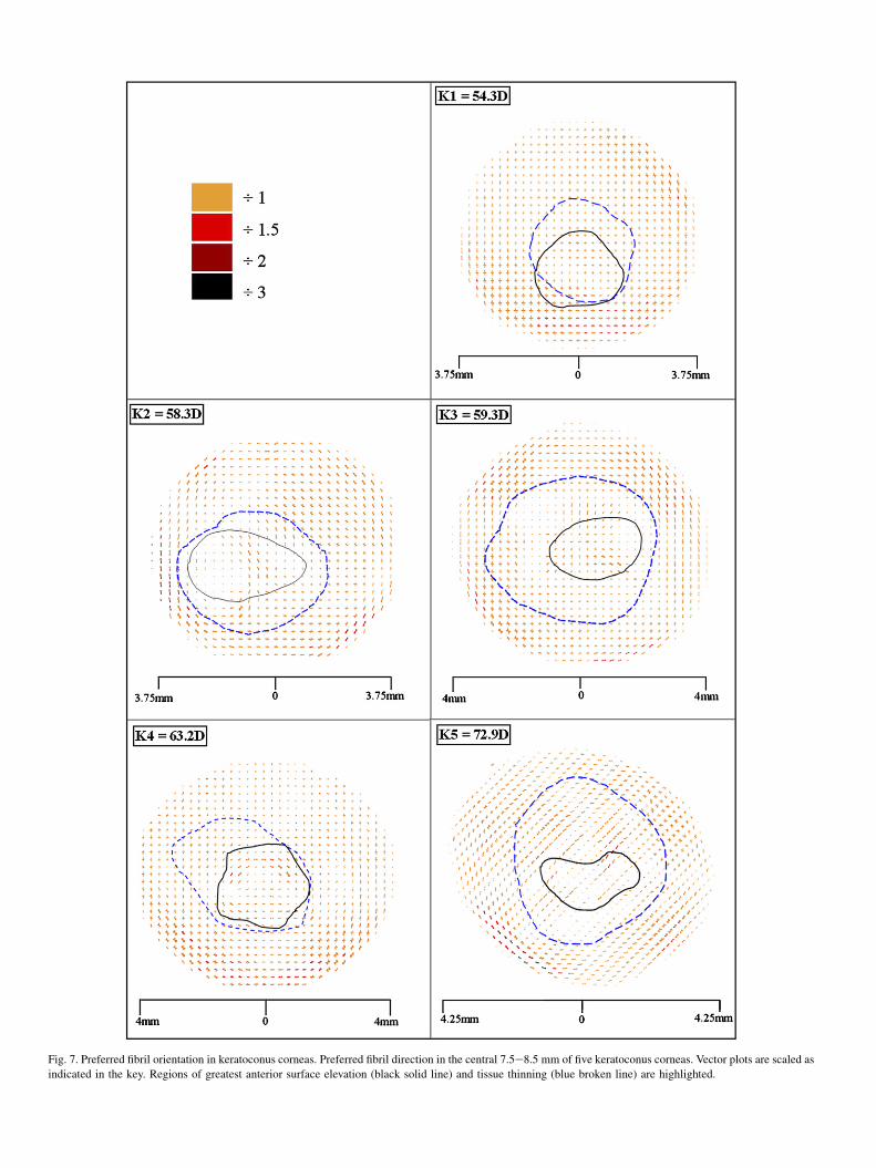
Fig. 7. Preferred fibril orientation in keratoconus corneas. Preferred fibril direction in the central 7.5e8.5 mm of five keratoconus corneas. Vector plots are scaled as
indicated in the key. Regions of greatest anterior surface elevation (black solid line) and tissue thinning (blue broken line) are highlighted.

433S. Hayes et al. / Experimental Eye Research 84 (2007) 423e434
Sawaguchi et al., 1998) this study goes further, to examine forthe first time the relationship between the altered arrangementand distribution of collagen and the specific shape and thick-ness of the diseased cornea. In normal cornea the total colla-gen scatter is essentially constant across the central 8 mm,while, in keratoconus, an uneven distribution of collagen isinvariably found. As would be expected, the region of greatesttissue thinning coincided with a decrease in total collagenscatter. However, the present study shows that in some casesthe structural changes are far more complex than the videoker-atography might suggest. For example, in one keratoconusbutton, the videokeratography showed a large region of re-duced tissue thickness, whereas X-ray scattering data revealedthat this region was actually comprised of two distinct regionsof stromal thinning (K3 in Figs. 3 and 4). The pre-set step sizeassigned to the colour scale of the Orbscan topography systemmay be responsible for its failure to detect such subtle localchanges in pachymetry.
Previously it was found that stromal thinning in keratoco-nus was not accompanied by a compaction of collagen fibrils(Fullwood et al., 1992), as is the case in macular corneal dys-trophy, another corneal disease in which the corneal stromathins (Quantock et al., 1990). It was reasoned, therefore, thatstromal thinning in keratoconus is most likely due to a lossof fibrils from the central cornea. This is verified by the currentfindings. However, the plots in Fig. 3 also imply that someparts of the keratoconus buttons show increased scatter. TheX-ray maps are two-dimensional projections of the curved cor-nea and, as such, increased corneal steepness will lead to anincrease in the fibrillar mass sampled. Our previous calcula-tions (Meek et al., 2005) suggest that this effect is small, sothe possibility cannot be excluded that regions of increasedscatter are those to which mass has been redistributed, perhapsby a process of lamella slippage (Meek et al., 2005; Polack,1976). Unfortunately, it is not easy to correlate these regionswith Orbscan thickness data for the reasons outlined above.
When the distribution of aligned lamellae is presented as anindex of orientation (Fig. 5), normal corneas show highly re-producible increases in the orientation in three quadrants esuperiorenasal, inferioretemporal and inferiorenasal (Booteet al., 2006). The least advanced cornea (K1) showed thesame pattern, but the other keratoconus buttons did not. Achange in the local index of orientation may be explained ei-ther by a preferential loss of isotropic or aligned collagen dur-ing stromal thinning or by a redistribution of collagen mass.The latter is more likely since abnormalities in the distributionof collagen are also accompanied by changes in the preferredorientation of lamellae (Fig. 7).
It has been shown in this paper and elsewhere (Aghamoha-madzadeh et al., 2004; Boote et al., 2006) that the increase inaligned collagen mass in the periphery of the normal corneaforms a highly reproducible skewed diamond shaped arrange-ment. This specific arrangement is thought to be due to thepresence of anchoring fibrils that traverse the peripheralcornea and contribute to the flattening of the cornea insidethe limbus (Aghamohamadzadeh et al., 2004). Abnormalitiesin the distribution of aligned collagen were observed in the
peripheral region of all keratoconus corneas with the possibleexception of the least advanced (K1) (Fig. 4), although theextent of the disruption appeared to be independent of the dis-ease severity and the size and shape of the cone. The absenceof the skewed diamond shape arrangement of aligned collagenin keratoconus suggests that the anchoring fibrils are not pres-ent or that they occur at a distance beyond the central 8 mmregion under investigation.
We have shown that in less advanced keratoconus, cornealectasia can be present with little loss of tissue mass and onlyminor alterations in collagen lamellar orientation. As thedisease progresses, there is a variability between keratoconuscorneas in terms of the relationship between the severity ofthe disease and the extent of the structural alterations. Irre-spective of the initial pathogenesis, which probably involvesan up-regulation of degradative enzymes (Kenney et al.,1994; Kenney and Brown, 2003; Sawaguchi et al., 1989;Zhou et al., 1998) leading to some loss of collagen (Yueet al., 1984), the findings presented in this paper supporta mechanism of tissue thinning in keratoconus corneas thatalso involves lamellar or fibrillar slippage. This idea is sup-ported by the observation of abnormal collageneproteoglycanarrangement in keratoconus corneas (Fullwood et al., 1990,1992). It is also supported by the observation of less lamellarinterweaving, a factor which may influence the biomechanicalproperties of the tissue and facilitate such a mechanism ofstromal thinning (Radner et al., 1998b). As a consequencewe propose that the development of interventional cross-link-ing strategies that not only protect the collagen from degrada-tion but that may also limit collagen fibrillar slippage(Wollensak et al., 2003), should be beneficial to delay or retardthe progression of keratoconus.
Acknowledgements
The authors wish to thank Dr. Mike MacDonald and the staffat the UK Synchrotron X-ray Source (Daresbury, UK) for helpwith data collection. We also thank Tom Kelly (ManchesterEye Hospital, UK) and Val Smith (UK Corneal Transplant Ser-vice Eye Bank, Bristol, UK) for provision of specimens. Thisstudy received programme grant support from the MedicalResearch Council (Grant G0001033) and the Council for theCentral Laboratory of Research Councils (Grant 18).
References
Aghamohamadzadeh, H., Newton, R.H., Meek, K.M., 2004. X-ray scattering
used to map the preferred collagen orientation in the human cornea and
limbus. Structure 12, 249e256.
Andreassen, T., Simonsen, A., Oxlund, H., 1980. Biomechanical properties of
keratoconus and normal cornea. Experimental Eye Research 31, 435e441.
Benedek, G.B., 1971. Theory of transparency of the eye. Applied Optics 10,
459e472.
Boote, C., Hayes, S., Abahussin, M., Meek, K., 2006. Mapping collagen orga-
nisation in the human cornea: left and right eyes are structurally distinct.
Investigative Ophthalmology and Visual Science 47, 901e908.
Borcherding, M.S., Balccik, L.J., Sittig, R.A., Bizzel, J.W., Breen, M.,
Weinstein, H.G., 1975. Proteoglycans and collagen fibre organisation in
human corneoscleral tissue. Experimental Eye Research 21, 59e70.

434 S. Hayes et al. / Experimental Eye Research 84 (2007) 423e434
Brookes, N.H., Loh, I.P., Clover, G.M., Poole, C.A., Sherwin, T., 2003.
Involvement of corneal nerves in the progression of keratoconus. Experi-
mental Eye Research 77, 515e524.
Buxton, J.N., Keates, R.H., Hoefle, F.B., 1984. The contact lens correction of ker-
atoconus. In: Dabezies, O.H.J. (Ed.), Contact lenses: The CLAO Guide to Ba-
sic Science and Clinical Practice. Grune and Stratton, Orlando, p. 55.5.
Chakravarti, S., Magnuson, T., Lass, J.H., Jespen, K.L., LaMantia, C.,
Carroll, H., 1998. Lumican regulates collagen fibril assembly: skin fragil-
ity and corneal opacity in the absence of lumican. The Journal of Cell
Biology 141, 1277e1286.
Connon, C.J., Meek, K., 2003. Organization of corneal collagen fibrils during
the healing of trephined wounds in rabbits. Wound Repair and Regenera-
tion 11, 71e78.
Daxer, A., Fratzl, P., 1997. Collagen fibril orientation in the human corneal
stroma and its implications in keratoconus. Investigative Ophthalmology
and Visual Science 38, 121e129.
Fini, M.E., Yue, B.Y., Sugar, J., 1992. Collagenolytic/gelatinolytic metallopro-
teinases in normal and keratoconus corneas. Current Eye Research 11,
849e862.
Fratzl, P., Daxer, A., 1993. Structural transformation of collagen fibrils in the
corneal stroma during drying: an X-ray scattering study. Biophysical
Journal 64, 1210e1214.
Fullwood, N.J., Meek, K.M., 1994. An ultrastructural, time-resolved study
of freezing in the corneal stroma. Journal of Molecular Biology 236,
749e758.
Fullwood, N.J., Meek, K.M., Malik, N.S., Tuft, S.J., 1990. A comparison of
proteoglycan arrangement in normal and keratoconus human corneas. Bio-
chemical Society Transactions 18, 961e962.
Fullwood, N.J., Tuft, S.J., Malik, N.S., Meek, K.M., Ridgway, A.E.A.,
Harrison, R.J., 1992. Synchrotron X-ray diffraction studies of keratoconus
corneal stroma. Investigative Ophthalmology and Visual Science 33,
1734e1741.
Funderburgh, J.L., Panjwani, N., Conrad, G.W., Baum, J., 1989. Altered
keratan sulphate epitopes in keratoconus. Investigative Ophthalmology
and Visual Science 30, 2278e2281.
Kao, W.W.Y., Liu, C.Y., 2003. Roles of lumican and keratocan on corneal
transparency. Glycoconjugate Journal 19, 275e285.
Kenney, M., Chwa, M., Escobar, M., Brown, D., 1989. Altered gelatinolytic
activity by keratoconus corneal cells. Biochemical and Biophysical
Research Communications 161, 353e357.
Kenney, M., Chwa, M., Opbroek, A.J., Brown, D.J., 1994. Increased gelatino-
lytic activity in keratoconus cultures. A correlation to an altered matrix
metalloproteinase-2/tissue inhibitor of metalloproteinase ratio. Cornea
13, 114e124.
Kenney, M.C., Brown, D.J., 2003. The cascade hypothesis of keratoconus.
Contact Lens and Anterior Eye 26, 139e146.
Kim, W.J., Rabinowitz, Y.S., Meisler, D.M., Wilson, S.E., 1999. Keratocyte
apoptosis associated with keratoconus. Experimental Eye Research 69,
475e481.
Klintworth, G.K., 1994. Degenerations, depositions and miscellaneous reac-
tions of the ocular anterior segment. In: Klintworth, G.K., Garner, A.
(Eds.), Pathobiology of Ocular Disease: A Dynamic Approach, second
ed. Marcel Dekker, New York.
Klug, H., Alexander, L.E., 1974. X-ray Diffraction Procedures for Crystalline
and Amorphous Materials. New York.
Komai, Y., Ushiki, T., 1991. The three-dimensional organisation of collagen
fibrils in the human cornea and sclera. Investigative Ophthalmology and
Visual Science 32, 2244e2258.
Malik, N.S., Moss, S.J., Ahmed, N., Furth, A.J., Wall, R.S., Meek, K.M., 1992.
Aging of the human corneal stroma e structural and biochemical changes.
Biochimica Et Biophysica Acta 1138, 222e228.
Maurice, D.M., 1957. The structure and transparency of the cornea. Journal of
Physiology 136, 263e286.
Meek, K., Blamires, T., Elliot, G., Gyi, T.J., Nave, C., 1987. The organisation
of collagen fibrils in the human corneal stroma: a synchrotron X-ray dif-
fraction study. Current Eye Research 6, 841e846.
Meek, K.M., Fullwood, N.J., Cooke, P.H., Elliott, G.F., Maurice, D.M.,
Quantock, A.J., Wall, R.S., Worthington, C.R., 1991. Synchrotron X-ray-
diffraction studies of the cornea, with implications for stromal hydration.
Biophysical Journal 60, 467e474.
Meek, K.M., Leonard, D.W., Connon, C.J., Dennis, S., Khan, S., 2003. Trans-
parency, swelling and scarring in the corneal stroma. Eye 17, 927e936.
Meek, K.M., Tuft, S.J., Huang, Y., Gill, P.S., Hayes, S., Newton, R.H.,
Bron, A.J., 2005. Changes in collagen orientation and distribution in
keratoconus corneas. Investigative Ophthalmology and Visual Science
46, 1948e1956.
Muller, L.J., Pels, E., Vrensen, G.F.J.M., 2001. The specific architecture of the
anterior stroma accounts for maintenance of corneal curvature. British
Journal of Ophthalmology 85, 437e443.
Nash, I., Greene, P., Foster, S., 1982. Comparison of mechanical properties of
keratoconus and normal cornea. Experimental Eye Research 35, 413e423.
Newton, R.H., Meek, K.M., 1998. Circumcorneal annulus of collagen fibrils in
the human limbus. Investigative Ophthalmology and Visual Science 39,
1125e1134.
Polack, F.M., 1976. Contributions of electron microscopy to the study of
corneal pathology. Survey of Ophthalmology 20, 375e414.
Quantock, A.J., Meek, K.M., Chakravarti, S., 2001. An X-ray diffraction
investigation of corneal structure in lumican-deficient mice. Investigative
Ophthalmology and Visual Science 42, 1750e1756.
Quantock, A.J., Meek, K.M., Ridgway, A.E.A., Bron, A.J., Thonar, E.J.M.A.,
1990. Macular corneal dystrophy e reduction in both corneal thickness
and collagen interfibrillar spacing. Current Eye Research 9, 393e398.
Radner, W., Mallinger, R., 2002. Interlacing of collagen lamellae in the
midstroma of the human cornea. Cornea 21, 598e601.
Radner, W., Zehetmayer, M., Aufreiter, R., Mallinger, R., 1998a. Interlacing
and cross-angle distribution of collagen lamellae in the human cornea.
Cornea 17, 537e543.
Radner, W., Zehetmayer, M., Skorpik, C., Mallinger, R., 1998b. Altered
organization of collagen in the apex of keratoconus corneas. Ophthalmic
Research 30, 327e332.
Rehany, U., Lahav, M., Shoshan, S., 1982. Collagenolytic activity in keratoco-
nus. Annals of Ophthalmology 14, 751e754.
Sawaguchi, S., Fukuchi, T., Abe, H., Kaiya, T., Sugar, J., Yue, B.Y., 1998.
Three dimensional electron microscopic study of keratoconus. Archives
of Ophthalmology 116, 62e98.
Sawaguchi, S., Twining, S.S., Yue, B.Y., Wilson, P.M., Sugar, J., Chan, S.K.,
1990. Alpha-1 proteinase inhibitor levels in keratoconus. Experimental
Eye Research 50, 549e554.
Sawaguchi, S., Twining, S.S., Yue, B.Y., Chang, S.H., Zhou, X., Loushin, G.,
Sugar, J., Feder, R.S., 1994. Alpha-2-Macro-globulin levels in normal and
keratoconus corneas. Investigative Ophthalmology and Visual Science 35,
4008e4014.
Sawaguchi, S., Yue, B.Y., Sugar, J., Gilboy, J.E., 1989. Lysosomal enzyme
abnormalities in keratoconus. Archives of Ophthalmology 107, 1507e
1510.
Sherwin, T., Brookes, N.H., Loh, I.P., Poole, C.A., Clover, G.M., 2002. Cellu-
lar incursion into Bowman’s membrane in the peripheral cone of the
keratoconic cornea. Experimental Eye Research 74, 473e482.
Smolek, M.K., McCarey, B.E., 1990. Interlamellar adhesive strength in human
eyebank corneas. Investigative Ophthalmology and Visual Science 31,
1087e1095.
Somodi, S., Hahnel, C., Slowik, C., Richter, A., Weiss, D.G., Guthoff, R.F.,
1996. Confocal in vivo microscopy and confocal laser-scanning fluores-
cence microscopy in keratoconus. German Journal of Ophthalmology 5,
518e525.
Teng, C.C., 1963. Electron microscope study of the pathology of keratoconus:
part 1. American Journal of Ophthalmology 55, 18e47.
Wollensak, G., Sporl, E., Seiler, T., 2003. Behandlung von Keratokonus durch
kollagenvernetzung. Ophthalmologe 100, 44e49.
Yue, B.Y., Sugar, J., Benveniste, K., 1984. Heterogenity in keratoconus:
possible biochemical basis. Proceedings of the Society for Experimental
Biology and Medicine 175, 336e341.
Zhou, L.L., Sawaguchi, S., Twining, S.S., Sugar, J., Feder, R.S., Yue, B.Y.,
1998. Expression of degradative enzymes and protease inhibitors in
corneas with keratoconus. Investigative Ophthalmology and Visual
Science 39, 1117e1124.