a sarcomeric akap, tethers protein kinase a at the
TRANSCRIPT
1
CARDIAC TROPONIN T: A SARCOMERIC AKAP, TETHERS PROTEIN KINASE A AT THE MYOFILAMENTS
C. Amelia Sumandea, Mary L. Garcia-Cazarin, Catherine H. Bozio, Gail A. Sievert, C. William Balke, Marius P. Sumandea
From the Department of Physiology, Center for Muscle Biology, University of Kentucky, Lexington, KY 40536
Running title: Characterization of cTnT as a sarcomeric AKAP
*Address correspondence to: Marius P. Sumandea, Ph.D., 426 Sanders Brown Building, 800 South Limestone Street, Lexington, KY 40536. Fax: 859-257-3235; E-mail: [email protected] Efficient and specific phosphorylation of PKA substrates, elicited in response to beta-adrenergic stimulation, require spatially confined pools of PKA anchored in proximity of its substrates. PKA-dependent phosphorylation of cardiac sarcomeric proteins has been the subject of intense investigations. Yet, the identity, composition, and function of PKA complexes at the sarcomeres have remained elusive. Here we report the identification and characterization of a novel sarcomeric AKAP (A kinase anchoring protein), cardiac troponin T (cTnT). Using the yeast two-hybrid technology in screening two adult human heart cDNA libraries, we identified the regulatory subunit of PKA as interacting with human cTnT bait. Immunoprecipitation studies show that cTnT is a dual specificity AKAP, interacting with both PKA-regulatory subunits type I and II. The disruptor peptide Ht31, but not Ht31P (control), abolished cTnT/PKA-R association. Truncations and point mutations identified an amphipathic helix domain in cTnT as the PKA binding site. This was confirmed by a peptide SPOT assay in the presence of Ht31 or Ht31P (control). Gelsolin-dependent removal of thin filament proteins also reduced myofilament-bound PKA-type II. Using a cardiac troponin (cTn) exchange procedure that substitutes the endogenous cTn complex with a recombinant cTn complex we show PKA-type II is troponin-bound in the myofilament lattice. Displacement of PKA-cTnT complexes correlates with a
significant decrease in myofibrillar PKA activity. Taken together, our data propose a novel role for cTnT as a dual-specificity sarcomeric AKAP.
Activation of β-adrenergic (β-AR) receptors in the heart is a major mechanism responsible for enhancing myocardial contractility. β-AR receptor stimulation is quickly followed by a sharp rise in cyclic AMP levels evoking a plethora of physiological and biochemical reactions, such as positive chronotropic, inotropic or lusitropic effects (1,2). The effector arm of cAMP signaling is PKA, which translates the extracellular stimuli into timely and specific responses.
PKA is a heterotetramer formed of two catalytic subunits kept in an inactive conformation by two regulatory subunits (either type I or II)(3). Another function of the regulatory (R) subunits is to facilitate tethering of the holoenzyme in the vicinity of relevant substrates thereby ensuring the fidelity of the cAMP signaling (4,5). cAMP binds cooperatively to two sites termed A and B, located at the interface between a regulatory and catalytic subunit, releasing catalytic subunit, which become catalytically active.
Cardiomyocyte contraction-relaxation cycles are regulated by fluctuating intracellular Ca2+ levels (6,7). Contraction is initiated by the opening of voltage sensitive L-type Ca2+ channels (LTCC) as a result of sarcolemmal depolarization. This causes a small influx of Ca2+ that activate ryanodine receptors (RyR) promoting a massive release of Ca2+ from the sarcoplasmic reticulum (SR) stores into the cytosol (8). The rise of intracellular Ca2+ levels is sensed by troponin
http://www.jbc.org/cgi/doi/10.1074/jbc.M110.148684The latest version is at JBC Papers in Press. Published on November 5, 2010 as Manuscript M110.148684
Copyright 2010 by The American Society for Biochemistry and Molecular Biology, Inc.
by guest on April 5, 2018
http://ww
w.jbc.org/
Dow
nloaded from

2
C (cTnC), which upon Ca2+ binding undergoes a conformational change inducing dissociation of troponin I (cTnI) from actin. This movement is transmitted through cTnT to tropomyosin (Tm) exposing myosin binding sites on actin and promoting contraction. Relaxation occurs when Ca2+ is pumped out of the cytosol by SERCA (sarcoplasmic reticulum calcium ATPase) or by Na+/Ca2+ exchanger.
PKA regulates the strength and frequency of contraction through phosphorylation of key proteins, such as LTCC, RYR, phospholamban, cTnI, myosin binding protein C (MyBP-C), titin (9). Considering the extent and diversity of PKA targets within the cardiomyocyte, a tightly regulated and spatially segregated activity of the cAMP/PKA pathways is critical for the specificity of the response to the inciting stimulus (10,11). Both, speed and precision are achieved through: i) targeting and pre-assembly of PKA signaling components in clusters, at discrete subcellular locations, through the help of A-kinase anchoring proteins (12,13); and ii) generation of local gradients of cAMP through spatially confined, phosphodiesterases (PDE) (14). PDEs, by degrading cAMP, regulate the amplitude, duration, and compartmentation of intracellular cyclic nucleotide signaling (10). The ingeniously assembled macromolecular complexes, containing AKAP, PKA, and other factors, confer efficient and specific transduction of cAMP signal.
Growing data over the last decade, have shed new light on the role of AKAP/PKA complexes bound to sarcoplasmic reticulum (regulating SERCA and RyR), t-tubules (regulating LTCC) and nuclear envelope (15). Despite the fact that cardiac myofilaments make-up > 50% of cellular volume (16) and that PKA-dependent phosphorylation of sarcomeric proteins cTnI, MyBP-C, titin appear to be dominant in control of myofilament function by β-AR stimulation (9), very little is known about the identity, composition, or function of AKAP/PKA complexes at the sarcomeres.
Here we report the identification and characterization of a novel AKAP, cardiac
TnT, which docks PKA at the thin filaments in proximity of its main sarcomeric substrates.
EXPERIMENTAL PROCEDURES
Yeast two-hybrid screening of human heart cDNA libraries. Human heart cDNA libraries (cat # 638865 and HL4042AH) were from Clontech. For library 1 screen, MATα yeast strain Y187, pretransformed with the cDNA library in pGADT7 vector, was mated with the MATa yeast strain AH109 containing human cTnT subcloned into pGBKT7 vector, at the NdeI and BamHI restriction endonuclease sites (see oligonucleotide Table 1). For library 2 screen, AH109 yeast cells were co-transformed with pACT2-cDNA library and pGBKT7-cTnT using the Yeastmaker Yeast transformation System 2 (Clontech) according to the manufacturer’s instructions. Resulting diploid colonies from both screens were evaluated for positive interactions on quadruple selective medium (-Ade, -His, -Leu, -Trp). Positive colonies were subjected to three rounds of additional selection on quadruple selective medium plates. Only yeast cells carrying both plasmids grew on selective medium plates. Protein-protein interactions were further confirmed by both α- and β-galactosidase colorimetric assays. Once a positive colony was identified, the plasmid carrying the library clone was isolated from yeast, transformed in E. coli strain XL10-Gold (Stratagene) for amplification, and subjected to DNA sequencing. The confirmed cDNA clones were identified from human genomic databases using the BLAST search engine (http://blast.ncbi.nlm.nih.gov/Blast.cgi). Positive hits were re-tested, a minimum of 3 times in separate experiments, by co-transforming with pGBKT7-cTnT in AH109 yeast cells, plated on quadruple selective medium, and verified for blue color development by α- and β-gal colorimetric assays. α- and β-Gal Colorimetric Assays. For the α-gal assays, yeast transformants were spread on quadruple selective plates
by guest on April 5, 2018
http://ww
w.jbc.org/
Dow
nloaded from

3
containing X-α-gal (40µg/mL, Sigma), incubated at 30oC and then analyzed for color development. For the β-gal (or colony-lift) assays, freshly transformed yeast cells were replica-plated to filter papers, immersed into liquid nitrogen to lyse the cells, placed on filters pre-soaked in 49 mM X-β-gal solution and incubated at 30oC for up to 2 hr. Colonies that turned blue indicated positive protein-protein interactions. Co-expression and immunoprecipitation of cTnT/PKA-R from HEK293 cells. cTnT was subcloned into pCMV6-AN-MYC vector (N-terminal Myc tag). PKA-RI and –RII were subcloned into both pCMV6-AN-HA (N-terminal HA tag) and pCMV6-AC-HA (C-terminal HA tag) mammalian expression vectors from Origene (see oligonucleotide Table 1). Both amino-terminal and carboxy-terminal tags were used for PKA to rule out the role of the free carboxy terminus. PKA constructs were individually co-transfected with the cTnT construct into HEK293 cells using the MBS mammalian transfection kit (Stratagene), following the manufacturers’ instructions. To determine specificity of PKA-TnT interaction some HEK293 cells were incubated with 30 µM membrane permeable PKA/AKAP disruptor peptide stearate-Ht31 (St-Ht31) or St-Ht31P (inactive peptide, control) (Promega). 48 hours after transfection cells were lysed in an ice-cold modified RIPA Buffer. To reduce nonspecific
protein binding, the cell lysate was first incubated with protein A/G agarose beads (Santa Cruz) for 4 h at 4°C. After the removal of the protein A/G beads by centrifugation (1,000 g), the cleared supernatant was incubated with 4 µg anti-Myc antibody (clone 9E10, Santa Cruz Biotechnology) or anti-HA antibody (clone F-7, Santa Cruz Biotechnology). Immuno-complexes were allowed to form overnight on a rotating platform at 4°C, and then precipitated following incubation with protein A/G agarose for 1-2 hr. Pelleted beads were washed (5x) with TBST, resolved by SDS-PAGE, and analyzed by immunoblotting. Anti-PKA antibodies were from BD Transduction Labs,
and anti-cTnT antibodies were either from Sigma (clone JLT-12) or University of Iowa Hybridoma Bank (clone CT3). In vitro translation and immuno-precipitation. Human cTnT and PKA-regulatory subunits (RI and RII) interactions were confirmed by immunoprecipitation (IP). Epitope tagged myc-cTnT and HA-PKA-R proteins were expressed individually in vitro using a rabbit reticulocyte lysate system in the presence of a modified lysine tRNA labeled with the fluorophore BODIPY®-FL (Promega). After expression, the two protein-cocktails (i.e cTnT and PKA-R) were mixed and interaction was determined by IP with anti-Myc and anti-HA antibodies (Santa Cruz Biotechnology), as detailed above. Using this system, fluorescently labeled lysine residues were incorporated into nascent proteins during translation and were detected in-gel after SDS-PAGE on a Typhoon 9410 molecular imager. Specificity of cTnT - PKA-R binding was assessed in the presence of 20 µM Ht31 or Ht31P (inactive peptide). Gluathione S-Transferase (GST)-cTnT Expression and Purification. cTnT was subcloned into the GST vector pGEX 5X-1 (GE Healthcare) to generate the construct GST-cTnT (see primer table - Table 1), which was sequence verified. GST-cTnT protein expression in E. coli and purification were performed as described (17). GST Pull-down Assays. Heart lysates were prepared from freshly excised hearts of 6 month-old Sprague Dawley rats from Harlan. Hearts were quickly removed under deep anesthesia (sodium pentobarbital; 100 mg/kg IP) and rinsed free of blood in ice-cold saline (0.9% NaCl) or Tyrodes buffer. Left ventricular tissue was minced into small pieces and homogenized in a Dounce homogenizer in ice-cold Homogenization Buffer consisting of 20 mM HEPES, pH7.4, 150 mM NaCl, 15% Glycerol, 5 mM MgCl2, 1 mM EGTA,1 mM EDTA, 1 mM Na3VO4, 10 mM Na pyrophosphate, 50 mM NaF, 1% Triton X-100, 1% sodium deoxycholate, 1 mM DTT, 0.1% SDS, 50 µg/ml leupeptin, 50 µg/ml
by guest on April 5, 2018
http://ww
w.jbc.org/
Dow
nloaded from

4
aprotinin, 50 µg/ml pepstatin A, 1 mM AEBSF. Lysates were cleared by centrifugation prior to use. For GST pull-down assays, 10 µg of GST-cTnT protein was added to 200 µg of cell lysate (pre-cleared with 50% glutathione-agarose slurry for 1 hr) and incubated overnight at 4°C. Then 50 µl of 50% glutathione-agarose slurry (GE Healthcare) was added and incubated for 1-2 hrs at 4°C. Glutathione agarose containing bound GST-cTnT complexes were collected by centrifugation and washed five times with ice-cold TBST buffer. Specificity of interaction was assessed in the presence of 20 µM Ht31 or Ht31P. The resulting pellets were resolved by SDS-PAGE and analyzed by immunoblotting. Anti-GST antibody used in control experiments was from Santa Cruz Biotechnology. Immunoprecipitation of endogenous cTnT/PKA-R complexes from rat hearts. Heart lysates were prepared as above. The IP protocol was essentially as described above (HEK293 cells). Mapping PKA binding region. cTnT truncations or mutations (see oligonucleotide Table 1) were generated by PCR and cloned into pGBKT7 vector. pGBKT7-cTnTs (wt or truncation/mutation) were co-transformed with either pGADT7-hcTnI (positive control), pGADT7-PKA-RI or pGADT7-PKA-RII followed by selective media plating and α-gal and β-gal assays. To determine specificity of PKA-TnT interaction, co-transformed yeast cells were incubated with 50 µM membrane permeable stearate-Ht31 (St-Ht31) or St-Ht31P (inactive peptide, control) (Promega). PKA-RI and -RII bacterial expression and purification. cDNA for PKA-RI (BC036285) and RII (BC002763) was subcloned into pGADT7 dual-purpose vector (see primer table - Table 1) and transformed into BL21(DE3) E. coli strain for protein expression. PKA-RI and RII (regulatory subunits) were purified by affinity chromatography on a resin prepared by crosslinking 8-AEA-cAMP (BioLog) to N-
hydroxysuccimide (NHS)-activated Sepharose (GE Healthcare), according to a modified procedure from (18). Purified PKA proteins were used for the peptide SPOTS assays (see below) at concentrations of 10-20 µg/ml. SPOTS blot solid phase assay. 139 overlapping 13-mer peptides frame shifted by two residues, representing the entire cTnT sequence, were synthesized on a membrane by the SPOT technology (Jerini Peptide Technology). Binding of PKA-RI, -RII to cTnT spots was determined as recommended by the manufacturer. Briefly, the membrane was rehydrated in methanol, washed three times for 10 minutes with TBS and blocked for 2 hrs. PKA-R (10-20 µg/ml in TBS) was incubated with the blocked membrane overnight at 4oC. Unbound protein was removed by three washes with TBS-T. Spot-bound PKA reacted with anti-PKA-RI or -RII antibodies (BD Transduction Labs) and was detected using the ECL kit (GE Healthcare). Gelsolin treatment of isolated myofibrils. Purified bovine gelsolin (Sigma) was solubilized in ice-cold Reaction Buffer (20 mM MOPS, pH 7.3, 100 mM KCl, 5 mM MgCl2, 1 mM DTT) containing a cocktail of protease inhibitors (1 mM Leupeptin, 1 mM AEBSF, 1 mM E-64, 2 µg/ml Pepstatin A, 2 µg/ml Aprotinin). Soluble gelsolin fraction was dialyzed overnight at 4oC in Reaction Buffer supplemented with 2 mM Leupeptin and was used immediately to treat isolated myofibrils (19). Myofibrils were washed three times at 4oC with fresh reaction buffer containing a cocktail of protease inhibitors (see above) supplemented with 4 mM ATP and 0.2 mM CaCl2. Washed myofibrils were incubated ± gelsolin at 25oC for 5 hours with gentle shaking in a VorTemp 56™ Shaking Incubator (Alpco Diagnostics) to maintain myofibril suspension. Following centrifugation (10 min, 14000xg, 4oC) the pellet and supernatant were carefully separated. Samples were then resolved on 8-16% SDS-PAGE gels (Bio-Rad) and either stained with Sypro Ruby as previously reported (20) or
by guest on April 5, 2018
http://ww
w.jbc.org/
Dow
nloaded from

5
transferred to a PVDF membrane for immunoblotting. Cardiac troponin expression, purification, and complex reconstitution. Recombinant cardiac cTnT, cTnI and cTnC were expressed in E. coli and purified as previously described (20) (21). Recombinant heterotrimeric cTn complex was reconstituted by mixing equimolar amounts of cTnT, cTnI and cTnC in a Solubilization Buffer containing 6 M Urea, and 1M KCl as previously reported (20) (21). Troponin exchange experiments. Fiber bundles were prepared from hearts of 3-4 months old, C57BL6 mice purchased from Harlan or Charles Rivers laboratories. Hearts were quickly removed under deep anesthesia (sodium pentobarbital; 100 mg/kg IP) and rinsed free of blood in ice-cold saline (0.9% NaCl) or Tyrodes buffer. Muscle strips (200-300 µm wide and 3-4 mm long) were dissected from left ventricular papillary muscle. Fiber bundles were then detergent treated at 4oC, in a Relax Buffer (20). Following detergent extraction the fiber bundles were transferred to a bath containing a recombinant cTn complex (20-30 µM) in Exchange Buffer (20 mM MOPS pH 6.5, 190 mM KCl, 5 mM EGTA, 5 mM MgCl2, 1 mM DTT) and incubated overnight at 4oC. The extent of native cTn substitution by recombinant cTn was determined by immunoblot analysis of control vs. exchanged fiber bundles, as previously described (20,21). PKA activity assay. Permeabilized papillary muscle strips (native or exchanged) were resuspended in ice cold PKA assay buffer containing 50 mM MOPS, pH 7.0, 25 mM beta-glycerophosphate, 1 mM EGTA, 10 mM MgCl2, 0.25 mg/ml BSA, 20 µM cAMP, 1 mM DTT, and a cocktail of protease and phosphatase inhibitors. The mixtures were briefly sonicated in a chilled water bath and centrifuged at 16,000xg at 4oC to remove all myofilaments. The clarified supernatant containing active PKA (catalytic domain) was then supplemented with 0.25 mM Kemptide
(Sigma) and 0.25 mM [γ-32P]ATP, in the presence or absence of 15 µM PKA inhibitor fragment 6-22 amide (PKI, Sigma). Reactions were incubated for 15-30 min at 30oC and stopped by the addition of 75 mM phosphoric acid. PKA activity was determined by measuring the initial rate of radiolabeled phosphate incorporation in the Kemptide substrate (filter paper assay, modified from (21)). Statistical Analysis. All values are presented as means ± S.E.M. Differences among means were considered significant at p<0.05. Statistical evaluation was by student t-test (KaleidaGraph, Synergy Software). Animal Care. All animals were handled in accordance with the guidelines of the University of Kentucky Animal Care Committee.
RESULTS
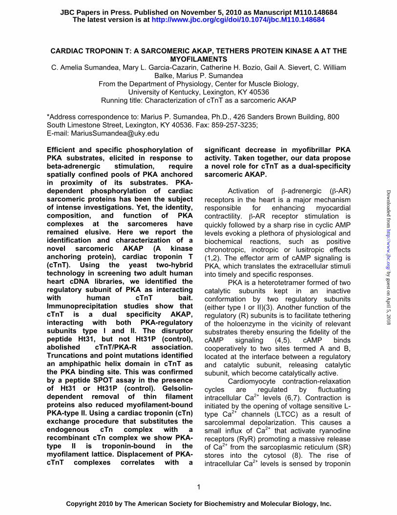
Yeast two-hybrid screens show human cardiac PKA interacts with cardiac TnT. To identify signaling proteins that bind and post-translationally modify human cardiac troponin, we used a yeast two-hybrid approach to screen two separate adult human heart cDNA libraries (Clontech) with human cardiac TnT as “bait”. The yeast two-hybrid system (Y2H) is a sensitive eukaryotic assay widely and successfully used to detect novel interactions between proteins (22). The Y2H is also suited to map the specific interacting domains of two proteins known to form a complex in which the strength of the interaction can be correlated with the level of expression of an appropriate reporter gene (23). Y2H screens of more than 4x10⁶ cDNA clones identified over 1000 colonies that grew on high-stringency plates. 375 colonies turned blue in the presence of α-gal, indicating positive interactions with cTnT (Fig. 1). DNA sequencing led to the identification of 19 gene products (see Table 2), including multiple hits for known binding partners of cTnT, cTnI and Tm.
Among the positive clones interacting with cTnT, we identified cAMP-dependent
by guest on April 5, 2018
http://ww
w.jbc.org/
Dow
nloaded from

6
protein kinase A regulatory subunit type I (Table 2). A characteristic feature of PKA regulatory subunits is the presence of docking domains that tether the holoenzyme in vicinity of substrates through interaction with an amphipathic helix of an anchoring protein (24). In general, PKA-R subunits interact with small aliphatic (Ala, Val, Leu, Ile) and basic amino acids (Arg, Lys) that decorate one side of the AKAP helix (25,26). In silico analysis of cTnT amino acid sequence using ClustalW multiple alignment algorithm (part of the MacVector 11 sequence analysis suite) identified a fragment of an amphipathic α-helix (spanning residues 212-224) as a putative PKA-R biding site (Fig. 2).
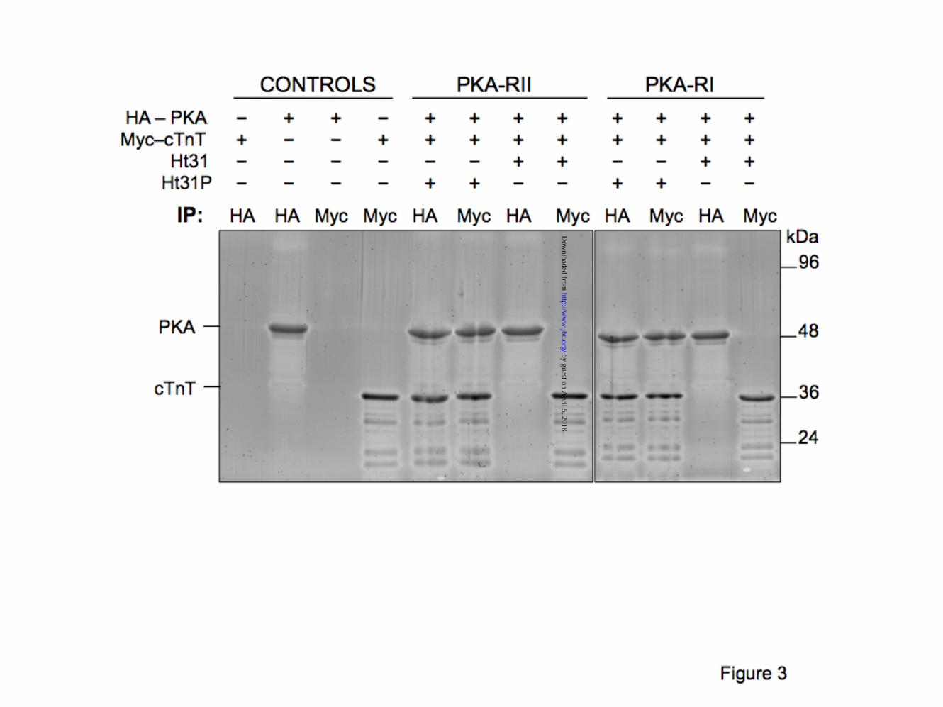
Human cTnT interacts directly with PKA-R. To establish whether the association between cTnT and PKA-R occurs through direct interaction or mediated through another protein, we assessed the ability of recombinant human cTnT to bind recombinant human PKA-RI and PKA-RII, in vitro. Using a previously validated cell free expression method, epitope tagged Myc-cTnT and HA-PKA-RI or HA-PKA-RII were expressed in vitro using a rabbit reticulocyte lysate system in the presence of a modified lysine tRNA labeled with the fluorophore BODIPY-FL. As a result, fluorescently labeled lysines (accounting for 11.4% of cTnT a.a, 5.8% of PKA-RI and 5.5% of PKA-RII) were incorporated into the nascent proteins during translation, allowing for convenient “in-gel” detection. Newly expressed, fluorescent Myc-cTnT was incubated with fluorescent HA-PKA-RI or -RII and the resulting complexes were immunoprecipitated using anti-Myc or anti-HA antibodies. Fig. 3 shows data from a representative experiment, confirming the Y2H results that cTnT and PKA-R interact directly with each other. Alignment of cTnT (212-230) and Ht31 (PKA/AKAP disruptor peptide) shows high homology between the two regions (Fig.2B), signifying that Ht31 peptide might be an efficient cTnT-PKA-R disruptor. Indeed, the addition of Ht31 peptide (Promega) completely abolished both cTnT-RI and cTnT-RII interactions. On the other
hand, the inactive peptide Ht31P had no effect on the cTnT-PKA-R interaction (Fig. 3).
Human cTnT associates with PKA-R
in a cellular environment. To ascertain whether human cTnT can interact with human PKA-R in a cellular environment, we co-expressed Myc-cTnT and HA-PKA-RI or -RII in the heterologous cell line HEK293 (Fig. 4). Immunoprecipitation with anti-Myc and anti-HA antibodies revealed that human cTnT specifically interacts with both PKA-RII and –RI (data not shown). The addition of membrane permeable St-Ht31 peptide disrupts both cTnT-RI and cTnT-RII interactions. Collectively, these data strongly suggest that cTnT and PKA-R form stable complexes in a cellular environment.
Characterization of PKA binding site on cTnT. To investigate the identity of PKA binding site on cTnT, Y2H assays were set up with human cTnT as “bait” and human PKA regulatory subunits RI or RII as “prey” (Fig. 5). Various cTnT truncations were generated to the left or to the right of the putative PKA binding site shown in Fig. 2. These fragments of cTnT (Fig. 5A) were previously confirmed as good functional representations of that particular fragment of the protein (27). Interactions between cTnT constructs and PKA-RI, or -RII, were assessed by two different colorimetric assays, using either α-gal (on selective media plates) or β-gal (in a colony lift assay), see Fig 1B,C. Our data from β-gal assays is summarized in Fig. 5 B, D. Identical results were obtained with the α-gal assays (data not shown). Truncation analysis of cTnT demonstrate that the region spanning amino acids 212-224 is required for PKA binding. St-Ht31 peptide, but not St-Ht31P, obstructs cTnT - PKA-R interaction (Fig. 5D).
To verify that the cTnT fragment spanning residues 212 and 224 is essential to PKA-R binding we designed point mutations to either disrupt the amphipathic helix (V218P) or to remove an essential hydrophobic residue (A220N) conserved in all AKAPs (Fig. 5 C, D). Our data demonstrate
by guest on April 5, 2018
http://ww
w.jbc.org/
Dow
nloaded from

7
that both mutants, V218P and A220N, successfully abolished cTnT - PKA-R association. Interestingly, a mutation that replaced Arg216 with an Ile (R216I) residue, found in other AKAPs at that position, seemed to be a gain-of–function mutant as it significantly enhanced interaction of cTnT with both PKA-Rs.
In addition to the loss or gain-of-function mutants, we also investigated the effect of a cardiomyopathy-causing deletion in cTnT at residue 210 (in the vicinity of PKA binding domain). Data from our recent study (19) indicate that deletion of Lys210 (ΔK210) affects PKA-dependent phosphorylation of MyBP-C and cTnI (decreased by over 40% in knock-in ΔK210 mouse hearts). Consistent with our previous findings, ΔK210 induces a ~65% decrease of PKA binding to cTnT (Fig.5 C, D).
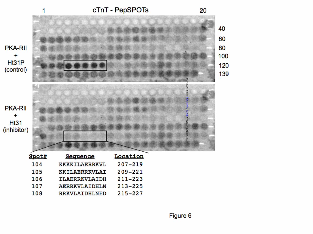
Solid phase peptide-scan binding assays corroborate PKA biding site on cTnT. PKA binding residues were confirmed using peptide SPOTS blot technology (Jerini Peptide Technology). The SPOTS blot displays the 288 amino acid sequence of cTnT as a set of 139 overlapping 13-mer peptides frame shifted by two residues (Fig. 6). Spot-bound PKA reacted with an anti-PKA-R antibody (BD Biosciences) and was detected using the ECL kit (GE Healthcare). Our results indicate PKA-RII interaction was strongest with cTnT peptides (#104-108) spanning the proposed PKA binding region (i.e. amino acids 212 - 224). Other, weaker interactions were due to nonspecific binding (Fig. 6 - top panel). The inhibitory peptide Ht31 selectively prevented PKA - cTnT peptides interaction (#104-108) (Fig. 6 - bottom panel).
Endogenously expressed PKA-R
interacts directly and specifically with cTnT. To determine whether endogenously expressed, rat heart PKA-R specifically interact with cTnT, we used a GST-cTnT pull-down assay. We monitored the ability of purified GST-tagged cTnT to associate with endogenous cardiac PKA-RI and RII from adult rat heart lysates (Fig. 7A). PKA-type I
and II are localized, in cardiomyocytes, in a variety of functionally and spatially distinct compartments such as nuclear envelope, sarcolemma, SR, cytoplasm, mitochondria, sarcomere, etc (28,29). Heart lysates contain PKA-R released from all these compartments (especially from membranous domains disrupted by detergents present in lysis buffer). A recent report indicates that homogenization of rodent hearts leads to augmentation of “free” PKA-R (non-AKAP bound), likely due to mechanical/chemical disruption (11). Therefore, rat heart lysates contain enough unbound PKA-R to associate with GST-cTnT (as shown in Fig 7A). Our results indicate that endogenously expressed, rat heart, PKA-RI and -RII interact with GST-cTnT. The specificity of the interaction was tested in the presence of Ht31 (ablates cTnT-PKA-R binding) and Ht31P (no effect), as reported in Fig. 7A.
Native PKA-R and cTnT form stable
complexes in the rat heart. To determine whether natively expressed PKA-R and cTnT form stable complexes, we performed co-immunoprecipitation assays using rat heart homogenates. As shown in Fig. 7B, native cTnT-PKA-RI and –RII complexes were immunoprecipitated with an anti-cTnT antibody, but not by a control IgG. Ht31 peptide, but not HT31P, prevented cTnT-PKA interaction. These data strongly suggest that native cTnT and PKA-R form stable complexes in the heart.
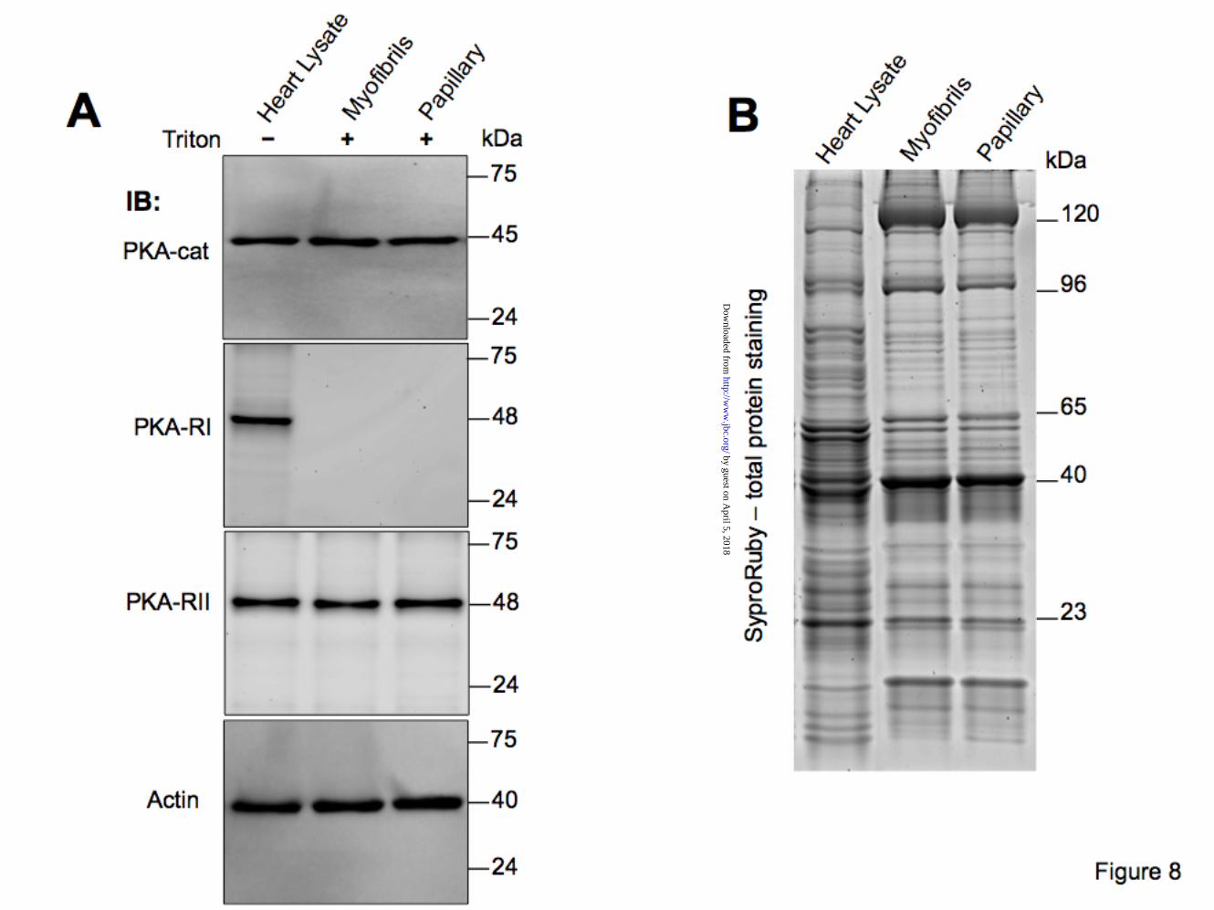
PKA-R have differential affinities to
detergent extracted cardiac myofibrils and papillary muscle strips. To elucidate whether PKA-R tether specifically to the cardiac myofilaments we used isolated myofibrils and papillary muscle strips that were treated with Triton X-100 to remove all membranes and membrane associate proteins. Interestingly, immunoblot analysis revealed PKA-RII, but not PKA-RI, associated with myofilaments from both preparations. Fig. 8 indicates a robust reactivity of myofilaments with anti-PKA-catalytic and anti-PKA-RII antibodies, but not anti-PKA-RI. This is not entirely unexpected since dual AKAPs
by guest on April 5, 2018
http://ww
w.jbc.org/
Dow
nloaded from

8
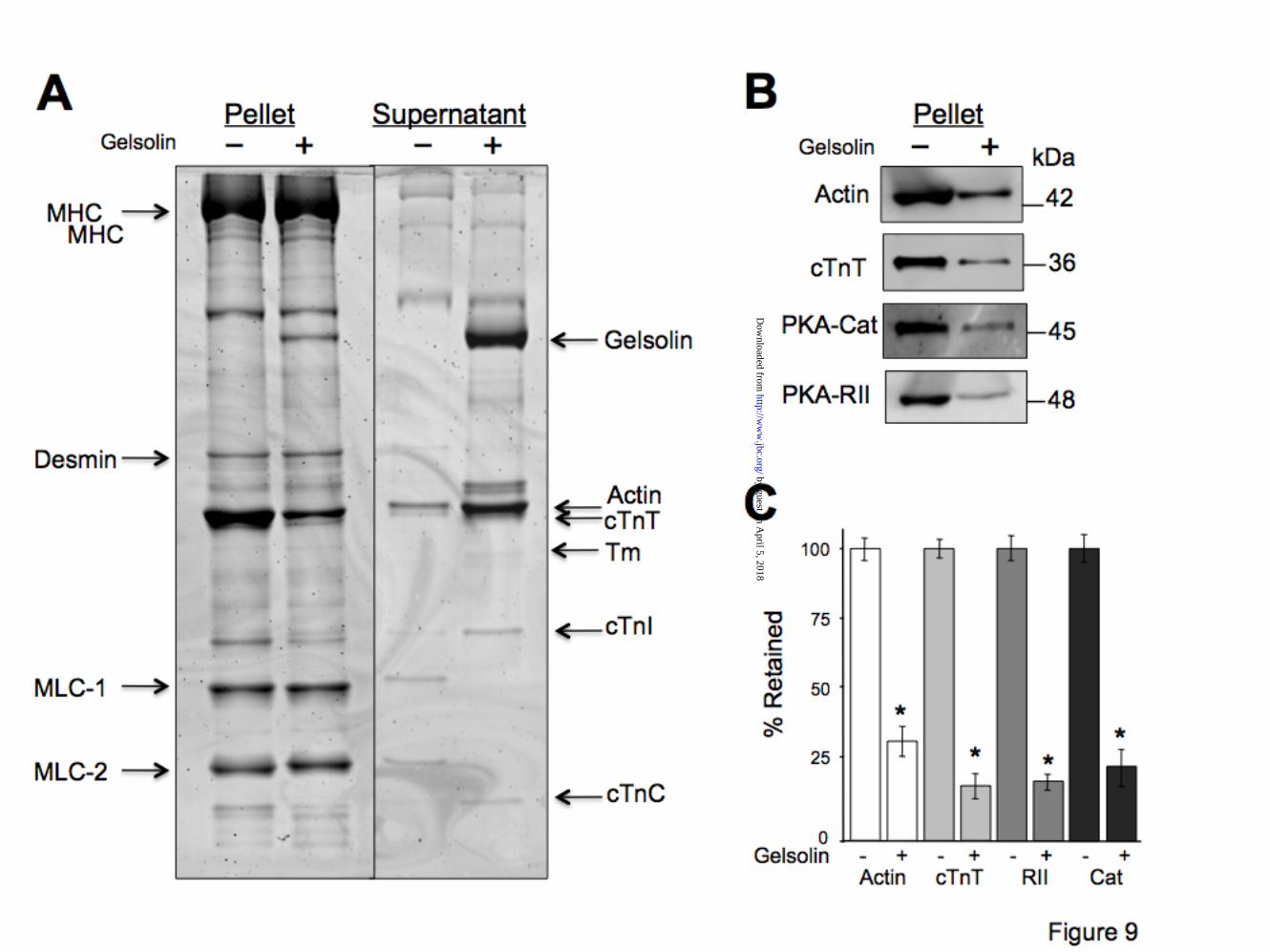
tend to bind PKA-RII with higher affinity than PKA-RI (30). It is likely that myofilament bound PKA-RI have been removed by the detergent treatment. Similar disruption of PKA-RI anchoring was reported in a recent study (11). PKA-type II is associated with cardiac thin filaments. To investigate whether endogenous cardiac PKA is associated with the thin filaments (i.e. cTnT) we have used a gelsolin assay. Gelsolin is a Ca2+-dependent actin filament - severing molecule (31). It selectively removes cardiac thin filaments leaving behind relatively intact thick, intermediate and titin filaments, anchored at the Z lines (32-34). Fig. 9 shows data from a representative experiment. Incubation of myofibrils with gelsolin leads to selective removal of actin filaments (thin filaments) as indicated by the extraction of actin, Tm, and troponins from the pellet fraction and their enrichment in the supernatant fraction (Fig. 9A). Immunoblot analyses of gelsolin treated myofilaments indicate a ~75% decrease in endogenous PKA-cat and PKA-RII protein concentrations. PKA extraction is proportional with the reduction of cTnT (Fig. 9 B, C). PKA-type II is bound to cardiac troponin in the myofibrillar milieu. To show that PKA holoenzyme (RII + cat) is part of the same complex with cTnT, in the myofibrillar milieu, we used a cTn exchange method to replace endogenous cTn and therefore displace any PKA bound to cTnT. The schematic of troponin exchange maneuver is shown in Fig. 10A. Immunoblot analysis of control (Cont) and exchanged (Exch) papillary muscle fibers show that approximately 72% of the endogenous cTn (Endo) was replaced by recombinant cTn (Rec) (Fig. 10 B,C). Notice that recombinant cTnT migrates with a different mobility (i.e. slower) on SDS-PAGE due to the presence of a Myc tag (used to differentiate between endogenous and recombinant cTnT). The cTn exchange procedure led to a considerable decrease in PKA bound to the myofilaments, indicating that ~51% of PKA-cat and ~55% of PKA-RII were removed together with endogenous cTn.
PKA activity was determined in the presence and absence of PKI (PKA inhibitor peptide) before and after the exchange (Fig. 10D). Our data indicate a significant blunting (by ~50%) of myofilament bound PKA activity after the cTn exchange procedure.
DISCUSSION
This study provides novel evidence
that cTnT is a dual-specificity AKAP, and that native cTnT forms stable complexes in the rat heart with PKA-RI and –RII. The disruptor peptide Ht31, but not Ht31P (control), abolished cTnT/PKA-R association. Using gelsolin and cTn exchange assays we show that PKA-RII associates with thin filaments and endogenous cTnT, in isolated rat myofibrils. In addition, our study indicates that PKA-R docks to an amphipathic domain on cTnT and mutation of specific residues abolished cTnT/PKA-R interaction.
PKA regulates many aspects of cardiac contractility. In healthy myocardium, stimulation of β-AR signaling pathway enhances the heart rate, force of contraction, and relaxation (35). At the cardiomyocyte level these events are mediated by PKA, which is anchored by various AKAPs in the vicinity of key Ca2+ handling and contractile proteins, and regulates their activity through phosphorylation. For example, AKAP18α, a small membrane-associated protein, has been shown to target PKA to the L-type Ca2+ channel (36). PKA-dependent phosphorylation of the channel increases its open probability. Similarly, muscle specific AKAP (mAKAP), a 255-kDa protein, has been shown to facilitate anchoring and PKA-dependent phosphorylation of RyR2 at the SR (15). Phosphorylation of RyR2 releases the inhibitory action of FKBP12.6 resulting in an increase open channel probability. Lastly, AKAP18δ, has been identified as the anchoring protein responsible for PKA-dependent regulation of SERCA2, through phosphorylation of its inhibitory protein phospholamban (PLB) (37). PLB phosphorylation relieves its inhibitory action on SERCA2 thereby boosting Ca2+ re-uptake into the SR during diastole.
by guest on April 5, 2018
http://ww
w.jbc.org/
Dow
nloaded from
9
PKA-dependent phosphorylation of cardiac sarcomeric proteins is more studied and better understood than all the other kinases targeting the sarcomere. PKA-dependent phosphorylation of cTnI and MyBP-C seem to dominate the control of myofilament function during β-AR stimulation (9). Phosphorylation of cTnI at its cardiac specific sites Ser23Ser24 depresses myofilament Ca2+ sensitivity and controls the kinetics of Ca2+ binding to cTnC (38). Like cTnI, MyBP-C has a N-terminal phosphorylation motif that is unique to the cardiac isoform. Phosphorylation of MyBP-C affects the radial movement of cross-bridges from the thick filament (39,40). PKA phosphorylation of both cTnI and MyBP-C is essential in shortening the heart beat cycle time required to adjust to augmented frequency in exercise (41,42). PKA also phosphorylates titin, the giant third filament protein, and controls its mechanical properties (reduces passive tension) (43).
In spite of the importance of PKA-mediated phosphorylation in the control of myofilament function, little is known regarding its targeting to the sarcomere (44). Docking of PKA near substrates is attained by interaction of regulatory domains with AKAPs. Both, PKA-RI and PKA-RII are ubiquitously expressed in cardiac myocytes. Studies over the past decade have illuminated our understanding of their non-redundant biological role, usually through sequestering each PKA type to distinct intracellular signaling compartments. Recent studies employing various imaging techniques show overlapping staining patterns at the cardiac sarcomere for both PKA-RI and PKA-RII (11,45). The striated staining is largely abolished in the presence of peptide disruptors. These data are in agreement with our present findings that PKA-R interact with cTnT and are anchored at the myofilaments.
However, what could be the functional advantage of having both PKA-types anchored to cardiac sarcomeres? Probably the two isoforms could impart a more dynamic modulation of myofilament function in response to varying cAMP levels, and the combined regulation could provide a more
refined physiological response. It is well known that the two PKA types possess distinct activation and docking properties. For example, PKA type I is more readily activated by cAMP (activation constant, Kact, of 50-100nM) than PKA type II (Kact of 200-400nM) (46). In addition, PKA-RI appear to have a weaker binding affinity to it’s AKAP(s) compared with PKA-RII, therefore it could be easier released from its binding site in a process related to mechanical state of the sarcomere, as shown for other proteins (47,48)
The interaction between cTnT and PKA-regulatory subunits is not unanticipated. Evidence from an Y2H screen of a human skeletal muscle cDNA library proposed that skeletal TnT interacts with the leucine zipper domain of the regulatory subunit of cGMP-dependent protein kinase I (or PKG) (49). PKG and PKA are part of the same family of kinases. A major difference is their respective regulatory subunits require distinct binding motifs.
All members of the diverse AKAP family share a conserved PKA-anchoring domain, consisting of an amphipathic helix of 10-16 residues, which interacts with a hydrophobic groove in the docking/dimerization domain of the regulatory subunits (25,26). Crystal structure of the core domain of cardiac troponin complex shows that cTnT residues 202-226 form an amphipathic helix, are solvent exposed, and apparently are not required for intra-troponin interactions (50). This region shows homology to amphipathic helices found in other AKAPs that serve as docking domains for PKA-R. It was shown that amino acid composition of the amphipathic helix dictates the binding preference for PKA-RI, –RII, or both (51).
In a recent study by Jarnaess et al (52) the authors demonstrate that some dual specificity AKAPs contain an additional PKA-RI binding region (termed RISR) that is generally located upstream of the amphipathic helix. The RISR motif was suggested to be an enhancer for PKA-RI binding to the amphipathic helix and that AKAP’s that contain both regions have a higher affinity for PKA-RI compared with
by guest on April 5, 2018
http://ww
w.jbc.org/
Dow
nloaded from

10
those that only have the amphipathic helix. Our study provide strong evidence that cardiac troponin T is a dual AKAP. To determine whether cTnT also possesses a possible RISR domain we took a closer look at its sequence. In silico sequence analysis identified a region in cTnT with high homology to the RISR minimal binding motif located just upstream of the amphipathic helix (see Fig. 11) spanning a critical phosphorylation region centered on Thr203 (20,21). Whether the putative RISR region is important in enhancing PKA-RI binding to cTnT, and whether Thr203 phosphorylation alters the complex formation, remains to be seen.
Recent data indicate that β-AR signaling abnormalities in heart failure include a profound decrease in PKA activation, likely due to defects in AKAP-based molecular complexes leading to impaired compartmentation of the cAMP/PKA signaling (2,28,53). As a result, studies of human and experimental heart failure models show significant decrease in the phosphorylation levels of PKA substrates cTnI, MyBP-C, and titin, which may account for a large part of the contractile dysfunction of the failing myocardium (9,54). Interestingly, targeted disruption of AKAP-PKA complexes by adenovirus-mediated gene transfer of Ht31 inhibitory peptide or TAT-conjugated AKAD (A kinase anchoring disruptor), elicited detrimental effects on chronotropy, inotropy and lusitropy via a redistribution of PKA away from AKAPs and its substrates, and a blunting of PKA-dependent phosphorylation of key substrates (45,55,56). PKA-dependent phosphorylation of myofilament proteins is necessary for optimal contractile function, is associated with cardioprotection, and confers
resistance to calpain-mediated proteolysis of cTnI and MyBP-C (38,57,58). Collectively, these studies strongly suggest that altered PKA anchoring at the myofilaments has a profound impact on cardiac function.
More work is needed to understand not only what PKA does in the heart to modulate contractile function, but also when and where it does it. Of particular importance is also to understand whether cTnT/PKA complexes are altered during heart disease progression, and whether cTnT phosphorylation has a regulatory role in complex formation (implicating a cross-talk between different kinases at the sarcomere).
Taken together our data indicate that cTnT is dual AKAP that tether PKA-R at the myofilaments. We predict that these macromolecular complexes are localized at the at the I-Z-I region away from myosin-actin cross-bridges. Generally, AKAP-PKA clusters are signaling modules composed of many other proteins such as PDEs and phosphatases, and even other kinases. While numerous studies discuss the important role of phosphorylation events in regulating cardiac myofilament function, essentially nothing is know about the mechanisms that promote the anchoring of these signaling molecules in the proximity of their substrates. This information is vital to a complete understanding of cardiac muscle regulation. In addition, direct and specific manipulation of sarcomeric proteins and signaling complexes, circumventing receptor-ligand pathways, could pave the way for myofilament-targeted therapeutic approaches in muscle diseases (59).
REFERENCES
1. Steinberg, S. F., and Brunton, L. L. (2001) Annual review of pharmacology and
toxicology 41, 751-773 2. El-Armouche, A., and Eschenhagen, T. (2009) Heart failure reviews 14, 225-241 3. Taylor, S. S., Kim, C., Vigil, D., Haste, N. M., Yang, J., Wu, J., and Anand, G. S. (2005)
Biochim Biophys Acta 1754, 25-37 4. Tasken, K., and Aandahl, E. M. (2004) Physiol Rev 84, 137-167 5. McConnachie, G., Langeberg, L. K., and Scott, J. D. (2006) Trends Mol Med 12, 317-
323
by guest on April 5, 2018
http://ww
w.jbc.org/
Dow
nloaded from

11
6. Kobayashi, T., and Solaro, R. J. (2005) Annu Rev Physiol 67, 39-67 7. Solaro, R. J. (2010) J Biomed Biotechnol 2010, 105648 8. Bers, D. M. (2002) Nature 415, 198-205 9. Solaro, R. J. (2008) The Journal of biological chemistry 283, 26829-26833 10. Fischmeister, R., Castro, L. R., Abi-Gerges, A., Rochais, F., Jurevicius, J., Leroy, J., and
Vandecasteele, G. (2006) Circ Res 99, 816-828 11. Di Benedetto, G., Zoccarato, A., Lissandron, V., Terrin, A., Li, X., Houslay, M. D., Baillie,
G. S., and Zaccolo, M. (2008) Circ Res 103, 836-844 12. Michel, J. J., and Scott, J. D. (2002) Annual review of pharmacology and toxicology 42,
235-257 13. Zaccolo, M. (2009) Br J Pharmacol 158, 50-60 14. Houslay, M. D., Baillie, G. S., and Maurice, D. H. (2007) Circ Res 100, 950-966 15. Dodge-Kafka, K. L., Bauman, A., and Kapiloff, M. S. (2008) Handb Exp Pharmacol, 3-14 16. David, H., Meyer, R., Marx, I., Guski, H., and Wenzelides, K. (1979) J Mol Cell Cardiol
11, 631-638 17. Wu, S. C., and Solaro, R. J. (2007) The Journal of biological chemistry 282, 30691-
30698 18. Canaves, J. M., Leon, D. A., and Taylor, S. S. (2000) Biochemistry 39, 15022-15031 19. Duke, L. S., Garcia-Cazarin, M. L., Sumandea, C. A., Sievert, G. A., Balke, C. W., Zhan,
D. Y., Morimoto, S., and Sumandea, M. P. (2010) J Mol Cell Cardiol 48, 934-942 20. Sumandea, M. P., Vahebi, S., Sumandea, C. A., Garcia-Cazarin, M. L., Staidle, J., and
Homsher, E. (2009) Biochemistry 48, 7722-7731 21. Sumandea, M. P., Pyle, W. G., Kobayashi, T., de Tombe, P. P., and Solaro, R. J. (2003)
J Biol Chem 278, 35135-35144 22. Fields, S., and Song, O. (1989) Nature 340, 245-246 23. Estojak, J., Brent, R., and Golemis, E. A. (1995) Mol Cell Biol 15, 5820-5829 24. Carr, D. W., Stofko-Hahn, R. E., Fraser, I. D., Bishop, S. M., Acott, T. S., Brennan, R. G.,
and Scott, J. D. (1991) The Journal of biological chemistry 266, 14188-14192 25. Kinderman, F. S., Kim, C., von Daake, S., Ma, Y., Pham, B. Q., Spraggon, G., Xuong, N.
H., Jennings, P. A., and Taylor, S. S. (2006) Mol Cell 24, 397-408 26. Gold, M. G., Lygren, B., Dokurno, P., Hoshi, N., McConnachie, G., Tasken, K., Carlson,
C. R., Scott, J. D., and Barford, D. (2006) Mol Cell 24, 383-395 27. Hinkle, A., Goranson, A., Butters, C. A., and Tobacman, L. S. (1999) The Journal of
biological chemistry 274, 7157-7164 28. Diviani, D. (2008) Curr Opin Pharmacol 8, 166-173 29. Mauban, J. R., O'Donnell, M., Warrier, S., Manni, S., and Bond, M. (2009) Physiology
(Bethesda) 24, 78-87 30. Pidoux, G., and Tasken, K. (2010) J Mol Endocrinol 44, 271-284 31. Sun, H. Q., Yamamoto, M., Mejillano, M., and Yin, H. L. (1999) The Journal of biological
chemistry 274, 33179-33182 32. Kawai, M., and Ishiwata, S. (2006) Journal of muscle research and cell motility 27, 455-
468 33. Trombitas, K., and Granzier, H. (1997) Am J Physiol 273, C662-670 34. Trombitas, K., Greaser, M. L., and Pollack, G. H. (1997) Journal of muscle research and
cell motility 18, 345-351 35. Ruehr, M. L., Russell, M. A., and Bond, M. (2004) J Mol Cell Cardiol 37, 653-665 36. Hulme, J. T., Lin, T. W., Westenbroek, R. E., Scheuer, T., and Catterall, W. A. (2003)
Proceedings of the National Academy of Sciences of the United States of America 100, 13093-13098
by guest on April 5, 2018
http://ww
w.jbc.org/
Dow
nloaded from

12
37. Lygren, B., Carlson, C. R., Santamaria, K., Lissandron, V., McSorley, T., Litzenberg, J., Lorenz, D., Wiesner, B., Rosenthal, W., Zaccolo, M., Tasken, K., and Klussmann, E. (2007) EMBO Rep 8, 1061-1067
38. Layland, J., Solaro, R. J., and Shah, A. M. (2005) Cardiovascular research 66, 12-21 39. Colson, B. A., Bekyarova, T., Fitzsimons, D. P., Irving, T. C., and Moss, R. L. (2007)
Journal of molecular biology 367, 36-41 40. Stelzer, J. E., Fitzsimons, D. P., and Moss, R. L. (2006) Biophysical journal 90, 4119-
4127 41. Kentish, J. C., McCloskey, D. T., Layland, J., Palmer, S., Leiden, J. M., Martin, A. F., and
Solaro, R. J. (2001) Circ Res 88, 1059-1065 42. Stelzer, J. E., Patel, J. R., Walker, J. W., and Moss, R. L. (2007) Circ Res 101, 503-511 43. Yamasaki, R., Wu, Y., McNabb, M., Greaser, M., Labeit, S., and Granzier, H. (2002) Circ
Res 90, 1181-1188 44. Russell, M. A., Lund, L. M., Haber, R., McKeegan, K., Cianciola, N., and Bond, M.
(2006) Arch Biochem Biophys 456, 204-215 45. Patel, H. H., Hamuro, L. L., Chun, B. J., Kawaraguchi, Y., Quick, A., Rebolledo, B.,
Pennypacker, J., Thurston, J., Rodriguez-Pinto, N., Self, C., Olson, G., Insel, P. A., Giles, W. R., Taylor, S. S., and Roth, D. M. (2010) Journal of Biological Chemistry 285, 27632-27640
46. Dostmann, W. R. G., and Taylor, S. S. (1991) Biochemistry 30, 8710-8716 47. Hoshijima, M. (2006) Am J Physiol Heart Circ Physiol 290, H1313-1325 48. Frank, D., Kuhn, C., Katus, H. A., and Frey, N. (2006) J Mol Med 84, 446-468 49. Yuasa, K., Michibata, H., Omori, K., and Yanaka, N. (1999) The Journal of biological
chemistry 274, 37429-37434 50. Takeda, S., Yamashita, A., Maeda, K., and Maeda, Y. (2003) Nature 424, 35-41 51. Feliciello, A., Gottesman, M. E., and Avvedimento, E. V. (2001) Journal of molecular
biology 308, 99-114 52. Jarnaess, E., Ruppelt, A., Stokka, A. J., Lygren, B., Scott, J. D., and Tasken, K. (2008)
Journal of Biological Chemistry 283, 33708-33718 53. Zakhary, D. R., Fink, M. A., Ruehr, M. L., and Bond, M. (2000) The Journal of biological
chemistry 275, 41389-41395 54. Hamdani, N., Kooij, V., van Dijk, S., Merkus, D., Paulus, W. J., Remedios, C. D.,
Duncker, D. J., Stienen, G. J., and van der Velden, J. (2008) Cardiovascular research 77, 649-658
55. Fink, M. A., Zakhary, D. R., Mackey, J. A., Desnoyer, R. W., Apperson-Hansen, C., Damron, D. S., and Bond, M. (2001) Circ Res 88, 291-297
56. McConnell, B. K., Popovic, Z., Mal, N., Lee, K., Bautista, J., Forudi, F., Schwartzman, R., Jin, J. P., Penn, M., and Bond, M. (2009) The Journal of biological chemistry 284, 1583-1592
57. Nagayama, T., Takimoto, E., Sadayappan, S., Mudd, J. O., Seidman, J. G., Robbins, J., and Kass, D. A. (2007) Circulation 116, 2399-2408
58. Sadayappan, S., Osinska, H., Klevitsky, R., Lorenz, J. N., Sargent, M., Molkentin, J. D., Seidman, C. E., Seidman, J. G., and Robbins, J. (2006) Proceedings of the National Academy of Sciences of the United States of America 103, 16918-16923
59. Solaro, R. J. (2009) IDrugs 12, 243-251
FOOTNOTES
This work was supported in part by a NIH Ruth L. Kirschstein National Research Service Award (HL82120) to CAS, and by NIH grants HL071865, HL68733 and AG032009.
by guest on April 5, 2018
http://ww
w.jbc.org/
Dow
nloaded from

13
The abbreviations used are: cTnT, cardiac troponin T; PKA, Protein Kinase A; PKA-R – regulatory domain of PKA; AKAP, A-kinase anchoring protein; Y2H, yeast two-hybrid;
FIGURE LEGENDS Figure 1. Yeast two-hybrid library screens identify cTnT-PKA-R novel interaction. A) schematic representation of main steps of the two yeast two-hybrid assays including high stringency screening on quadruple selective media. B) β-galactosidase assay shows the interaction between cTnT and Laminin C (negative control – top panel), cTnI (positive control – middle panel) and PKA-RI (bottom panel) as monitored by shades of color blue. The intense blue color developed by yeast cells harboring cTnT-cTnI indicates a stronger interaction between these two proteins compared with cTnT-PKA RI (lighter blue). C) quantitative analysis of the interaction was determined using Image J software and normalized to that of control. Data shown is from a single experiment and is representative of 4 separate experiments. Figure 2. Cardiac TnT contains a highly conserved PKA docking site. A) schematic illustration shows the location of PKA binding site and of cardiac specific cTnI-Ser23Ser24 phosphorylation sites (star). Drawing of troponin complex is based on the crystal structure of the troponin core domain (50). N-terminal region of cTnT (residues 1-204) was not solved in the crystal structure. Rectangles represent helical structures. cTnC is colored red, cTnI is green and cTnT is blue. B) ClustalW multiple sequence alignment of cTnT with nine other AKAPs. Conserved residues responsible for tethering PKA are shown in white. The high homology between cTnT and Ht31 is also shown (boxed). C) surface representation of the cTnT (pdb 1J1D) helix 203-224 shows the position of hydrophobic residues (red) involved in PKA docking. Figure 3. Human cTnT interacts directly with both PKA-RI and PKA-RII. Interaction between epitope tagged Myc-cTnT and HA-PKA-RI or HA-PKA-RII was determined by immunoprecipitation (IP). Fluorescently labeled, recombinant proteins were expressed in a cell-free system (Promega). Proteins were mixed and the interaction was verified by IP using anti-c-Myc and anti-HA antibodies. Content of each lane is shown on top. 20 µM Ht31 or Ht31P (control) peptides were used as shown to assess the specificity of the cTnT-PKA interactions. Similar data were obtained in 3 separate experiments. Molecular weight markers are shown on the left. Figure 4. Characterization of cTnT-PKA interaction in a cellular environment. Co-expression of cTnT and PKA-RII in HEK293 cells, in the presence or absence of membrane permeable St-Ht31 or St-Ht31P, was followed by IP with agarose conjugated anti-my or anti-HA antibodies. Co-precipitation was detected with anti-cTnT antibody and anti-PKA-RII antibody. Molecular weight markers are shown on the left. Data shown is from a single experiment and is representative of 4 separate experiments. Figure 5. Residues 212-224 of cTnT are essential for PKA-R binding. A) schematic representation of cTnT truncations tested with two-hybrid assays. PKA-R docking site and cTnI binding region are also shown. B) interaction between cTnT-wt (1-288) or cTnT truncations with PKA-RI and PKA-RII was determined by the β-gal assay. Data normalized to cTnI – cTnT-wt binding (arbitrary set to 100). Only cTnT fragments that contained the proposed PKA binding site interacted positively with PKA-RI and PKA-RII. C) schematic representation of cTnT mutants tested in the Y2H system: V218P and A220N (loss-of-function), R216I (gain-of-function), K210del (cardiomyopathy-causing deletion). D) interaction between cTnT-wt or -mutants with PKA-RI and PKA-RII assessed by β-gal assay. Membrane permeable St-Ht31 (50
by guest on April 5, 2018
http://ww
w.jbc.org/
Dow
nloaded from
14
µM) disrupts cTnT-PKA interaction. cTnI was used as a positive control and laminin C as a negative control. Each screen was repeated at least 5 times. (*, p<0.05) Figure 6. Fine mapping of cTnT residues interacting with PKA-R. The 288 amino acid residues of human cTnT were displayed on a SPOTS blot membrane as series of 139 overlapping 13-mer peptides frame shifted by two residues. The cTnT SPOTS blot was tested for interaction with PKA-RII in a solid phase assay (in the presence of 20 µM Ht31 or Ht31P). PKA-RII has highest affinity for the peptides spanning amino acids 207-227, as predicted from multiple sequence alignment and by the Y2H data. The other spots represent non-specific interactions. Data shown is from a single experiment and is representative of 3 separate experiments. Figure 7. Endogenously expressed PKA-R interacts directly and specifically with cTnT. A) rat heart homogenates were used to test for endogenous cardiac PKA-R binding to cTnT. GST-TnT interaction with PKA-RI and RII was detected by immunoblotting with specified anti-PKA antibodies. GST alone shows no interaction with PKA-R. Ht31 and Ht31P peptides were used to test the specificity of interaction. Anti-GST antibody was used as control. B) rat heart homogenates were used to immunoprecipitate native cTnT-PKA-R complexes. Immunoblots of the IP complexes show endogenous association of cTnT-PKA-R in stable complexes. Data shown is from a single experiment and is representative of 3 separate experiments. Molecular weight markers are shown on the left. Figure 8. PKA-R have differential affinities to detergent extracted cardiac myofibrils and papillary muscle strips. A) Triton X-100 extracted rat cardiac myofibrils or left ventricular (LV) papillary muscle fibers indicate that PKA type II holoenzyme (RII and cat) is associated with cardiac myofilaments. PKA-RI appears to have been removed by the detergent treatment. Similar data were obtained in 3 separate experiments. B) Representative gel used to resolve detergent treated myofibrils and papillary fibers prior to transfer to PVDF membranes. SyproRuby - total protein staining indicates similar loading of myofibrils and papillary muscle strips. Molecular weight markers are shown on the left. Figure 9. Gelsolin-dependent removal of cardiac thin filaments significantly reduces sarcomeric PKA abundance. A) rat cardiac myofibrillar proteins (pellets and supernatants) resolved by SDS-PAGE following gelsolin treatment. Myosin heavy chain (MHC), myosin light chains 1 and 2 (MLC), and desmin, indicate intact thick and intermediate filaments, while thin filament proteins (actin, troponins and tropomyosin) are selectively extracted by gelsolin treatment. B) immunoblot analysis of control (-) and gelsolin extracted (+) pellets. PKA-catalytic subunit and –RII are extracted together with actin and cTnT indicating the holoenzyme’s association with cardiac thin filaments. C) Analysis of optical densities of thin filament proteins retained after gelsolin extraction vs. control. Similar data were obtained in 3 separate experiments. (*, p<0.05) Figure 10. Exchanging endogenous cTn complexes with recombinant cTn displaces the myofilament bound PKA holoenzyme type II. A) schematic representation of the cTn exchange protocol. Recombinant cTn complex (gray) added to the detergent extracted mouse papillary muscle fibers replaces the endogenous cTn on the myofilaments. This action dislodges any PKA holoenzyme (2R-red and 2Cat-purple subunits) bound to endogenous cTnT. B) Immunoblot analysis of detergent extracted fibers (skinned) incubated in exchange buffer with and without (Cont) the recombinant cTn (Exch). Top panel shows the incorporation of recombinant Myc-cTnT (Rec) and the displacement of the endogenous TnT (End). Middle panels indicate that the levels of PKA-cat and PKA-RII decreased in the exchanged fibers. Actin
by guest on April 5, 2018
http://ww
w.jbc.org/
Dow
nloaded from

15
immunobloting was used as control. C) Optical density (O.D) of PKA immunoblots normalized to actin. D) Kinase activity assay shows significant decrease of PKA activity following cTn exchange. PKA inhibitor (15 µM PKI) verifies that the activity is PKA-specific and not from another kinase. Data shown is from a single experiment and is representative of 3 separate experiments. (*, p<0.05) Figure 11. Identification of a putative PKA-RI specifier region (RISR) in cTnT. Sequence alignment of cTnT residues 191-230 and RISR minimal binding region described in (52). Conserved residues are shown in white. Amphipathic helix location is also shown.
by guest on April 5, 2018
http://ww
w.jbc.org/
Dow
nloaded from

16
Table I. Oligonucleotide primers used for cloning, truncations, point-mutations of hcTnT, and cloning of PKA-RI and -RII.
Gene Vector Primer Sequence 5’-3’ Restriction Sites
hcTnT pGBKT7 GAAGGAGATATCATATGTCTGACATAGAAGAGGTG NdeI CTTTGTTAGCAGCCGGATCCCTATTTCCAGCGCCC BamHI hcTnT pCMV6-AN-Myc GTACCAGATGCGATCGCCATGTCTGACATAGAAGAG SgfI CTGCAGCTCAAGCTTGATGGATCCCTATTTCCAGCGC HindIII hcTnT-1-117 pGBKT7 GAGGAGGACCTGCATATGTCTGACATAGAAGAGGTG NdeI CTTTGAGAGAAACGAATTCCTACTCCTCTTTCTTCC EcoRI hcTnT-1-180 pGBKT7 GAGGAGGACCTGCATATGTCTGACATAGAAGAGGTG NdeI ATGTAACCCCCAGAATTCATCTAGTTGGACAAAGCC EcoRI hcTnT-118-228 pGBKT7 ACAGGAAGAAACATATGGAGGAGCTCGTTTCTCTCA NdeI CAGGTCGACGGATCCCTATTTCCAGCGC BamHI hcTnT-181-228 pGBKT7 AAGAAGGCTTTGTCCCATATGATGCATTTTGGGGGT NdeI CAGGTCGACGGATCCCTATTTCCAGCGC BamHI hcTnT-225-228 pGBKT7 GTGCTGGCCATTGACCATATGAATGAAGATCAGCTG NdeI CAGGTCGACGGATCCCTATTTCCAGCGC BamHI hcTnT-K210-del pGBKT7 GGGAAAAGAAGAAGATTCTGGCTGAGAGG NdeI CCTCTCAGCCAGAATCTTCTTCTTTTCCC BamHI hcTnT-V218P pGBKT7 GCTGAGAGGAGGAAGCCACTGGCCATTGACCAC NdeI GTGGTCAATGGCCAGTGGCTTCCTCCTCTCAGC BamHI hcTnT- R216I pGBKT7 ATTCTGGCTGAGAGGATCAAGGTGCTGGCCATT NdeI AATGGCCAGCACCTTGATCCTCTCAGCCAGAAT BamHI hcTnT-A220N pGBKT7 GAGGAGGAAGGTGCTGAACATTGACCACCTGAATG NdeI CATTCAGGTGGTCAATGTTCAGCACCTTCCTCCTC BamHI hPKA-RI pGADT7 CGTCCCCAGAGACATATGGAGTCTGGCAGTACCGCC NdeI AGGCACAGGAGGGGATCCTCAGACAGACAGTGACAC BamHI hPKA-RI pCMV6-AN-HA GTACCAGATGCGATCGCCATGGAGTCTGGCAGTACCG SgfI TCTGCAGCTCAAGCTTGATGGATCCTCAGACAGACAGT HindIII hPKA-RI pCMV6-AC-HA CATACGACGTACAAGCTTACGCTCATATGGAGTC HindIII GCTCGAGCTCGATGGACTCGAGGACAGACAGTG XhoI hPKA-RII pGADT7 CCTTCGGCCGCCGCCCATATGAGCCACATCCAGATC NdeI CTCTGGGGTGTGGGATCCCTACTGCCCGAGGTTGCC BamHI hPKA-RII pCMV6-AN-HA GTACCAGATGCGATCGCCATGAGCCACATCCAG SgfI GCAGCTCGAGCTCGAGGGATCCCTACTGCCCGAG XhoI hPKA-RII pCMV6-AC-HA TACCAGATTACGCGTATATGAGCCACATCCAGAT MluI GCTCGATGGACTCGAGCTGCCCGAGGTTGCCCAG XhoI
by guest on April 5, 2018
http://ww
w.jbc.org/
Dow
nloaded from

17
Table II. Summary of human heart cDNA prey clones interacting with human cTnT bait. Y2H screens of more than 4x10⁶ cDNA clones identified over 1000 colonies that grew on high-stringency plates, of which 375 turned blue in the presence of α-gal, indicating positive interactions with hcTnT. DNA sequencing led to the identification of 19 gene products, including multiple hits for cTnI and tropomyosin known binding partners of cTnT.
Clone ID Accession Gene Library A. Sarcomeric multiple NM_000363 Troponin I, type 3, (TNNI3) 1; 2 multiple NM_001018005 Tropomyosin 1, alpha, (TPM1) 2 63 EU106177 Tropomyosin beta, (TPM2b) 2 42 BC015821 Myosin light chain 2, regulatory, slow, (MYL2) 1 43 NM_001101151
5 PDZ and LIM domain 5, (PDLIM5) 1
1 NM_001824 Creatine kinase, muscle, (CKM) 1 B. Sarcoplasmic Reticulum 142 NM_002667 Phospholamban, (PLN) 2 C. Nuclear 38 BC005893 Atrial natriuretic factor, (ANF) 1 144 NM_012068 Activating transcription factor 5, (ATF5) 2 22, 26 NM_003074 SWI/SNF rel. matrix assoc. actin reg. of
chromatin C1, (SMARCC1) 2
76, 92 BC000898 Myeloid leukemia factor 2, (MLF2) 2 88 AK223404 Death-associated protein 6, (DAXX) 2 165 AF151852 UTP11-like ribonucleoprotein, (UTP11L) 1 E. Cytoskeleton 106 NM_006289 Talin 1, (TLN1) 2 194 BC111836 Dystrophin, (DMD) 1 F. Membrane/ Cytoplasm 6,7 AF036536 Mitofusin 2, (MFN2) 1 58 NM_002734 Protein kinase type I-alpha regulatory sub.,
(PRKAR1A) 1
G. Unknown 123 AC114489 chromosome 1 clone RP11-114B7 1 143 BC007531 Chimeric clone 2
by guest on April 5, 2018
http://ww
w.jbc.org/
Dow
nloaded from


WIlliam Balke and Marius P. SumandeaC. Amelia Sumandea, Mary Lolis Garcia-Cazarin, Catherine H. Bozio, Gail A. Sievert, C.
Cardiac troponin T: a sarcomeric AKAP, tethers protein kinase A at the myofilaments
published online November 5, 2010J. Biol. Chem.
10.1074/jbc.M110.148684Access the most updated version of this article at doi:
Alerts:
When a correction for this article is posted•
When this article is cited•
to choose from all of JBC's e-mail alertsClick here
by guest on April 5, 2018
http://ww
w.jbc.org/
Dow
nloaded from