a role for calmodulin-like proteins in herbivore defense...
TRANSCRIPT

Journal of Endocytobiosis and Cell Research (2016) 27 | 1 | 1-12 | International Society of Endocytobiology zs.thulb.uni-jena.de/content/main/journals/ecb/info.xml
Journal of Endocytobiosis and Cell Research VOL 27 | 1 | 2016 1
Journal of Endocytobiosis and Cell Research
A role for calmodulin-like proteins in herbivore defense path-ways in plants Sandra S. Scholz1, Monika Heyer1, Jyothilak- shmi Vadassery2 and Axel Mithöfer1* 1Bioorganic Chemistry Department; Max-Planck-Institute for Chemical Ecology, Jena, Germany; 2National Institute of Plant Genome Research, Aruna Asaf Ali Marg, New Delhi, In-dia; *correspondence to: [email protected] During their lifetime, plants need to adapt to various stimuli originating from the abiotic and biotic environ-ment, some of which represent stress factors. One major biotic stress factor is the attack of herbivorous insects feeding on the plant. But plants are not defenseless; they are equipped with an arsenal of different defense layers. Additionally to mechanical barriers, which are a first line of defense, the plant can produce a variety of chem-icals like toxic secondary metabolites, anti-herbivory proteins or compounds involved in indirect defense. The production of many of these defensive compounds is triggered by HAMPs, elicitors in insects’ oral secre-tions (OS), which come in contact with the wounded plant tissues while insects are feeding. The early events upon perception of these stimuli are still poorly under-stood. Elevations in cytosolic calcium are one of these early events, which activate the downstream defense signaling network, including certain phytohormones. To reach this, a proper decoding of calcium signals by different calcium sensor proteins is important. In Ara-bidopsis thaliana, several members of the calmodulin-like proteins (CMLs), one group of calcium sensor pro-teins, is induced upon treatment with OS of the general-ist herbivore Spodoptera littoralis. Some of these CMLs are involved in herbivore defense by modulating the jasmonate pathway. Journal of Endocytobiosis and Cell Research (2016) 27(1):1-12 Category: Review Keywords: defense, secondary metabolites, jasmonates, cal-cium, CML, herbivory Accepted: 30 October 2015 ____________________________________________________________________ Plant defense strategies against insect her-bivory During their lifespan, plants have to deal with a multitude of stress factors originating from the abiotic as well as the bio-tic environment. Main abiotic environmental cues influenc-ing the plants performance and fitness include drought and
salt stress, ozone and UV-radiation, cold stress and many others (Lawlor 2011). Biotic stress factors originate from many different groups of organisms like pathogens, nema-todes, microorganisms, and also from feeding insects. Given the fact that over 50% of all insects show herbivorous feed-ing behavior, plants have to adapt to them by developing and modulating different defense strategies (Schoonhoven et al. 1998; Van Poecke 2007). Attack of insects, especially with chewing feeding behavior, cause a massive loss of plant tis-sue and viability leading to low reproduction rate (Stowe et al. 2000). Attack of herbivorous insects combines different stress stimuli inducing plant defense. Perception of her-bivory by the plant consists of recognition of wounding of plant tissue and of elicitors provided by the insect’s oral se-cretion (OS) (Maffei et al. 2004; Mithöfer et al. 2005b; Mith-öfer and Boland 2008; Wu and Baldwin 2010).
The plant defense activated upon herbivory, is a complex network of different pathways, which are constitutively ex-pressed or induced upon stimuli perception. Both groups of defense pathways are composed of direct and indirect de-fenses (Howe and Jander 2008). Direct defense compounds like glucosinolates or protease inhibitors directly influence the insects performance and feeding behavior, while indirect defenses like emission of volatile organic compounds (VOCs) after herbivore attack function as attractant for parasitic wasps which in turn predate on the attacker (Van Poecke 2007). While plants develop new defense compounds or mechanisms to enhance the resistance against herbivores, their attackers find new ways to bypass or detoxify these (Jander 2014). Generalist herbivores are feeding on many different plant species and have to encounter different de-fenses, specialist insects are limited to a number of food plants and show a higher level of adaptation to the defense mechanism of these specific plants (Ali and Agrawal 2012). For example Manduca sexta larvae feeding on tobacco plants show a high grade of adaptation to otherwise toxic levels of nicotine (Steppuhn et al. 2004; Pluskota et al. 2007).
In this review we will focus on the interaction between the mouse-ear cress Arabidopsis thaliana (Brassicaceae), a well-known model plant and the generalist herbivore Spodoptera littoralis (the Egyptian cotton leaf worm, Lepi-doptera). S. littoralis is a major pest of cotton, vegetables, flowers and crop plants and causes high loss of yield in agri-culture (http://www.cabi.org/isc/datasheet/51070). Lar-vae of S. littoralis can be kept on a simple artificial diet (Ber-gomaz and Boppre 1986), making them a good tool to study herbivory in the lab.
Mechanical defenses The plant’s mechanical defenses are the first layer of defense that a herbivorous insect encounters while feeding on them.
Calmodulin-like proteins in herbivore defense, Scholz SS et al.
2 Journal of Endocytobiosis and Cell Research VOL 27 | 1 | 2016
In A. thaliana, the major component contributing to its me-chanical defenses are trichomes. These structures on the plant surface, which are formed by epidermal cells, show a high grade of branching. It was shown that trichomes nega-tively influence the herbivore feeding behavior via its effect on insect mobility (Reymond et al. 2004). Additionally it was shown that in a population of Arabidopsis lyrata, plants lack-ing trichomes are more susceptible to herbivory than plants with higher trichome density (Løe et al. 2007). The plant sur-face also harbors additional layers of mechanical defense in form of epicuticular waxes which are influencing insect’s feeding behavior and egg deposition (Blenn et al. 2012). These mechanical barriers are thus a first line of defense; the ma-jor part of the plant’s defense against herbivores is, however, made up by different chemical de-fenses. Chemical defenses A. thaliana processes a huge arse-nal of inducible chemical herbi-vore defense mechanisms which contribute to direct and indirect defense by influencing the insect’s feeding behavior and fitness. One well studied indirect defense of Arabidopsis plants is the emission of VOCs after herbivore attack (Van Poecke 2007). Main compo-nents of VOCs are the fatty acid derivative (Z)-3-hexenyl acetate (Hex-Ac), the phenolic compound methyl-salicylate (MeSA) and the monoterpene linalool (Dicke et al. 1990). The blend of volatiles dif-fers in Pieris rapae infested and undamaged plants and functions as attractant for parasitic wasps like Cotesia rubecula, which are specifically predating on P. rapae caterpillars (Van Poecke et al. 2001).
Most defensive compounds produced by plants in re-sponse to herbivory belong to secondary metabolites. The primary task of these metabolites is - in contrast to primary metabolites used for growth and biomass production - to de-fend the plant against herbivorous insects and pathogens (Bennett and Wallsgrove 1994). Secondary metabolites are both, constitutively stored in different plant tissues and highly induced by herbivore attack (War et al. 2012).
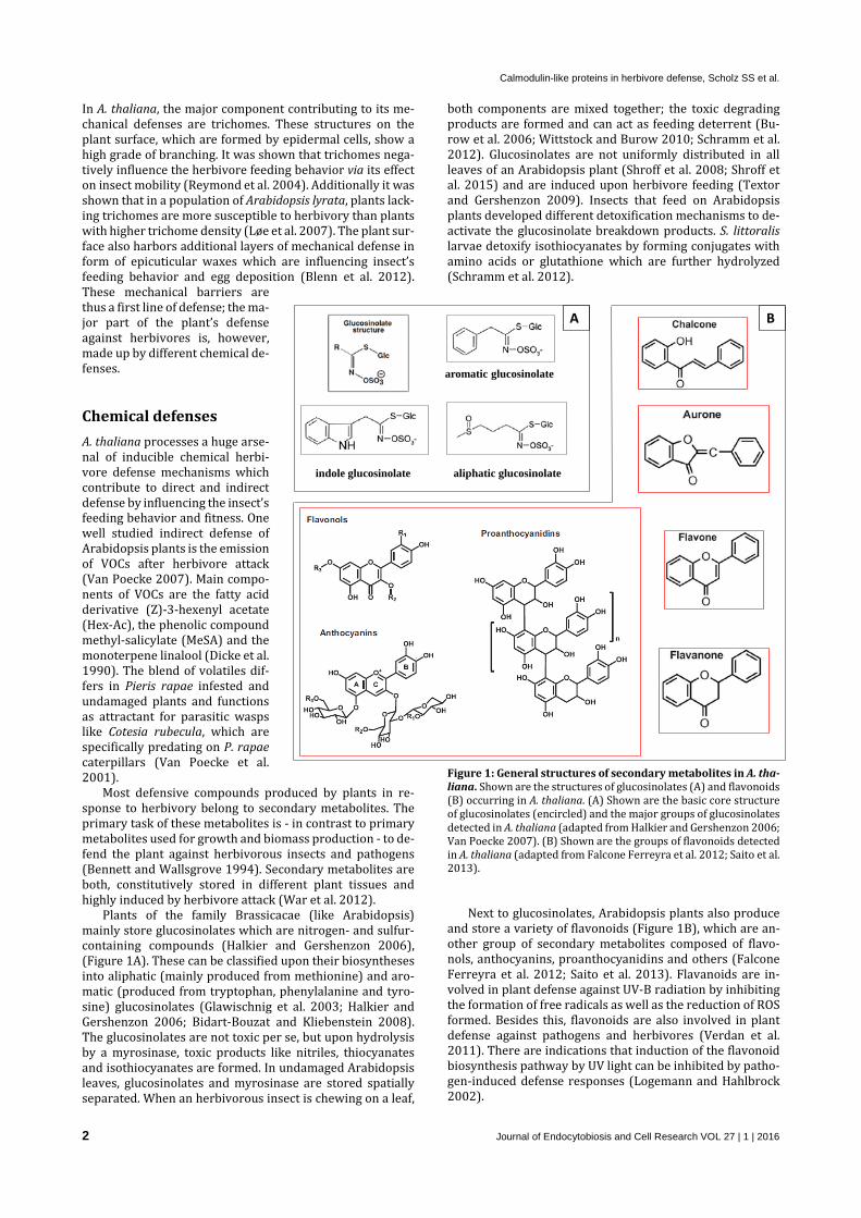
Plants of the family Brassicacae (like Arabidopsis) mainly store glucosinolates which are nitrogen- and sulfur-containing compounds (Halkier and Gershenzon 2006), (Figure 1A). These can be classified upon their biosyntheses into aliphatic (mainly produced from methionine) and aro-matic (produced from tryptophan, phenylalanine and tyro-sine) glucosinolates (Glawischnig et al. 2003; Halkier and Gershenzon 2006; Bidart-Bouzat and Kliebenstein 2008). The glucosinolates are not toxic per se, but upon hydrolysis by a myrosinase, toxic products like nitriles, thiocyanates and isothiocyanates are formed. In undamaged Arabidopsis leaves, glucosinolates and myrosinase are stored spatially separated. When an herbivorous insect is chewing on a leaf,
both components are mixed together; the toxic degrading products are formed and can act as feeding deterrent (Bu-row et al. 2006; Wittstock and Burow 2010; Schramm et al. 2012). Glucosinolates are not uniformly distributed in all leaves of an Arabidopsis plant (Shroff et al. 2008; Shroff et al. 2015) and are induced upon herbivore feeding (Textor and Gershenzon 2009). Insects that feed on Arabidopsis plants developed different detoxification mechanisms to de-activate the glucosinolate breakdown products. S. littoralis larvae detoxify isothiocyanates by forming conjugates with amino acids or glutathione which are further hydrolyzed (Schramm et al. 2012).
Figure 1: General structures of secondary metabolites in A. tha-liana. Shown are the structures of glucosinolates (A) and flavonoids (B) occurring in A. thaliana. (A) Shown are the basic core structure of glucosinolates (encircled) and the major groups of glucosinolates detected in A. thaliana (adapted from Halkier and Gershenzon 2006; Van Poecke 2007). (B) Shown are the groups of flavonoids detected in A. thaliana (adapted from Falcone Ferreyra et al. 2012; Saito et al. 2013).
Next to glucosinolates, Arabidopsis plants also produce
and store a variety of flavonoids (Figure 1B), which are an-other group of secondary metabolites composed of flavo-nols, anthocyanins, proanthocyanidins and others (Falcone Ferreyra et al. 2012; Saito et al. 2013). Flavanoids are in-volved in plant defense against UV-B radiation by inhibiting the formation of free radicals as well as the reduction of ROS formed. Besides this, flavonoids are also involved in plant defense against pathogens and herbivores (Verdan et al. 2011). There are indications that induction of the flavonoid biosynthesis pathway by UV light can be inhibited by patho-gen-induced defense responses (Logemann and Hahlbrock 2002).
B
aliphatic glucosinolateindole glucosinolate
aromatic glucosinolate
A

Calmodulin-like proteins in herbivore defense, Scholz SS et al.
Journal of Endocytobiosis and Cell Research VOL 27 | 1 | 2016 3
Anti-herbivore proteins Many defense compounds like anti-insect proteins produced by the plant act directly on the metabolism or development of feeding insects. So are by the plant produced protease in-hibitors (Molina-Rueda et al. 2015) able to disturb the diges-tion of ingested food material (Green and Ryan 1972) and, as a consequence of this, to slow down the development of the insect (Ryan 1990). Well studied defensive proteins pro-duced by Arabidopsis are the translated products encoded of JA-responsive genes VSP2, THI2.1 and PDF1.2. In previous studies it was demonstrated that VSP2 (VEGETATIVE STORAGE PROTEIN2) is induced by wounding, methyl jasmonate, insect feeding, and phosphate deprivation. The VSP2 protein shows phosphatase activity in acid pH range corresponding to the pH of insect gut lumen. Here, VSP2 could significantly delay development of the insects and in-crease their mortality (Berger et al. 1995; Liu et al. 2005). The expression of VSP2 could also be inhibited by neomycin application in Arabidopsis (Vadassery et al. 2014). Another JA-responsive gene induced by wounding of plant tissue and methyl jasmonate is THI2.1, encoding the antimicrobial pro-tein thionin which might also contribute to herbivore de-fense (Epple et al. 1997; Bohlmann et al. 1998; Vignutelli et al. 1998). PDF1.2, encoding another defensin in Arabidopsis is also activated upon methyl jasmonate and Spodoptera feeding (Manners et al. 1998; De Coninck et al. 2010; Kan-chiswamy et al. 2010). GABA as possible player in plant-herbivore defense γ-amino butyric acid is well studied as a neurotransmitter in invertebrates. After coupling, GABA-mediated Cl- channels are opened and the signal is transduced (Bown et al. 2006). In plants, the non-protein amino acid GABA (Figure 2A) plays a role in regulation of C/N balance and plant growth and development (Palanivelu et al. 2003; Bouche and Fromm 2004; Mirabella et al. 2008). Beside this, it was hy-pothesized that GABA has a possible role in plant defense. Excess supply of GABA could lead to hyper activation of the Cl- channels leading to paralysis of the attacking insect (Bown et al. 2006). So it was shown that high content of GABA in the insect’s diet causes developmental restrictions by increasing time to pupation (Bown et al. 2006).
GABA is mainly produced by decarboxylation of L-gluta-
mate catalyzed by glutamate decarboxylases (GADs) in the cytosol (Turano and Fang 1998; Zik et al. 1998). The catabo-lism of GABA into alanine and succinic semialdehyde (Figure 2B) is localized in the mitochondrial matrix, where a GABA transaminase (GABA-T) removes the amino group of GABA and transfers it onto pyruvate. The succinic semialdehyde
than is exported from mitochondria or oxidized to succinate by succinic semialdehyde dehydrogenase (SSADH) (Breitkreuz et al. 2003; Ludewig et al. 2008; Michaeli et al. 2011).
Under normal conditions, the activity of GADs and with this the accumulation of GABA, is regulated by Ca2+ and cal-modulin. Upon stimuli-induced cytosolic calcium elevation, calmodulins (CaMs) bind to calcium, and interact with GADs by coupling to their CaM-binding site (Snedden et al. 1995). Under stress conditions like disruption of plant tissue, GADs are strongly induced by an acidification of the cytosol (Wal-lace et al. 1984; Carroll et al. 1994; Ramputh and Bown 1996). This observation combined with the fact that Choris-toneura rosaceana larvae reared on GABA-containing diet show reduced weight gain, are hints for an involvement in herbivore defense (Ramputh and Bown 1996; Shelp et al. 1999; Bown et al. 2006). Additionally, it was observed that feeding and even walking behavior of Heliothis virescens lar-vae on Nicotiana tabacum leaves increases the content of GABA in the leaf tissue (Bown et al. 2002). Till now, the tem-poral and spatial accumulation of GABA after herbivore at-tack is still unknown.
Plant-herbivore interaction The recognition of a feeding herbivore starts seconds and minutes after the stimulus is perceived (Figure 3). Each her-bivore bears a number of herbivore-associated molecular patterns (HAMPs), which are – as first step in the signaling cascade - recognized by the plant through an array of spe-cialized putative receptors (Mithöfer and Boland 2008). Af-ter the receptor binding, a depolarization of the membrane occurs which is associated with an influx of calcium ions from external and internal stores into the cytosol (Maffei et al. 2007a; Vadassery et al. 2012a).
The spikes in cytosolic calcium levels [Ca2+]cyt are de-coded by different calcium sensor proteins, which interact with their target proteins to initiate the downstream signal-ing (DeFalco et al. 2010). An accumulation of herbivory- and wounding-related phytohormones like jasmonates, or the production of reactive oxygen species (ROS) are part of this cascade. As a consequence, metabolic changes like produc-tion of anti-herbivore peptides (Ryan 1990; Zavala et al. 2004) or defensive substances like nicotine (Steppuhn et al. 2004) and glucosinolates (Müller et al. 2010) are induced.
Figure 2: Structure, syntheses and metabolism of γ-amino bu-tyric acid (GABA, adapted from Ludewig et al. 2008). Shown is the molecular structure of the non-protein amino acid GABA (A) and the metabolism and catabolism of GABA (B). GAD glutamate decar-boxylase, GABA-T GABA transaminase, SSA succinic semialdehyde, SSADH succinic semialdehyde dehydrogenase.

Calmodulin-like proteins in herbivore defense, Scholz SS et al.
4 Journal of Endocytobiosis and Cell Research VOL 27 | 1 | 2016
Figure 3: Early events in plant-herbivore interaction (adapted from Maffei et al. 2007a). Shown are the first steps of plant herbi-vore perception, which occurs in the first minutes and hours after attack. After the stimulus perception, the membrane depolarizes and initiates a spike in the cytosolic Ca2+ level. This activates a sig-naling cascade which leads among others to ROS production and the accumulation of phytohormones. These induce the expression of re-sponsive genes and transcripts which in turn can modulate the plants metabolism. Herbivore-associated molecular patterns (HAMPs) and receptors The plant recognizes attacks by perception of different mo-lecular patterns (MPs), which are associated with the outer surface or released components of the aggressor (Taylor et al. 2004; Ausubel 2005; Mithöfer and Boland 2008). It is known that the conserved microbe-specific molecules, re-ferred to as microbe- or pathogen-associated molecular pat-terns (MAMPs or PAMPs), are recognized by pattern recog-nition receptors (PRRs). MAMPs like flagellin (Flg22), elon-gation factor Tu (EF-Tu), peptidoglycan (PGN), lipopolysac-charides (LPS), Ax21 (Activator of Xa 21-mediated immunity in rice), fungal chitin, and β-glucans from oomycetes are rec-ognized by plant surface localized PRRs (Jones and Dangl 2006; Newman et al. 2013; Ranf et al. 2015). Herbivore-as-sociated molecular patterns (HAMPs) are postulated to be present in insect oral secretions and are of two kinds: (i) chemical elicitors derived from insect oral secretions and oviposition fluids; and (ii) plant-derived self-recognition fac-tors, DAMPs (damage-associated molecular patterns) occur-ring due to a specific pattern of wounding (Mithöfer and Bo-land 2008; Heil and Land 2014). Insect OS contain elicitors, notable examples are inceptins, which are peptides formed as proteolytic products of plant chloroplastic ATP synthase formed in caterpillar midgut, and fatty acid-amino acid con-jugates (FACs) such as volicitin in maize (Alborn et al. 1997).
Upon herbivore attack, the plants encounter two main components of herbivore feeding: the wounding of plant tis-sue and recognition of elicitors in OS. HAMPs include the oral secretion of the larvae composed of saliva and reguritant,
damaged plant parts, ingested and me-tabolized phytohormones and other components like volicitin, (Alborn et al. 1997; Maffei et al. 2004; Wu and Bald-win 2010). Recently, a Porin-like pro-tein was identified as elicitor in S. litto-ralis OS that originated from the insects gut microbiota (Guo et al. 2013). The released quantity of these HAMPs and the leaf area injured may be different for distinct insect feeding styles, which causes a different plant response (Ali and Agrawal 2012). While insects with piercing sucking mouthparts like aphids cause only small wounds on plant tissue, chewing insects induce a much stronger lesion. The binding of all these HAMPs to unidentified PRRs is the first step of a complex signaling cas-cade, enabling the plant to react fast and efficient to different environmen-tal stimuli. Membrane depolarization
Next to the disrupted cells at the brink of the leaf area fed on, neighboring plant cells also respond to environmental stresses by changes in plasma transmembrane potential (Ebel and Mithöfer 1998; Maffei et al. 2004)). For Lima bean (Phaseolus lunatus) it was shown, that Vm changes induced by herbivores were much greater compared to those of sin-gle wounding and could travel throughout the whole leaf (Maffei et al. 2006; Maffei et al. 2007b). Vm changes are rec-orded as action potentials or system potentials, which can propagate the signal over longer distances (Maffei et al. 2007b; Zimmermann et al. 2009). Stress induced Vm changes (depolarization) can also modulate ion fluxes at the plasma membrane by activation of voltage-dependent chan-nels, like Ca2+ channels (White 2000; Maffei et al. 2007a). Mousavi et al. (2013) showed in Arabidopsis that for the propagation of electrical signals probably glutamate recep-tor-like genes are necessary. Finally, the electrical signals are able to induce JA-Ile elevation in systemic leaves (Mousavi et al. 2013). The second messenger calcium ions (Ca2+) The calcium ion (Ca2+) plays an important role as a second messenger in varied signaling networks of plant cells (Dodd et al. 2010). Plant cells maintain a level of 100-200 nM free cytosolic calcium [Ca2+]cyt, the so called Ca2+ homeostasis. This incident is due to the fact that high concentrations of cytosolic Ca2+ have a cytotoxic effect on phosphate-contain-ing components, including proteins and nucleic acids. To maintain this low level of Ca2+ in the cytosol, several active transporters like Ca2+-ATPases (ACAs) located in organelle- and cell membranes pump the Ca2+ into the stores (Sze et al. 2000). The Ca2+ is stored in high concentrations (105 times higher than cytosolic concentration) in different intra- and extracellular stores. While the apoplast serves as external calcium store, different organelles like the vacuole or chlo-roplasts store Ca2+ inside the cell (Knight et al. 1996; Peiter 2011; Stael et al. 2011). This high gradient of Ca2+ concentra-tions is the basis for a fast response to stress stimuli. Here an

Calmodulin-like proteins in herbivore defense, Scholz SS et al.
Journal of Endocytobiosis and Cell Research VOL 27 | 1 | 2016 5
influx of Ca2+ from the stores into the cytosol induces a cal-cium signature, whose specific shape, amplitude and dura-tion encode the information perceived (Lecourieux et al. 2006; McAinsh and Pittman 2009; Dodd et al. 2010). To achieve a specific decoding of Ca2+ signals both in the nu-cleus and the cytosol, the plant processes an arsenal of dif-ferent calcium sensor proteins (DeFalco et al. 2010). Calcium sensors In Arabidopsis the most studied groups of calcium sensor proteins are calmodulins (CaMs), calmodulin-like proteins (CMLs), calcineurin B-like pro-teins (CBLs) and calcium-dependent protein ki-nases (CDPKs, now renamed as CPKs), shown in Figure 4 (DeFalco et al. 2010).
In general, calcium sensor proteins found in Arabidopsis can be classified - in sense of mode of action - into two groups: sensor responders and sensor relays (DeFalco et al. 2010). Sensor responders bind the cytosolic free Ca2+, un-dergo conformational changes and actively reg-ulate downstream signaling by their own enzy-matic activity. The family of CPKs, Ca2+ sensors involved in e.g. ABA and herbivore defense sig-naling, belongs to this group (Wu and Baldwin 2010; Romeis and Herde 2014). So it was shown, that Arabidopsis cpk3 and cpk13 mu-tants express significantly less JA-responsive genes making them more susceptible to Spodoptera feeding (Kanchiswamy et al. 2010). Silencing of CPK4 and CPK5 in Nicotiana attenuata plants in contrast caused higher accu-mulation of JA and reduced growth of Manduca sexta larvae (Hettenhausen et al. 2013b; Yang et al. 2014).
Sensor relay proteins in contrast do not contain any en-zymatic domain. After binding of calcium and conforma-tional shift, they need to physically interact with target pro-teins to transfer the signal perceived. CaMs/CMLs and CBLs can be assigned to this group (DeFalco et al. 2010).
CBLs form complexes with CIPKs (CBL-interacting pro-tein kinases) and regulate membrane channels and trans-porters (Batistič and Kudla 2004). The function of CBLs is still not well understood since the knowledge about CBL-interacting proteins is limited. First results show that CBLs are involved in salt stress signaling (Batistič and Kudla 2009). Here, CBL1 and CBL9 are involved in K+ uptake by ac-tivation of a K+ -transporter under low-K+ conditions (Xu et al. 2006) and CBL4 (also SOS3) activates an H+/Na+ ex-changer (also SOS1) under high salt stress (Halfter et al. 2000).
The induction pattern of CAMs and CMLs is better under-stood (McCormack and Braam 2003; McCormack et al. 2005). CAMs, which are very similar to animal CAMs, do not show strong transcript abundance changes in the response to diverse stimuli. Only for CAM2 (also TCH1) it was ob-served that the expression was induced by touch (Braam and Davis 1990; Lee et al. 2005). The group of CMLs is in-volved in the regulation of diverse signaling pathways (McCormack et al. 2005).
Calmodulin-like proteins CMLs are one class of calcium sensor proteins, which act as sensor relays where they are propagating the Ca2+ signal. To
achieve this, CMLs contain a number of EF-hands (1-6), he-lix-loop-helix structures, which are responsible for high-af-finity cooperative binding of Ca2+. After binding, CMLs un-dergo a conformational change and can interact with their target proteins (Kawasaki et al. 1998; McCormack and Braam 2003; Clapham 2007; Gifford et al. 2007). In Ara-bidopsis, the class of CMLs consists of 50 members (Figure 5), which show at least 16% sequence identity to CAMs. Analysis of a neighbor-joining tree, based on amino acid sim-ilarities, showed that CMLs cluster in 9 groups (McCormack et al. 2005).
Figure 4. Different classes of calcium sensor proteins activated upon abiotic and biotic stimuli (adapted from DeFalco et al. 2010). Shown are the classes of Ca2+ sensor used to decode cytosolic Ca2+ spikes induced by diverse stimuli. Here, calcium sensor pro-teins function as signal relays (CaM/CMLs and CBLs) or primary re-sponders (CDPKs).
While the seven CAM genes in Arabidopsis are very uni-
formly expressed at a high transcript level, the CMLs show various expression patterns over different tissues and devel-opmental stages of the plant while the transcript levels are quite low. These observed expression patterns do not corre-late with the identified CML groups (McCormack et al. 2005). While CMLs like CML8, 9, 24, 42 are expressed in all major plant organs (Delk et al. 2005; Magnan et al. 2008; Park et al. 2010; Vadassery et al. 2012a), other CMLs show a very spe-cific expression in a single plant organ. So it was shown that in A. thaliana, CML43 is only expressed in roots (Bender et al. 2014). Other CMLs show a specific subcellular localisa-tion, for example CML30 is targeted to mitochondria and CML3 to peroxisomes (Chigri et al. 2012). CML39 is mostly expressed during early seedling establishment (Bender et al. 2013) and CML12 (also TCH3) is expressed in growing tis-sues (Sistrunk et al. 1994). These observations indicate that CMLs might be involved in a tissue- and growth stage-spe-cific decoding of Ca2+ signals.
It was demonstrated that the expression of CMLs is in-duced by diverse abiotic as well as biotic stimuli. So is CML8 induced by SA and salt stress (Park et al. 2010). CML9 is also induced by SA as well as by infection with P. syringae and can alter plant responses to ABA and abiotic stress (Magnan et al. 2008; Leba et al. 2012). CML24 modulates ABA level dur-ing ion stress, regulates pollen tube growth and can induce changes in flowering time (Delk et al. 2005; Hubbard et al. 2008; Yang et al. 2014). Additionally it was shown that ex-pression of CML37, CML38 and CML39 are regulated by salt- and drought stress, phytohormones and P. syringae infection

Calmodulin-like proteins in herbivore defense, Scholz SS et al.
6 Journal of Endocytobiosis and Cell Research VOL 27 | 1 | 2016
(Vanderbeld and Snedden 2007) and CML42 is involved in trichome branching (Dobney et al. 2009). Recently, it was shown that one member of the CML-family, CML42, is involved in A. thaliana defense against S. littoralis herbivory.
CML42 acts as a negative regulator of plant defense against herbivory and affects JA perception of the plant. CML42 gene expression is herbivore elicitor-specific and is not activated upon mechanical wounding (Vadassery et al. 2012a). It was additionally observed that the gene expres-sion of eight CMLs is induced by insect OS (Figure 5, arrows, Vadassery et al. 2012b; Scholz et al. 2014). In contrast,
CML37 was identified as positive regulator of plant defense against herbivory. This CML is strongly induced by mechan-ical wounding and was shown to downregulate the most bi-oactive form of jasmonates, the isoleucine-jasmonic acid conjugate JA-Ile (Scholz et al. 2014). The exact position of CMLs in the signaling cascade and the further processing of the signal by target proteins are still unknown.
Figure 5: Neighbour-joining tree of CMLs based on amino-acid similarities. CMLs induced by OS are indicated by arrows (Vadassery et al. 2012b; Scholz et al 2014).
CML37
CML42
CML16 CML17
CML12
CML11 CML9
CML23

Calmodulin-like proteins in herbivore defense, Scholz SS et al.
Journal of Endocytobiosis and Cell Research VOL 27 | 1 | 2016 7
Downstream signaling The downstream signaling components of plant herbivore defense are not completely known, but it became obvious that several signaling pathways are activated. So are activa-tion of mitogen-activated protein kinases (MAPKs), accumu-lation of JA and expression of JA-dependent genes, and the production of ROS involved (Wu and Baldwin 2010).
The production of ROS, which include superoxide anion (O2−), hydrogen peroxide (H2O2), singlet oxygen (1O2), and hydroxyl radical (·OH), is well studied as a part of plant re-sponse to pathogens (Lamb and Dixon 1997). In recent stud-ies it became clear that ROS production is also involved in herbivore defense. Medicago truncatula plants accumulated ROS only after herbivory while wounding did not induce ROS production (Leitner et al. 2005). In lima bean plants (Phaseolus lunatus) it was similarly shown that the produc-tion of ROS after herbivory was much higher than that after mechanical wounding alone (Maffei et al. 2006). So showed soybean plants challenged with Helicoverpa zea an elevated lipid peroxidation and OH radical formation (Bi and Felton 1995).
Another early signaling event after herbivore attack is also the activation of MAPKs, which play critical roles in plant resistance to herbivores by reshaping the JA pathway and the transcriptome (Hettenhausen et al. 2015). These ac-tivated MAPKs phosphorylate their substrates, which in-clude transcription factors and enzymes (Hazzalin and Ma-hadevan 2002). It was shown that fatty acid conjugates (FACs), elicitors in insect OS, induce the MAPKs in the wounded leaf of treated Nicotiana attenuata plants (Wu et al. 2007). Interestingly, activation of MAPK4 in N. attenuata shows herbivore specific pattern. While OS of M. sexta in-duced MAPK4 and decreased JA accumulation, S. littoralis OS did not induce a change in JA level (Hettenhausen et al. 2013a). In A. thaliana, grasshopper (Schistocerca gregaria) OS was also able to activate MAPKs, MPK3 and MPK6 (Schäfer et al. 2011).
Very powerful tools mediating plant defense are phyto-hormones, endogenous signaling compounds. Several groups of phytohormones (Figure 6) play important roles in plant growth and development. Next to the regulation and coordination of developmental processes, plant hormones are essential for the adaption to the abiotic and biotic envi-ronment (Bari and Jones 2009).
In plant defense against herbivory, the most important and most studied class of phytohor-mones is the one of JAs (Wasternack 2007). JAs are lipid-derived molecules originating from plastid membrane-bound α-linolenic acid. The JA biosyn-thetic pathway is well understood and the enzymes partici-pating in it are well characterized (Vick and Zimmerman 1984; Schaller and Stintzi 2009). In the chloroplast, the re-leased α-linolenic acid is metabolized in several steps to form OPC-8:0 followed by cis-OPDA, which is catalyzed by
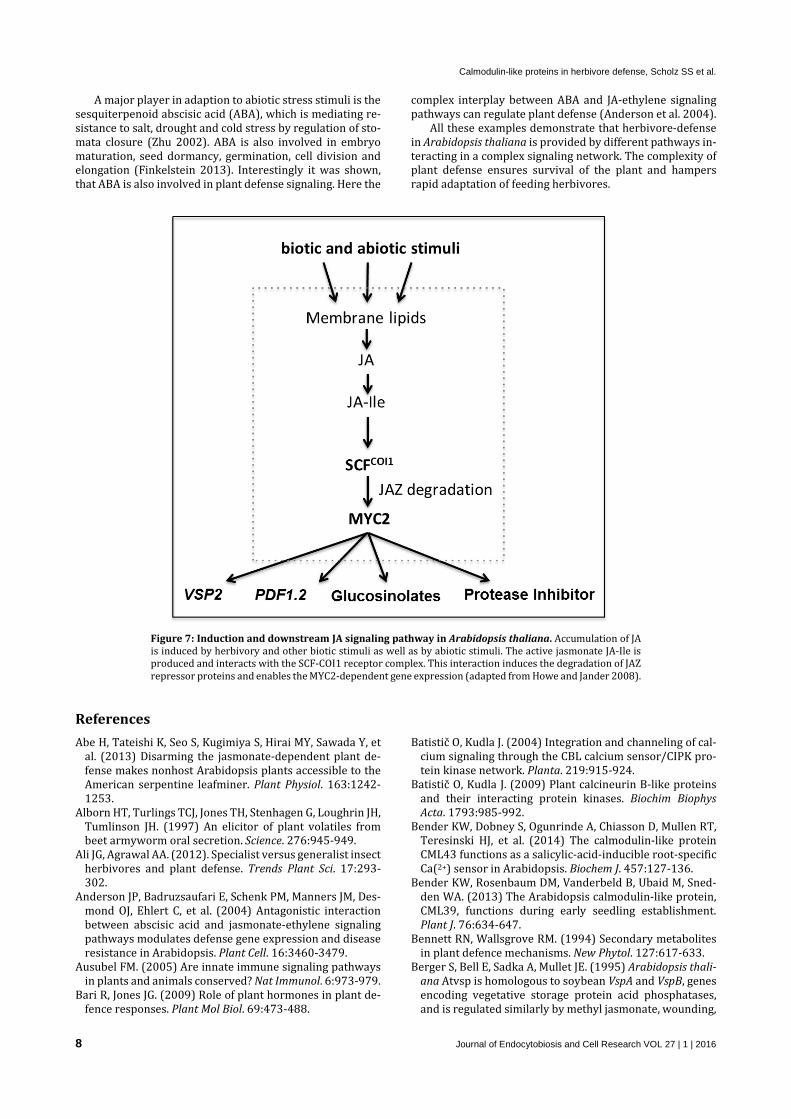
lipoxygenase (LOX), allene oxide synthase (AOS), and allene oxide cyclase (AOC). After a translocation to the peroxisome, the cis-OPDA is further processed to form JA (Schaller and Stintzi 2009). The active phytohormone JA-Ile is formed by a conjugation of JA and the amino acid isoleucine catalyzed by JASMONATE RESISTANT1, JAR1 (Staswick and Tiryaki 2004). Activation of the receptor complex SCF-COI1 by JA-Ile (Figure 7) triggers the degradation of JAZ proteins, the tran-scriptional repressors of JA responsive genes. This removal of repression leads to activation of the transcription factor MYC2 and the expression of anti-insect JA-responsive genes including PDF1.2, Thi2.1 and VSP2 (Wasternack and Kombrink 2010). Mutants of receptor COI1 like coi1-1 and coi1-16, jar1 and jaz1 show higher susceptibility to herbi-vore feeding (Feys et al. 1994; Chung et al. 2008; Westphal et al. 2008; Chung et al. 2009; Abe et al. 2013).
The production of JAs and the subsequent signaling in re-sponse to herbivore attack is triggered by wounding and as-sociated osmotic stress as well as by elicitors originating from the insect (Turner et al. 2002; Maffei et al. 2004; Mith-öfer et al. 2005a). Production and accumulation of JAs is a very strong and effective defense reaction against feeding in-sects, since the response to triggers starts very fast. So it was shown for Arabidopsis leaves that JA accumulation starts al-ready 2-5 minutes after wounding of the plant tissue (Glauser et al. 2008). Additionally it was shown in lima bean leaves that the area fed by S. littoralis larvae, contained a high level of JAs, while the surrounding plant tissue showed lower content of JAs (Schulze et al. 2007). To further analyze the dynamic and downstream signaling of JAs, structural mimics like coronalon (Figure 2 inlet) were applied in previ-ous studies. It was demonstrated that coronalon could suc-cessfully induce plant defense reactions like secondary me-tabolites and the expression of defense-genes (Schüler et al. 2001; Schüler et al. 2004; Pluskota et al. 2007; Nakamura et al. 2014).
In recent studies, it was shown that also cytokinins (CK), which are involved in resistance to abiotic stress like drought or nutrient availability and senescence signaling, have a possible role in plant herbivore defense. In N. attenu-ata, CK levels and several genes in the signaling cascade were induced by Manduca sexta OS and wounding (Schäfer et al. 2015).
Figure 6: Structure of different phytohormone classes. Shown are the basic structures of phytohormone classes and the structural JA-Ile mimic coronalon in comparison to JA-Ile itself (inlet).
Calmodulin-like proteins in herbivore defense, Scholz SS et al.
8 Journal of Endocytobiosis and Cell Research VOL 27 | 1 | 2016
A major player in adaption to abiotic stress stimuli is the sesquiterpenoid abscisic acid (ABA), which is mediating re-sistance to salt, drought and cold stress by regulation of sto-mata closure (Zhu 2002). ABA is also involved in embryo maturation, seed dormancy, germination, cell division and elongation (Finkelstein 2013). Interestingly it was shown, that ABA is also involved in plant defense signaling. Here the
complex interplay between ABA and JA-ethylene signaling pathways can regulate plant defense (Anderson et al. 2004).
All these examples demonstrate that herbivore-defense in Arabidopsis thaliana is provided by different pathways in-teracting in a complex signaling network. The complexity of plant defense ensures survival of the plant and hampers rapid adaptation of feeding herbivores.
Figure 7: Induction and downstream JA signaling pathway in Arabidopsis thaliana. Accumulation of JA is induced by herbivory and other biotic stimuli as well as by abiotic stimuli. The active jasmonate JA-Ile is produced and interacts with the SCF-COI1 receptor complex. This interaction induces the degradation of JAZ repressor proteins and enables the MYC2-dependent gene expression (adapted from Howe and Jander 2008).
References Abe H, Tateishi K, Seo S, Kugimiya S, Hirai MY, Sawada Y, et
al. (2013) Disarming the jasmonate-dependent plant de-fense makes nonhost Arabidopsis plants accessible to the American serpentine leafminer. Plant Physiol. 163:1242-1253.
Alborn HT, Turlings TCJ, Jones TH, Stenhagen G, Loughrin JH, Tumlinson JH. (1997) An elicitor of plant volatiles from beet armyworm oral secretion. Science. 276:945-949.
Ali JG, Agrawal AA. (2012). Specialist versus generalist insect herbivores and plant defense. Trends Plant Sci. 17:293-302.
Anderson JP, Badruzsaufari E, Schenk PM, Manners JM, Des-mond OJ, Ehlert C, et al. (2004) Antagonistic interaction between abscisic acid and jasmonate-ethylene signaling pathways modulates defense gene expression and disease resistance in Arabidopsis. Plant Cell. 16:3460-3479.
Ausubel FM. (2005) Are innate immune signaling pathways in plants and animals conserved? Nat Immunol. 6:973-979.
Bari R, Jones JG. (2009) Role of plant hormones in plant de-fence responses. Plant Mol Biol. 69:473-488.
Batistič O, Kudla J. (2004) Integration and channeling of cal-cium signaling through the CBL calcium sensor/CIPK pro-tein kinase network. Planta. 219:915-924.
Batistič O, Kudla J. (2009) Plant calcineurin B-like proteins and their interacting protein kinases. Biochim Biophys Acta. 1793:985-992.
Bender KW, Dobney S, Ogunrinde A, Chiasson D, Mullen RT, Teresinski HJ, et al. (2014) The calmodulin-like protein CML43 functions as a salicylic-acid-inducible root-specific Ca(2+) sensor in Arabidopsis. Biochem J. 457:127-136.
Bender KW, Rosenbaum DM, Vanderbeld B, Ubaid M, Sned-den WA. (2013) The Arabidopsis calmodulin-like protein, CML39, functions during early seedling establishment. Plant J. 76:634-647.
Bennett RN, Wallsgrove RM. (1994) Secondary metabolites in plant defence mechanisms. New Phytol. 127:617-633.
Berger S, Bell E, Sadka A, Mullet JE. (1995) Arabidopsis thali-ana Atvsp is homologous to soybean VspA and VspB, genes encoding vegetative storage protein acid phosphatases, and is regulated similarly by methyl jasmonate, wounding,

Calmodulin-like proteins in herbivore defense, Scholz SS et al.
Journal of Endocytobiosis and Cell Research VOL 27 | 1 | 2016 9
sugars, light and phosphate. Plant Mol Biol. 27:933-942. Bergomaz R, Boppre M. (1986) A simple instant diet for rear-
ing arctiidae and other moths. J Lepid Soc. 40:131-137. Bi JL, Felton GW. (1995) Foliar oxidative stress and insect
herbivory: Primary compounds, secondary metabolites, and reactive oxygen species as components of induced re-sistance. J Chem Ecol. 21:1511-1530.
Bidart-Bouzat MG, Kliebenstein DJ. (2008) Differential levels of insect herbivory in the field associated with genotypic variation in glucosinolates in Arabidopsis thaliana. J Chem Ecol. 34:1026-1037.
Blenn B, Bandoly M, Küffner A, Otte T, Geiselhardt S, Fatou-ros N, et al. (2012) Insect egg deposition induces indirect defense and epicuticular wax changes in Arabidopsis thali-ana. J Chem Ecol. 38:882-892.
Bohlmann H, Vignutelli A, Hilpert B, Miersch O, Wasternack C, Apel K. (1998) Wounding and chemicals induce expres-sion of the Arabidopsis thaliana gene Thi2.1, encoding a fungal defense thionin, via the octadecanoid pathway. FEBS Lett. 437:281-286.
Bouche N, Fromm H. (2004) GABA in plants: just a metabo-lite? Trends Plant Sci. 9:110-115.
Bown AW, Hall DE, MacGregor KB. (2002) Insect footsteps on leaves stimulate the accumulation of 4-Aminobutyrate and can be visualized through increased chlorophyll fu-orescence and superoxide production. Plant Physiol. 129:1430-1434.
Bown AW, Macgregor KB, Shelp BJ. (2006) Gamma-amino-butyrate: defense against invertebrate pests? Trends Plant Sci. 11:424-427.
Braam J, Davis RW. (1990) Rain-, wind-, and touch-induced expression of calmodulin and calmodulin-related genes in Arabidopsis. Cell. 60:357-364.
Breitkreuz KE, Allan WL, Van Cauwenberghe OR, Jakobs C, Talibi D, Andre B, et al. (2003) A novel gamma-hydroxy-butyrate dehydrogenase: identification and expression of an Arabidopsis cDNA and potential role under oxygen de-ficiency. J Biol Chem. 278:41552-41556.
Burow M, Muller R, Gershenzon J, Wittstock U. (2006) Al-tered glucosinolate hydrolysis in genetically engineered Arabidopsis thaliana and its influence on the larval devel-opment of Spodoptera littoralis. J Chem Ecol. 32:2333-2349.
Carroll AD, Fox GG, Laurie S, Phillips R, Ratcliffe RG, Stewart GR. (1994) Ammonium asimilation and the role of γ-Ami-nobutyric Acid in pH homeostasis in carrot cell suspen-sions. Plant Physiol. 106:513-520.
Chigri F, Flosdorff S, Pilz S, Kolle E, Dolze E, Gietl C, et al. (2012) The Arabidopsis calmodulin-like proteins AtCML30 and AtCML3 are targeted to mitochondria and peroxisomes, respectively. Plant Mol Biol. 78:211-222.
Chung HS, Koo AJK, Gao XL, Jayanty S, Thines B, Jones AD, et al. (2008) Regulation and function of Arabidopsis JASMONATE ZIM-domain genes in response to wounding and herbivory. Plant Physiol. 146:952-964.
Chung HS, Niu YJ, Browse J, Howe GA. (2009) Top hits in con-temporary JAZ: An update on jasmonate signaling. Phyto-chemistry. 70:1547-1559.
Clapham DE. (2007) Calcium Signaling. Cell. 131:1047-1058. De Coninck BMA, Sels J, Venmans E, Thys W, Goderis IJWM, Carron D, et al. (2010) Arabidopsis thaliana plant defensin AtPDF1.1 is involved in the plant response to biotic stress. New Phytol. 187:1075-1088.
DeFalco TA, Bender KW, Snedden WA. (2010) Breaking the code: Ca2+ sensors in plant signalling. Biochem J. 425:27-40.
Delk NA, Johnson KA, Chowdhury NI, Braam J. (2005) CML24, regulated in expression by diverse stimuli, en-codes a potential Ca2+ sensor that functions in responses to abscisic acid, day length, and ion stress. Plant Physiol. 139:240-253.
Dicke M, Van Beek TA, Posthumus MA, Ben Dom N, Van Bokhoven H, De Groot A. (1990) Isolation and identifica-tion of volatile kairomone that affects acarine preda-torprey interactions Involvement of host plant in its pro-duction. J Chem Ecol. 16:381-396.
Dobney S, Chiasson D, Lam P, Smith SP, Snedden WA. (2009) The calmodulin-related calcium sensor CML42 plays a role in trichome branching. J Biol Chem. 284:31647-31657.
Dodd AN, Kudla J, Sanders D. (2010) The language of calcium signaling. Annu Rev Plant Biol. 61:593-620.
Ebel J, Mithöfer A. (1998) Early events in the elicitation of plant defence. Planta. 206:335-348.
Epple P, Apel K, Bohlmann H. (1997) Overexpression of an endogenous thionin enhances resistance of Arabidopsis against Fusarium oxysporum. Plant Cell. 9:509-520.
Falcone Ferreyra ML, Rius SP, Casati P. (2012) Flavonoids: biosynthesis, biological functions, and biotechnological applications. Front Plant Sci. 3:222.
Feys BJF, Benedetti CE, Penfold CN, Turner JG. (1994) Ara-bidopsis mutants selected for resistance to the phytotoxin Coronatine are male-sterile, insensitive to methyl jasmonate and resistant to a bacterial pathogen. Plant Cell. 6:751-759.
Finkelstein R. (2013) Abscisic Acid Synthesis and Response. The Arabidopsis Book. American Society of Plant Biolo-gists. 11:e0166.
Gifford JL, Walsh MP, Vogel HJ. (2007) Structures and metal-ion-binding properties of the Ca2+-binding helix-loop-helix EF-hand motifs. Biochem J. 405:199-221.
Glauser G, Grata E, Dubugnon L, Rudaz S, Farmer EE, Wolfender JL. (2008) Spatial and temporal dynamics of jasmonate synthesis and accumulation in Arabidopsis in response to wounding. J Biol Chem. 283:16400-16407.
Glawischnig E, Mikkelsen MD, Halkier BA. (2003) Glucosin-olates: Biosynthesis and Metabolism. In: Sulphur in Plants-Abrol Y, Ahmad A, eds.: Springer Netherlands. 145-162.
Green TR, Ryan CA. (1972) Wound-induced proteinase in-hibitor in plant leaves: A possible defense mechanism against insects. Science. 175:776-777.
Guo H, Wielsch N, Hafke JB, Svatos A, Mithöfer A, Boland W. (2013) A porin-like protein from oral secretions of Spodop-tera littoralis larvae induces defense-related early events in plant leaves. Insect Biochem Mol Biol. 43:849-858.
Halfter U, Ishitani M, Zhu J-K. (2000) The Arabidopsis SOS2 protein kinase physically interacts with and is activated by the calcium-binding protein SOS3. Proc Nat Acad Sci USA. 97:3735-3740.
Halkier BA, Gershenzon J. (2006) Biology and biochemistry of glucosinolates. Annu Rev Plant Biol. 57:303-333.
Hazzalin CA, Mahadevan LC. (2002) MAPK-regulated tran-scription: a continuously variable gene switch? Nat Rev Mol Cell Biol. 3:30-40.
Heil M, Land WG. (2014) Danger signals - damaged-self recognition across the tree of life. Front Plant Sci. 5.
Hettenhausen C, Schuman MC, Wu J. (2015) MAPK signaling: A key element in plant defense response to insects. Insect Sci. 22:157-164.
Hettenhausen C, Baldwin IT, Wu J. (2013a) Nicotiana atten-uata MPK4 suppresses a novel jasmonic acid (JA) signal-ing-independent defense pathway against the specialist in-sect Manduca sexta, but is not required for the resistance

Calmodulin-like proteins in herbivore defense, Scholz SS et al.
10 Journal of Endocytobiosis and Cell Research VOL 27 | 1 | 2016
to the generalist Spodoptera littoralis. New Phytol. 199:787-799.
Hettenhausen C, Yang DH, Baldwin IT, Wu J. (2013b) Cal-cium-dependent protein kinases, CDPK4 and CDPK5, affect early steps of jasmonic acid biosynthesis in Nicotiana at-tenuata. Plant Signal Behav. 8:e22784.
Howe GA, Jander G. (2008) Plant immunity to insect herbi-vores. Annu Rev Plant Biol. 59:41-66.
Hubbard K, Hotta C, Gardner M, Braam J, Webb A. (2008) The Arabidopsis thaliana Calmodulin-like protein CML24 is a regulator of rhythmic Ca2+ signalling and flowering time. Comp Biochem Physiol A Mol Integr Physiol.150:S153-153.
Jander G. (2014) Revisiting plant-herbivore co-evolution in the molecular biology era. In: Annual Plant Reviews: John Wiley & Sons, Ltd. 361-384.
Jones JD, Dangl JL. (2006) The plant immune system. Nature. 444:323-329.
Kanchiswamy CN, Takahashi H, Quadro S, Maffei ME, Bossi S, Bertea C, et al. (2010) Regulation of Arabidopsis defense responses against Spodoptera littoralis by CPK-mediated calcium signaling. BMC Plant Biol. 10:97.
Kawasaki H, Nakayama S, Kretsinger RH. (1998). Classifica-tion and evolution of EF-hand proteins. BioMetals. 11:277-295.
Knight H, Trewavas AJ, Knight MR. (1996) Cold calcium sig-naling in Arabidopsis involves two cellular pools and a change in calcium signature after acclimation. Plant Cell. 8:489-503.
Lamb C, Dixon RA. (1997) The oxidative burst in plant dis-ease resistance. Annu Rev Plant Physiol Plant Mol Biol. 48:251-275.
Lawlor D. (2011) Abiotic stress adaptation in plants. physio-logical, molecular and genomic foundation. Ann Bot. 107:vii-ix.
Leba L, Cheval C, Ortiz-Martin I, Ranty B, Beuzon CR, Galaud JP, et al. (2012) CML9, an Arabidopsis calmodulin-like pro-tein, contributes to plant innate immunity through a flagel-lin-dependent signalling pathway. Plant J. 71:976-989.
Lecourieux D, Ranjeva R, Pugin A. (2006) Calcium in plant defence-signalling pathways. New Phytol. 171:249-269.
Lee D, Polisensky DH, Braam J. (2005). Genome-wide identi-fication of touch- and darkness-regulated Arabidopsis genes: a focus on calmodulin-like and XTH genes. New Phy-tol. 165:429-444.
Leitner M, Boland W, Mithöfer A. (2005) Direct and indirect defences induced by piercing-sucking and chewing herbi-vores in Medicago truncatula. New Phytol. 167:597-606.
Liu Y, Ahn JE, Datta S, Salzman RA, Moon J, Huyghues-De-spointes B, et al. (2005) Arabidopsis vegetative storage protein is an anti-insect acid phosphatase. Plant Physiol. 139:1545-1556.
Løe G, Toräng P, Gaudeul M, Ågren J. (2007) Trichome pro-duction and spatiotemporal variation in herbivory in the perennial herb Arabidopsis lyrata. Oikos. 116:134-142.
Logemann E, Hahlbrock K. (2002) Crosstalk among stress re-sponses in plants: pathogen defense overrides UV protec-tion through an inversely regulated ACE/ACE type of light-responsive gene promoter unit. Proc Natl Acad Sci USA. 99:2428-2432.
Ludewig F, Hüser A, Fromm H, Beauclair L, Bouché N. (2008) Mutants of GABA transaminase (POP2) suppress the se-vere phenotype of succinic semialdehyde dehydrogenase (ssadh) mutants in Arabidopsis. PLoS ONE. 3:e3383.
Maffei M, Bossi S, Spiteller D, Mithöfer A, Boland W. (2004) Effects of feeding Spodoptera littoralis on lima bean leaves. I. Membrane potentials, intracellular calcium variations,
oral secretions, and regurgitate components. Plant Physiol. 134:1752-1762.
Maffei ME, Mithöfer A, Arimura G, Uchtenhagen H, Bossi S, Bertea CM, et al. (2006) Effects of feeding Spodoptera litto-ralis on lima bean leaves. III. Membrane depolarization and involvement of hydrogen peroxide. Plant Physiol. 140:1022-1035.
Maffei ME, Mithöfer A, Boland W. (2007a) Before gene ex-pression: early events in plant-insect interaction. Trends Plant Sci. 12:310-316.
Maffei ME, Mithöfer A, Boland W. (2007b) Insects feeding on plants: rapid signals and responses preceding the induc-tion of phytochemical release. Phytochemistry. 68:2946-2959.
Magnan F, Ranty B, Charpenteau M, Sotta B, Galaud JP, Aldon D. (2008). Mutations in AtCML9, a calmodulin-like protein from Arabidopsis thaliana, alter plant responses to abiotic stress and abscisic acid. Plant J. 56:575-589.
Manners J, Penninckx IMA, Vermaere K, Kazan K, Brown R, Morgan A, et al. (1998) The promoter of the plant defensin gene PDF1.2 from Arabidopsis is systemically activated by fungal pathogens and responds to methyl jasmonate but not to salicylic acid. Plant Mol Biol. 38:1071-1080.
McAinsh MR, Pittman JK. (2009) Shaping the calcium signa-ture. New Phytol. 181:275-294.
McCormack E, Braam J. (2003) Calmodulins and related po-tential calcium sensors of Arabidopsis. New Phytol. 159:585-598.
McCormack E, Tsai YC, Braam J. (2005) Handling calcium sig-naling: Arabidopsis CaMs and CMLs. Trends Plant Sci. 10:383-389.
Michaeli S, Fait A, Lagor K, Nunes-Nesi A, Grillich N, Yellin A, et al. (2011) A mitochondrial GABA permease connects the GABA shunt and the TCA cycle, and is essential for normal carbon metabolism. Plant J. 67:485-498.
Mirabella R, Rauwerda H, Struys EA, Jakobs C, Tri-antaphylides C, Haring MA, et al. (2008) The Arabidopsis her1 mutant implicates GABA in E-2-hexenal responsive-ness. Plant J. 53:197-213.
Mithöfer A, Boland W. (2008) Recognition of herbivory-as-sociated molecular patterns. Plant Physiol. 146:825-831.
Mithöfer A, Maitrejean M, Boland W. (2005a) Structural and biological diversity of cyclic octadecanoids, jasmonates, and mimetics. J Plant Growth Regul. 23:170-178.
Mithöfer A, Wanner G, Boland W. (2005b) Effects of feeding Spodoptera littoralis on lima bean leaves. II. Continuous mechanical wounding resembling insect feeding is suffi-cient to elicit herbivory-related volatile emission. Plant Physiol. 137:1160-1168.
Molina-Rueda JJ, Pascual MB, Pissarra J, Gallardo F. (2015) A putative role for gamma-aminobutyric acid (GABA) in vas-cular development in pine seedlings. Planta. 241:257-267.
Mousavi SAR, Chauvin A, Pascaud F, Kellenberger S, Farmer EE. (2013) GLUTAMATE RECEPTOR-LIKE genes mediate leaf-to-leaf wound signalling. Nature. 500:422-426.
Müller R, de Vos M, Sun JY, Sonderby IE, Halkier BA, Witt-stock U, et al. (2010) Differential effects of indole and ali-phatic glucosinolates on lepidopteran herbivores. J Chem Ecol. 36:905-913.
Nakamura Y, Paetz C., Brandt W, David A, Rendon-Anaya M, Herrera-Estrella A, et al. (2014) Synthesis of 6-substituted 1-oxoindanoyl isoleucine conjugates and modeling studies with the COI1-JAZ co-receptor complex of lima bean. J Chem Ecol. 40:687-699.
Newman MA, Sundelin T, Nielsen JT, Erbs G. (2013) MAMP (microbe-associated molecular pattern) triggered immun-

Calmodulin-like proteins in herbivore defense, Scholz SS et al.
Journal of Endocytobiosis and Cell Research VOL 27 | 1 | 2016 11
ity in plants. Front Plant Sci. 4:139. Palanivelu R, Brass L, Edlund AF, Preuss D. (2003) Pollen
tube growth and guidance is regulated by POP2, an Ara-bidopsis gene that controls GABA levels. Cell. 114:47-59.
Park HC, Park CY, Koo SC, Cheong MS, Kim KE, Kim MC, et al. (2010) AtCML8, a calmodulin-like protein, differentially activating CaM-dependent enzymes in Arabidopsis thali-ana. Plant Cell Rep. 29:1297-1304.
Peiter E. (2011) The plant vacuole: emitter and receiver of calcium signals. Cell Calcium. 50:120-128.
Pluskota WE, Qu N, Maitrejean M, Boland W, Baldwin IT. (2007) Jasmonates and its mimics differentially elicit sys-temic defence responses in Nicotiana attenuata. J Exp Bot. 58:4071-4082.
Ramputh AI, Bown AW. (1996) Rapid [gamma]-aminobu-tyric acid synthesis and the inhibition of the growth and development of oblique-banded leaf-roller larvae. Plant Physiol. 111:1349-1352.
Ranf S, Gisch N, Schaffer M, Illig T, Westphal L, Knirel YA, et al. (2015) A lectin S-domain receptor kinase mediates lip-opolysaccharide sensing in Arabidopsis thaliana. Nat Im-munol. 16:426-433.
Reymond P, Bodenhausen N, Van Poecke RMP, Krishna-murthy V, Dicke M, Farmer EE. (2004) A conserved tran-script pattern in response to a specialist and a generalist herbivore. Plant Cell. 16:3132-3147.
Romeis T, Herde M. (2014) From local to global: CDPKs in systemic defense signaling upon microbial and herbivore attack. Curr Opin Plant Biol. 20:1-10.
Ryan CA. (1990) Protease inhibitors in plants: Genes for im-proving defenses against insects and pathogens. Annu Rev Phytopathol. 28:425-449.
Saito K, Yonekura-Sakakibara K, Nakabayashi R, Higashi Y, Yamazaki M, Tohge T, et al. (2013) The flavonoid biosyn-thetic pathway in Arabidopsis: Structural and genetic di-versity. Plant Physiol Biochem. 72:21-34.
Schäfer M, Fischer C, Meldau S, Seebald E, Oelmüller R, Bald-win IT. (2011) Lipase activity in insect oral secretions me-diates defense responses in Arabidopsis. Plant Physiol. 156:1520-1534.
Schäfer M, Meza-Canales ID, Navarro-Quezada A, Brütting C, Vanková R, Baldwin IT, et al. (2015) Cytokinin levels and signaling respond to wounding and the perception of her-bivore elicitors in Nicotiana attenuata. J. Integr Plant Biol. 57:198-212.
Schaller A, Stintzi A. (2009) Enzymes in jasmonate biosyn-thesis - structure, function, regulation. Phytochemistry. 70:1532-1538.
Scholz SS, Vadassery J, Heyer M, Reichelt M, Bender KW, Snedden WA, Boland W, Mithöfer, A. (2014) Mutation of the Arabidopsis Calmodulin-Like Protein CML37 deregu-lates the jasmonate pathway and enhances susceptibility to herbivory. Mol Plant. 7:1712-26.
Schoonhoven LM, Jermy T, van Loon JJA. (1998) Insect-plant biology: from physiology to evolution. In: Insect-plant bi-ology: from physiology to evolution. London: Chapman & Hall. 1-409.
Schramm K, Vassao DG, Reichelt M, Gershenzon J, Wittstock U. (2012) Metabolism of glucosinolate-derived isothiocya-nates to glutathione conjugates in generalist lepidopteran herbivores. Insect Biochem Mol Biol. 42:174-182.
Schüler G, Görls H, Boland W. (2001) 6-Substituted indanoyl isoleucine conjugates mimic the biological activity of coro-natine. Eur J Org Chem. 2001:1663-1668.
Schüler G, Mithöfer A, Baldwin IT, Berger S, Ebel J, Santos JG, et al. (2004) Coronalon: a powerful tool in plant stress physiology. FEBS Lett. 563:17-22.
Schulze B, Dabrowska P, Boland W. (2007) Rapid enzymatic isomerization of 12-oxophytodienoic acid in the gut of lep-idopteran larvae. ChemBioChem. 8:208-216.
Shelp BJ, Bown AW, McLean MD. (1999) Metabolism and functions of gamma-aminobutyric acid. Trends Plant Sci. 4:446-452.
Shroff R, Schramm K, Jeschke V, Nemes P, Vertes A, Gershen-zon J, et al. (2015) Quantification of plant surface metabo-lites by matrix-assisted laser desorption-ionization mass spectrometry imaging: glucosinolates on Arabidopsis tha-liana leaves. Plant J. 81:961-972.
Shroff R, Vergara F, Muck A, Svatoš A, Gershenzon J. (2008) Nonuniform distribution of glucosinolates in Arabidopsis thaliana leaves has important consequences for plant de-fense. Proc Nat Acad Sci USA. 105:6196-6201.
Sistrunk ML, Antosiewicz DM, Purugganan MM, Braam J. (1994) Arabidopsis TCH3 encodes a novel Ca2+ binding protein and shows environmentally induced and tissue-specific regulation. Plant Cell. 6:1553-1565.
Snedden WA, Arazi T, Fromm H, Shelp BJ. (1995) Cal-cium/calmodulin activation of soybean glutamate decar-boxylase. Plant Physiol. 108:543-549.
Stael S, Wurzinger B, Mair A, Mehlmer N, Vothknecht UC, Teige M. (2011) Plant organellar calcium signalling: an emerging field. J Exp Bot. 63:1525-42.
Staswick PE, Tiryaki I. (2004) The oxylipin signal jasmonic acid is activated by an enzyme that conjugates it to isoleu-cine in Arabidopsis. Plant Cell. 16:2117-2127.
Steppuhn A, Gase K, Krock B, Halitschke R, Baldwin IT. (2004) Nicotine’s defensive function in nature. PLoS Biol. 2:e217.
Stowe KA, Marquis RJ, Hochwender CG, Simms EL. (2000) The evolutionary ecology of tolerance to consumer dam-age. Annu Rev Ecol Syst. 31:565-595.
Sze H, Liang F, Hwang I, Curran AC, Harper JF. (2000) Diver-sity and regulation of plant Ca2+ pumps: insights from ex-pression in yeast. Annu Rev Plant Physiol Plant Mol Biol. 51:433-462.
Taylor JE, Hatcher PE, Paul ND. (2004) Crosstalk between plant responses to pathogens and herbivores: a view from the outside in. J Exp Bot. 55:159-168.
Textor S, Gershenzon J. (2009) Herbivore induction of the glucosinolate-myrosinase defense system: major trends, biochemical bases and ecological significance. Phytochem Rev. 8:149-170.
Turano FJ, Fang TK. (1998) Characterization of two gluta-mate decarboxylase cDNA clones from Arabidopsis. Plant Physiol. 117:1411-1421.
Turner JG, Ellis C, Devoto A. (2002) The jasmonate signal pathway. Plant Cell Online. 14:S153-S164.
Vadassery J, Reichelt M, Jimenez-Aleman GH, Boland W, Mithöfer A. (2014) Neomycin inhibition of (+)-7-iso-jasmonoyl-L-isoleucine accumulation and signaling. J Chem Ecol. 40:676-686.
Vadassery J, Reichelt M, Hause B, Gershenzon J, Boland W, Mithöfer A. (2012a) CML42-mediated calcium signaling coordinates responses to Spodoptera herbivory and abi-otic stresses in Arabidopsis. Plant Physiol. 159:1159-1175.
Vadassery J, Scholz SS, Mithöfer A. (2012b) Multiple calmod-ulin-like proteins in Arabidopsis are induced by insect-de-rived (Spodoptera littoralis) oral secretion. Plant Signal Be-

Calmodulin-like proteins in herbivore defense, Scholz SS et al.
12 Journal of Endocytobiosis and Cell Research VOL 27 | 1 | 2016
hav. 7:1277-1280. Van Poecke RMP. (2007) Arabidopsis-insect interactions. In:
The Arabidopsis book. The American Society of Plant Biol-ogists. 1-34.
Van Poecke RP, Posthumus M, Dicke M. (2001) Herbivore-induced volatile production by Arabidopsis thaliana leads to attraction of the parasitoid Cotesia rubecula: Chemical, behavioral, and gene-expression analysis. J Chem Ecol. 27:1911-1928.
Vanderbeld B, Snedden WA. (2007) Developmental and stimulus-induced expression patterns of Arabidopsis cal-modulin-like genes CML37, CML38 and CML39. Plant Mol Biol. 64:683-697.
Verdan AM, Wang HC, Garcia CR, Henry WP, Brumaghim JL. (2011) Iron binding of 3-hydroxychromone, 5-hydroxy-chromone, and sulfonated morin: Implications for the an-tioxidant activity of flavonols with competing metal bind-ing sites. J Inorg Biochem. 105:1314-1322.
Vick BA, Zimmerman DC. (1984) Biosynthesis of jasmonic acid by several plant species. Plant Physiol. 75:458-461.
Vignutelli A, Wasternack C, Apel K, Bohlmann H. (1998) Sys-temic and local induction of an Arabidopsis thionin gene by wounding and pathogens. Plant J. 14:285-295.
Wallace W, Secor J, Schrader LE. (1984) Rapid accumulation of γ-aminobutyric acid and alanine in soybean leaves in re-sponse to an abrupt transfer to lower temperature, dark-ness, or mechanical manipulation. Plant Physiol. 75:170-175.
War AR, Paulraj MG, Ahmad T, Buhroo AA, Hussain B, Igna-cimuthu S, Sharma HC. (2012) Mechanisms of plant de-fense against insect herbivores. Plant Signal Behav. 7:1306-1320.
Wasternack C. (2007) Jasmonates: An update on biosynthe-sis, signal transduction and action in plant stress response, growth and development. Ann Bot. 100:681-697.
Wasternack C, Kombrink E. (2010) Jasmonates: structural requirements for lipid-derived signals active in plant stress responses and development. ACS Chem Biol. 5:63-77.
White P J. (2000). Calcium channels in higher plants. Biochim Biophys Acta. 1465:171-189.
Wittstock U, Burow M. (2010) Glucosinolate breakdown in Arabidopsis: Mechanism, regulation and biological signifi-cance. The Arabidopsis Book. American Society of Plant Bi-ologists. 8:e0134.
Wu J, Hettenhausen C, Meldau S, Baldwin IT. (2007) Her-bivory rapidly activates MAPK signaling in attacked and unattacked leaf regions but not between leaves of Nicoti-ana attenuata. Plant Cell. 19:1096-1122.
Wu JQ, Baldwin IT. (2010) New insights into plant responses to the attack from insect herbivores. Annu Rev Genet. 44:1-24.
Xu J, Li H-D., Chen L-Q, Wang Y, Liu L-L, He L, et al. (2006) A protein kinase, interacting with two calcineurin B-like pro-teins, regulates K+ transporter AKT1 in Arabidopsis. Cell. 125:1347-1360.
Yang X, Wang SS, Wang M, Qiao Z, Bao CC, Zhang W. (2014) Arabidopsis thaliana calmodulin-like protein CML24 regu-lates pollen tube growth by modulating the actin cytoskel-eton and controlling the cytosolic Ca(2+) concentration. Plant Mol Biol. 86:225-236.
Zavala JA, Patankar AG, Gase K, Hui D, Baldwin IT. (2004) Manipulation of endogenous trypsin proteinase inhibitor production in Nicotiana attenuata demonstrates their function as antiherbivore defenses. Plant Physiol. 134:1181-1190.
Zhu JK. (2002) Salt and drought stress signal transduction in plants. Annu Rev Plant Biol. 53:247-273.
Zik M, Arazi T, Snedden W, Fromm H. (1998) Two isoforms of glutamate decarboxylase in Arabidopsis are regulated by calcium/calmodulin and differ in organ distribution. Plant Mol Biol. 37:967-975.
Zimmermann MR, Maischak H, Mithöfer A, Boland W, Felle HH. (2009) System potentials, a novel electrical long-dis-tance apoplastic signal in plants, induced by wounding. Plant Physiol. 149:1593-1600.
Westphal L, Scheel D, Rosahl S. (2008) The coi1-16 mutant harbors a second site mutation rendering PEN2 nonfunc-tional. Plant Cell. 20:824-826.