a protein interaction node at the neurotransmitter release site
TRANSCRIPT
Cellular/Molecular
A Protein Interaction Node at the Neurotransmitter ReleaseSite: Domains of Aczonin/Piccolo, Bassoon, CAST, and RimConverge on the N-Terminal Domain of Munc13-1
Xiaolu Wang,1 Bin Hu,1 Agata Zieba,2 Nicole G. Neumann,2 Monika Kasper-Sonnenberg,1 Annegret Honsbein,2
Greta Hultqvist,2 Tim Conze,1,2 Wolfgang Witt,1 Christoph Limbach,2 Matthis Geitmann,3 Helena Danielson,3
Richard Kolarow,4 Gesa Niemann,4 Volkmar Lessmann,4,5 and Manfred W. Kilimann1,2
1Institut fur Physiologische Chemie, Ruhr-Universitat Bochum, D-44780 Bochum, Germany, Departments of 2Neuroscience and 3Biochemistry andOrganic Chemistry, Uppsala University, S-75124 Uppsala, Sweden, 4Institute for Physiology, University of Mainz, D-55128 Mainz, Germany, and 5Instituteof Physiology, Otto-von-Guericke University, D-39120 Magdeburg, Germany
Multidomain scaffolding proteins organize the molecular machinery of neurotransmitter vesicle dynamics during synaptogenesis andsynaptic activity. We find that domains of five active zone proteins converge on an interaction node that centers on the N-terminal regionof Munc13-1 and includes the zinc-finger domain of Rim1, the C-terminal region of Bassoon, a segment of CAST1/ELKS2, and the thirdcoiled-coil domain (CC3) of either Aczonin/Piccolo or Bassoon. This multidomain complex may constitute a center for the physical andfunctional integration of the protein machinery at the active zone. An additional connection between Aczonin and Bassoon is mediatedby the second coiled-coil domain of Aczonin. Recombinant Aczonin-CC3, expressed in cultured neurons as a green fluorescent proteinfusion protein, is targeted to synapses and suppresses vesicle turnover, suggesting involvements in synaptic assembly as well as activity.Our findings show that Aczonin, Bassoon, CAST1, Munc13, and Rim are closely and multiply interconnected, they indicate that Aczonin-CC3 can actively participate in neurotransmitter vesicle dynamics, and they highlight the N-terminal region of Munc13-1 as a hub ofprotein interactions by adding three new binding partners to its mechanistic potential in the control of synaptic vesicle priming.
IntroductionNeurotransmitter release is confined to a specialized area of thepresynaptic plasma membrane known as the “active zone.”Calcium-dependent exocytosis at the synapse is an elaborate,multistep process that is fast and precise yet highly restrained andsubject to modulation (presynaptic plasticity) (Wojcik and Brose,2007). Moreover, it is functionally coupled to re-endocytosis,which maintains the homeostasis of presynaptic membranes bythe retrieval of proteins and lipids into the vesicle compartment.A cytoskeletal-like, solubilization-resistant lattice of proteins as-sociated with the inner face of the presynaptic plasma membrane,the cytomatrix at the active zone (CAZ), is apparent in electronmicroscopy and seems to integrate and organize many proteins ofthe presynaptic machinery. However, our understanding of theprecise molecular architecture of active zones and of the mecha-nistic steps through which their molecular constituents interact isstill rudimentary (for review, see Gundelfinger et al., 2003; Zivand Garner, 2004; Schoch and Gundelfinger, 2006).
Five large multidomain proteins, or small protein families,have been identified that are believed to be important for theorganization and functioning of the molecular machinery ofmammalian active zones, both during the assembly of synapsesand in acute vesicle dynamics: Aczonin/Piccolo (550 kDa),Bassoon (420 kDa), CAST/ERC/ELKS (�100 kDa), Munc13(�190 kDa), and Rim (�170 kDa). Several physical interactionsof these five CAZ proteins with other proteins have been identi-fied. These include interactions between each other (Munc13with Rim; Rim with CAST; CAST with Bassoon and Aczonin),interactions with other proteins of presumed scaffolding func-tions (Munc13 with �-spectrin III �1; Aczonin with GIT; Bas-soon with CtBP1/Ribeye; CAST with liprin-� and syntenin-1;Rim with Rim-BPs, 14-3-3, and liprin-�), and interactions withproteins involved in vesicle traffic or fusion (Rim with Rab3,Rab6, synaptotagmin, SNAP-25, and the �1B calcium channelsubunit; Munc13 with syntaxin and DOC2; Aczonin and Bassoonwith PRA1), in cytoskeletal remodeling (Aczonin with profilinand Abp1), or in synaptic plasticity (Rim with cAMP–GEFII;Munc13 with calmodulin) (for references, see Schoch andGundelfinger, 2006). Numerous additional interactions can beexpected to be found, given the large sizes and multidomain ar-chitectures of the proteins involved. The emerging picture is thatof a highly interwoven protein network, and the giant proteinsAczonin and Bassoon are presumed to be of particular impor-tance as scaffolds for the structural and functional integration ofthe protein machinery of the active zone.
Received March 8, 2009; revised Aug. 19, 2009; accepted Aug. 21, 2009.This work was supported by grants from Deutsche Forschungsgemeinschaft, Fonds der Chemischen Industrie,
the intramural research funding program (FoRUM) of the University of Bochum Medical School, and startup fundsfrom the Faculty of Science and Technology of Uppsala University (M.W.K.).
Correspondence should be addressed to Manfred W. Kilimann, Department of Neuroscience, Box 593, UppsalaUniversity, S-75124 Uppsala, Sweden. E-mail: [email protected].
X. Wang’s present address: Evotec AG, NeuroScience, D-22525 Hamburg, Germany.DOI:10.1523/JNEUROSCI.1255-09.2009
Copyright © 2009 Society for Neuroscience 0270-6474/09/2912584-13$15.00/0
12584 • The Journal of Neuroscience, October 7, 2009 • 29(40):12584 –12596
In the present study, we have identified several new molecularinteractions among the CAZ proteins Aczonin, Bassoon, CAST1,and Munc13-1. Strikingly, in addition to the known binding ofthe Rim Zn-finger domain, several interactions converge on theN-terminal region of Munc13-1, highlighting it as a potential hubfor multilateral crosstalk between these proteins and suggesting ahigh degree of supramolecular organization at the neurotrans-mitter release site.
Materials and MethodscDNA sequences and vectors. The complete coding cDNA sequences ofmouse CAST1, Munc13-1, and Rim1 were amplified in overlapping seg-ments from mouse brain RNA by reverse transcription (RT)-PCR, ini-tially with rat primers. Sequences, including multiple splice variants ofCAST1 and Rim1, were deposited in GenBank under accession numbersAY356530-8 and AY753536. Glutathione S-transferase (GST) fusionproteins were expressed from the vector pGEX– 4T-2 (SmaI site; GEHealthcare). Fusion proteins with N-terminal His tags were expressedfrom pQE32 (SmaI site; Qiagen). Constructs with an N-terminal thiore-doxin fusion and a C-terminal His tag were in pBAD/Thio-TOPO (In-vitrogen). Construct Mu1:pet and the series Mu1 � 2-1 to Mu1 � 2-6were cloned into a derivative of pET3 (Novagen) engineered in our lab-oratory such that the recombinant protein begins with the natural startcodon of Munc13-1 and carries a C-terminal His tag. Enhanced GFP(EGFP) fusion constructs were cloned into the SmaI site of pEGFP–C1(Clontech).
Antibodies. cDNAs encoding Aczonin, Bassoon, CAST1, Munc13-1,and Rim1 partial sequences were amplified from mouse brain RNA byRT-PCR and inserted into the SmaI sites of the His-tagged vector pQE32(Qiagen) or the GST vector pGEX– 4T-2 (GE Healthcare). Recombinantproteins were expressed in bacteria and used for the immunization ofrabbits and guinea pigs. The following constructs were used as antigens:AczN (amino acids 63-218, LPK . . . IPK), Aczp18p19 (amino acids1808-2150, QKE . . . GVS), Cast4a (amino acids 711-957, EAS . . . IWA),Rim43 (amino acids 9-387�83-105, GPR . . . RYP), and Mu1 [amino acids2-320, SLL . . . QDE, as a thioredoxin fusion protein in pBAD/Thio-TOPO(Invitrogen)]. Sera were affinity purified on the same recombinant proteinsused for immunization. Anti-Aczonin immunoprecipitations (IPs) andWestern blot developments were performed with anti-Aczp18p19 unlessindicated otherwise. A mouse monoclonal antibody against rat Bassoon(amino acids 738-1035) was obtained from Nventa Biopharmaceuticals. ARim1 monoclonal antibody (clone 26; immunogen, amino acids 602-723 ofrat Rim1) was from BD Biosciences (“Rim1-BD”), and Munc13 monoclonalantibodies were purchased from BD Biosciences [clone 32; immunogen,amino acids 621-834 of rat Munc13-1 (“Munc13-BD”); described by thevendor as panMunc13 reactive] and Synaptic Systems [immunogen, aminoacids 1399-1736 of rat Munc13-1 (“Munc13-SS”); described by the vendoras Munc13-1 specific]. A rabbit antiserum against neurobeachin (Nbea) hasbeen described previously (Wang et al., 2000). Most other antibodies werepurchased from BD Biosciences.
Immunoprecipitation. Mouse brains were homogenized with a glass–Teflon homogenizer in 5 v/w of IP lysis buffer (20 mM Tris, pH 7.4, 150mM NaCl, 0.5 mM EDTA, 1 mM Na3VO4, 10 mM NaF, 2 mM PMSF, 2�g/ml pepstatin A, 2 �g/ml leupeptin, 2 �g/ml aprotinin, 2 mM benza-midine, 0.5% Triton X-100, and 0.5% BSA) and centrifuged at 120,000 �g and 4°C for 30 min. Supernatants were precleared with 1⁄60 vol of Pan-sorbin (Calbiochem) at 4°C for 1 h. Aliquots (400 �l) of precleared lysatewere incubated at 4°C for 1 h with 5–10 �l of crude sera, 5–10 �l ofmonoclonal antibodies, or 50 –100 �l of affinity-purified sera. A total of60 �l of 10% Pansorbin (preblocked with 3% BSA in IP lysis buffer at 4°Cfor 1 h) were added, and samples were incubated with rotation for anadditional hour. Pansorbin was sedimented at 10,000 � g for 1 min, andpellets were washed five times with 1 ml of IP lysis buffer and analyzed bySDS-PAGE and Western blotting.
Brain lysate pull-down assay. Rat brains were homogenized with aglass–Teflon homogenizer in pull-down tissue lysis buffer (50 mM Tris,pH 7.4, 150 mM NaCl, 4 mM MgCl2, 0.5 mM EDTA, 1 mM DTT, 0.5%Triton X-100, 0.5 mM PMSF, and 2 �g/ml each of pepstatin A, leupeptin,
and aprotinin) and centrifuged at 120,000 � g and 4°C for 30 min.Supernatants were precleared with 1⁄10 vol of glutathione agarose beads(prewashed with pull-down lysis buffer) for 4 h at 4°C and adjusted to aprotein concentration of 10 mg/ml with pull-down lysis buffer. Glutathi-one agarose beads were loaded with GST fusion proteins or unmodifiedGST (�20 �g of protein per 20 �l of beads), preblocked with 3% BSA inlysis buffer for 2 h at 4°C, and washed with 1 ml of lysis buffer. Aliquots(20 �l) of loaded beads were incubated with 300 �l aliquots of preclearedbrain lysate at 4°C overnight under end-over-end agitation. Beads werespun down, washed rapidly with 3� 1 ml of ice-cold lysis buffer, boiled inSDS-PAGE sample buffer, and analyzed by Western blotting using thechemiluminescence detection system. His-tagged fusion proteins wereimmobilized for pull-down assays by covalent coupling. Affi-Gel 15 (40�l; Bio-Rad) were incubated with 200 �l of fusion protein (0.4 –1 mg/ml)in coupling buffer (100 mM NaCl and 100 mM NaHCO3, pH 8.0) withrotation at 4°C overnight. Beads were washed with 1 ml of couplingbuffer, and remaining functional groups were blocked by incubationwith 400 �l of 0.2 M ethanolamine in coupling buffer at room tempera-ture for 2 h. After preblocking with 3% BSA and washing with lysis buffer,20 �l aliquots of beads (loaded with 15–30 �g of protein) were incubatedwith brain lysate as above.
Pull-down assays with recombinant proteins as soluble ligands. GST fu-sion proteins were expressed and bound to glutathione agarose accord-ing to the instructions of the manufacturer. They were not eluted fromthe affinity matrix after washing but kept under recombinant pull-downbuffer (20 mM Tris, pH 8.0, 150 mM NaCl, 1 mM EDTA, and 1% TritonX-100) with 5% BSA until use. His-tagged proteins (pQE, pBAD/Thio-TOPO, or pET based) were purified on nickel agarose, eluted with 300mM NaCl, 500 mM imidazole, 1% Triton X-100, and 50 mM Na-phosphate, pH 8.0, and kept in elution buffer until use on the next day. Ina pull-down reaction, typically 10 �l packed beads carrying 200 – 400pmol of GST fusion protein were incubated with 300 –1100 pmol ofHis-tagged ligand proteins, individually and in combinations added di-rectly in imidazole elution buffer, in 500 �l total volume [pull-downbuffer, adjusted to equal volumes of imidazole elution buffer as intro-duced by the ligand protein samples (45–75 �l) and supplemented with0.3– 0.75% BSA] for 2 h at room temperature under head-over-end ro-tation. After removal of the supernatant, beads were washed three timesfor 90 s in 1 ml of pull-down buffer without BSA, resuspended in SDSsample buffer, and processed for SDS-PAGE, Coomassie blue staining,and immunoblotting. Pull-down experiments with the thioredoxinMu1–Mu5 series (see Fig. 3A) and the thioredoxin Cast3c-1 to Cast3c-9series (see Fig. 3B) were performed with crude bacterial lysates of thesoluble ligands. C-terminal His-tagged fusion proteins (in pET andpBAD) were detected with anti-His(C-Term) monoclonal antibody(R930-25; Invitrogen) and N-terminal His-tagged fusion proteins (inpQE) with anti-RGS–His monoclonal (34610; Qiagen).
Surface plasmon resonance interaction analysis. The biosensor experi-ments were performed using a BIAcore 2000 instrument (GE Health-care). Flow cells on a CM5 chip (GE Healthcare) were activated with a 1:1mixture of 50 mM N-hydroxysuccinimide and 200 mM N-ethyl-N�-((dimethylamino)propyl)carbodiimide for 7 min. A polyclonal anti-GST antibody (27-4577-01; GE Healthcare) was diluted to 0.1 mg/mlinto 50 mM sodium acetate, pH 5, and injected over the surface to pro-duce antibody densities of 10 –12 kRU. The surfaces were subsequentlyblocked with ethanolamine (1 M, pH 8.5) and then postconditioned withthree 1-min injections of 50 mM NaOH. All immobilization steps wereperformed using a flow rate of 5 �l/min. Binding experiments were per-formed at 25°C using a running buffer composed of 20 mM Tris, pH 8.0,200 mM NaCl, 1 mM EDTA, and 0.1% Triton X-100. GST fusion proteins(0.5 mg/ml) were loaded on the antibody surface for 3 min at a flow rateof 10 �l/min, whereupon His6 –Cast3c or Mu1:pet was injected for 1 minat a flow rate of 30 ml/min. For analysis of the ternary complex (see Fig.4 B, C), an injection of 0.15 mg/ml His6 –Cast3c with a contact time of 2min was followed by a 1 min injection of Mu1:pet. The dissociation wasmonitored for 5 min before the surface was regenerated with 1 min pulsesof 50 mM glycine, pH 2.5, and 50 mM NaOH. Analytes were tested at fourconcentrations using a series of fivefold dilutions. For each experiment,response data were processed using a reference surface to correct for bulk
Wang et al. • Multidomain Complex of Active Zone Proteins J. Neurosci., October 7, 2009 • 29(40):12584 –12596 • 12585
refractive index changes and any nonspecific binding. Data were alsoreferenced using responses from blank analyte injections. To determinethe binding constants, association and dissociation phase data were glo-bally fitted to a heterogeneous interaction model (A � B � AB, A* � B �A*B) using the Biaevaluation software (GE Healthcare). The presentedSDs are based on replicates performed with two different surfaces andprotein preparations and sets of three and four concentrations in theglobal data analysis.
Expression of fluorescent-tagged recombinant proteins and FM dye label-ing in hippocampal microcultures, live-cell imaging. Dissociated postnatalrat hippocampal microcultures were prepared as described previously(Brigadski et al., 2005). Primary postnatal [postnatal day 0 (P0) to P2]neocortical astrocytes were isolated and cultured for 2-4 weeks inDMEM containing 10% fetal calf serum (FCS) until confluence. Astro-cytes were passaged and seeded on glass coverslips at a density of 80,000cells per 3.5 cm dish in DMEM/10% FCS, to yield spherical astrocyteislands of 100 –300 �m in diameter after 7–14 d in vitro (DIV). Postnatalrat (P0 –P2) hippocampal neurons were plated in DMEM/10% FCS at adensity of 1-10 neurons per astrocyte island onto the astrocyte coverslips.After 20 h, the plating medium was exchanged to serum-free medium(Neurobasal with 2% B27 supplement; Invitrogen).
Cultures were transfected with the respective expression plasmids at8 –9 DIV, using the Ca 2� phosphate precipitation method as describedpreviously (Haubensak et al., 1998). During incubation (3 h), 10 �M
DNQX and 100 �M D,L-APV were added. Cells were used for experiments1-2 d after transfection (9 –11 DIV). The accumulation of EGFP–Acz7-2and EGFP–Rim5/8 at synapses became visible 12 h after transfection.However, additional diffuse labeling of the soma, axons, and dendriteswas also evident throughout the expression period. Neurons did nottolerate well expression of the fusion constructs beyond 60 h after trans-fection. DsRed2–VAMP2 and EGFP–VAMP2 were generated by PCRfrom a rat VAMP2 cDNA plasmid (Brigadski et al., 2005). To identifypresynaptic structures in EGFP–Acz7-2- and EGFP–Rim5/8-expressinghippocampal neurons, cells were cotransfected with DsRed2–VAMP(DNA ratio of EGFP/DsRed, 1:1). Activity-dependent labeling of presyn-aptic terminals in hippocampal neurons was performed as describedpreviously (Klau et al., 2001; Mohrmann et al., 2003). Briefly, coverslipswith hippocampal neurons were incubated for 1 min at room tempera-ture in HEPES-buffered saline (HBS) containing 50 mM K � (replacingan equal amount of Na �), 2 mM Ca 2�, 1 mM Mg 2�, and 10 �M FM4-64[N-(3-triethylammoniumpropyl)-4-(6-(4- diethylamino)phenyl)hexatrie-nyl)pyridinium dibromide] (Invitrogen). Cells were washed four times indye-free HBS with low K� (4 mM) and without Ca2� and were transferredinto the recording chamber of an upright microscope (BX51WI; Olympus).
Colocalization of EGFP-labeled presynaptic proteins with DsRed2–VAMP2 or FM4-64 was investigated with a Nipkow spinning-diskconfocal system (Visitech), attached to a conventional fluorescencemicroscope (BX51WI; Olympus), and equipped with a 60� water-immersion objective (numerical aperture 0.90). Glass coverslips withtransfected cells were mounted in the recording chamber of the micro-scope. Full-frame images were taken with a cooled CCD camera (Cool-Snap HQ; Roper Scientific). Green and red fluorescence was excited withthe 488 and 568 nm lines of a krypton/argon laser (Laser Physics), respec-tively. Exposure times were chosen such that saturation was avoided.Image capture and data analysis was performed using MetaMorph soft-ware (Molecular Devices ) and Adobe Photoshop software (Adobe Sys-tems), without compromising the evident primary image information.However, in the figures, fluorescence is enhanced close to saturation tofacilitate the visibility of synaptic structures. Red and green fluorescentpictures, respectively, were taken from the identical field of view, thresh-olded, and converted to binary pictures. The percentage of greenpunctate structures colocalizing with the red fluorescence signal wascalculated for each field of view. Average values were determined from atleast five independent experiments (n � 3 cells per experiment analyzed)for each combination of construct and staining procedure. All calcula-tions were performed with MetaMorph software (Molecular Devices)and Excel (Microsoft).
ResultsAczonin, Bassoon, CAST, Munc13, and Rimcoimmunoprecipitate from brain lysateThe molecular layout of Aczonin as a giant multidomain proteinsuggests a role as a scaffolding protein in the molecular organiza-tion of the active zone. We therefore probed for binding betweenAczonin and other presynaptic proteins by coimmunoprecipita-tion from brain lysate. The majority of Aczonin cosediments withan insoluble subcellular fraction (Wang et al., 1999; Fenster et al.,2000), but �10% remain in the 100,000 � g supernatant of abrain lysate, which was used in the following immunoprecipita-tion and pull-down experiments. Anti-Aczonin antibodies selec-tively coprecipitated Bassoon, CAST, Munc13, and Rim (Fig.1A). Conversely, antibodies against Bassoon, Munc13, and Rimcoprecipitated the respective other proteins (Fig. 1B). These re-sults were confirmed with precipitating antibodies against twoother sequence regions of Aczonin, a second sequence region ofMunc13-1 and a second epitope of Rim1 (Fig. 1B, and data notshown). The corresponding preimmune sera and several otherantibodies [cadherin, syntaxin, CASK, neurobeachin, palmdel-phin, protein kinase A (PKA) subunit RII�] did not coprecipitateAczonin, Bassoon, CAST, Munc13, or Rim (Fig. 1A,B, and datanot shown).
We probed the immunoprecipitates with antibodies againstnumerous presynaptic proteins (Fig. 1A, and data not shown:synaptophysin, synaptogyrin, synaptobrevin-2, SNAP-25, cadherin,syntaxin, CASK, synapsin-1, Munc18, rabphilin-3, syndapin-1,complexin-2, tomosyn, NSF, �/�-SNAP, neurobeachin, �-NAP,and PKA–RII�) but did not detect additional binding partners.This does not exclude that some of these proteins do bind toAczonin, Bassoon, Munc13, or Rim but evade detection in thisassay. For example, we did not detect the published binding ofsyntaxin to Munc13 (Betz et al., 1997) or of SNAP-25 to Rim(Coppola et al., 2001) under our conditions. However, we con-firmed the coprecipitation of synaptobrevin-2 and tomosyn withanti-syntaxin (Fujita et al., 1998; Hatsuzawa et al., 2003). Theobserved association between Aczonin, Bassoon, CAST, Munc13,and Rim therefore seems to be very robust. Note, in particular,the strong enrichment of Bassoon and CAST from the lysate byAczonin immunoprecipitation. These results indicate the exis-tence of a very stable complex encompassing all five proteinsthat are characteristic for and highly enriched at active zones(in brief, “active zone-specific”): Aczonin, Bassoon, CAST,Munc13, and Rim.
The second coiled-coil domain of Aczonin precipitatesBassoon, a region around the third coiled-coil domain ofAczonin precipitates CAST, Munc13, and Rim, and a regionnear the Bassoon C-terminus precipitates Munc13 and RimImmobilized recombinant partial Aczonin sequences, collec-tively representing most of the Aczonin molecule (Fig. 1C)(supplemental Table 1, available at www.jneurosci.org as sup-plemental material), were used in pull-down experiments withbrain lysate (Fig. 1D). Aczonin construct Acz5 (amino acids3059-3336, DIN . . . PRN) precipitated native Bassoon, andAczonin construct Acz7 � 8 (amino acids 3593-4121; SRA . . . SSS)precipitated native CAST, Munc13, and Rim. As positive con-trols, fusion proteins with the zinc-finger domain of Rim1 pre-cipitated Munc13 only (Betz et al., 2001; Wang et al., 2001).Confirming the specificity and avidity of these interactions, noother Aczonin binding partners could be detected in the precip-itates with antibodies against numerous additional presynap-tic candidate proteins [Aczonin itself, synapsin-1 (Fig. 1D),
12586 • J. Neurosci., October 7, 2009 • 29(40):12584 –12596 Wang et al. • Multidomain Complex of Active Zone Proteins
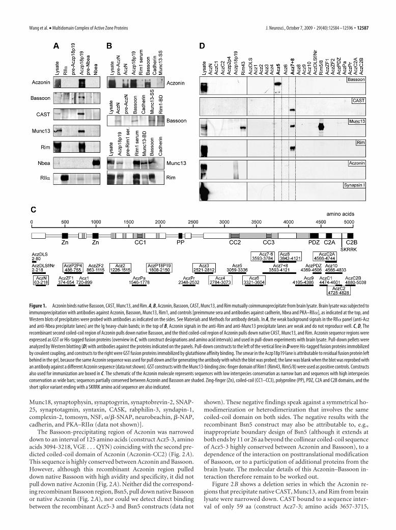
Munc18, synaptophysin, synaptogyrin, synaptobrevin-2, SNAP-25, synaptotagmin, syntaxin, CASK, rabphilin-3, syndapin-1,complexin-2, tomosyn, NSF, �/�-SNAP, neurobeachin, �-NAP,cadherin, and PKA–RII� (data not shown)].
The Bassoon-precipitating region of Aczonin was narroweddown to an interval of 125 amino acids (construct Acz5-3, aminoacids 3094-3218, VGE . . . QYN) coinciding with the second pre-dicted coiled-coil domain of Aczonin (Aczonin-CC2) (Fig. 2A).This sequence is highly conserved between Aczonin and Bassoon.However, although this recombinant Aczonin region pulleddown native Bassoon with high avidity and specificity, it did notpull down native Aczonin (Fig. 2A). Neither did the correspond-ing recombinant Bassoon region, Bsn5, pull down native Bassoonor native Aczonin (Fig. 2A), nor could we detect direct bindingbetween the recombinant Acz5-3 and Bsn5 constructs (data not
shown). These negative findings speak against a symmetrical ho-modimerization or heterodimerization that involves the samecoiled-coil domain on both sides. The negative results with therecombinant Bsn5 construct may also be attributable to, e.g.,inappropriate boundary design of Bsn5 (although it extends atboth ends by 11 or 26 aa beyond the collinear coiled-coil sequenceof Acz5-3 highly conserved between Aczonin and Bassoon), to adependence of the interaction on posttranslational modificationof Bassoon, or to a participation of additional proteins from thebrain lysate. The molecular details of this Aczonin–Bassoon in-teraction therefore remain to be worked out.
Figure 2B shows a deletion series in which the Aczonin re-gions that precipitate native CAST, Munc13, and Rim from brainlysate were narrowed down. CAST bound to a sequence inter-val of only 59 aa (construct Acz7-3; amino acids 3657-3715,
Figure 1. Aczonin binds native Bassoon, CAST, Munc13, and Rim. A, B, Aczonin, Bassoon, CAST, Munc13, and Rim mutually coimmunoprecipitate from brain lysate. Brain lysate was subjected toimmunoprecipitation with antibodies against Aczonin, Bassoon, Munc13, Rim1, and controls [preimmune sera and antibodies against cadherin, Nbea and PKA–RII�], as indicated at the top, andWestern blots of precipitates were probed with antibodies as indicated on the sides. See Materials and Methods for antibody details. In A, the weak background signals in the RII� panel (anti-Aczand anti-Nbea precipitate lanes) are the Ig heavy-chain bands; in the top of B, Aczonin signals in the anti-Rim and anti-Munc13 precipitate lanes are weak and do not reproduce well. C, D, Therecombinant second coiled-coil region of Aczonin pulls down native Bassoon, and the third coiled-coil region of Aczonin pulls down native CAST, Munc13, and Rim. Aczonin sequence regions wereexpressed as GST or His-tagged fusion proteins (overview in C, with construct designations and amino acid intervals) and used in pull-down experiments with brain lysate. Pull-down pellets wereanalyzed by Western blotting (D) with antibodies against the proteins indicated on the panels. Pull-down constructs to the left of the vertical line in D were His-tagged fusion proteins immobilizedby covalent coupling, and constructs to the right were GST fusion proteins immobilized by glutathione affinity binding. The smear in the Aczp18p19 lane is attributable to residual fusion protein leftbehind in the gel, because the same Aczonin sequence was used for pull down and for generating the antibody with which the blot was probed; the lane was blank when the blot was reprobed withan antibody against a different Aczonin sequence (data not shown). GST constructs with the Munc13-binding zinc-finger domain of Rim1 (Rim43, Rim5/8) were used as positive controls. Constructsalso used for immunization are boxed in C. The schematic of the Aczonin molecule represents sequences with low interspecies conservation as narrow bars and sequences with high interspeciesconservation as wide bars; sequences partially conserved between Aczonin and Bassoon are shaded. Zing-finger (Zn), coiled-coil (CC1–CC3), polyproline (PP), PDZ, C2A and C2B domains, and theshort splice variant ending with a SKRRK amino acid sequence are also indicated.
Wang et al. • Multidomain Complex of Active Zone Proteins J. Neurosci., October 7, 2009 • 29(40):12584 –12596 • 12587
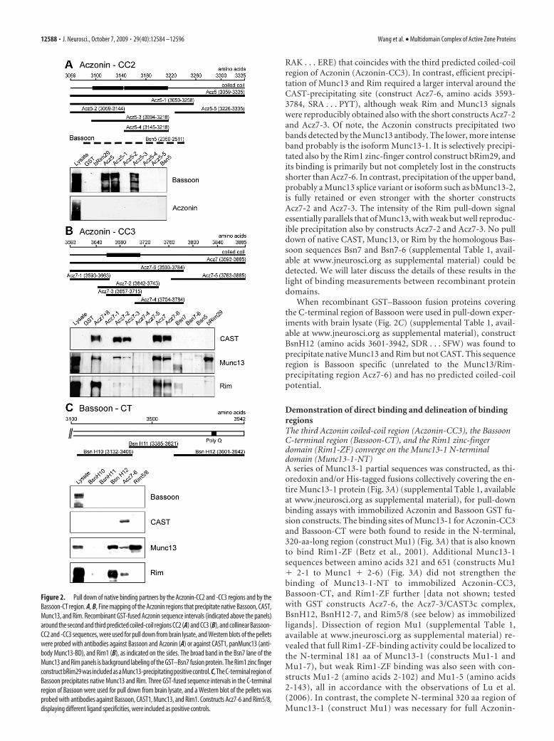
RAK . . . ERE) that coincides with the third predicted coiled-coilregion of Aczonin (Aczonin-CC3). In contrast, efficient precipi-tation of Munc13 and Rim required a larger interval around theCAST-precipitating site (construct Acz7-6, amino acids 3593-3784, SRA . . . PYT), although weak Rim and Munc13 signalswere reproducibly obtained also with the short constructs Acz7-2and Acz7-3. Of note, the Aczonin constructs precipitated twobands detected by the Munc13 antibody. The lower, more intenseband probably is the isoform Munc13-1. It is selectively precipi-tated also by the Rim1 zinc-finger control construct bRim29, andits binding is primarily but not completely lost in the constructsshorter than Acz7-6. In contrast, precipitation of the upper band,probably a Munc13 splice variant or isoform such as bMunc13-2,is fully retained or even stronger with the shorter constructsAcz7-2 and Acz7-3. The intensity of the Rim pull-down signalessentially parallels that of Munc13, with weak but well reproduc-ible precipitation also by constructs Acz7-2 and Acz7-3. No pulldown of native CAST, Munc13, or Rim by the homologous Bas-soon sequences Bsn7 and Bsn7-6 (supplemental Table 1, avail-able at www.jneurosci.org as supplemental material) could bedetected. We will later discuss the details of these results in thelight of binding measurements between recombinant proteindomains.
When recombinant GST–Bassoon fusion proteins coveringthe C-terminal region of Bassoon were used in pull-down exper-iments with brain lysate (Fig. 2C) (supplemental Table 1, avail-able at www.jneurosci.org as supplemental material), constructBsnH12 (amino acids 3601-3942, SDR . . . SFW) was found toprecipitate native Munc13 and Rim but not CAST. This sequenceregion is Bassoon specific (unrelated to the Munc13/Rim-precipitating region Acz7-6) and has no predicted coiled-coilpotential.
Demonstration of direct binding and delineation of bindingregionsThe third Aczonin coiled-coil region (Aczonin-CC3), the BassoonC-terminal region (Bassoon-CT), and the Rim1 zinc-fingerdomain (Rim1-ZF) converge on the Munc13-1 N-terminaldomain (Munc13-1-NT)A series of Munc13-1 partial sequences was constructed, as thi-oredoxin and/or His-tagged fusions collectively covering the en-tire Munc13-1 protein (Fig. 3A) (supplemental Table 1, availableat www.jneurosci.org as supplemental material), for pull-downbinding assays with immobilized Aczonin and Bassoon GST fu-sion constructs. The binding sites of Munc13-1 for Aczonin-CC3and Bassoon-CT were both found to reside in the N-terminal,320-aa-long region (construct Mu1) (Fig. 3A) that is also knownto bind Rim1-ZF (Betz et al., 2001). Additional Munc13-1sequences between amino acids 321 and 651 (constructs Mu1� 2-1 to Munc1 � 2-6) (Fig. 3A) did not strengthen thebinding of Munc13-1-NT to immobilized Aczonin-CC3,Bassoon-CT, and Rim1-ZF further [data not shown; testedwith GST constructs Acz7-6, the Acz7-3/CAST3c complex,BsnH12, BsnH12-7, and Rim5/8 (see below) as immobilizedligands]. Dissection of region Mu1 (supplemental Table 1,available at www.jneurosci.org as supplemental material) re-vealed that full Rim1-ZF-binding activity could be localized tothe N-terminal 181 aa of Munc13-1 (constructs Mu1-1 andMu1-7), but weak Rim1-ZF binding was also seen with con-structs Mu1-2 (amino acids 2-102) and Mu1-5 (amino acids2-143), all in accordance with the observations of Lu et al.(2006). In contrast, the complete N-terminal 320 aa region ofMunc13-1 (construct Mu1) was necessary for full Aczonin-
Figure 2. Pull down of native binding partners by the Aczonin-CC2 and -CC3 regions and by theBassoon-CT region. A, B, Fine mapping of the Aczonin regions that precipitate native Bassoon, CAST,Munc13, and Rim. Recombinant GST-fused Aczonin sequence intervals (indicated above the panels)around the second and third predicted coiled-coil regions CC2 (A) and CC3 (B), and collinear Bassoon-CC2 and -CC3 sequences, were used for pull down from brain lysate, and Western blots of the pelletswere probed with antibodies against Bassoon and Aczonin (A) or against CAST1, panMunc13 (anti-body Munc13-BD), and Rim1 (B), as indicated on the sides. The broad band in the Bsn7 lane of theMunc13 and Rim panels is background labeling of the GST–Bsn7 fusion protein. The Rim1 zinc fingerconstructbRim29wasincludedasaMunc13-precipitatingpositivecontrol. C,TheC-terminalregionofBassoon precipitates native Munc13 and Rim. Three GST-fused sequence intervals in the C-terminalregion of Bassoon were used for pull down from brain lysate, and a Western blot of the pellets wasprobed with antibodies against Bassoon, CAST1, Munc13, and Rim1. Constructs Acz7-6 and Rim5/8,displaying different ligand specificities, were included as positive controls.
12588 • J. Neurosci., October 7, 2009 • 29(40):12584 –12596 Wang et al. • Multidomain Complex of Active Zone Proteins
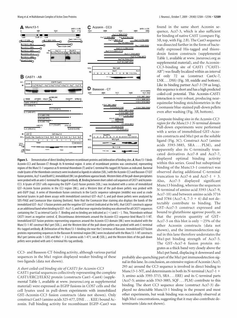
CC3- and Bassoon-CT-binding activity, although various partialsequences in the Mu1 region displayed weaker binding of thesetwo ligands (data not shown).
A short coiled-coil binding site of CAST1 for Aczonin-CC3CAST1 partial sequences collectively representing the completeCAST1/ERC2/ELKS2 protein (constructs Cast1–Cast4) (supple-mental Table 1, available at www. jneurosci.org as supplementalmaterial) were expressed as EGFP fusions in COS7 cells and thecell lysates used in pull-down experiments with immobilizedGST–Aczonin-CC3 fusion constructs (data not shown). Onlyconstruct Cast3 (amino acids 325-677, DNE . . . KEE) bound Ac-zonin. Full binding activity for recombinant EGFP–Cast3 was
found in the same short Aczonin se-quence, Acz7-3, which is also sufficientfor binding of native CAST (compare Fig.3B, top, with Fig. 2B). The Cast3 sequencewas dissected further in the form of bacte-rially expressed His-tagged and thiore-doxin fusion constructs (supplementalTable 1, available at www. jneurosci.org assupplemental material), and the Aczonin-CC3-binding site of CAST1 (“CAST1-AB”) was finally localized within an intervalof only 72 aa (construct Cast3c-7,LNK . . . DSS) (Fig. 3B, middle and bottom).Like its binding partner Acz7-3 (59 aa long),this sequence is short and has a high predictedcoiled-coil potential. This Aczonin–CAST1interaction is very robust, producing near-equimolar binding stoichiometries in theCoomassie blue-stained pull-down pelletseven after washing (Fig. 3B, bottom).
Composite binding sites in the Aczonin-CC3region for the Munc13-1 N-terminal domainPull-down experiments were performedwith a series of immobilized GST–Aczo-nin constructs and Mu1:pet as the solubleligand (Fig. 3C). Construct Acz7 (aminoacids 3593-3885, SRA . . . PLM), andapparently also its C-terminally trun-cated derivatives Acz7-8 and Acz7-7,displayed optimal binding activitywithin this series. Good but suboptimalbinding of the Munc13-1 construct wasobserved during additional C-terminaltruncation to Acz7-6 and Acz7-1 � 3.Also, Acz7-5 displayed significantMunc13 binding, whereas the sequencesN-terminal of amino acid 3593 (Acz7-9,Acz7-10) and between amino acids 3716and 3784 (Acz7-4, 7-3 � 4) did not de-tectably contribute to binding. TheGST–Acz7-5 construct expressed andbound to glutathione agarose poorly, sothat the protein quantity of GST–Acz7-5 on this blot is only �25% of theother Aczonin constructs (data notshown), and the immunodetection sig-nal in this lane therefore understates theMu1:pet binding strength of Acz7-5.The GST–Acz7-6 fusion protein mi-grates as a thick band very closely above theMu1:pet band, displacing it downward and
probably also quenching part of the Mu1:pet immunodetection sig-nal in this lane. In conclusion, an extensive region of Aczonin (Acz7;293 aa) around the CC3 sequence is involved in direct binding toMunc13-1-NT, and determinants in both its N-terminal (Acz7-1 �3; amino acids 3593-3715, SRA . . . ERE) and its C-terminal parts(Acz7-5; amino acids 3763-3885, SQF . . . PLM) contribute to thisbinding. The short CC3 sequence alone (construct Acz7-3) dis-played no detectable Munc13-1 binding in the present and mostother experiments, but weak binding was occasionally observed athigh Mu1 concentrations, suggesting that it may also contribute de-terminants (data not shown).
Figure 3. Demonstration of direct binding between recombinant proteins and delineation of binding sites. A, Munc13-1 bindsAczonin-CC3 and Bassoon-CT through its N-terminal region. A series of recombinant proteins was constructed, representingregions of the Munc13-1 sequence as N-terminal thioredoxin (T) and/or C-terminal His-tagged (H) fusions as indicated. Bacterialcrude lysates of the thioredoxin constructs were incubated as ligands in solution (SOL.) with the Aczonin-CC3 and Bassoon-CT GSTfusion proteins, Acz7-6 and BsnH12, immobilized (IM.) on glutathione agarose beads. Western blots of the pull-down precipitateswere probed with an anti-C-terminal His-tagged antibody. B, Binding between short coiled-coil sequences of CAST1 and Aczonin-CC3. A lysate of COS7 cells expressing the EGFP–Cast3 fusion protein (SOL.) was incubated with a series of immobilizedGST–Aczonin fusion proteins in the CC3 region (IM.), and a Western blot of the pull-down pellets was probed withanti-EGFP (top). A series of thioredoxin fusion constructs in the Cast3c sequence subregion (middle) was used as crudebacterial lysates in pull-down assays with immobilized construct GST–Acz7-3, and pull-down pellets were analyzed bySDS-PAGE and Coomassie blue staining (bottom). Note that the Coomassie blue staining also displays the bands of theimmobilized GST–Acz7-3 fusion protein and the negative GST control (indicated at the left), that CAST1 constructs appearas an additional band when binding to GST–Acz7-3, and that near-equimolar binding was observed for all CAST1 sequencescontaining the 72 aa interval Cast3c-7. Binding and no binding are indicated as (�) and (). Thio, Thioredoxin withoutCAST1 insert as negative control. C, Discontinuous determinants around the Aczonin-CC3 sequence bind Munc13-1-NT.Immobilized GST fusion proteins representing sequences around the Aczonin-CC3 domain (IM.) were incubated with theMunc13-1-NT construct Mu1:pet (SOL.), and the Western blot of the pull-down pellets was probed with anti-C-terminalHis-tagged antibody. D, Delineation of the Munc13-1-binding site near the C terminus of Bassoon. Immobilized GST fusionproteins representing sequences in the Bassoon N-terminal region (IM.) were incubated with the Munc13-1-NT constructsMu1:pet (amino acids 1-320) and Mu1 � 2-6 (amino acids 1-471; see A) (SOL.), and the Western blots of the pull-downpellets were probed with anti-C-terminal His-tag antibody.
Wang et al. • Multidomain Complex of Active Zone Proteins J. Neurosci., October 7, 2009 • 29(40):12584 –12596 • 12589
The Bassoon C-terminal binding site forthe Munc13-1 N-terminal domainA series of immobilized GST–Bassoon fu-sion constructs (Fig. 3D) (supplementalTable 1, available at www.jneurosci.org assupplemental material) was used in pull-down assays with the recombinantMunc13-1 N-terminal constructs Mu1:pet (amino acids 1-320) and Mu1 � 2-6(amino acids 1-471) (shown in Fig. 3D),and also with Mu1 � 2-4 (amino acids1-384) and thioredoxin-Mu1 (amino ac-ids 2-320) (data not shown). ConstructsBsnH12, H12-7, C1, and C3 displayed thestrongest binding with no reproducibledifferences between each other; the bind-ing strengths of BsnC4 and C5 wereslightly suboptimal; BsnH12-5 boundmoderately but clearly suboptimally, andweaker binding was also detected withH12-4 and H12-6 and very weakly withH12-1. It appears that the C-terminal corebinding site of Bassoon for Munc13-1 re-sides in construct BsnH12-5 (amino acids3601-3746), with determinants mediatingpartial binding activity in both halves ofthis sequence (constructs H12-4, H12-6,and H12-1), but that neighboring se-quences either on the N-terminal (con-struct BsnC1) or the C-terminal side(BsnH12-7) are required for full bindingactivity (possibly as folding aids). In the experiments of thepresent study, we mostly used BsnH12-7 as the construct repre-senting the C-terminal Munc13-1-binding site of Bassoon. Verysimilar binding patterns were obtained with all four Munc13-1constructs tested, suggesting that Munc13-1 amino acids 321-471do not significantly contribute to binding to the Bassoon-CTregion.
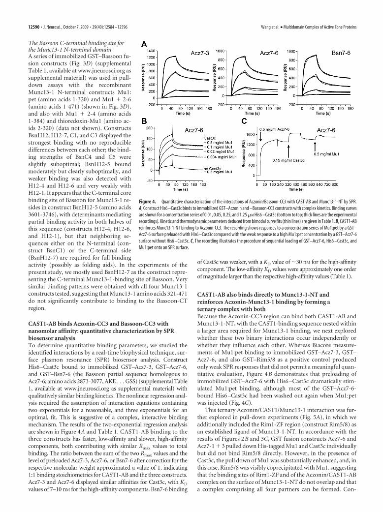
CAST1-AB binds Aczonin-CC3 and Bassoon-CC3 withnanomolar affinity: quantitative characterization by SPRbiosensor analysisTo determine quantitative binding parameters, we studied theidentified interactions by a real-time biophysical technique, sur-face plasmon resonance (SPR) biosensor analysis. ConstructHis6–Cast3c bound to immobilized GST–Acz7-3, GST–Acz7-6,and GST–Bsn7-6 (the Bassoon partial sequence homologous toAcz7-6; amino acids 2873-3077, AKE . . . GSS) (supplemental Table1, available at www.jneurosci.org as supplemental material) withqualitatively similar binding kinetics. The nonlinear regression anal-ysis required the assumption of interaction equations containingtwo exponentials for a reasonable, and three exponentials for anoptimal, fit. This is suggestive of a complex, interactive bindingmechanism. The results of the two-exponential regression analysisare shown in Figure 4A and Table 1. CAST1-AB binding to thethree constructs has faster, low-affinity and slower, high-affinitycomponents, both contributing with similar Rmax values to totalbinding. The ratio between the sum of the two Rmax values and thelevel of preloaded Acz7-3, Acz7-6, or Bsn7-6 after correction for therespective molecular weight approximated a value of 1, indicating1:1 binding stoichiometries for CAST1-AB and the three constructs.Acz7-3 and Acz7-6 displayed similar affinities for Cast3c, with KD
values of 7–10 nM for the high-affinity components. Bsn7-6 binding
of Cast3c was weaker, with a KD value of �30 nM for the high-affinitycomponent. The low-affinity KD values were approximately one orderof magnitude larger than the respective high-affinity values (Table 1).
CAST1-AB also binds directly to Munc13-1-NT andreinforces Aczonin-Munc13-1 binding by forming aternary complex with bothBecause the Aczonin-CC3 region can bind both CAST1-AB andMunc13-1-NT, with the CAST1-binding sequence nested withina larger area required for Munc13-1 binding, we next exploredwhether these two binary interactions occur independently orwhether they influence each other. Whereas Biacore measure-ments of Mu1:pet binding to immobilized GST–Acz7-3, GST–Acz7-6, and also GST–Rim5/8 as a positive control producedonly weak SPR responses that did not permit a meaningful quan-titative evaluation, Figure 4B demonstrates that preloading ofimmobilized GST–Acz7-6 with His6 –Cast3c dramatically stim-ulated Mu1:pet binding, although most of the GST–Acz7-6-bound His6 –Cast3c had been washed out again when Mu1:petwas injected (Fig. 4C).
This ternary Aczonin/CAST1/Munc13-1 interaction was fur-ther explored in pull-down experiments (Fig. 5A), in which weadditionally included the Rim1-ZF region (construct Rim5/8) asan established ligand of Munc13-1-NT. In accordance with theresults of Figures 2B and 3C, GST fusion constructs Acz7-6 andAcz7-1 � 3 pulled down His-tagged Mu1 and Cast3c individuallybut did not bind Rim5/8 directly. However, in the presence ofCast3c, the pull down of Mu1 was substantially enhanced, and, inthis case, Rim5/8 was visibly coprecipitated with Mu1, suggestingthat the binding sites of Rim1-ZF and of the Aczonin/CAST1-ABcomplex on the surface of Munc13-1-NT do not overlap and thata complex comprising all four partners can be formed. Con-
Figure 4. Quantitative characterization of the interactions of Aczonin/Bassoon-CC3 with CAST-AB and Munc13-1-NT by SPR.A, Construct His6 –Cast3c binds to immobilized GST–Aczonin and –Bassoon-CC3 constructs with complex kinetics. Binding curvesare shown for a concentration series of 0.01, 0.05, 0.25, and 1.25 �M His6 –Cast3c (bottom to top; thick lines are the experimentalrecordings). Kinetic and thermodynamic parameters deduced from bimodal curve fits (thin lines) are given in Table 1. B, CAST1-ABreinforces Munc13-1-NT binding to Aczonin-CC3. The recording shows responses to a concentration series of Mu1:pet by a GST–Acz7-6 surface preloaded with His6 –Cast3c compared with the weak response to a high Mu1:pet concentration by a GST–Acz7-6surface without His6 –Cast3c. C, The recording illustrates the procedure of sequential loading of GST–Acz7-6, His6 –Cast3c, andMu1:pet onto an SPR surface.
12590 • J. Neurosci., October 7, 2009 • 29(40):12584 –12596 Wang et al. • Multidomain Complex of Active Zone Proteins
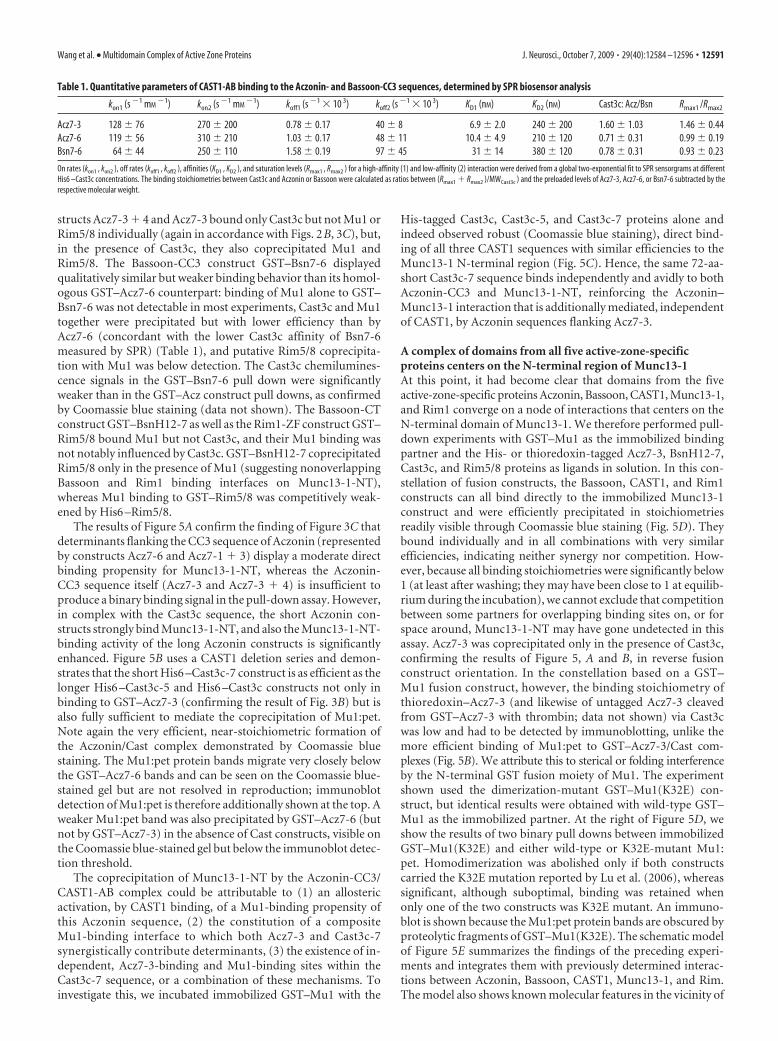
structs Acz7-3 � 4 and Acz7-3 bound only Cast3c but not Mu1 orRim5/8 individually (again in accordance with Figs. 2B, 3C), but,in the presence of Cast3c, they also coprecipitated Mu1 andRim5/8. The Bassoon-CC3 construct GST–Bsn7-6 displayedqualitatively similar but weaker binding behavior than its homol-ogous GST–Acz7-6 counterpart: binding of Mu1 alone to GST–Bsn7-6 was not detectable in most experiments, Cast3c and Mu1together were precipitated but with lower efficiency than byAcz7-6 (concordant with the lower Cast3c affinity of Bsn7-6measured by SPR) (Table 1), and putative Rim5/8 coprecipita-tion with Mu1 was below detection. The Cast3c chemilumines-cence signals in the GST–Bsn7-6 pull down were significantlyweaker than in the GST–Acz construct pull downs, as confirmedby Coomassie blue staining (data not shown). The Bassoon-CTconstruct GST–BsnH12-7 as well as the Rim1-ZF construct GST–Rim5/8 bound Mu1 but not Cast3c, and their Mu1 binding wasnot notably influenced by Cast3c. GST–BsnH12-7 coprecipitatedRim5/8 only in the presence of Mu1 (suggesting nonoverlappingBassoon and Rim1 binding interfaces on Munc13-1-NT),whereas Mu1 binding to GST–Rim5/8 was competitively weak-ened by His6 –Rim5/8.
The results of Figure 5A confirm the finding of Figure 3C thatdeterminants flanking the CC3 sequence of Aczonin (representedby constructs Acz7-6 and Acz7-1 � 3) display a moderate directbinding propensity for Munc13-1-NT, whereas the Aczonin-CC3 sequence itself (Acz7-3 and Acz7-3 � 4) is insufficient toproduce a binary binding signal in the pull-down assay. However,in complex with the Cast3c sequence, the short Aczonin con-structs strongly bind Munc13-1-NT, and also the Munc13-1-NT-binding activity of the long Aczonin constructs is significantlyenhanced. Figure 5B uses a CAST1 deletion series and demon-strates that the short His6 –Cast3c-7 construct is as efficient as thelonger His6 –Cast3c-5 and His6 –Cast3c constructs not only inbinding to GST–Acz7-3 (confirming the result of Fig. 3B) but isalso fully sufficient to mediate the coprecipitation of Mu1:pet.Note again the very efficient, near-stoichiometric formation ofthe Aczonin/Cast complex demonstrated by Coomassie bluestaining. The Mu1:pet protein bands migrate very closely belowthe GST–Acz7-6 bands and can be seen on the Coomassie blue-stained gel but are not resolved in reproduction; immunoblotdetection of Mu1:pet is therefore additionally shown at the top. Aweaker Mu1:pet band was also precipitated by GST–Acz7-6 (butnot by GST–Acz7-3) in the absence of Cast constructs, visible onthe Coomassie blue-stained gel but below the immunoblot detec-tion threshold.
The coprecipitation of Munc13-1-NT by the Aczonin-CC3/CAST1-AB complex could be attributable to (1) an allostericactivation, by CAST1 binding, of a Mu1-binding propensity ofthis Aczonin sequence, (2) the constitution of a compositeMu1-binding interface to which both Acz7-3 and Cast3c-7synergistically contribute determinants, (3) the existence of in-dependent, Acz7-3-binding and Mu1-binding sites within theCast3c-7 sequence, or a combination of these mechanisms. Toinvestigate this, we incubated immobilized GST–Mu1 with the
His-tagged Cast3c, Cast3c-5, and Cast3c-7 proteins alone andindeed observed robust (Coomassie blue staining), direct bind-ing of all three CAST1 sequences with similar efficiencies to theMunc13-1 N-terminal region (Fig. 5C). Hence, the same 72-aa-short Cast3c-7 sequence binds independently and avidly to bothAczonin-CC3 and Munc13-1-NT, reinforcing the Aczonin–Munc13-1 interaction that is additionally mediated, independentof CAST1, by Aczonin sequences flanking Acz7-3.
A complex of domains from all five active-zone-specificproteins centers on the N-terminal region of Munc13-1At this point, it had become clear that domains from the fiveactive-zone-specific proteins Aczonin, Bassoon, CAST1, Munc13-1,and Rim1 converge on a node of interactions that centers on theN-terminal domain of Munc13-1. We therefore performed pull-down experiments with GST–Mu1 as the immobilized bindingpartner and the His- or thioredoxin-tagged Acz7-3, BsnH12-7,Cast3c, and Rim5/8 proteins as ligands in solution. In this con-stellation of fusion constructs, the Bassoon, CAST1, and Rim1constructs can all bind directly to the immobilized Munc13-1construct and were efficiently precipitated in stoichiometriesreadily visible through Coomassie blue staining (Fig. 5D). Theybound individually and in all combinations with very similarefficiencies, indicating neither synergy nor competition. How-ever, because all binding stoichiometries were significantly below1 (at least after washing; they may have been close to 1 at equilib-rium during the incubation), we cannot exclude that competitionbetween some partners for overlapping binding sites on, or forspace around, Munc13-1-NT may have gone undetected in thisassay. Acz7-3 was coprecipitated only in the presence of Cast3c,confirming the results of Figure 5, A and B, in reverse fusionconstruct orientation. In the constellation based on a GST–Mu1 fusion construct, however, the binding stoichiometry ofthioredoxin–Acz7-3 (and likewise of untagged Acz7-3 cleavedfrom GST–Acz7-3 with thrombin; data not shown) via Cast3cwas low and had to be detected by immunoblotting, unlike themore efficient binding of Mu1:pet to GST–Acz7-3/Cast com-plexes (Fig. 5B). We attribute this to sterical or folding interferenceby the N-terminal GST fusion moiety of Mu1. The experimentshown used the dimerization-mutant GST–Mu1(K32E) con-struct, but identical results were obtained with wild-type GST–Mu1 as the immobilized partner. At the right of Figure 5D, weshow the results of two binary pull downs between immobilizedGST–Mu1(K32E) and either wild-type or K32E-mutant Mu1:pet. Homodimerization was abolished only if both constructscarried the K32E mutation reported by Lu et al. (2006), whereassignificant, although suboptimal, binding was retained whenonly one of the two constructs was K32E mutant. An immuno-blot is shown because the Mu1:pet protein bands are obscured byproteolytic fragments of GST–Mu1(K32E). The schematic modelof Figure 5E summarizes the findings of the preceding experi-ments and integrates them with previously determined interac-tions between Aczonin, Bassoon, CAST1, Munc13-1, and Rim.The model also shows known molecular features in the vicinity of
Table 1. Quantitative parameters of CAST1-AB binding to the Aczonin- and Bassoon-CC3 sequences, determined by SPR biosensor analysis
kon1 (s 1 mM1) kon2 (s 1 mM
1) koff1 (s 1 � 10 3) koff2 (s 1 � 10 3) KD1 (nM) KD2 (nM) Cast3c: Acz/Bsn Rmax1 /Rmax2
Acz7-3 128 76 270 200 0.78 0.17 40 8 6.9 2.0 240 200 1.60 1.03 1.46 0.44Acz7-6 119 56 310 210 1.03 0.17 48 11 10.4 4.9 210 120 0.71 0.31 0.99 0.19Bsn7-6 64 44 250 110 1.58 0.19 97 45 31 14 380 120 0.78 0.31 0.93 0.23
On rates (kon1 , kon2 ), off rates (koff1 , koff2 ), affinities (KD1 , KD2 ), and saturation levels (Rmax1 , Rmax2 ) for a high-affinity (1) and low-affinity (2) interaction were derived from a global two-exponential fit to SPR sensorgrams at differentHis6 –Cast3c concentrations. The binding stoichiometries between Cast3c and Aczonin or Bassoon were calculated as ratios between (Rmax1 � Rmax2 )/MWCast3c ) and the preloaded levels of Acz7-3, Acz7-6, or Bsn7-6 subtracted by therespective molecular weight.
Wang et al. • Multidomain Complex of Active Zone Proteins J. Neurosci., October 7, 2009 • 29(40):12584 –12596 • 12591
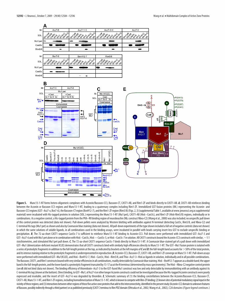
Figure 5. Munc13-1-NT forms hetero-oligomeric complexes with Aczonin/Bassoon-CC3, Bassoon-CT, CAST1-AB, and Rim1-ZF and binds directly to CAST1-AB. A, CAST1-AB reinforces bindingbetween the Aczonin or Bassoon CC3 regions and Munc13-1-NT, leading to a quaternary complex including Rim1-ZF. Immobilized GST fusion proteins (IM.) representing the Aczonin- andBassoon-CC3 regions (GST–Acz7-x, Bsn7-6), the Bassoon-CT region (BsnH12-7), and the Rim1-ZF region (Rim5/8) (Figs. 2, 3) (supplemental Table 1, available at www.jneurosci.org as supplementalmaterial) were incubated with His-tagged proteins in solution (SOL.) representing the Munc13-1-NT (Mu1:pet), CAST1-AB (His6 –Cast3c), and Rim1-ZF (His6-Rim5/8) regions, individually or incombinations. As a negative control, a His-tagged protein from the PKA–RII binding region of neurobeachin (Nb; construct Nbea-C2) (Wang et al., 2000) was also included; no unspecific pull downof this control protein was detected (data not shown). Pull-down pellets were analyzed by Western blotting with antibodies against N-terminal (detecting Cast3c, Rim5/8, and Nbea-C2) andC-terminal His tags (Mu1:pet) as shown and also by Coomassie blue staining (data not shown). All pull-down experiments of the type shown included a full set of negative controls (data not shown)in which the same solutions of soluble ligands, in all combinations used in the binding assays, were incubated in parallel with beads carrying insert-less GST to exclude unspecific binding orprecipitation. B, The 72-aa-short CAST1 sequence Cast3c-7 is sufficient to reinforce Munc13-1-NT binding to Aczonin-CC3. Pull downs were performed with immobilized GST–Acz7-3 andGST–Acz7-6 and with Mu1:pet alone or in combination with His6 –Cast3c, His6 —Cast3c-5, or His6 –Cast3c-7 in solution. All CAST1 constructs bound the Aczonin-CC3 constructs with similar, �1:1stoichiometries, and stimulated Mu1:pet pull down. C, The 72-aa-short CAST1 sequence Cast3c-7 binds directly to Munc13-1-NT. A Coomassie blue-stained gel of a pull down with immobilizedGST–Mu1 (dimerization-deficient mutant K32E) demonstrates that all CAST1 constructs bind with similarly high efficiencies directly to Munc13-1-NT. The GST–Mu1 fusion protein is isolated witha series of proteolytic fragments in addition to the full-length protein at the top, as indicated by brackets at the left margins of C and D; the full-length band accounts for �50% of the total protein,and its intense staining relative to the proteolytic fragments is underrepresented in reproduction. D, Aczonin-CC3, Bassoon-CT, CAST1-AB, and Rim1-ZF converge on Munc13-1-NT. Pull-down assayswere performed with immobilized GST–Mu1(K32E), and His6 –BsnH12-7, His6 –Cast3c, His6 –Rim5/8, and Thio–Acz7-3–His6 as ligands in solution, individually and in all possible combinations.The Bassoon, CAST1, and Rim1 constructs bound with very similar efficiencies in all combinations, readily detectable by Coomassie blue staining. His6 –BsnH12-7 appears as a double band; the upperband is the full-length protein, and the lower band is a proteolytic fragment truncated by 13-17 aa at the N terminus (determined by mass spectrometry). The His6 –Nbea-C2 negative control protein(see A) did not bind (data not shown). The binding efficiency of thioredoxin–Acz7-3 to the GST-fused Mu1 construct was low and only detectable by immunoblotting with an antibody against itsC-terminal His tag (shown at the bottom). Direct binding, to GST–Mu1, of Acz7-6 or other longer Aczonin constructs could not be investigated because the His-tagged Aczonin constructs were poorlyexpressed and insoluble, and the insert of GST–Acz7-6 was degraded by thrombin. E, Schematic summary of (1) the binding constellations between the Aczonin/Bassoon-CC3, Bassoon-CT,CAST1-AB, Munc13-1-NT, and Rim1-ZF regions, including homodimerization of Munc13-1-NT, which is known to compete with Rim-ZF binding; (2) known sites of potential modulatory signal input in thevicinity of these regions; and (3) interactions between other regions of these five active zone proteins that add to the interconnectivity, identified in the present study (Aczonin-CC2 domain to unknown featuresof Bassoon, possibly indirectly through a third partner) or as published previously [CAST C terminus to Rim PDZ domain (Ohtsuka et al., 2002; Wang et al., 2002), C2A domains (Figure legend continues.)
12592 • J. Neurosci., October 7, 2009 • 29(40):12584 –12596 Wang et al. • Multidomain Complex of Active Zone Proteins
the interacting domains that may provide regulatory input (seelegend and Discussion).
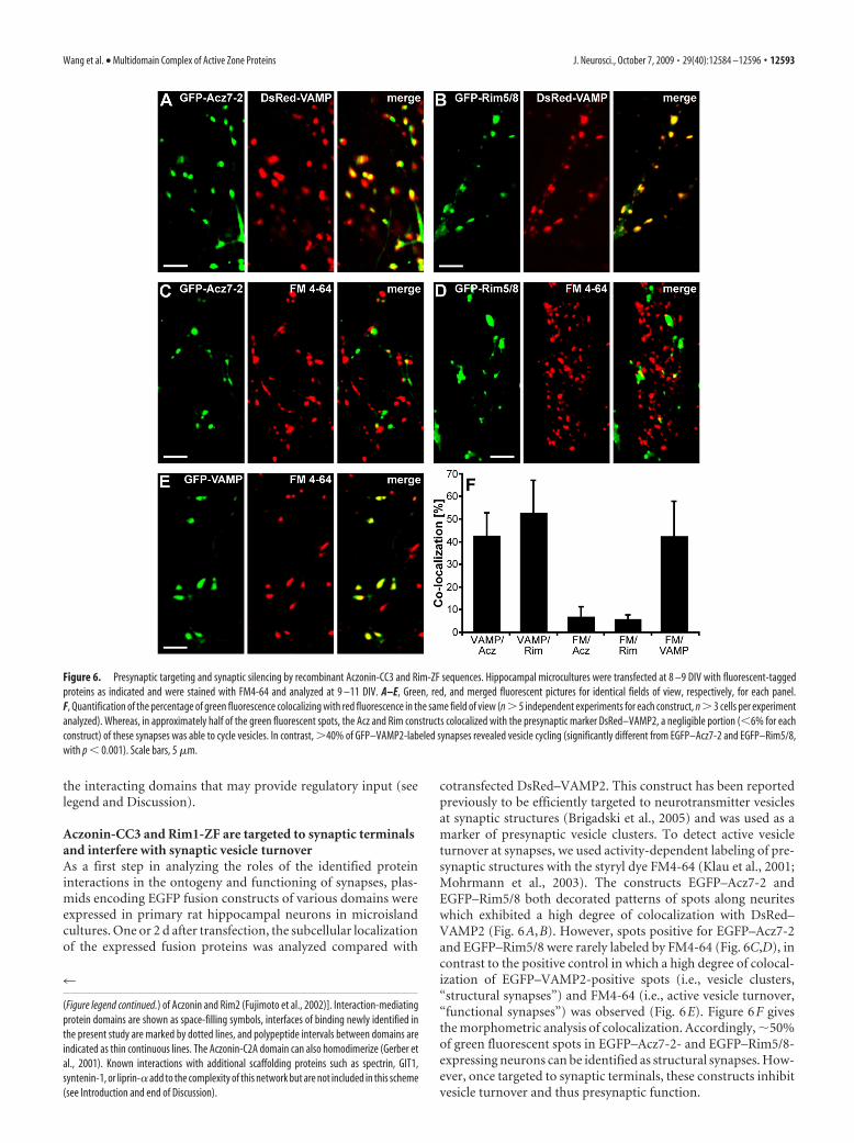
Aczonin-CC3 and Rim1-ZF are targeted to synaptic terminalsand interfere with synaptic vesicle turnoverAs a first step in analyzing the roles of the identified proteininteractions in the ontogeny and functioning of synapses, plas-mids encoding EGFP fusion constructs of various domains wereexpressed in primary rat hippocampal neurons in microislandcultures. One or 2 d after transfection, the subcellular localizationof the expressed fusion proteins was analyzed compared with
cotransfected DsRed–VAMP2. This construct has been reportedpreviously to be efficiently targeted to neurotransmitter vesiclesat synaptic structures (Brigadski et al., 2005) and was used as amarker of presynaptic vesicle clusters. To detect active vesicleturnover at synapses, we used activity-dependent labeling of pre-synaptic structures with the styryl dye FM4-64 (Klau et al., 2001;Mohrmann et al., 2003). The constructs EGFP–Acz7-2 andEGFP–Rim5/8 both decorated patterns of spots along neuriteswhich exhibited a high degree of colocalization with DsRed–VAMP2 (Fig. 6A,B). However, spots positive for EGFP–Acz7-2and EGFP–Rim5/8 were rarely labeled by FM4-64 (Fig. 6C,D), incontrast to the positive control in which a high degree of colocal-ization of EGFP–VAMP2-positive spots (i.e., vesicle clusters,“structural synapses”) and FM4-64 (i.e., active vesicle turnover,“functional synapses”) was observed (Fig. 6E). Figure 6F givesthe morphometric analysis of colocalization. Accordingly, �50%of green fluorescent spots in EGFP–Acz7-2- and EGFP–Rim5/8-expressing neurons can be identified as structural synapses. How-ever, once targeted to synaptic terminals, these constructs inhibitvesicle turnover and thus presynaptic function.
4
(Figure legend continued.) of Aczonin and Rim2 (Fujimoto et al., 2002)]. Interaction-mediatingprotein domains are shown as space-filling symbols, interfaces of binding newly identified inthe present study are marked by dotted lines, and polypeptide intervals between domains areindicated as thin continuous lines. The Aczonin-C2A domain can also homodimerize (Gerber etal., 2001). Known interactions with additional scaffolding proteins such as spectrin, GIT1,syntenin-1, or liprin-� add to the complexity of this network but are not included in this scheme(see Introduction and end of Discussion).
Figure 6. Presynaptic targeting and synaptic silencing by recombinant Aczonin-CC3 and Rim-ZF sequences. Hippocampal microcultures were transfected at 8 –9 DIV with fluorescent-taggedproteins as indicated and were stained with FM4-64 and analyzed at 9 –11 DIV. A–E, Green, red, and merged fluorescent pictures for identical fields of view, respectively, for each panel.F, Quantification of the percentage of green fluorescence colocalizing with red fluorescence in the same field of view (n � 5 independent experiments for each construct, n � 3 cells per experimentanalyzed). Whereas, in approximately half of the green fluorescent spots, the Acz and Rim constructs colocalized with the presynaptic marker DsRed–VAMP2, a negligible portion (�6% for eachconstruct) of these synapses was able to cycle vesicles. In contrast, �40% of GFP–VAMP2-labeled synapses revealed vesicle cycling (significantly different from EGFP–Acz7-2 and EGFP–Rim5/8,with p � 0.001). Scale bars, 5 �m.
Wang et al. • Multidomain Complex of Active Zone Proteins J. Neurosci., October 7, 2009 • 29(40):12584 –12596 • 12593
These findings suggest that both Acz7-2 (this sequence bindsCAST-AB but not, by itself, Munc13-1-NT) (Figs. 2B, 3C) andRim5/8 (this sequence binds Munc13-1-NT but not Rab3) areefficiently targeted to synaptic terminals and impair vesicle turn-over, presumably by inhibiting exocytosis through dominant-negative replacement of holo-Aczonin and holo-Rim from theirbinding sites on CAST1 and Munc13-1, respectively.
DiscussionAll five active-zone-specific multidomain proteins aremultiply interconnectedIn the present study, we have identified several new interactionsbetween domains of proteins of the presynaptic active zone. Weshow that the Munc13-1 N-terminal domain, in addition to itsestablished homodimerization and to its heterodimerizationwith the Rim zinc-finger domain (Lu et al., 2006), binds a regionnear the Bassoon C terminus, a short sequence stretch in themiddle of CAST1, and determinants around the third coiled-coildomain of Aczonin. Moreover, high-affinity binding of theAczonin-CC3 sequence to CAST1 considerably strengthens, in-directly, the adherence of Aczonin to Munc13-1-NT. An addi-tional connection between Aczonin and Bassoon, whosemolecular details remain to be worked out, is mediated by thesecond coiled-coil domain of Aczonin. Our pull-down resultswith recombinant proteins (Fig. 5D) suggest that Aczonin-CC3,Rim-ZF, CAST1-AB, and Bassoon-CT can bind simultaneouslyto Munc13-1-NT (model of Fig. 5E). However, whereas the oc-currence of a quaternary complex of Aczonin-CC3, CAST1-AB,Munc13-1-NT, and Rim1-ZF, and of a ternary complex ofBassoon-CT, Munc13-1-NT, and Rim1-ZF, seem established(Fig. 5A), it requires additional clarification whether or notBassoon-CT competes with CAST1-AB or Aczonin-CC3 forbinding to Munc13-1-NT. Munc13-1-NT homodimerizationand its heterodimerization with Rim-ZF have been shown pre-viously to compete (Lu et al., 2006), and it remains to beinvestigated whether Aczonin-CC3, CAST1-AB, or Bassoon-CTinterfere with the homodimerization of Munc13-1-NT. Thesefindings demonstrate that the five large multidomain proteins ofthe active zone, Aczonin, Bassoon, CAST, Munc13, and Rim, areclosely and multiply interconnected, physically and presumablyalso functionally. In particular, the organization of this complexaround the N-terminal domain of Munc13-1 emphasizes a keyrole of this domain in the orchestration of molecular events at theactive zone. Although Rab3-Rim and Rim-Munc13-1 bindinghave been known for many years, no downstream effector mech-anisms have been identified for them. Our results now place thesebinary interactions into a larger and much more elaborate mo-lecular context (Fig. 5E), which may be essential for additionalprogress in mechanistic understanding.
The Bassoon-CC3 domain was reported previously to bindCAST1 (Takao-Rikitsu et al., 2004), and the shortest CAST1-binding Bassoon sequence described there (BsnCasBD) is almostprecisely collinear and has high sequence similarity with our min-imal CAST1-binding Aczonin construct, Acz7-3. However, inSPR and pull-down measurements between recombinant li-gands, we find that CAST1 binds more strongly to Aczonin-CC3than to Bassoon-CC3 (Figs. 4A, 5A; Table 1). Moreover, in ourbrain lysate pull-down experiments, we detected no precipitationof native CAST1, Munc13, or Rim by the GST–Bsn7/Bsn7-6 con-structs, whereas the collinear GST–Aczonin constructs producedstrong CAST1, Munc13, and Rim signals in the same experiments(Fig. 2B). Even long overexposure of the Western blot of Figure2B showed no trace of CAST1 binding to Bsn7 or Bsn7-6, along-
side massive CAST1 signals in the Acz7/7-6 lanes (detection limit:�1% of CAST1 binding to the Aczonin constructs). We note thatTakao-Rikitsu et al. (2004) describe precipitation by BsnCasBDonly for recombinant, heterologously overexpressed CAST1, notfor native CAST1 from a brain lysate. The approximately three-fold lower affinity of Bassoon-CC3, relative to Aczonin-CC3, forCAST1-AB measured by SPR (Table 1) thus translates into asignificantly poorer performance in the pull down of recombi-nant CAST1 (Fig. 5A) and a complete failure to pull down nativeCAST1 from brain lysate, at low ligand concentration and incompetition with the native Aczonin in the lysate, suggesting thatthis affinity difference may be functionally significant.
The CAST1-binding domain of Aczonin (construct Acz7-3)and the Aczonin- and Munc13-1-binding domain of CAST1(construct Cast3c-7) are small (maximally 60 –70 aa each) andengage robustly in a 1:1 complex in vitro. In contrast, the otherprotein binding modules characterized here are large (Munc13-1-NT, 320 aa; Bsn-CT, 220 –310 aa; the Aczonin region aroundCC3 capable of Munc13 binding without CAST1, 230 –293 aa),and their dissection produces fragments with partial binding ac-tivities. Their complexes are substoichiometric in the pull-downassay and give weak or undetectable signals in SPR. This suggeststhat the respective binding interfaces are extensive and complex,composed of several determinants and dependent on optimalfolding.
In light of the binding data with the recombinant proteins, wecan now reinspect the details of the pull down of holoproteinsfrom brain lysate (Fig. 2B). Holo-CAST1 was precipitated by theshort and long Acz-CC3 constructs with similar efficiencies. Incontrast, Munc13-1 precipitation was strong only with the longconstructs, whereas the short Aczonin-CC3 constructs producedonly weak Munc13-1 signals. This may reflect a weak residualMunc13-1-binding activity of Acz7-3 alone (which we observedin some recombinant pull-down experiments), or a small frac-tion of the immobilized Acz7-3 construct (present in vast excessover the individual native proteins in the lysate) has acquiredhigh Munc13-1 affinity through forming a complex with nativeCAST1 from the brain lysate. Of note, the second Munc13-immunopositive band of slower electrophoretic migration pre-cipitated by the Aczonin constructs, but not by the recombinantRim1-ZF construct bRim29, is pulled down with equal or evenhigher efficiency by the short than by the long Acz-CC3 con-structs. This suggests that this ligand, presumably a Munc13splice variant or isoform such as bMunc13-2, is bound with fullstrength also by the short Acz-CC3 constructs. The identity ofthis second Munc13-like ligand and the details of its mode ofbinding to Acz7-3 remain to be elucidated.
We interpret the pull down of Rim from brain lysate by theAczonin-CC3 and Bassoon-CT constructs (Figs. 1D, 2B,C) to beindirect, primarily via the Munc13-1 N-terminal region, whichcan be demonstrated in pull-down experiments with the recom-binant constructs (Fig. 5A). Binding between the CAST1 C ter-minus and the Rim PDZ domain (Ohtsuka et al., 2002; Wang etal., 2002) may additionally contribute to an indirect pull down ofRim from brain lysate, in particular by the short Aczonin-CC3constructs Acz7-2 and Acz7-3, which bind Munc13-1 poorly. Itremains possible that Rim1 sequences downstream of theMunc13- and Rab3-binding regions (amino acids 1-228) alsobind directly to the Acz7 region or other partners of the complex.Fusion proteins representing these Rim1 sequences were poorlysoluble and did not allow us to conclusively answer this question.
The close encounter of domains of Aczonin, Bassoon, CAST1,Munc13-1, and Rim1 in a “node” of interactions suggests a high
12594 • J. Neurosci., October 7, 2009 • 29(40):12584 –12596 Wang et al. • Multidomain Complex of Active Zone Proteins
potential for crosstalk. Their juxtaposition may poise these orother domains of their respective holoproteins for fast and pre-cise multilateral or sequential interactions. At least the binaryinteraction of Aczonin with Munc13-1-NT is profoundly rein-forced by the presence of CAST1, which binds avidly to both.Moreover, the domains participating in this interaction node areneighbored by multiple molecular features that may mediate reg-ulatory signal input (Fig. 5E). The Aczonin and Bassoon CC3sequences are flanked on both sides by in vivo phosphorylationsites (Collins et al., 2005). Munc13-1 carries a Ca 2�/calmodulin(CaM) binding site at mouse amino acids 456-473 (Xu et al.,1998; Junge et al., 2004) and a diacylglycerol/phorbol ester bind-ing C1 zinc-finger domain at amino acids 564-614 (Betz et al.,1998). Amino acids 19-50 and the SGAWFF motif around tryp-tophan 201 of mouse Rim1 interact with Rab3–GTP (Sun et al.,2001; Wang et al., 2001; Dulubova et al., 2005), amino acids 56-82and 83-105 of Rim1 can be deleted individually or together bydifferential splicing (Wang et al., 2001), and phosphorylation hasbeen detected at Rim1 serines 241, 287, and 413 and a Rim2residue corresponding to Rim1 serine 379 (Lonart et al., 2003;Sun et al., 2003). The Rim1–Rab3 interaction is thought to beinvolved in the synaptic vesicle life cycle, and the S413 phos-phorylation of Rim1 and the interactions of Munc13 withCaM and diacyl glycerol in synaptic plasticity. For none ofthese molecular modifications or interactions of Aczonin, Bassoon,Munc13, and Rim, however, have downstream effector mecha-nisms been identified. It is tempting to suspect that some of theinteractions described here might be influenced by them. TheC-terminal half of Munc13-1, in particular, is responsible forthe synaptic vesicle priming activity of the protein, which in turnis thought to be regulated by signal input through the N-terminalhalf (Basu et al., 2005; Stevens et al., 2005). Our findings add threenew molecular interactions of the Munc13-1 N-terminal half(with CAST1, Aczonin-CC3, and Bassoon-CT) to those alreadyknown (with itself, Rim-ZF, Ca 2�/calmodulin, and diacylglyc-erol), significantly expanding the scope of its regulatory poten-tial. The identification of the multidomain complex describedhere provides a basis for additional, in-depth analysis of its struc-tural organization, of its molecular dynamics and regulatorymodulation, and for selectively addressing the roles of individualbinary interactions in neurotransmission.
Involvement of interacting domains in synaptogenesis andsynaptic activityFigure 6 demonstrates that the Acz7-2 and Rim5/8 fusion pro-teins with EGFP (1) are targeted to and concentrated at synapticterminals and (2) suppress synaptic vesicle turnover, suggestingfunctions of the Aczonin-CC3/CAST1-AB and the Rim1-ZF/Munc13-1-NT interactions in both the assembly and activity ofsynapses. The suppression of vesicle turnover by our Rim5/8 con-struct is consistent with the �50% reduction of neurotransmitterrelease that was achieved by instillation into the calyx of Heldsynaptic terminal, of a ZF domain from the Rim2� isoformclosely collinear with our Rim1 construct (Dulubova et al., 2005).The effect of Acz7-2 on vesicle turnover is in line with the �50%suppression of synaptic transmission that Takao-Rikitsu et al.(2004) observed within 100 min after microinjection into supe-rior cervical ganglion cells, of a CAST1-binding GST–Bassoonfusion protein almost exactly collinear with our Acz7-3 sequence(which in turn is nested within Acz7-2). Takao-Rikitsu et al.(2004) also inhibited synaptic transmission by microinjecting arecombinant CAST1 sequence that binds Aczonin-CC3 orBassoon-CC3 but also, as we show here for the first time,
Munc13-1-NT. Because of the density of binding interfaces inthis multidomain complex, future functional experiments willhave to address the various binary interactions individually.
The presynaptic targeting and vesicle turnover suppressingactivities of our constructs may occur by “acute” displacement ofthe endogenous holoprotein in the terminal, particularly duringactivity; in the course of the in situ turnover of the active zoneproteins [which can have time constants in the range of minutes(Munc13-1) (Kalla et al., 2006) to hours (Bassoon) (Tsuriel et al.,2009)], during the de novo assembly of active zone material be-tween the Golgi complex and nascent synapses, or a combinationof these mechanisms. Assembly of the active zone machinery, inassociation with membranes, begins as early as at the Golgi com-plex or the trans-Golgi network (Wang et al., 1999; Dresbach etal., 2006), followed by dispatch as a coat on dense-core secretoryvesicles or multi-vesicle aggregates toward the neuronal periph-ery in which it can be rapidly deposited at nascent synapses(Ahmari et al., 2000; Zhai et al., 2001; Shapira et al., 2003; Tao-Cheng, 2007). The hierarchy of protein associations in the courseof this assembly is an interesting question, but, given the highlyinterwoven structure of this macromolecular aggregate, it maynot be a simple linear algorithm with molecular “leaders” and“followers.” With the respective other four active-zone-specific pro-teins alone, Aczonin, Bassoon, CAST, and Rim are connectedthrough at least two separate domains each [Aczonin-CC2 and -CC3,Bassoon-CC3 and -CT, CAST1-AB (binding both Aczonin-CC3and Munc13-NT) and -CT, and Rim-ZF and -PDZ], theMunc13-1-NT binds all four partners, and homodimerization isknown for Munc13-1-NT and CAST1 (Fig. 5E). The interactionbetween Munc13-NT and Rim-ZF, for example, is important butnot exclusively responsible for the active zone recruitment ofMunc13 (Andrews-Zwilling et al., 2006). Interactions with addi-tional multivalent scaffolding proteins such as those of Rim andCAST1 with liprin-� (Schoch et al., 2002; Ko et al., 2003; Dai etal., 2006), of CAST1 with syntenin-1 (Ko et al., 2006), of Munc13with �-spectrin (Sakaguchi et al., 1998), and of Aczonin withGIT1 (Kim et al., 2003) add additional layers of complexity to themolecular architecture of the active zone and to the formation ofa polymeric, highly interconnected protein lattice. Hence, the“targeting code” of active zone proteins is probably combina-torial, and, through their early association along the secretorypathway, they can find the way to synaptic terminals as fellowtravelers, although each of them may possess only part of thetargeting information.
ReferencesAhmari SE, Buchanan J, Smith SJ (2000) Assembly of presynaptic active
zones from cytoplasmic transport packets. Nat Neurosci 3:445– 451.Andrews-Zwilling YS, Kawabe H, Reim K, Varoqueaux F, Brose N (2006)
Binding to Rab3A-interacting molecule RIM regulates the presynapticrecruitment of Munc13-1 and ubMunc13-2. J Biol Chem281:19720 –19731.
Basu J, Shen N, Dulubova I, Lu J, Guan R, Guryev O, Grishin NV, RosenmundC, Rizo J (2005) A minimal domain responsible for Munc13 activity.Nat Struct Mol Biol 12:1017–1018.
Betz A, Okamoto M, Benseler F, Brose N (1997) Direct interaction of the ratunc-13 homologue Munc13-1 with the N terminus of syntaxin. J BiolChem 272:2520 –2526.
Betz A, Ashery U, Rickmann M, Augustin I, Neher E, Sudhof TC, Rettig J,Brose N (1998) Munc13-1 is a presynaptic phorbol ester receptor thatenhances neurotransmitter release. Neuron 21:123–136.
Betz A, Thakur P, Junge HJ, Ashery U, Rhee JS, Scheuss V, Rosenmund C,Rettig J, Brose N (2001) Functional interaction of the active zone pro-teins Munc13-1 and RIM1 in synaptic vesicle priming. Neuron30:183–196.
Brigadski T, Hartmann M, Lessmann V (2005) Differential vesicular target-
Wang et al. • Multidomain Complex of Active Zone Proteins J. Neurosci., October 7, 2009 • 29(40):12584 –12596 • 12595
ing and time course of synaptic secretion of the mammalian neurotro-phins. J Neurosci 25:7601–7614.
Collins MO, Yu L, Coba MP, Husi H, Campuzano I, Blackstock WP,Choudhary JS, Grant SGN (2005) Proteomic analysis of in vivo phos-phorylated synaptic proteins. J Biol Chem 280:5972–5982.
Coppola T, Magnin-Luthi S, Perret-Menoud V, Gattesco S, Schiavo G,Regazzi R (2001) Direct interaction of the Rab3 effector RIM with Ca 2�
channels, SNAP-25, and synaptotagmin. J Biol Chem 276:32756–32762.Dai Y, Taru H, Deken SL, Grill B, Ackley B, Nonet ML, Jin Y (2006) SYD-2
liprin-� organizes presynaptic active zone formation through ELKS. NatNeurosci 9:1479 –1487.
Dresbach T, Torres V, Wittenmayer N, Altrock WD, Zamorano P, Zuschrat-ter W, Nawrotzki R, Ziv NE, Garner CC, Gundelfinger ED (2006) As-sembly of active zone precursor vesicles: obligatory trafficking ofpresynaptic cytomatrix proteins Bassoon and Piccolo via a trans-Golgicompartment. J Biol Chem 281:6038 – 6047.
Dulubova I, Lou X, Lu J, Huryeva I, Alam A, Schneggenburger R, Sudhof TC,Rizo J (2005) A Munc13/RIM/Rab3 tripartite complex: from priming toplasticity? EMBO J 24:2839 –2850.
Fenster SD, Chung WJ, Zhai R, Cases-Langhoff C, Voss B, Garner AM,Kaempf U, Kindler S, Gundelfinger ED, Garner CC (2000) Piccolo, apresynaptic zinc finger protein structurally related to Bassoon. Neuron25:203–214.
Fujimoto K, Shibasaki T, Yokoi N, Kashima Y, Matsumoto M, Sasaki T,Tajima N, Iwanaga T, Seino S (2002) Piccolo, a Ca 2� sensor in pancre-atic �-cells - Involvement of cAMP-GEFII. Rim2.Piccolo complex incAMP-dependent exocytosis. J Biol Chem 277:50497–50502.
Fujita Y, Shirataki H, Sakisaka T, Asakura T, Ohya T, Kotani H, Yokoyama S,Nishioka H, Matsuura Y, Mizoguchi A, Scheller RH, Takai Y (1998) To-mosyn: a syntaxin-1-binding protein that forms a novel complex in theneurotransmitter release process. Neuron 20:905–915.
Gerber SH, Garcia J, Rizo J, Sudhof TC (2001) An unusual C2 domain in theactive-zone protein piccolo: implications for Ca 2� regulation of neuro-transmitter release. EMBO J 20:1605–1619.
Gundelfinger ED, Kessels MM, Qualmann B (2003) Temporal and spatialcoordination of exocytosis and endocytosis. Nat Rev Mol Cell Biol4:127–139.
Hatsuzawa K, Lang T, Fasshauer D, Bruns D, Jahn R (2003) The R-SNAREmotif of tomosyn forms SNARE core complexes with syntaxin 1 andSNAP-25 and down-regulates exocytosis. J Biol Chem 278:31159 –31166.
Haubensak W, Narz F, Heumann R, Lessmann V (1998) BDNF-GFP con-taining secretory granules are localized in the vicinity of synaptic junc-tions of cultured cortical neurons. J Cell Sci 111:1483–1493.
Junge HJ, Rhee JS, Jahn O, Varoqueaux F, Spiess J, Waxham MN, Rosenmund C,Brose N (2004) Calmodulin and Munc13 form a Ca 2� sensor/effectorcomplex that controls short-term synaptic plasticity. Cell 118:389–401.
Kalla S, Stern M, Basu J, Varoqueaux F, Reim K, Rosenmund C, Ziv NE, BroseN (2006) Molecular dynamics of a presynaptic active zone protein stud-ied in Munc13-1-enhanced yellow fluorescent protein knock-in mutantmice. J Neurosci 26:13054 –13066.
Kim S, Ko J, Shin H, Lee JR, Lim C, Han JH, Altrock WD, Garner CC,Gundelfinger ED, Premont RT, Kaang BK, Kim E (2003) The GIT fam-ily of proteins forms multimers and associates with the presynaptic cyto-matrix protein Piccolo. J Biol Chem 278:6291– 6300.
Klau M, Hartmann M, Erdmann KS, Heumann R, Lessmann V (2001) Re-duced number of functional glutamatergic synapses in hippocampal neuronsoverexpressing full-length TrkB receptors. J Neurosci Res 66:327–336.
Ko J, Na M, Kim S, Lee JR, Kim E (2003) Interaction of the ERC family ofRIM-binding proteins with the liprin-� family of multidomain proteins.J Biol Chem 278:42377– 42385.
Ko J, Yoon C, Piccoli G, Chung HS, Kim K, Lee JR, Lee HW, Kim H, Sala C,Kim E (2006) Organization of the presynaptic active zone by ERC2/CAST1-dependent clustering of the tandem PDZ protein syntenin-1.J Neurosci 26:963–970.
Lonart G, Schoch S, Kaeser PS, Larkin CJ, Sudhof TC, Linden DJ (2003)Phosphorylation of RIM1a by PKA triggers presynaptic long-term poten-tiation at cerebellar parallel fiber synapses. Cell 115:49 – 60.
Lu J, Machius M, Dulubova I, Dai H, Sudhof TC, Tomchick DR, Rizo J(2006) Structural basis for a Munc13-1 homodimer to Munc13-1/RIMheterodimer switch. PLoS Biol 4:e192.
Mohrmann R, Lessmann V, Gottmann K (2003) Developmental matura-tion of synaptic vesicle cycling as a distinctive feature of central glutama-tergic synapses. Neuroscience 117:7–18.
Ohtsuka T, Takao-Rikitsu E, Inoue E, Inoue M, Takeuchi M, Matsubara K,Deguchi-Tawarada M, Satoh K, Morimoto K, Nakanishi H, Takai Y(2002) CAST: a novel protein at the cytomatrix at the active zone ofsynapses that forms a ternary complex with RIM1 and Munc13-1. J CellBiol 158:577–590.
Sakaguchi G, Orita S, Naito A, Maeda M, Igarashi H, Sasaki T, Takai Y (1998)A novel brain-specific isoform of �-spectrin: isolation and its interactionwith Munc13. Biochem Biophys Res Commun 248:846 – 851.
Schoch S, Gundelfinger ED (2006) Molecular organization of the presynap-tic active zone. Cell Tissue Res 326:379 –391.
Schoch S, Castillo PE, Jo T, Mukherjee K, Geppert M, Wang Y, Schmitz F,Malenka RC, Sudhof TC (2002) RIM1� forms a protein scaffold for regu-lating neurotransmitter release at the active zone. Nature 415:321–326.
Shapira M, Zhai RG, Dresbach T, Bresler T, Torres VI, Gundelfinger ED, ZivNE, Garner CC (2003) Unitary assembly of presynaptic active zonesfrom Piccolo-Bassoon transport vesicles. Neuron 38:237–252.
Stevens DR, Wu ZX, Matti U, Junge HJ, Schirra C, Becherer U, Wojcik SM,Brose N, Rettig J (2005) Identification of the minimal protein domainrequired for priming activity of Munc13-1. Curr Biol 15:2243–2248.
Sun L, Bittner MA, Holz RW (2001) Rab3a binding and secretion-enhancing domains in Rim1 are separate and unique. Studies in adrenalchromaffin cells. J Biol Chem 276:12911–12917.
Sun L, Bittner MA, Holz RW (2003) Rim, a component of the presynapticactive zone and modulator of exocytosis, binds 14-3-3 through itsN-terminus. J Biol Chem 278:38301–38309.
Takao-Rikitsu E, Mochida S, Inoue E, Deguchi-Tawarada M, Inoue M, Oht-suka T, Takai Y (2004) Physical and functional interaction of the activezone proteins, CAST, RIM1, and Bassoon, in neurotransmitter release.J Cell Biol 164:301–311.
Tao-Cheng JH (2007) Ultrastructural localization of active zone and synap-tic vesicle proteins in a preassembled multi-vesicle transport aggregate.Neuroscience 150:575–584.
Tsuriel S, Fisher A, Wittenmayer N, Dresbach T, Garner CC, Ziv NE (2009)Exchange and redistribution dynamics of the cytoskeleton of the activezone molecule Bassoon. J Neurosci 29:351–358.
Wang X, Kibschull M, Laue MM, Lichte B, Petrasch-Parwez E, Kilimann MW(1999) Aczonin, a 550-kD putative scaffolding protein of presynapticactive zones, shares homology regions with Rim and Bassoon and bindsprofilin. J Cell Biol 147:151–162.
Wang X, Herberg FW, Laue MM, Wullner C, Hu B, Petrasch-Parwez E,Kilimann MW (2000) Neurobeachin: a protein kinase A-anchoring,beige/Chediak-Higashi protein homolog implicated in neuronal mem-brane traffic. J Neurosci 20:8551– 8565.
Wang X, Hu B, Zimmermann B, Kilimann MW (2001) Rim1 andRabphilin-3 bind Rab3-GTP by composite determinants partially relatedthrough N-terminal �-helix motifs. J Biol Chem 276:32480 –32488.
Wang Y, Liu X, Biederer T, Sudhof TC (2002) A family of RIM-bindingproteins regulated by alternative splicing: implications for the genesis ofsynaptic active zones. Proc Natl Acad Sci U S A 99:14464 –14469.
Wojcik SM, Brose N (2007) Regulation of membrane fusion in synapticexcitation-secretion coupling: speed and accuracy matter. Neuron 55:11–24.
Xu XZ, Wes PD, Chen H, Li HS, Yu M, Morgan S, Liu Y, Montell C (1998)Retinal targets for calmodulin include proteins implicated in synaptictransmission. J Biol Chem 273:31297–31307.
Zhai RG, Vardinon-Friedman H, Cases-Langhoff C, Becker B, Gundelfinger ED,Ziv NE, Garner CC (2001) Assembling the presynaptic active zone: a char-acterization of an active zone precursor vesicle. Neuron 29:131–143.
Ziv NE, Garner CC (2004) Cellular and molecular mechanisms of presyn-aptic assembly. Nat Rev Neurosci 5:385–399.
12596 • J. Neurosci., October 7, 2009 • 29(40):12584 –12596 Wang et al. • Multidomain Complex of Active Zone Proteins