a pictorial guide for the identification of mold fungi on...
TRANSCRIPT

A Pictorial Guide for the Identification
of Mold Fungi on Sorghum Grain
S S Navi, R Bandyopadhyay, A J Hall, and Paula J Bramel-Cox
Information Bulletin no. 59
Natural Resources Institute International Crops Research Institute
for the Semi-Arid Tropics

Citation: Navi, S.S., Bandyopadhyay, R., Hall, A.J., and Bramel-Cox, P.J. 1999. A pictorial guide for the identification
of mold fungi on sorghum grain. Information Bulletin no. 59 (In En. Summaries in En, Fr). Patancheru 502 324,
Andhra Pradesh, India: International Crops Research Institute for the Semi-Arid Tropics. 118 pp. ISBN 92-9066-416-9.
Order code IBE 059.
Abstract
Sorghum is one of the main staple food crops of the world's poorest and most food-insecure people. Approximately
90% of the world's sorghum areas are located in Africa and Asia. During 1992-94, 42% of the total sorghum
produced worldwide was utilized for food, and 48% for animal feed. A preliminary study was conducted to
understand the various storage conditions of sorghum grain, and the potential occurrence of mold fungi under such
conditions. A total of 67 sorghum grain samples were collected from two surveys, 15 samples from the 1996 rainy
season harvest, and 11 from the 1996/97 postrainy season harvest collected in June 1997, and 19 samples from
the 1996/97 postrainy season and 22 from 1997 rainy season harvest collected in October 1997. Approximately
1 kg grain from each of the grain lots stored under various conditions (gunny bags, mud-lined baskets, metallic
containers, polypropylene bags, and grains piled in a corner of a room) by farmers in rural India was collected.
Each grain sample (200 grains treatment1) was examined to identify fungi up to the species level. Grains with and
without surface sterilization were transferred separately to pre-sterilized petri dish humid chambers under aseptic
conditions. The petri dishes were incubated for 5 days at 28±1 °C in an incubator with a 12-h light cycle. Under each
treatment, 200 grains (25 grains dish-1) were examined for 49 mold fungi, including the species of Aspergillus and
Penicillium. The major fungi observed on the grains included species of Alternaria, Curvularia, Drechslera,
Fusarium, and Rhizopus. The frequency of occurrence of the various fungi on each grain sample under the various
treatments was analyzed. This bulletin reports some new mold fungi on sorghum grain in India: Alternaria longipes,
Bipolaris zeicola, Curvularia affinis, C. clavata, C. fallax, C. geniculata, C. harveyi, C. ovoidea, C. pallescens,
C. tuberculata, Drechslera halodes, Gonatobotrys simplex, Nigrospora oryzae, Periconia macrospinosa, Spadicoides
obovata, Torula graminis, and Trichothecium roseum.
Abstrait
Cover Micrograph of Aspergillus flavus. (Note: The sample was critical point dried and observed under JSM35
CF Scanning Electron Microscope at 10kV.)
Front Spore head containing spiny conidia on rough conidiophore of 15 μm width.
Back Conidiophores (15 μrn width) bearing spore heads with spiny conidia.

A Pictorial Guide for the Identification
of Mold Fungi on Sorghum Grain
S S Navi, R Bandyopadhyay, A J Hall, and Paula J Bramel-Cox
Information Bulletin no. 59
ICRISAT
International Crops Research Institute for the Semi-Arid Tropics
Patancheru 502 324, Andhra Pradesh, India
Natural Resources Institute
Central Avenue, Chatham Maritime, Kent ME4 4TB, UK
1999

Authors
ICRISAT, Patancheru, Andhra Pradesh, India
S S Navi, Scientific Officer (Pathology), Genetic Resources and Enhancement Program (GREP)
R Bandyopadhyay, Senior Scientist (Pathology), GREP
Paula J Bramel-Cox, Principal Scientist, GREP
Natural Resources Institute, UK
A J Hall, Principal Scientist
The designations employed and the presentation of the material in this publication do not imply the
expression of any opinion whatsoever on the part of ICRISAT concerning the legal status of any country,
territory, city, or area, or of its authorities, or concerning the delimitation of its frontiers or boundaries.
Where trade names are used this does not constitute endorsement of or discrimination against any
product by the Institute.
Copyright® 1999 by the International Crops Research Insitute for the Semi-Arid Tropics (ICRISAT).
All rights reserved. Except for quotations of short passages for the purpose of criticism and review, no
part of this publication may be reproduced, stored in retrieval systems, or transmitted in any form or by
any means, electronic, mechanical, photocopying, recording, or otherwise, without prior permission
from ICRISAT. The Institute does not require payment for the noncommercial use of its published works,
and hopes that this Copyright declaration will not diminish the bonafide use of its research findings in
agricultural research and development.
Photography
Figures 1a & b: L Vidyasagar, Partnerships and Information Management Division
Photomicrography
Figures 2-19a, 20-27, 29-33, 35-70, 73-88a, and 89-95: S S Navi
Figures 19b, 28, 34, 71-72, and 88b: K M Ahmed and Ravinder Reddy, GREP
Cover: AK Murthy, Electron Microscope Unit, GREP
Acknowledgement
This publication is an output from two research projects funded by the United Kingdom Department for
International Development (DFID) for the benefit of developing countries. The views expressed are not
necessarily those of DFID [R6767, R7506, the Crop Protection Programme, and the Crop Post-Harvest
Programme].

Contents
Foreword 1
Introduct ion 2
Col lect ion of s o r g h u m samples and storage condi t ions 3
Detect ion technique 4
Identif ication and photomicrography of fungi 6
S y m p t o m s and morphology 7
Acladium conspersum 8
Acremonium strictum 10
Alternaria alternata 12
Alternaria brassicicola 14
Alternaria longipes 16
Alternaria longissima 18
Alternaria tenuissima 20
Aspergillus candidus 22
Aspergillus flavus 24
Aspergillus niger 26
Bipolaris australiensis 28
Bipolaris halodes 30
Bipolaris maydis 32
Bipolaris sacchari 34
Bipolaris spicifera 36
Bipolaris zeicola 38
Botrytis cinerea 40
Chaetomium oryzae 42
Cladosporium oxysporum 44
Cladosporium sphaerospermum 46
Colletotrichum graminicola 48
Curvularia affinis 50
Curvularia clavata 52
Curvularia eragrostidis 54
Curvularia fallax 56
Curvularia geniculata 58
Curvularia harveyi 60
Curvularia lunata 62
Curvularia lunata var aeria 64
Curvularia ovoidea 66
Curvularia pallescens 68
Curvularia trifolii 70
Curvularia tuberculata 72
Epicoccum nigrum 74
Exserohilum rostratum 76
Exserohilum turcicum 78
Fusarium moniliforme 80
Fusarium semitectum 82
Gloecercospora sorghi 84
Gonatobotrys simplex 86
Nigrospora oryzae 88
Penicillium citrinum 90
Penicillium griseofulvum 92
Periconia macrospinosa 93
Phoma sorghina 96
Rhizopus stolonifer 98
Spadicoides obovata 100
Torula graminis 102
Trichothecium roseum 104
References 106
Appendix 1 112
Glossary 114

Foreword
The International Crops Research Institute for the Semi-Arid Tropics (ICRISAT) aims
to help the poor by increasing the productivity of resources commit ted to its mandate
crops while protecting the environment, through agricultural research and in concert
with national agricultural research systems.
Germplasm improvement continues to be ICRISAT's main line of work, responding to
a predicted increase in demand for advanced germplasm products and for source
populations containing special traits. For this reason ICRISAT also serves as a world
storage and trust facility for the genetic resources of sorghum, pearl millet, f inger millet,
pigeonpea, chickpea, and groundnut.
By recognizing and reducing the enormous crop losses that occur between harvesting
and final utilization a significant contribution can be made to improving the supply of
agricultural products above and beyond what may be achieved by increased primary
production. Historically, the study of postharvest crop losses has largely been
associated with protection of food stocks, particularly emergency grain supplies,
during wart ime and especially where more developed temperate countries have been
involved.
The main objective of this bulletin was to compile and collate information of practical
value which plant pathologists, plant quarantine experts, and seed technologists could
use in handling such seed stocks both in the field and in the laboratory. This publication
is the result of a fruitful cooperation between ICRISAT, India, and the Food Security
Department, Natural Resources Institute (NRI), UK.
The study conducted by the authors at ICRISAT was to understand the various storage
condit ions of sorghum grain and the potential occurrence of mold fungi under such
condit ions, and the importance of individual fungi including production of mycotoxins.
The information in this bulletin is based on observations of the sorghum grain samples
collected from grain lots stored by farmers in gunny bags, polypropylene bags,
mud-l ined baskets, a corner of a room, and metallic containers in rural India. This
bulletin is a ready reference for researchers working on sorghum grain mold.
Director General
International Crops Research Institute
for the Semi-Arid Tropics
Director
Genet ic Resources and
Enhancement Program
1

Introduction
People need food, and a crop is not food until it is eaten. A program to reduce storage losses
probably could result in an increase of available food in some developing countries, and might
also assure that whatever increases in production occur in future would be used for the
nourishment of people, not for feeding pests. Overall postharvest losses of cereals, oilseeds,
and pulses have been estimated at 20% of the harvested crop in Africa, Asia, and Latin
America. The Food and Agriculture Organization of the United Nations (FAO) has estimated
losses of these commodities at 10% on a worldwide basis (FAO/ICRISAT 1996). In individual
cases losses may be much greater and it is suggested that losses at the farm-level of 35-50%
followed by 10-12% in traders' stores and further 5% in centralized stores may not be
uncommon (Booth and Burden 1983).
There is little doubt that grain mold in its broadest sense constitutes one of the most important
biotic constraints to sorghum (Sorghum bicolor (L.) Moench) improvement and production. The
real and potential importance of grain mold has been emphasized for Africa, the Americas, and
India (Forbes et al. 1992). Grain mold fungi have repeatedly been associated with losses in
seed mass, grain density, and germination and other damage relating to storage quality, food
and feed processing quality, and market value of the grain. More specifically, the effects of
fungi in quality loss in stored grains are: (1) decrease in germinability; (2) discoloration of part
or all of the seed or kernel; (3) heating and mustiness; (4) various biochemical changes; and
(5) production of toxins that if consumed may be injurious to humans and to domestic animals.
Grain mold continues to receive much attention because of the growing concern for deleterious
nature of subacute dosages of mycotoxins on animals. Mycotoxin content of grains
contaminated during pre-harvest increases when the grains are stored. There are species of
32 dematiaceous hyphomycetes which produce mycotoxins and other metabolites. More
species in the genera Alternaria, Bipolaris, Curvularia, Drechslera, Exserohilum, and Fusarium
have been investigated for mycotoxins than those in the other fungal genera (Sivanesan
1991). In addition, species of Aspergillus can produce aflatoxins (Pitt 1991).
Seeds carry mycoflora which vary with the host species. This is especially true for the more
deeply seated mycoflora, whilst on the surface many "accidental guests" may be carried as
well. The seedborne mycoflora can be identified through the use of seed health tests. The tests
are used for several purposes:
• To assess the incidence of a seedborne pathogen that may affect seed quality.
• To detect organisms of quarantine concern.
• To determine seed quality in terms of germinability and or vigor.
• To determine if pesticide treatment of the seed is necessary.
In this study, efforts were made to compile information on symptoms of 49 grain mold fungi, to
detail their morphology, provide quick clues for identification, and describe their importance in
terms of diseases, and mycotoxin and metabolite production.
2

Collection of Sorghum Samples and
Storage Conditions
A total of 67 sorghum grain samples, representing hybrids, varieties, and local cultivars were
collected in two surveys in rural areas of the states of Andhra Pradesh, Karnataka, and
Maharashtra in India. The grain samples were collected from lots stored by farmers for food
purpose in five types of storage conditions: gunny bags, mud-lined baskets, metallic
containers, polypropylene bags, and piled in a corner of a room. During the first survey in June
1997, 15 samples were collected from grain lots stored after harvest in the 1996 rainy season
and 11 from the 1996/97 postrainy season harvest. During the second survey in October
1997, 19 samples were obtained from 1996/97 postrainy season harvest and 22 samples
from the 1997 rainy season harvest. Approximately 1 kg grain samples were collected from
each lot using compartment probe (80 cm long x 2.5 cm diameter) where there was open
access to the grain bulk (mud-lined basket and loose grain piles) and where access was more
difficult (stacks of gunny bags and polypropylene bags), a short probe (27 cm long x 1.5 cm
diameter) was used. Farmers were paid for their grain at the market rate. Care was taken not
to mention to farmers that a further sample would be taken at a later stage. This was done to
ensure that their subsequent behavior would not be influenced by the opportunity to sell grain.
3

Detection Technique
Eight hundred grains from each sample were examined to identify fungi up to the species
level. Each grain sample was subjected to four treatments (200 grains treatment1):
1. Grains were surface sterilized in 1 % sodium hypochlorite (NaOCI) [prepared from Clorox®
(Clorox Company, Oakland, CA 94612, USA) containing 5.25% NaOCI] without fungicide
treatment.
2. Grains were sterilized in NaOCI, and treated with benomyl (0.05%) [Benefit® 50 WP
(benomyl 50% WP), EID Parry (India)].
3. Grains were sterilized in NaOCI and treated with benomyl.
4. Grains were not sterilized and no benomyl treatment.
The grains were transferred to pre-sterilized petri dish humid chambers @ 25 grains dish1
(Fig. 1 a, b) under aseptic conditions, and were incubated for 5 days at 28±1 °C in an incubator
(Percival®) with a 12-h light cycle for observation. The fungi mentioned in this bulletin were
encountered across the treatments, storage conditions, seasons, and cultivars. The effects of
all these factors on mean frequency of occurrence of individual fungi are published separately.
4

Figure 1b. After incubation.
5
Figure 1a. Before incubation.

Identification and Photomicrography of Fungi
Each of the grains in the four treatments were examined under a stereoscopic microscope
(Olympus C01) for grain colonization and a compound microscope (Olympus BH2) for
proper identification of fungi using the scotch-tape method (Appendix 1). This method was
mainly to preserve attachment of conidia to conidiophores. It was particularly useful for
those fungi in which the conidia readily dislodge from conidiophores under normal
procedures for slide preparation. Photomicrographs were made of the colonization of
grains either by an individual fungus, or by a group of fungi using the stereoscopic
microscope and for fungal structures using the compound microscope. The proper
identification of fungi was confirmed by comparison with the details available in the
literature, and the knowledge acquired by the senior author in the international course on
identification of fungi of agricultural and environmental significance at the International
Mycological Institute, Egham, Surrey, UK in 1996. In addition, most descriptions of each
fungus included in this bulletin are from Standen (1945), Nelson (1959), Whitehead and
Calvert (1959), Simmons (1967), Barron (1968), Ellis (1971, 1976), Barnett and Hunter
(1972), Raper and Fennel (1973), Sutton (1980), Zillinsky (1983), Sivanesan (1987), Pitt
(1988), Hanlin (1990), Champ et al. (1991), and Hawksworth et al. (1995).
6

Symptoms and Morphology

Acladium conspersum Link ex Pers.
Symptoms on grain. Colonies are effuse, often very large, cottony and pale at first, later
becoming velvety and fulvous or snuff-colored (Fig. 2).
Morphology. Mycelium is mostly superficial. Conidiophores and hyphae have same
thickness (6-9 μm), up to 350 μm long but usually shorter, and are subhyaline; cylindrical
denticles are numerous especially on the upper part. Conidia are ellipsoidal, papillate at the
base, smooth, individually subhyaline or straw-colored, fulvous in mass, 15-20 (average 17)
μm x 9-14 (average 12) μm (Fig. 3).
Quick clue. Lemon-shaped conidia are present on the conidiophore.
Importance. Acladium conspersum is very common on dead wood and bark of many different
trees and shrubs in Canada, Europe including Great Britain, and USA. Occurrence of this
fungus and also the method to kill the fungus adhering to the grains for its safe consumption
has been reported on sorghum by Navi et al. (1997).
Figure 2 x67
8

Acladium conspersum
Figure 3 x956
9

Acremonium strictum W. Gams
Teleomorph. Cephalosporium acremonium Corda
Cephalosporium madurae Padhye, Sukapure, & Thirumalachar
Symptoms on grain. Colony on grain is compact, slow-growing, white to pale and becomes
slate gray or black with age (Fig. 4). Hyphae are hyaline, septate, simple or branched, and are
often grouped together forming threads and along the sides of the threads numerous solitary
conidiophores are formed, each with a globule of spores. Infected grain may show white
streaks on the grain surface.
Morphology. Conidiophores, arising directly and singly at right angles from the vegetative
hyphae, are hyaline, short, tapered towards the tip, and measure 30-60 μm in length and
1.5 μm in width at the base (Fig. 5).
Quick clue. The characteristic of Acremonium is the ball of spores produced at the apex of
solitary, tapering conidiophores, usually borne at right angles to the hyphae.
(Note: The genus can be readily confused with other genera such as Gliomastix, Verticillium,
and microconidial Fusarium or Cylindrocarpon. Nevertheless, it is perhaps one of the easiest
fungi to identify at the genus level and one of the most difficult in which to make species
determinations.)
Importance. Acremonium strictum is distributed worldwide, but is more frequent in the tropics.
It causes acremonium wilt of sorghum (Bandyopadhyay et al. 1987) and black bundle disease
of maize (Zea mays L ) . The latter is a late season disease which is common in USA
and other countries.
10

Figure 4 x12
Figure 5 x 5085
11
Acremonium strictum

Alternaria alternata (Fr.) Keissler
Symptoms on grain. The fungus produces woolly or powdery chains of dark brown conidia
of variable lengths and shapes. The color of the colony is usually extremely variable between
olive green to dark brown (Fig. 6a, b).
Morphology. The mycelium may be either sparse or abundant and variable in color, usually
light olive green to brown. Hyphae are dark brown, thick, septate, and branched.
Conidiophores are simple, erect, 40-50 μm long, 2-6 μm thick, and often clustered.
Conidiophores produce dark pigmented conidia in an acropetal succession of simple or
branched chains. These chains normally branch at the beak of a spore, or sometimes from
the short lateral projection of the beak. Conidia have transverse and oblique septa, measure
10-18 x 20-65 μm, and are ovoid to obovoid, obclavate, obpyriform, ellipsoidal, uniform,
with an elongated terminal cell (Fig. 7). Conidia often have a short conical or cylindrical beak
which is about one third the length of the conidium, and measure 2-5 x 10-20 μm. Surface
walls are either smooth or verrucose and pale to mid-golden brown.
Quick clue. Chains of conidia are produced at the beak of a spore, or sometimes from the
short lateral projection of the beak.
Importance. The fungus is distributed worldwide and is usually seedborne. It causes leaf
spot on several hosts and blight of pigeonpea (Cajanus cajan (L.) Millsp.), chickpea (Cicer
arietinum L ) , and groundnut (Arachis hypogaea L ) . Several metabolites and toxins have
been isolated from A. alternata: tentoxin (Templeton 1972), AF-toxins I and II (Maekawa et al.
1984), alkaloids (Rizk et al. 1985), alternariol (Logrieco et al. 1990), and mannitol
(Combe et al. 1970).
12

Figure 7 X 1 8
13
Figure 6a x17 Figure 6b x42
Alternaria alternata

Alternaria brassicicola (Schwein.) Wiltshire
Helminthosporium brassicicola Schweinitz
Macrosporium cheiranthi Fr. var circinans Berk. & Curt.
Alternaria circinans (Berk. & Curt.) Bolle.
Alternaria oleracea Milbraith.
Symptoms on grain. Colonies are amphigenous, effuse, dark olivaceous brown to dark
blackish brown, and velvety. Dark brown to almost black, circular (1-10 mm diameter), zonate
spots are formed (Fig. 8).
Morphology. The mycelium is immersed; hyphae are branched, septate, hyaline at first, later
turn brown or olivaceous brown, inter- and intracellular, smooth, and 1.5-7.5 μm thick. The
conidiophores arise singly or in groups of 2-12 or more, and emerge through the stomata. They
are usually simple, erect or ascending, straight or curved, occasionally geniculate, more or less
cylindrical but often slightly swollen at the base, septate, pale to mid-olivaceous brown, smooth,
70 μm long, and 5-8 μm thick. The conidia are usually produced in chains of 20 or more,
sometimes branched, acropleurogenous, and arise through small pores in the conidiophore wall.
They are straight, nearly cylindrical, usually tapering, slightly towards the apex or obclavate, with
the basal cell rounded, the beak usually almost non-existent, the apical cell being more or less
rectangular or resembling a truncated cone, occasionally better developed but then always short
and thick, with 1-11, mostly less than 6 transverse septa and usually few but up to 6 longitudinal
septa, often slightly constricted at the septa, pale to dark olivaceous brown, smooth or becoming
slightly warted with age, 18-130 μm long, 8-20 μm thick in the broadest part, with the beak 1/6
the length of the conidium and 6-8 μm thick (Fig. 9).
Quick clue. Conidia are nearly cylindrical, usually tapering, the beak usually almost non-
existent.
Importance. "Brassicicolon A" metabolite was isolated from Alternaria brassicicola (Ciegler and
Lindenfelser 1969). The fungus causes leaf spot of crucifers.
14

Figure 9 x 1749
15
x17 Figure 8
Alternaria brassicicola

Figure 10 x56
16
Alternaria longipes (Ellis & Everh.) Mason
Macrosporium longipes Ellis & Everh.
Symptoms on grain. Colonies are amphigenous. The spots which appear first are orbicular,
brown, and frequently zonate (Fig. 10). The entire grain eventually becomes brown and the
spots then appear a shade paler than the surrounding areas (Fig. 10).
Morphology. Conidiophores arise singly or in groups, erect or ascending, simple or loosely
branched, straight or flexuous, cylindrical, septate, rather pale olivaceous brown, 80 μm long,
3-5 μm thick, with 1 or several conidial scars. Conidia are sometimes solitary but usually in
chains, obclavate, rostrate, pale to mid-pale brown, smooth or verruculose, overall length
35-110 (average 69) μm, body of conidium 11-21 (average 14) μm thick in the broadest part,
tapering gradually into the pale brown beak which is usually 1/3 to 1/2 the total length, 2-5 μm
thick and often slightly swollen at the tip; there are 3-7, usually 5-6 transverse septa and 1 to
several longitudinal or oblique septa (Fig. 11).
Quick clue. Refer Figure 11.
Importance. On tobacco (Nicotiana tabacum L) , A. longipes causes brown spot. But this is
the first report of its occurrence on sorghum in India.

Figure 11 x686
17
Alternaria longipes

Figure 12
18
x13
Alternaria longissima Deighton & MacGarvie
Symptoms on grain. Colony on grain is brown to blackish brown (Fig. 12).
Morphology. Mycelium is partly superficial and partly immersed. Conidiophores are erect or
ascending, simple or occasionally branched, straight or slightly flexuous, sometimes geniculate,
somewhat swollen at the apex, septate, pale to mid-pale brown, smooth below, verruculose at
and sometimes below the apex, 150 μm long, 3-5 μm thick, with one to several conidial scars.
Conidia are solitary or catenulate, extremely variable in shape and size, pale straw colored to
brown. They are usually very long (up to 500 μm), Cercospora-like, obclavate or with a basal
sub-cylindric portion of few to several cells and a very long, narrow septate beak (Fig. 13). They
have 5-40 transverse septa. Conidia are 4-17 μm thick in the broadest part and about 2.5 μm
thick at the apex. Shorter conidia, variable in shape and often with a few longitudinal or oblique
septa, are also formed. Conidia are thin-walled, smooth except around the base where they are
often verruculose. Dark brown, multicellular, muriform chlamydospores 16-42 x 16-34 μm
sometimes occur, both on natural substrata and in culture.
Quick clue. Very long, Cercospora-like conidium is a distinct feature of A. longissima.
Importance. The fungus was previously reported on sorghum along with method(s) to kill the
fungus adhering to the grains for safe use of grains for consumption (Navi et al. 1997).
Metabolites isolated from A. longissima include tenuazonic acid, cellulase, and
polygalacturonase (von Ramm and Lucas 1963; Mikami et al. 1971).

Figure 13 x1102
19
Alternaria longissima

Figure 14 x51
20
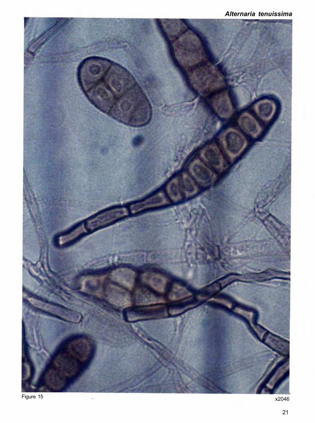
Alternaria tenuissima (Kunze ex Pers.) Wiltshire
Helminthosporium tenuissimum Kunze in C.G. & T.F.L. Nees
Macrosporium tenuissimum Fr.
Symptoms on grain. Golden brown to black growth on the seed surface (Fig. 14).
Morphology. Conidiophores are solitary or in groups, simple or branched, straight or
flexuous, more or less cylindrical, septate, pale or mid-pale brown, smooth, with one or several
conidial scars, up to 115 μrn long, and 4 μm thick. Conidia are solitary or in short chains,
straight or curved, obclavate or ellipsoidal tapering gradually to the beak which is up to half the
length of the conidium, usually shorter, sometimes tapered to a point but more frequently
swollen at the apex where there may be several scars; pale to clear mid-golden brown, usually
smooth, sometimes minutely verruculose generally with 4 -7 transverse and several
longitudinal or oblique septa, and slightly or not constricted at the septa; overall length 22-95
(average 54) μm, 8-19 (average 13.8) μm thick in the broadest part, beak 2-4 μm thick, and
swollen apex 4-5 μm wide (Fig. 15).
Quick clue. Refer Figure 15.
Importance. Alternaria tenuissima is extremely common and recorded on a wide range of
plant species, usually as a secondary invader rather than a primary parasite. It produces
tenuazonic acid (Davies et al. 1977). It has been reported to cause leaf spot of pigeonpea. It
produces the same toxins as A. alternata.
x2046
21
Figure 15
Alternaria tenuissima

Figure 16a x36 Figure 16b x436
22
Aspergillus candidus Link
Symptoms on grain. Conidial heads are persistently white or become yellowish cream with
age (Fig. 16a); typically globose when young, often splitting with age, or approaching columnar
in small heads (Fig. 16b).
Morphology. Conidiophores are smooth, colorless or slightly yellow in terminal areas.
Vesicles are typically globose to subglobose and fertile over the entire surface. Sterigmata
typically in two series, with primary series often much enlarged, sometimes varying greatly in
size within the same head (Fig. 17). Conidia are globose or subglobose and smooth.
Quick clue. Absence of pigmentation and smooth conidia. White conidial heads are present.
Importance. Aspergillus candidus is widely distributed in nature. It is encountered most
commonly on stored cereal grains and on grain products. It has been revealed frequently in
necropsies of birds and mammals at the Paris Zoological Gardens. It is a thermo-tolerant
fungus, capable of growing at 40-50°C, and is xerophilic (Raper and Fennel 1973).

Figure 17 x 1980
23
Aspergillus candidus

Aspergillus flavus Link
Symptoms on grain. Colony on seed is usually spreading and very light yellow-green, deep
yellow-green, olive brown, or brown (Fig. 18a). Conidiophores are swollen apically and bear
numerous conidia-bearing cells (phialides) with conidia in long, dry chains. Conidial heads are
typically spherical, splitting into several poorly defined columns, rarely exceeding 500-600 μm
diameter, but mostly 300-400 μm (Fig. 18b).
(Note: Severely infected sorghum grains are discolored and shrivelled.)
Morphology. Conidiophores are heavy walled, hyaline, coarsely roughened, and usually
<1 mm in length, with 10-20 μm diameter just below the apex. Apices are elongated when
young, becoming subspherical to spherical, 10-65 (am in diameter, but commonly 25-45 μm.
There can be one or two series of conidia-bearing cells (phialides and supporting cells)
depending on the species. Supporting cells are usually 6-10 x 4-6 μm but sometimes up to
15-16 x 8-9 μm in diameter. Phialides measure 6-10 x 3-5 μm (Fig. 19a). Conidia are
typically spherical to subspherical, conspicuously spiny, variable, 3-6 μm in diameter, and
sometimes oval or pear-shaped at first and occasionally remaining so (Fig. 19b).
Quick clue. Aspergillus flavus is recognized by the light yellow-green, deep yellow-green,
olive brown, or brown, compact, spherical or columnar spore heads.
Importance. Aflatoxins produced by A. flavus are toxic to humans and animals, and reduce
grain palatability for feed or food. Seed infection can reduce germination. Production of large
numbers of air-disseminated spores can cause respiratory diseases in humans and animals
(Raper and Fennel 1973). Aspergillus flavus has been used more widely in industry than any
other group of molds, particularly for the production of enzymes.
24

Figure 19a x502 Figure 19b X1130
25
Aspergillus flavus
Figure 18a x11 Figure 18b x37

Aspergillus niger van Tieghem
Symptoms on grain. Colony on seed grows slowly, consisting of a compact to fairly loose
white to faintly yellow basal mycelium, which bears abundant erect and usually crowded
conidial structures, typically carbon black but sometimes deep brown-black, covering the entire
colony except for a narrow growing margin (Fig. 20). Conidial heads are typically large and
black, compact at first, spherical, or split into two or more loose to reasonably well-defined
columns, and commonly reach 700-800 μm in diameter.
(Note: Severely infected sorghum grains are discolored and shrivelled.)
Morphology. Conidiophores are smooth, hyaline or faintly brownish near the apex and up to 3
μm in length and 15-20 μrn in diameter. Apices are spherical or nearly so, up to 75 μm in
diameter but often quite small. Two series of conidia-bearing cells (supporting cells and
phialides) are produced, but in some heads only phialides are present. Supporting cells are of
varying lengths and sometimes septate, but when mature usually 20-30 μrn long. Phialides
are more uniform in length, usually 7-10 x 2-3 μm. Conidia are typically spherical at maturity,
often very rough or spiny, mostly 4—5 μm diameter, and very dark in color or with conspicuous
longitudinal striations (Fig. 21).
Quick clue. Aspergillus niger is recognized by the production of compact, greenish black,
brownish black, purplish black, or carbon black, spherical or columnar spore heads.
Importance. Seed infection can reduce germination. Production of large numbers of air-
disseminated spores can cause respiratory diseases in man and animals. Aspergillus niger is
worldwide in distribution and occurs in and upon the greatest variety of substrata including
grains, forage products, spoiled fruits and vegetables, exposed cotton textiles and fabrics,
leather, dairy products, and other protein-rich substrata (Raper and Fnnel 1973).
26

Aspergillus niger
Figure 20 x14
Figure 21 x1617
27

Bipolaris australiensis (M.B. Ellis) Tsuda & Ueyama
(Bipolaris species "with" Cochliobolus teleomorph)
Drechslera australiensis M.B. Ellis
Helminthosporium australiense Bugnicourt
Teleomorph. Cochliobolus australiensis (Tsuda & Ueyama) Alcorn
Symptoms on grain. Conidial colonies are effuse, gray to blackish brown, and velvety.
Hyphae are pale to dark brown, smooth, and septate. Stromata are erect, straight, cylindrical,
and black (Fig. 22).
Morphology. Conidiophores are single, flexuous, geniculate, septate, smooth, cylindrical,
reddish brown, up to 150 μm long and 3-7 μm thick, having verruculose, conidiogenous nodes.
Conidia are straight, ellipsoidal or oblong, rounded at the ends, pale brown to mid-reddish
brown, usually 3-, rarely 4-5 distoseptate, 14—40 x 6-11 μm (Fig. 23).
The species is heterothallic and the teleomorph is obtained by pairing opposite compatible
monoconidial isolates in Sach's agar media with sterilized rice straw. Ascomata on rice straw
are globose to subglobose, black, superficial on columnar to flat stromata, 375-940 μm in
diameter with a long cylindrical ostiolar beak 250-1250 x 90-125 μm. Pseudoparaphyses are
filamentous, hyaline, septate, and branched. Asci are cylindrical to long, 100-182 x
8.5-15 μm clavate, vestigial bitunicate, short pedicellate, with 1-8 spores. Ascospores are
parallel to partly or closely coiled in a helix in the ascus, filiform, somewhat tapering towards the
ends, flagelliform at the ends, hyaline to very pale brown, 3-13 septate, 81-206 x
2.5-5.6 μm.
Quick clue. Verruculose conidiogenous nodes are present.
Importance. Production of mycotoxin by the fungus is unknown. Cochliobolus australiensis
causes leaf spot of pearl millet (Pennisetum glaucum (L.) R. Br.) (Chand and Singh 1966) and
leaf blight of citronella grass (Cymbopogan winterianus Jowitt.) (Ramaiah and Chandrashekar
1981) in India.
28

Bipolaris australiensis
Figure 22 x49
Figure 23 x1452
29

Bipolaris halodes (Drechsler) Shoem.
(Bipolaris species "without" Cochliobolus teleomorph)
Drechslera halodes (Drechsler) Subram. & Jain
Bipolaris rostrata (Drechsler) Shoem.
Drechslera rostrata (Drechsler) Richardson & Fraser
Exserohilum halodes (Drechsler) Leonard & Suggs
Exserohilum rostratum (Drechsler) Leonard & Suggs Imp.
Helminthosporium appatternae K.S. Deshpande & K.S. Deshpande
Helminthosporium halodes Drechsler
Helminthosporium rostratum Drechsler
Helminthosporium halodes Drechsler var tritici Mitra
Helminthosporium halodes Drechsler var elaeidicola Kovachich.
Luttrellia rostrata (Drechsler) Gonorstai
Symptoms on grain. Stromata are formed on seeds and are erect, simple or branched,
cylindrical, dark, blackish brown to start, up to 2 x 1 μm (Fig. 24).
Morphology. Conidiophores are up to 200 μm long, 5-8 μm thick, septate, cylindrical,
olivaceous brown, paler towards the apex, simple, and geniculate. Conidia are straight to
slightly curved, ellipsoidal to narrowly obclavate or rostrate, brown or olivaceous, thick-walled,
except in a small subhyaline region at the apex and a similar region surrounding the hilum
which protrudes as a darkened cylinder or truncate cone from the end of the basal cell, basal
septum darker and thicker than the other septa, up to 18-distoseptate, 15-200 x 7-29 μm
(Fig. 25). Germination occurs from the subhyaline region of the end cells and germ tubes
grow semiaxially.
(Note: Teleomorph is absent.)
Quick clue. A small subhyaline region is present at the apex of the conidium.
Importance. It is a seedborne fungus and is widely distributed. Mycotoxin production by this
fungus is unknown. It commonly occurs on grasses, and many other plant species, soil, and
textiles (Sivanesan 1987).
30

Bipolaris halodes
Figure 24 x26
Figure 25 x1320
31
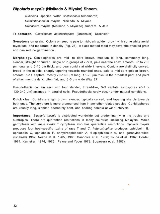
Bipolaris maydis (Nisikado & Miyake) Shoem.
(Bipolaris species "with" Cochliobolus teleomorph)
Helminthosporium maydis Nisikado & Miyake
Drechslera maydis (Nisikado & Miyakae) Subram. & Jain
Teleomorph. Cochliobolus heterostrophus (Drechsler) Drechsler
Symptoms on grain. Colony on seed is pale to mid-dark golden brown with some white aerial
mycelium, and moderate in density (Fig. 26). A black matted mold may cover the affected grain
and can reduce germination.
Morphology. Conidiophores are mid- to dark brown, medium to long, commonly long,
slender, straight or curved, single or in groups of 2 or 3, pale near the apex, smooth, up to 700
μm long, and 5-10 μm thick, and bear conidia at wide intervals. Conidia are distinctly curved,
broad in the middle, sharply tapering towards rounded ends, pale to mid-dark golden brown,
smooth, 5-11 septate, mostly 70-160 μm long, 15-20 μm thick in the broadest part; and point
of attachment is dark, often flat, and 3-5 μm wide (Fig. 27).
Pseudothecia contain asci with four slender, thread-like, 5-9 septate ascospores (6-7 x
130-340 μm) arranged in parallel coils. Pseudothecia rarely occur under natural conditions.
Quick clue. Conidia are light brown, slender, typically curved, and tapering sharply towards
both ends. The curvature is more pronounced than in any other related species. Conidiophores
are usually long, slender, alternately bent, and bearing conidia at wide intervals.
Importance. Bipolaris maydis is distributed worldwide but predominantly in the tropics and
subtropics. There are quarantine restrictions in many countries including Malaysia. Maize
germplasm with male sterile T cytoplasm also has quarantine restrictions. Bipolaris maydis
produces four host-specific toxins of race T and C. heterostrophus produces ophiobolin B,
ophiobolin C, ophiobolin F, anhydroophiobolin A, 6-epiophiobolin A, and geranylnerolidol
(Ishibashi 1962; Nozoe et al. 1965, 1966; Canonica et al. 1966; Tsuda et al. 1967; Cordell
1974; Karr et al. 1974, 1975; Payne and Yoder 1978; Sugawera et al. 1987).
32

Figure 27 x568
33
x22 Figure 26
Bipolaris maydis

Bipolaris sacchari (E. Butler) Shoem.
(Bipolaris species "without" Cochliobolus teleomorph)
Helminthosporium sacchari E. Butler
Drechslera sacchari (E. Butler) Subram. & Jain
Symptoms on grain. Stromata are formed on seeds and are erect, simple or branched,
cylindrical, dark, blackish brown to start, up to 2 x 1 mm (Fig. 28).
Morphology. Conidiophores are single or in small groups, often from groups of dark cells
which form a loose stroma, straight to flexuous, mid- to dark brown or olivaceous brown, paler
towards the apex, septate, smooth, cylindrical, up to 200 μm long, 5-8 μm thick; in culture up to
700 μm long and 10 μm thick. Conidiogenous nodes are smooth to slightly verruculose.
Conidia are slightly curved, rarely straight, cylindrical or narrowly ellipsoidal, mid-pale to mid-
yellow golden brown, 5-9 (commonly 8) distoseptate, 35-96 x 9-17 μm, hilum 2-3 μm wide
(Fig. 29).
(Note: Teleomorph is absent.)
Quick clue. Groups of dark cells and slightly curved distoseptate conidia are formed.
Importance. Bipolaris sacchari produces helminthosporoside (Beier et al. 1982) and three
isomeric host-specific toxins (Macko et al. 1983). It causes eye spot and seedling blight of
sugarcane (Saccharum officinarum L.) and leaf spots of grasses.
Fiqure 28 x521
34

Figure 29 x1980
35
Bipolaris sacchari

Bipolaris spicifera (Bainier) Subram.
(Bipolaris species "with" Cochliobolus teleomorph)
Helminthosporium spiciferum (Bainier) Nicot
Helminthosporium tetramera McKinney
Curvularia spicifera (Bainier) Boedijn
Teleomorph. Cochliobolus spicifer Nelson
Symptoms on grain. Colony on seed is brown, gray or black, hairy, cottony or cushion-like
and spreads loosely with abundant brownish conidiophores, single or in clusters of 2-3
(Fig. 30). Many small conidia are produced at very short intervals, giving rise to a bottle-brush
appearance. Colonies strongly resemble those of Curvularia spp.
Morphology. Conidiophores are brown and curved, with obvious and numerous scars
resulting in an irregular zigzag appearance. Conidia are short, typically 3-septate, light to dark
brown, oval, curved to straight with rounded ends, and measure 20-40 μm x 9-14 μm. Conidia
are lighter in color towards the terminal cells.
Ascomata are black, spherical to oval, curved, 460-710 x 350-650 μm, with an inverted cone-
shaped neck and pore. Asci are cylindrical to club-shaped, straight to slightly curved, with 1-8
spores and 130-160 x 12-20 μm. Ascospores are parallel to closely coiled in the ascus, thread-
like, somewhat tapered at the ends, 6-16 septate, hyaline, and 135-240 x 3-7 μm
(Fig. 31).
Quick clue. Under the dissecting microscope, conidia appear to be clustered for some length
on the conidiophores, giving the appearance of a bottle-brush. Conidia are very small and
typically 3-septate, almost cylindrical, more or less uniform in size, and the end cells have
subhyaline areas towards their terminal ends.
Importance. Bipolaris spicifera is distributed worldwide and is very common in tropical and
subtropical areas. The mycotoxins isolated from B. spicifera are spiciferone A and cynodontin
metabolites and those from C. spicifera are curvularin and D-mannitol (Combe et al. 1968;
Nakajima et al. 1989). The main diseases caused by B. spicifera are foot rot (or common root
rot) of winter wheat (Triticum aestivum L.) and mycotic keratitis in humans. A subcutaneous
mycosis in cat and horses is also induced by C. spicifer.
36

Figure 31 x1353
37
x10 Figure 30
Bipolaris spicifera

Bipolaris zeicola (Stout) Shoem.
(Bipolaris species "with" Cochliobolus teleomorph)
Helminthosporium carbonum Ullstrup
Helminthosporium zeicola Stout
Drechslera carbonum (Ullstrup) Sivan
Drechslera zeicola (Stout) Subram. & Jain
Teleomorph. Cochliobolus carbonum Nelson
Symptoms on grain. Grains are covered by very dark brown to black mycelium which gives a
characteristic charcoal appearance. Conidia are also visible (Fig. 32).
Morphology. Conidiophores are single or in small groups, straight to flexuous, mid- to dark
brown or olivaceous brown, up to 250 μm long, 5-8 μm thick, smooth, septate, and cylindrical.
Conidiogenous nodes are verruculose with the surface wall below them granulose. Conidia are
curved or sometimes straight, occasionally almost cylindrical but usually broad in the middle
and tapering towards the rounded ends, 6-12 (commonly 7-8) distoseptate, 30-100 x 12-18
μm, often finally becoming dark or very dark brown or olivaceous brown, with the end cells
sometimes remaining tapered than the middle cells (Fig. 33). The surface is often granulose
and hilum is not very conspicuous.
The species is heterothallic and the teleomorph is obtained by pairing opposite mating single
conidial isolates in Sach's agar media holding sterilized maize leaf segments or barley
(Hordeum vulgare L.) grains at 24°C (Nelson 1959). Ascomata are black, globose to
ellipsoidal, 355-550 x 320-420 μm, with setae over the upper half of the wall mixed with
conidiophores, and with a well-defined sub-conical to paraboloid ostiolar beak 60-200 μm
long. Pseudoparaphyses are filiform, hyaline, septate, and branched. Asci are cylindrical to
clavate, short-stalked, straight to slightly curved, 1-8 spored, vestigial bitunicate, 160-257 x
18.0-27.5 (am. Ascospores are filiform or flagelliform, somewhat tapering towards the ends,
hyaline, 5-9 septate, 180-307 x 6-10 μm, often surrounded by a thin hyaline mucilaginous
sheath.
Quick clue. Distoseptate dark to dark brown conidia are present.
Importance. Bipolaris zeicola is distributed worldwide. There are quarantine restrictions for
Indonesia, Egypt, and Chile. Bipolaris zeicola produces HC-toxins I, II, III, IV, and CHS
polypeptide (Ramussen and Scheffer 1988), and C carbonum produces carbtoxinine and
victoxinine (Nishimura et al. 1966; Pringle and Scheffer 1967). Cochliobolus carbonum is
reported on maize from many countries including India. This is the first report on sorghum in
India.
38
r

x1320
39
Figure 33
Figure 32 x53
Bipolaris zeicola

Botrytis cinerea Pers. ex Pers.
Teleomorph. Botryotinia fuckeliana (de Bary) Whetzel
Symptoms on grain. Colony on seed is white or gray or grayish-brown, and spreading for a
short distance around the affected seed (Fig. 34).
Morphology. Conidiophores are brown, tall, upright or nearly so, septate and branched, up to
30 μm wide and 2 μm long. The branches are constricted at their point of origin and quickly
collapse when removed from a moist atmosphere. Conidia occur in clusters at the swollen
rounded apices and at intervals along with conidiophores on short blunt teeth. Conidia are oval
or egg-shaped, often with a slightly projecting point of attachment, colorless to pale brown, and
measure 6-18 x 4 - 1 μm (Fig. 35).
Fairly large, black, irregular sclerotia can be produced, but not normally within the period of a
seed health test. They are rather flat in appearance and measure 5 x 2 x 2 μm.
Quick clue. The funugs is characterized by stout, brown, branched conidiophores supporting
glistening gray heads of pale conidia, which can be observed under low magnification of a
binocular microscope.
Importance. The fungus is a common gray mold, frequently parasitic, and produces abscisic
acid, botrydial, botrylacton, citric acid, and thermostable toxins (Fehlhaber et al. 1974; Kamoen
and Jamart 1974; Lyon 1977; Welmer et al. 1979; Morooko et al. 1986). However, it is not
noted as a toxigenic species.
Figure 34 x131
40

Figure 35 x858
41
Botrytis cinerea

Chaetomium oryzae
Symptoms on grain. Colony on seed is white with the density of mycelium varying from light
to dense. The perithecia are found on the seed surface beneath the aerial white mycelium (Fig.
36).
Morphology. Perithecia are spherical or elongate, with a pore opening, and a dark,
membranous, cellular wall which is covered with conspicuous hairs of various types (Fig. 37).
Asci are hyaline, usually club-shaped but in a few cases cylindrical, and contain eight
ascospores. Ascospores are one-celled and in most cases lemon-shaped. They are extruded
through the pore opening either as a mass amongst the hairs or as a column depending on
conditions.
Quick clue. Colonies of Chaetomium species can be readily recognized by the presence of
perithecia with many stiff dark terminal hairs with ornamentation.
Importance. Chaetomium is distributed worldwide. It has no significance in crop production.
However, it is a common saprophyte and secondary invader. Seeds of low germinating capacity
are sometimes found to be heavily contaminated with Chaetomium (Skolko and Groves 1953).
Figure 36
42
x23

Figure 37 x396
43
Chaetomium oryzae

Cladosporium oxysporum Berk. & Curt.
Symptoms on grain. Colonies are effuse, pale gray or grayish brown, thinly hairy on natural
substrata (Fig. 38); cottony or loosely felted in culture.
Morphology. Conidiophores are macronematous, straight or slightly flexuous, distinctly
nodose, pale or mid-pale brown, smooth, up to 500 μm long or sometimes even longer in
culture, 3-5 μm thick, with terminal and intercalary swellings of 6-8 μm diameter. Conidia
arise from terminal swellings, which later become intercalary, in simple or branched chains.
Conidia are cylindrical, rounded at the ends, ellipsoidal, limoniform or subspherical, subhyaline
or pale olivaceous brown, smooth, 5-30 x 3-6 μm (Fig. 39).
Quick clue. Cladosporium is characterized by erect, pigmented conidiophores with chains of
conidia in tree-like heads. This genus can frequently be identified by the distinctive lemon-
shaped conidia, which have well marked, dark attachment scars and show considerable
variation in size and septation within and between species.
Importance. Heavily infected sorghum grains may have dark green to black blotches, or
streaks that extend from the grain tips. The fungus is common, widely distributed in the tropics
on dead leaves and stems of herbaceous and woody plants. Many saprophytic species are
commonly encountered on seeds. Cladosporium is usually associated with frost damage and
wet weather. Black head molds are caused by saprophytic or weakly parasitic species and are
usually associated with insect infestations, lodging, nutrient deficiencies, and/or wet weather at
maturation and harvest.
x52 Figure 38
44

Figure 39 x3102
45
Cladosporium oxysporum

Cladosporium sphaerospermum Penz.
Symptoms on grain. Colony on seed spreads loosely or occasionally small, point-like,
cushion-like, cotton-like groups or with tufts, or hairy (Fig. 40a). It is often olive green but also
sometimes gray, light brownish yellow, brown or dark blackish brown (Fig. 40b). Colonies are
relatively slow growing and produce little aerial mycelium but normally sporulate freely.
Conidiophores are produced in dense stands from the seed.
(Note: Heavily infected sorghum grains may have dark green to black blotches, or streaks that
extend from the grain tips.)
Morphology. Mycelium is hyaline, becoming dark, septate, smooth or finely rough, 3-4 μm
wide. Conidiophores arise laterally from the mycelium or are formed terminally on the hyphae,
brown, smooth or finely roughened, septate, variable in length, up to about 160 μm long, 3-4
μm wide. Conidial heads are composed of branched chains of spores, a large proportion of
which are globose. Conidia are brown, echinulate (echinulation not readily seen at x600), the
majority globose or subglobose or rather ellipsoidal, continuous, 4 -6 μm in diameter; a smaller
number of larger spores are more irregular in shape, globose, ovoid, ellipsoidal with both ends
pointed or pointed at one end and with two or more pretensions at the other, sometimes
septate, 6-14 x 4-6 μm (Fig. 41).
Quick clue. Cladosporium sphaerospermum is characterized by erect, pigmented
conidiophores with chains of conidia in tree-like heads. The genus can frequently be identified
by the distinctive lemon-shaped conidia, which have well marked, dark attachment scars and
show considerable variation in size and septation within and between species. Tree-like heads
of conidiophores can be readily observed by using the scotch-tape method (see Appendix 1)
under the microscope at low power (x100).
Importance. The fungus is a very common cosmopolitan species. It occurs as secondary
invader on many plant species and has been isolated from air, soil, foodstuff, paint, textiles,
and occasionally from man and animals.
46

Figure 41 x2640
47
x46 X 1 5 Figure 40b Figure 40a
Cladosporium sphaerospermum

Colletotrichum graminicola (Cesat i ) W i l s o n
Colletotrichum sublineolum Henn. Kab & Bubak
Teleomorph. Glomerella graminicola Politis
Symptoms on grain. Visible symptoms are dark brown to black acervuli scattered on grain
surface (Fig. 42). These acervuli are irregular in shape and consist of dark setae. Sometimes
acervuli are also formed on the glumes.
Morphology. Acervuli are rounded or elongate, separate or confluent, superficial, erumpent,
with conspicuous multicellular, darkly pigmented setae, and 70-300 μm in diameter. The
acervuli consist of a gelatinous or mucoid, salmon orange colored conidial mass.
Conidiophores are hyaline, single-celled, falcate, fusiform, spindle shaped, with acute apices,
and measure 19-28.9 x 3.3-4.8 μm. Setae are brown with a dark swollen base and a pale
rounded tip (Sutton 1980) (Fig. 43).
Quick clue. Conidia are sickle-shaped and single celled.
Importance. Colletotrichum graminicola is widespread. It causes anthracnose of sorghum
and many other plant species.
Figure 42 x37
48

x396
49
Figure 43
Colletotrichum graminicola

Curvularia affinis Boedijn
(Curvularia species "without" Cochliobolus teleomorph)
Symptoms on grain. Colonies are effuse, gray, brown or blackish brown, hairy, cottony or
cushion-like and spread loosely (Fig. 44). Stromata are cylindrical, black, and unbranched.
Morphology. Conidiophores arise singly or in groups, terminally and laterally on the hyphae,
also on stromata when these are present. On natural substrata, conidiophores are erect,
simple, straight or flexuous, sometimes geniculate, septate, brown, paler near the apex,
smooth, up to 200 μm long, often swollen at the base (9-11 μm), 6-8 μm thick just above the
basal swelling, and 3-4 μm at the apex; in culture simple or loosely branched, flexuous, often
geniculate, septate, pale brown to brown, smooth, up to 400 μm long, 2-3 μm thick at the base
broadening to 4-5 μm near the apex. Conidia are straight or curved, broadly fusiform to
ellipsoidal, usually 4-, occasionally 5-distoseptate, cell at each end pale brown, intermediate
cells brown, middle cell sometimes darker, 27-49 (average 32) μm long, 8-13 (average 10)
μm thick in the broadest part (Fig. 45).
(Note: Teleomorph is absent.)
Quick clue. Conidia are often curved but seldom geniculate, 32 x 10 μm.
Importance. Curvularia affinis is isolated from rice (Oryza sativa L ) , maize, and some
dicotyledon hosts, and soil. This probably is a new report on sorghum grain from India.
Figure 44
50
x16

Figure 45 x3300
51
Curvularia affinis

Curvularia clavata Jain
(Curvularia species "without" Cochliobolus teleomorph)
Symptoms on grain. Colonies are grayish brown or brown and cottony (Fig. 46).
Morphology. Conidiophores arise terminally and laterally on the hyphae, simple, straight or
flexuous, sometimes geniculate, septate, pale brown to brown, smooth, up to 150 μm long,
2-6 μm thick, narrower at the base, and thicker towards the apex. Conidia are straight or
occasionally slightly curved, usually clavate, sometimes truncate at the base, 3-distoseptate,
smooth, 17-29 (average 23) μm long, 7-13 (average 9.6) μm thick in the broadest part
(Fig. 47). The hilum is not or very slightly protuberant, basal cell is pale brown and other cells
are brown or dark brown.
(Note: Teleomorph is absent.)
Quick clue. Conidia are straight or almost straight, symmetrical, and clavate.
Importance. Curvularia clavata is distributed worldwide especially in the tropics and is
frequently encountered as a pathogen or saprophyte. It causes serious losses in tropical
regions, but is a minor pathogen in temperate regions. An unidentified toxin produced by C.
clavata has been reported (Olufolaji1986).
Figure 46 x29
52

Figure 47 x2739
53
Curvularia clavata

Curvularia eragrostidis (Henn.)
(Curvularia species "with" Cochliobolus teleomorph)
Teleomorph. Cochliobolus eragrostidis (Tsuda & Ueyama) Sivanesan comb. nov.
Pseudocochliobolus eragrostidis Tsuda & Ueyama
Brachysporium eragrostidis P. Hennings
Spondylocladium maculans Bancroft
Symptoms on grain. Colony on seed is brown, gray, or black, hairy, cottony or cushion-like
and spreads loosely (Fig. 48).
Morphology. Conidiophores are solitary or in groups, simple or rarely branched, straight or
curved, sometimes geniculate near the apex, multiseptate, brown to light brown, variable in
length up to 5 μm diameter. Conidia are 3-distoseptate, ellipsoidal or barrel-shaped, the middle
septum almost median appearing as a black band, with brown to dark brown central cells and
paler end cells, rather smooth, 18-37x 11-20 μm (Fig. 49). Stromata are formed on rice straw
or other substrata.
The species is heterothallic and the teleomorph is obtained by pairing compatible conidial
isolates in Sach's agar media containing sterilized rice straw (Tsuda and Ueyama 1985).
Ascomata are superficial, globose, black, 375-750 x 375-750 μm, with protruding ostiolar
beaks, developing from columnar or flat stromata firmly adhering to the substrate at the base;
ostiolar beak 250-1125 x 85-190 μm, with a hyaline apex. Asci are vestigial bitunicate, almost
cylindrical with a short stalk, 1-8 spored, 150-240 x 12.5-22 μm, among filamentous
pseudoparaphyses. Ascospores are hyaline, filiform or flagelliform, 175-240 x 3.8-6.3 μm,
12-22 septate, parallel to loosely coiled in the ascus or rarely coiled in a helix.
Quick clue. Conidia are symmetrical, and middle septum is usually truly median appearing as
a black band.
Importance. The fungus was also isolated by Adiver and Anahosur (1994) from sorghum
grain samples. Mycotoxin production of this fungus is unknown. This fungus is widely
distributed on cereals, dicotyledons, and other substrata.
54

x1419
55
Figure 49
x28 Figure 48
Curvularia eragrostidis

Curvularia fallax Boedijn
(Curvularia species "without" Cochliobolus teleomorph)
Symptoms on grain. Colonies are effuse, blackish brown, velvety or cottony. Stromata are up
to 7 mm long, often branched, black, formed frequently on potato-dextrose agar and always on
grains.
Morphology. Conidiophores arise singly or in groups, terminally and laterally on the hyphae,
also on stromata, simple or loosely branched, straight or flexuous, sometimes geniculate,
reddish brown, often paler near the apex, smooth, septate; on natural substrata up to 250 μm
long and swollen at the base (11-16 μm diameter), and in culture up to 1 mm long and 4 -6 μm
thick. Conidia are straight or slightly curved, broadly fusiform or ellipsoidal, almost always
4-distoseptate, smooth; cell at each end is subhyaline or very pale brown, and intermediate
cells are mid-pale brown to brown. On natural substrata conidia are 24-26 (average 30) μm
long, 10-16 (average 12.2) μm thick in the broadest part, in culture 24-38 (average 30.6) μm x
9-15 (average12.3) μm ( Fig. 50).
(Note: Teleomorph is absent.)
Quick clue. Conidia are often curved but seldom geniculate, 30 x 12.2 μm. Stromata are
branched.
Importance. The fungus has a wide host range (species of Oryza, Panicum, Sorghum, and a
variety of dicotyledonous hosts). It is also isolated from air, house dust, soil, and wood.
Probably this is a new report of the occurrence of C. fallax on sorghum grain in India. However,
C. fallax has been reported on rice in India.
56

x1980
57
Figure 50
Curvularia fallax

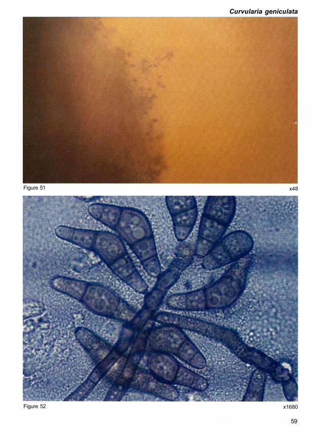
Curvularia geniculata (Tracy & Earle) Boedijn
(Curvulaha species "with" Cochliobolus teleomorph)
Teleomorph. Cochliobolus geniculatus Nelson
Symptoms on grain. Colony on seed is brown, gray, or black, hairy, cottony or cushion-like
and spreads loosely (Fig. 51).
Morphology. Conidiophores are up to 600 μm long. Conidia are usually curved, geniculate,
fusiform, 3-4 distoseptate but almost always 4-distoseptate, rarely 5-distoseptate, smooth,
26-48 x 8-13 μm on natural substrata and 18-37 x 8-14 μm in culture (Fig. 52). The end cells
are subhyaline or very pale brown, intermediate cells brown to dark brown, and the central cell
usually dark brown and swollen.
The species is heterothallic and the teleomorph is obtained by pairing compatible conidial
isolates in Sach's agar media containing sterilized barley grains at 24°C under constant
artificial light (Nelson 1964). Ascomata are free or frequently develop on a columnar stroma,
up to 830 μm broad. Asci are 1-8 spored, cylindrical, vestigial bitunicate, and 170-290 x
15-20 μm among filamentous pseudoparaphyses. Ascospores are somewhat tapered at the
ends, filiform, 6-16 septate, 160-270 x 4-7 μm, coiled in a helix inside the ascus.
Quick clue. Conidia are often distinctly geniculate, curved, and tapering gradually towards
each end.
Importance. Curvularia geniculata and its teleomorph is known to produce 1,4,5,8-
tetrahydroxy-2,6-dimethylanthraquinone metabolite (Combe et al. 1968). This is a new report
of its occurrence on sorghum grain in India. However, the frequency of occurrence was less
(only 24 grains were colonized out of 20,800 grains).
58
x1680
59
Figure 52
x48 Figure 51
Curvularia geniculata

Curvularia harveyi Shipton
(Curvularia species "without" Cochliobolus teleomorph)
Symptoms on grain. Colonies are effuse, grayish brown, cottony to velvety (Fig. 53).
Morphology. Conidiophores arise singly or in groups, terminally and laterally on the hyphae,
simple or occasionally branched, straight or flexuous, sometimes geniculate, septate, pale
brown to brown, smooth, up to 250 μm long, 3-7 μm thick. Conidia are straight or slightly
curved, cylindrical to ellipsoidal, with a markedly protuberant hilum at the base, rounded at the
apex, and almost always 3-distoseptate, but rarely 1-4 distoseptate (Fig. 54).
(Note: Teleomorph is absent.)
Quick clue. Conidia are cylindrical to ellipsoidal with protuberant hilum at the base.
Importance. Occurrence of C. harveyi has been reported only on Triticum sp from Australia.
This is a new report of its occurrence on sorghum grain in India.
Figure 53
60
x11

Figure 54 x2508
61
Curvularia harveyi

Curvularia lunata (Wakker) Boedijn
(Curvularia species "with" Cochliobolus teleomorph)
Teleomorph. Cochliobolus lunatus Nelson & Haasis
Pseudocochliobolus pallescens Tsuda & Ueyama
Curvularia leonensis M.B. Ellis
Symptoms on grain. Colony on seed is brown, gray, or black, hairy, cottony or cushion-like
and spreads loosely (Fig. 55).
Morphology. Conidiophores arise singly or in groups, simple or rarely branched, straight or
sometimes geniculate near the apex, brown to dark brown, multiseptate, variable in length, up
to 5-6 μm diameter. Conidia are mostly 3-distoseptate, ellipsoidal to fusiform, or often
disproportionately enlarged in the third cell and markedly geniculate or hook-shaped, pale to
somewhat colored, almost concolorous, 17-32 x 7-12.5 μm, and smooth (Fig. 56). Conidia are
sparse in culture, and variable in shape and size among isolates.
Teleomorph is produced when compatible conidial isolates are paired in Sach's agar media
(Tsuda and Ueyama 1983). Ascomata are superficial, globose to subglobose, black, 250-750
x 250-750 μm, with protruding ostiolar beaks, developing from columnar or flat stromata, firmly
adhering to the substrate at the base; ostiolar beak 190-690 x 60-160 μm, with a hyaline apex.
Asci are vestigial bitunicate, almost cylindrical with a short stalk, 140-215 x 12.5-19.0 μm,
produced among the filamentous pseudoparaphyses, arising from the base of the locule.
Ascospores are flagelliform or filiform, hyaline, tapering towards either end, 125-215 x
2.5-6.3 μm, 6-13 septate, parallel or coiled in a certain portion of the ascus.
Quick clue. Stromata are very rarely formed; conidia are 18-32 x 8-16 μm, always curved at
the third cell.
Importance. Curvularia lunata is distributed worldwide especially in the tropics and is
frequently encountered as a pathogen or saprophyte. It causes serious losses in the tropical
regions but is a minor pathogen in temperate regions. Curvularia lunata and C. lunatus are
known to produce the metabolites brefeldin A, D-mannitol, anthraquinone, cytochalasin B,
cynadontin, and radicinol (Bohlmann et al. 1961; Combe et al. 1968; Nukina and Marumo
1976; van Eijk and Roeymans 1977; Wells et al. 1981).
62

x1815
63
Figure 56
Curvularia lunata
x23 Figure 55

Curvularia lunata var aeria (Bat., Lima, & Vasconcelos) M.B. Ellis
(Curvularia species "without" Cochliobolus teleomorph)
Malustela aeria Bat., Lima, & Vasconcelos
Curvularia caricae-papayae Srivastava & Bilgrami
Curvularia lycopersici Tandon & Kakkar
Symptoms on grain. Colonies are floccose, brown, dark brown to black, often zonate,
showing reverse alternating bands of red, yellow, or gray. Stromata are large, black, cylindrical,
simple or branched, formed abundantly on grains (Fig. 57).
Morphology. Conidiophores are terminal and lateral on hyphae and stromata, simple or
branched, straight or flexuous, often geniculate, septate, pale brown to brown, smooth, up to
800 μm thick. Conidia are straight to curved, ellipsoidal, obovoid or clavate, often truncate at
the scar, almost always 3-distoseptate, rarely 4-distoseptate, with one or more septa
sometimes thicker and darker than the others, smooth, with walls often rather thicker, 18-32 x
8-16 μm (Fig. 58). The third cell from base is frequently larger and darker than the others, end
cells are usually pale brown, and intermediate cells are brown or dark brown.
(Note: Teleomorph is absent.)
Quick clue. Stromata are large, black, cylindrical, simple or branched, formed abundantly on
grains.
Importance. The fungus is distributed worldwide especially in the tropics and is frequently
encountered as a pathogen or saprophyte. It causes serious losses in tropical regions but is a
minor pathogen in temperate regions. It produces a thermostable toxin (Bisen 1983).
Figure 57
64
x45

Figure 58 x 2739
65
Curvularia lunata var aeria

Curvularia ovoidea (Hiroe & Watan) Muntanola
(Curvularia species "without" Cochliobolus teleomorph)
Brachysporium ovoideum Hiroe & Watan
Symptoms on grain. Colonies are circular to irregular, pale brown to dark brown, and velvety.
Stromata are not seen (Fig. 59a, b).
Morphology. Conidiophores are straight to flexuous, multiseptate, cylindrical, smooth, pale
brown, geniculate above, up to 400 μm long, 4-9 μm thick. Conidia are ovoid, 1-3
distoseptate, straight or curved, 16-29 x 10-17 μm, commonly 20-25 x 13-16 μm, brown with
paler end cells (Fig. 60).
(Note: Teleomorph is absent. Tsuda et al. (1985) treated this species as a synonym of C.
lunata.)
Quick clue. Stromata are absent and often symmetrical conidia are produced.
Importance. Occurrence of Curvularia ovoidea on species of Capsicum, Pennisetum, and
Zea has been reported from Egypt, India, and Japan. This is a new report of C. ovoidea on
sorghum grain from India.
Figure 59a x15 Figure 59b x59
66

Curvularia ovoidea
x2277
67
Figure 60

Curvularia pallescens Boedijn
(Curvularia species "with" Cochliobolus teleomorph)
Teleomorph. Cochliobolus pallescens (Tsuda & Ueyama) Sivan.
Symptoms on grain. Colony on seed is brown, gray, or black, hairy, cottony or cushion-like
and spreads loosely (Fig. 61).
Morphology. Conidiophores arise singly or in groups, simple, rarely branched, straight or
sometimes geniculate near the apex, brown to dark brown, multiseptate, variable in length, up
to 5-6 μm. Conidia are mostly 3-distoseptate, ellipsoidal to fusiform, or often disproportionately
enlarged in the third cell, markedly geniculate or hook-shaped, pale to somewhat colored,
almost concolorous, 17-32 x 7-12.5 μm, smooth (Fig. 62). Conidia are sparse in culture, and
variable in shape and size among isolates.
Ascomata are superficial, globose to subglobose, black, 250-750 x 250-750 μm, with
protruding ostiolar beaks, developing from columnar or flat stromata, firmly adhering to the
substrate at the base; ostiolar beak 190-690 x 60-160 μm, with a hyaline apex. Asci are
vestigial bitunicate, almost cylindrical with a short stalk, 140-215 x 12.5-19.0 μm, among the
pseudoparaphyses, arising from the base of the locule. Ascospores are flagelliform or filiform,
hyaline, tapering towards either end, 125-215 x 2.5-6.3 μm, 6-13 septate, parallel or coiled in
certain portion of the ascus.
Quick clue. Conidia are usually straight or only slightly curved, hook-shaped; all conidial cells
are usually pale or very pale brown.
Importance. The fungus is distributed worldwide especially in the tropics and is frequently
encountered as a pathogen or saprophyte. It causes serious losses in tropical regions, but is a
minor pathogen in temperate regions. The production of an unidentified toxin by this fungus
has been reported (Olufolaji 1986).
68

Figure 61 x53
x2079
69
Figure 62
Curvularia pallescens

Curvularia trifolii (Kauffm.) Boedijn
(Curvularia species "without" Cochliobolus teleomorph)
Symptoms on grain. Colonies are effuse, brown or grayish brown, hairy or dark blackish
brown, cottony, sometimes floccose (Fig. 63). Stromata are cylindrical, black, sometimes
formed in old cultures.
Morphology. Conidiophores arise singly or in groups, terminally and laterally on the hyphae,
simple or branched, straight or flexuous, sometimes geniculate, septate; on natural substrata
rather pale brown, seldom up to 150 μm long, with a swollen base of 8-13 u.m, 5-17 μm thick
just above the basal swelling, 3-5 μm at the apex; in culture pale brown to brown, smooth or
verrucose, up to 400 μm long, 3-8 μm thick. Conidia are 3-distoseptate, smooth, almost
always curved at the third cell from the base which is usually larger than the others. The hilum
is protuberant, cell at each end is subhyaline or pale brown, intermediate cells are brown or
dark brown, and the third cell from the base is often the darkest. On natural substrata conidia
are 28-38 (average 33.3) μm long, 12-16 (average 14) μm thick in the broadest part
whereas in culture they are 20-34 (average 27.7) μm x 8-14 (average 11.5) μm (Fig. 64).
(Note: Teleomorph is absent.)
Quick clue. Conidia are 3-distoseptate, <40 μm, almost always curved at the third cell from
the base which is usually larger than the others.
Importance. The fungus has a wide host range and is distributed widely. It produces 1,4,5,8-
tetrahydroxy-2,6-dimethylanthraquinone metabolite (Combe et al. 1968).
x31 Figure 63
70

x1980
71
Figure 64
Curvularia trifolii

Curvularia tuberculata Jain
(Curvularia species "with" Cochliobolus teleomorph)
Teleomorph. Cochliobolus tuberculatus Sivan.
Symptoms on grain. Colony on seed is brown, gray, or black, hairy, cottony or cushion-like
and spreads loosely (Fig. 65).
Morphology. Conidiophores arise singly or in groups, terminal or lateral on hyphae, stromata,
and ascomata, simple or branched, straight or flexuous, smooth, pale to mid-brown, septate,
up to 300 μm long, 2-7 μm thick. Conidia are straight, ovoid, obclavate or ellipsoidal, 3-5
(sometimes 8, but mostly 3) septate, intermediate cells brown to dark brown, end cells
subhyaline to pale or dark brown, mature conidia tuberculate, 23-52 x 13-20 μm (Fig. 66).
Young conidia are smooth and subhyaline. First septum in the conidium is usually median,
second septum often delimiting the basal cell but variations in septal formation may occur.
Germination is both by bipolar and lateral germ tubes.
The species is heterothallic and the teleomorph is obtained by pairing monoconidial
compatible isolates (Sivanesan 1985). Ascomata are black, globose, often borne on a
columnar basal stroma or a flattened crust, 500-720 μm high, 400-490 μm wide, with a conical
truncate beak up to 300 μm high, 115-140 μm wide at the base, often hairy in the globose part
with simple, brown, septate hyphae. Conidiophores arise from the globose part of the ascoma
but are not formed abundantly. Pseudoparaphyses are hyaline, filiform, and branched above.
Asci are cylindrical, short-stalked, with 2-8 spored, vestigial bitunicate, 170-340 x 13.5 μm.
The stalk is cylindrical with or without a bifurcate base, with a wall that does not stain in
lactophenol cotton blue. Ascospores are filiform, hyaline, helically coiled in the ascus and often
straight at one or both ends, commonly tapering at both ends but more so at the base,
sometimes with a truncate apex, with hyaline mucilaginous sheath up to 4 μm thick (only visible
in water mounts), not constricted, 13-23 distoseptate, 160-460 x 3-4.5 μm.
Quick clue. Conidia are straight, 3-septate, tuberculate (having tubercles) or rough-walled
unlike other Curvularia species.
Importance. Curvularia tuberculata is distributed worldwide especially in the tropics and is
frequently encountered as a pathogen or saprophyte. It causes serious losses in tropical
regions but is a minor pathogen in temperate regions. The production of an unidentified toxin
by this fungus has been reported (Olufolaji 1986). This is a new report of C. tuberculata on
sorghum grain in India.
72

Curvularia tuberculata
x48 Figure 65
x1650
73
Figure 66

Epicoccum nigrum Link
Epicoccum purpurascens Ehrenb.
Symptoms on grain. Colony on seed grows rapidly, often producing a yellow, amber to
orange, or red/black pigmentation within but particularly surrounding the white, compact
mycelium (Fig. 67). Due to these features, the fungus is occasionally confused with Fusarium
spp and frequently mistaken as Ustilaginales.
(Note: Infected sorghum grains may become red.)
Morphology. Epicoccum nigrum is a mitosporic fungus. Conidiophores are compact or
occasionally branched, loose, dark, smooth, short, occurring in tight clusters from the hyphae
and produce a single, terminal conidium. Mature conidia are dark brown to black, mostly
spherical but also pear-shaped, irregularly septate, and may appear to be very coarsely
marked like a net. The septa are often hidden by the thick, rough spore wall, which appears to
be covered by short, blunt projections. Conidia measure 15-25 μm in diameter and often occur
in dark, cushion shaped spore masses of variable size within and on the surface of the
mycelium (Fig. 68).
Quick clue. Dark spore masses look like black spots scattered over the mycelium. Individual
spores resemble dark, rough soccer balls, and may be confused with spores of smuts and
bunts.
Importance. Occurrence of E. nigrum on sorghum grains has been reported along with
method(s) to kill the fungus adhering to the grains for safe use of grains for consumption (Navi
et al. 1997). The fungus is distributed worldwide. It is a common saprophyte and secondary
invader. Its quarantine importance is not known. Unidentified toxins have been isolated from
this fungus (Schol-Schwarz 1959).
74

Figure 67 x21
x1452
75
Figure 68
Epicoccum nigrum

Exserohilum rostratum (Drechsler) Leonard & Suggs
Helminthosporium rostratum Drechsler
Drechslera rostrata (Drechsler) Richardson & Fraser
Bipolaris rostrata (Drechsler) Shoemaker
Teleomorph. Setosphaeria rostrata Leonard
Symptoms on grain. Colony on seed appears mid- to dark brown or golden brown with very
little white, aerial mycelium. Conidiophores are formed together in a dense mat covering the
seed. Infected sorghum grains show pink discoloration or are charcoal black when severely
colonized (Fig. 69).
Morphology. Conidiophores are solitary or in groups, straight or bending, mid- to dark brown
or olive brown, up to 200 μm long and 8 μm thick. Conidia are straight or slightly curved,
tapering at both ends with one end typically wider, and the narrow end terminating in a
pronounced beak. Conidia have golden brown intermediate cells, 6-16 transverse septa,
hyaline or pale end cells with a thick dark septum, and measure 40-180 x 14-22 μm (Fig. 70).
Ascocarps are spherical, black, 340-600 x 330-580 μm, with pore opening and upper part
surrounded with dark brown, blunt spine-like projections. Asci have a slimy sheath and are
short-stalked, club-shaped to cylindrical, 1-8 spored, and measure 105-260 x 26-42 μm.
Ascospores are hyaline to pale brown, straight to curved, 2-5 septate, narrowed at septa,
29-85 x 9-21 μm.
Quick clue. Conidia have a distinctive shape and are straight or slightly curved, with a
pronounced beak, and visible, dark, end septa.
Importance. Infected sorghum grains show pink discoloration or are charcoal black when
severely colonized. Exserohilum rostratum causes leaf blight of sorghum and produces
glyceollin toxin (Kumar et al. 1984) and cynodontin toxin (van Eijk and Roeymans 1977).
76

Exserohilum rostratum
x48 Figure 69
x729
77
Figure 70

Exserohilum turcicum (Pass.) Leonard & Suggs
Helminthosporium turcicum Pass.
Drechslera turcica (Pass.) Subram. & Jain
Helminthosporium inconspicuum Cooke & Ellis
Teleomorph. Setosphaeria turcica (Luttrell) Leonard & Suggs
Symptoms on grain. Colony on seed is pale to mid-dark brown with very little white, aerial
mycelium (Fig. 71a, b).
Morphology. Conidiophores are single or in groups of 2-6, straight or bent, light to dark olive
brown, medium to long, sometimes very long, and measure 150-300 x 7-11 μm. Conidia are
straight or slightly curved, club-shaped or widest near the middle, tapering towards the ends,
with a rounded apex, and basal cell swollen at the point of attachment. Conidia are pale to mid-
straw colored or yellowish brown or olive gray in color, 4-9 septate, and 50-144 x 18-33 μm
(Fig. 72).
(Note: Perithecia rarely occur in nature.)
Quick clue. Conidia arise from long conidiophores and are large, yellowish brown, straight, or
slightly curved, narrowing towards both ends (almost cigar shaped), with the basal cell bulging
at the point of attachment. '
Importance. The fungus is distributed worldwide but predominantly in subtropical to
temperate climates. There are quarantine restrictions for some countries. Mycotoxins
produced by this fungus are monocerin, ophiobolin A (Ishibashi 1961; Nozoe et al. 1965;
Canonica et al. 1966; Robeson and Strobel 1982), and ravenelin (Raistrick et al. 1936).
78

Figure 71a x72 Figure 71b x237
Exserohilum turcicum
x2706
79
Figure 72

Fusarium moniliforme J. Sheld. Lisea fujikuroi Sawada
Fusarium verticilloides (Sacc.) Nirenberg
Teleomorph. Gibberella fujikuroi (Sawada) Ito
Gibberella moniliforme Wineland
Symptoms on grain. Colony on grain grows rapidly with white aerial mycelium often
becoming tinged with purple, particularly on the blotting paper in the petri dish. Mycelium has a
powdery appearance due to the presence of chains of microconidia. Tan to orange spore
masses of irregular shape and size are occasionally present (Fig. 73).
Morphology. Abundant microconidia are formed. They are hyaline, usually one-celled but
occasionally two-celled, 5-12 x 1-3 μm, oval to club-shaped, and slightly flattened at each end
(Fig. 74). Macroconidia are formed infrequently. They are hyaline, delicate with thin walls,
curved to almost straight, 3-7 septate, 25-60 x 2-4 μm, and have a foot-shaped basal cell
(Fig. 74). Chlamydospores are never present in the mycelium or conidia.
Perithecia, which occur rarely, are spherical, blue-black, and 250-350 x 220-300 μm. Asci are
oval to club-shaped with 4-8 ascospores. Ascospores are hyaline, straight, mostly one-
septate, and measure 4-7 x 12-17 μm.
Quick clue. Abundant uniform microconidia are formed in long chains that can readily be
observed using the scotch-tape method (see Appendix 1) under the microscope at low
power (X100) . Chlamydospores are never formed.
Importance. The fungus produces the mycotoxin fumonisin which is toxic to humans and
livestock when heavily infected grain is consumed. It is widespread in both humid and sub-
humid, temperate zones and subtropical and tropical zones. There are quarantine restrictions
for this fungus in Egypt.
80

Figure 73 x12
x594
81
Figure 74
Fusarium moniliforme

Fusarium semitectum Berk. & Rav. [W&R, G,B,J]
Fusarium roseum LK. emend. Snyd. & Hans. Pro Parte [S&H]
Fusarium roseum LK. emend. Snyd. & Hans. var arthrosponioides (Sherb) Messiaen
& Cassini Pro Parte [M&C]
Teleomorph. Not known.
Symptoms on grain. Colony on grain is pink or orange in color and often turns white (Fig. 75).
Morphology. Microconidia are rarely produced. However, two types of macroconidia are
produced. Some are borne on mycelium and are spindle-shaped, straight to slightly curved.
The other type are sickle-shaped and are borne in sporodochia. These are slightly curved, with
a foot-shaped basal cell. Conidiophores are unbranched and monophialides and polyphialides
are branched (Fig. 76).
Quick clue. Polyphialides are present in the aerial mycelium and spindle-shaped
macroconidia are produced in the aerial mycelium.
Importance. Fusarium semitectum has been reported to be toxigenic (Nelson et al. 1983).
x25 Figure 75
82

Fusarium semitectum
Figure 76 x600
83

Gloecercospora sorghi Bain & Edgerton ex Deighton
Symptoms on grain. Black, shiny, spindle or irregular shaped sclerotia, about 0.1-0.2 mm
diameter are seen on infected grains. The sclerotia are embedded in the pericarp, and often
become errumpent by rupturing it (Fig. 77).
Morphology. The fungus produces dark brown to charcoal black sclerotia, and pink to reddish
orange sporodochia. Sometimes only sclerotia are produced. Mycelium is scanty or abundant,
white to dull white, thin, and branched. Sporodochia are pink to salmon pink and are visible to
the naked eye. Each sporodochium consists of numerous hyaline conidiophores and conidia
that can be seen under a compound microscope. Conidiophores are hyaline, branched or
unbranched, septate, short, 5-10 μm long, with a somewhat swollen apex. Conidia are borne
in a pinkish, slimy matrix, and are hyaline, elongate to filiform, 1.4-3.2 x 20-195 μm, and
septate (Fig. 78).
Quick clue. Dark brown to charcoal black sclerotia, and pink to reddish orange sporodochia
are seen on the grain. Hyaline, elongate to filiform conidia are produced in a slimy matrix.
Importance. The fungus is widely distributed. It causes grain discoloration and also zonate
leaf spot of sorghum.
Figure 77 x27
84

x1452
85
Figure 78
Gloecercospora sorghi

Gonatobotrys simplex Corda
Gonatobotrys zeae Futrell & Bain (nomen nudum)
Symptoms on grain. Colony on seed is white and usually on the surface of other fungal
species, e.g., Alternaria, Cladosporium, Curvularia, and Fusarium (Fig. 79a,). Mycelium
appears as a mass of strings with clusters of "flower-like" bunches of conidia (Fig. 79b).
Morphology. Conidiophores are erect, sometimes tall, septate, simple or occasionally
branched, with inflated cells covered with a series of blunt teeth bearing conidia, inserted at
intervals and terminally on the hyphae. Conidia are borne singly on the blunt teeth. They are
1-celled, hyaline, oval to subspherical, and measure 10-22 x 6-12 μm (Fig. 80).
Quick clue. Gonatobotrys simplex is distinguished by the cluster of large, hyaline, conidia
arising from "nodes" along the length of the conidiophores, and appearing like a "string of beads".
Importance. Gonatobotrys simplex has worldwide distribution. Its quarantine significance is
not known. It is a parasite on Alternaria spp and Cladosporium spp (Whaley and Barnett 1963).
Figure 79a x45
86

Gonatobotrys simplex
Figure 79b x58
Figure 80 x2211
87

Nigrospora oryzae (Berk. & Br.) Petch
Teleomorph. Khuskia oryzae Hudson
Symptoms on grain. Colony on seed is initially white and the shiny, black conidia standing
out in sharp contrast give the colonies a striking appearance under the binocular dissecting
microscope (Fig. 81). In older cultures the hyphae darken and the colonies appear black, with
profuse conidial production.
(Note: Infected seeds have white streaks with black spore masses near the tips.)
Morphology. Conidiophores are short, pale brown, inflated and borne at right angles to
hyphae, bearing conidia singly and terminally. Conidia are smoky brown or jet black, spherical
or egg-shaped, 10-16 x 10-13 μm, and commonly measure 12-14 μm in diameter (Fig. 82).
Perithecia are formed in clusters of 1-7 in series or irregular rows, up to 2 μm long. They are
spherical or oval and up to 250 μm in diameter with protruding pore openings. Asci are short-
stalked, club-shaped, and measure 55-75 x 8-12 μm, with 8 ascospores. Ascospores are
hyaline, granular, curved, 16-21 x 5-7 μm, and tapering to the base with rounded ends. They
are initially one-celled but after discharge from the ascus may develop a single transverse
septum dividing the spore unequally into two cells.
Quick clue. Very dark conidia, slightly longer in the horizontal axis are borne on very short,
pale brown conidiophores with a characteristic bulge.
Importance. The fungus is distributed worldwide. It occurs commonly on Oryza spp and
maize but there are reports of isolation from air and soil (Hudson 1983). It is a new report on
sorghum grain from India. Nigrospora oryzae produces aphidicolin metabolite (Startratt and
Loschiavo 1974).
88

Figure 81 x46
x1485
89
Figure 82
Nigrospora oryzae

Penicillium citrinum Thorn.
Symptoms on grain. The fungus is readily recognized by its penicilli, which consist of 3-5
divergent and usually vesiculate metulae, bearing long, well-defined columns of conidia.
Colonies are often dominated by copious, clear to yellow or brown exudate at the centers
(Fig. 83). On malt extract agar, the growth is slower and usually dense, with heavy conidial
production.
[Note: Lactofuchsin stain was used for microscopical observations (Carmichael 1955) (see
Appendex 1).]
Morphology. Conidiophores are borne from subsurface or surface hyphae, with stipes
100-300 μm long, smooth walled, characteristically terminating in well defined verticils of 3-5
divergent metulae, less commonly with a divergent ramus, or subterminal or intercalary
metulae. Metulae are usually of uniform length, 12-15 μm long, commonly spathulate or
terminally vesiculate, up to 5 μm diameter; phialides are ampulliform, 7-8 (sometimes 12) μm
long. Conidia are spherical to subspheroidal, 2.2-3.0 μm with walls smooth or very finely
roughened, typically borne in long, well defined columns, one per metula, arranged in a
characteristic whorl on each conidiophore (Fig. 84).
Quick clue. Penicillium citrinum is an isolated species. Occasionally isolates show a few
characteristics suggesting a relationship to P. corylophilum Dierckx, i.e., faster growth on malt
extract agar and metulae of unequal length.
Importance. Like several other Penicillium metabolites, citrinin produced by P. citrinum is
known to be a potentially hazardous mycotoxin. Citrinin causes watery diarrhoea, increased
food consumption, and reduced weight gain due to kidney degeneration in chickens,
ducklings, and turkeys. The effect of citrinin on humans is not documented. However, kidney
damage appears to be a likely result of prolonged ingestion. Penicillium citrinum may well be
one of the most common eukaryotic life forms of earth. It is ubiquitous in soil, decaying
vegetation, and the air. It is also a powerful biodeteriogen, commonly causing decay and
losses in foods, textiles, paints, and plastics (Pitt 1991).
90

Penicillium citrinum
x20 Figure 83
x1683
91
Figure 84

Penicillium griseofulvum
Penicillium griseofulvum Dierckx
Penicillium palulem Bainier
Penicillium urticae Bainier
Symptoms on grain. The fungus produces very short phialides and it bears them on highly
branched conidiophores. Colonies on Czapek yeast extract agar and malt extract agar are gray
with only weak greenish overtones; and surface texture is fasciculate to minutely coremial.
Morphology. Conidiophores are borne in fascicles, with stipes of indeterminate length, often
sinuous, smooth walled, brownish, terminating in distinctive penicilli, sometimes terverticillate,
more commonly a quaterverticillate, and not infrequently with 5 or even more branch points
between stipe and phialide; rami are 15-25 (sometimes 30) μm long and ramuli are 10-15 μm
long. Metulae are 7-10 μm long, sometimes apically inflated; phialides are ampulliform, closely
packed, exceptionally short, 4.5-6.0 μm, abruptly tapering to short collula. Conidia are
ellipsoidal, 3.0-3.5 μm long, smooth walled, borne in closely packed, disordered chains.
[Note: Lactofuchsin stain was used for microscopical observations (Carmichael 1955)
(see Appendix 1).]
Quick clue. The fungus is a stable species, with little isolate to isolate variation. Penicillium
griseofulvum has several features which set it apart from the other species, especially the
highly branched conidiophores, brown walled stipes, and very short phialides. It may provide a
link with the genus Nomuraea.
Importance. Penicillium griseofulvum is a very commonly occurring species, with worldwide
distribution. It plays a major role in the decay of vegetation, and of seeds (cereals), food, and
feed. The fungus produces the antibiotic griseofulvin (Pitt 1991) and the mycotoxins patulin,
cyclopiazonic acid, and roquefortine C.
(Note: Figures could not be reproduced due to technical reasons.)
92

Periconia macrospinosa
Periconia macrospinosa Lefebvre & A.G. Johnson
Symptoms on grain. Colonies are effuse, gray, brown, and hairy. The mycelium is mostly
immersed but sometimes partly superficial (Fig. 85).
x53
93
Figure 85

Morphology. Conidiophores are very dark brown, up to 420 μm long, 7-12 μm thick at the
base, and 6-10 μm immediately below the head (Fig. 86a). Conidia are 18-35 μm in diameter,
coarsely echinulate; the spines are 2-7 μm long and sometimes adhere closely to one another
in groups (Fig. 86b).
Quick clue. Conidia are echinulated.
Importance. Periconia macrospinosa has been isolated from species of Chenopodium,
Prunus, Trifolium, and Triticum and soil in Australia, Canada, Europe, Hong Kong, India, Iraq,
and USA (Ellis 1971). However, this is a new report of its occurrence on sorghum grain in India.
x3531 Figure 86a
94

Periconia macrospinosa
x3531
95
Figure 86b

Phoma sorghina (Sacc.) Boerema, Dorenbosch, & van Kesteren
Phoma insidiosa Tassi
Symptoms on grain. Colony on seed has very little white or gray mycelium but produces
large numbers of dark brown or black pycnidia on seed surface or on the blotting paper in the
petri dish. Grains with large number of pycnidia appear shrivelled (Fig. 87).
Morphology. Pycnidia are almost spherical, dark brown, thin-walled, and variable in size
(100-300 μm diameter), with one conspicuous protruding pore opening. Conidia are released
from the pycnidia in the form of a creamy colored curved tendril (Fig. 88a). Conidia are
unicellular, oblong to oval, hyaline, and measure 5-8 x 2-4 μm (Fig. 88b).
Quick clue. Spherical, dark brown pycnidia release unicellular, hyaline conidia through a
pronounced pore opening in the form of a curved tendril. The pycnidia of Phoma species often
develop in compact colonies and produce spores profusely. Unicellular conidia distinguish
Phoma species from the pycnidial fungi of the Septoria complex.
Importance. The fungus is distributed worldwide. It occurs as a pathogen after prolonged
periods of humid weather. It is frequently observed as a secondary invader. It produces
tenuaronic acid.
x58 Figure 87
96

x4972
97
Figure 88b
Phoma sorghina
x 118 Figure 88a

Rhizopus stolonifer (Ehrenb: Fr.) Lindner
Symptoms on grain. Colony on the seed spreads rapidly by means of stolons with abundant,
loose, gray mycelium (Fig. 89). Stolons produce numerous, brown sporangiophores and
rhizoids.
(Note; The fungus is so common on maize seeds, that tests for other pathogens often employ
precautionary measures to avoid growth of Rhizopus, e.g., by surface sterilization of seeds
with NaOCI.)
Morphology. Stolons are hyaline becoming brown towards nodes, near which a septum may
occur. Rhizoids are short, brown and sometimes absent. Sporangiophores arise singly or in
small groups from nodes on the stolons. They are brown, smooth or finely roughened, non-
septate, 1000-3500 μm long and up to 34 μm wide. Sporangia are spherical, initially white but
later black, and 100-350 μm in diameter with numerous spores (Fig. 90). Columellae are light
brown, subspherical, 63-224 x 70-140 μm, and umbrella-shaped when dehisced. Sporangio-
spores are yellow to dilute brown, spherical or oval, longitudinally striped, and measure 5-8 x
20-26 μm.
Quick clue. Dark, spherical sporangia can readily be seen under a dissecting microscope,
enabling identification of Rhizopus (without removal of the lid of the petri dish). The fungus is
often referred to as pin mold as the sporangia resemble black pinheads and are widely
interspersed in cotton wool-like mycelium.
Importance. The fungus is distributed worldwide. It is a common saprobe and facultative
parasite of mature fruits and vegetables. It is important in storage rot complex under high
moisture and temperature conditions.
Figure 89
98
x15

x1980
99
Figure 90
Rhizopus stolonifer
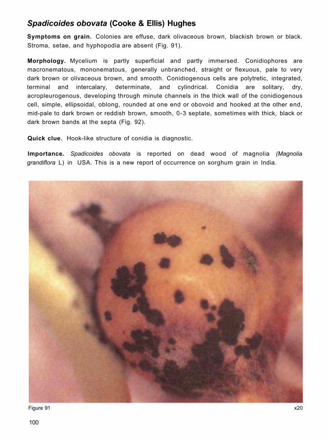
Spadicoides obovata (Cooke & Ellis) Hughes
Symptoms on grain. Colonies are effuse, dark olivaceous brown, blackish brown or black.
Stroma, setae, and hyphopodia are absent (Fig. 91).
Morphology. Mycelium is partly superficial and partly immersed. Conidiophores are
macronematous, mononematous, generally unbranched, straight or flexuous, pale to very
dark brown or olivaceous brown, and smooth. Conidiogenous cells are polytretic, integrated,
terminal and intercalary, determinate, and cylindrical. Conidia are solitary, dry,
acropleurogenous, developing through minute channels in the thick wall of the conidiogenous
cell, simple, ellipsoidal, oblong, rounded at one end or obovoid and hooked at the other end,
mid-pale to dark brown or reddish brown, smooth, 0-3 septate, sometimes with thick, black or
dark brown bands at the septa (Fig. 92).
Quick clue. Hook-like structure of conidia is diagnostic.
Importance. Spadicoides obovata is reported on dead wood of magnolia (Magnolia
grandiflora L) in USA. This is a new report of occurrence on sorghum grain in India.
x20 Figure 91
100

Spadicoides obovata
Figure 92 x3135
101

Torula graminis Desm.
Symptoms on grain. Colony on seed forms small, compact, olive green mounds which may
coalesce and when older tend to become brown. Colonies are round or oval up to 1.5 x 0.5 μm.
Morphology. Conidiophores are short including conidiogenous cells, 2-5 μm thick, or lacking,
and not readily distinguished, with conidia arising more or less directly from the vegetative
hyphae. Conidia develop in long chains, which break into segments from one to many cells
when mature, brown, minutely verruculose, 4-5 x 4-6 μm; cells or zero septate conidia are
almost spherical but often slightly broader than long. Conidia are barrel shaped, with the end
cells rounded, smooth to moderately rough surface, and dark brown to black (Fig. 93).
Quick clue. Torula graminis is characterized by simple or branched chains of dark conidia
which break up readily and which arise more or less directly from the vegetative hyphae.
Importance. The fungus is distributed worldwide. It is a common saprophyte and secondary
invader. It is predominant in wet harvests. It causes sooty head mold of wheat. Occurrence of
T. graminis on grasses in Europe has been reported. However, this is a new report of
occurrence on sorghum grain from India.
102

Figure 93 x792
103
Torula graminis

Trichothecium roseum Link
Symptoms on grain. Colony on seed usually appears as a salmon pink crust with the
production of numerous conidia (Fig. 94). Colonies can be cushion-like or powdery.
Morphology. Conidiophores are erect or suberect, produced singly or in groups, simple or
sparingly branched, long, slender, hyaline, and septate. Conidia are produced in short, fragile
chains. Conidia are large (12-18 x 8-10 μm), smooth, two-celled (slightly narrowed at the
septum), hyaline, more or less egg-shaped, with well marked attachment point and upper cell
somewhat larger than the lower one (Fig. 95).
Quick clue. Colony on seed superficially resembles the spore masses of Fusarium or
Gliocladium species. The short chains of two-celled conidia at the apex of a hyaline, simple
conidiophore are diagnostic.
Importance. The fungus is widespread. It is a common saprophyte and secondary invader. Its
quarantine significance is not known. It causes pink rot of apple (Malus pumila Miller). It
produces trichothecene mycotoxins, e.g., trichothecin and trichothecolon.
x66 Figure 94
104
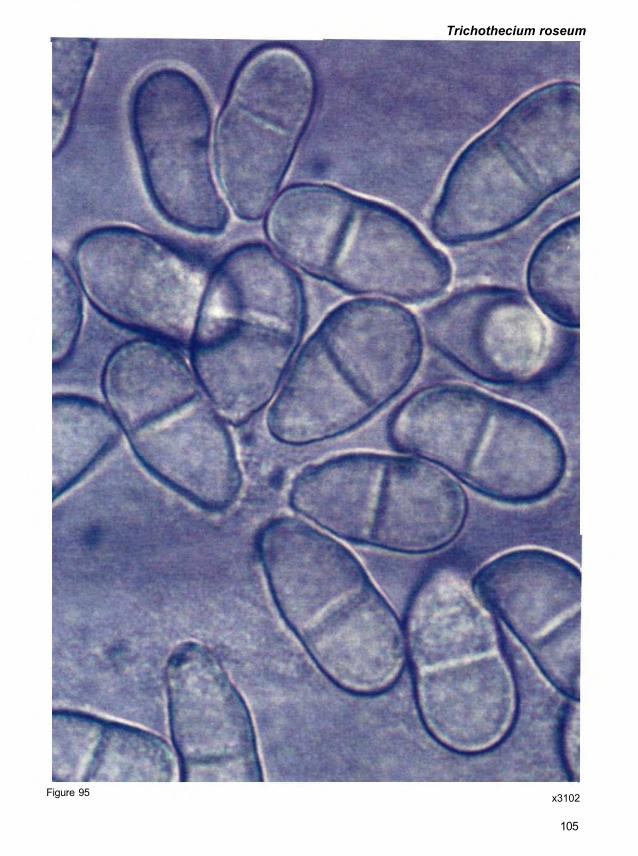
Trichothecium roseum
Figure 95 x3102
105

References
Adiver, S.S., and Anahosur, K.H. 1994. Association of Curvularia and Exserohilum species as
sorghum molds and their effect on seed germination and seedling growth. Current Research
23:3-4.
Bandyopadhyay, R., Mughogho, L.K., and Satyanarayana, M.V. 1987. Systemic infection
of sorghum by Acremonium strictum and its transmission through seed. Plant Disease 71:647-
650.
Barnett, H.L., and Hunter, B.B. 1972. Illustrated genera of imperfect fungi. Minneapolis,
Minnesota, USA: Burgess Publishing Co. 241 pp.
Barron, G.L. 1968. The genera of hyphomycetes from soil. New York, USA: Robert E Krieger
Publishing Co. 364 pp.
Beier, R.C., Mundy, B.P., and Strobel, G.A. 1982. Helminthosporoside, a host-specific toxin
from Helminthosporium sacchari: a structure review and a new portion structure. Experientia
38:1312-1314.
Bisen, P.S. 1983. Production of toxic metabolites by Curvularia lunata (Wakker) Boedijn var.
aeria and its role in leaf spot disease of bean (Phaseolus vulgaris L ) . Acta Botanica India
11:235-237.
Bohlmann, F., Luders, W., and Plettner, W. 1961. Uber einen Anthrachinon-farbstoff aus
Curvularia lunata. Archiv der Pharmazie 294:521-524.
Booth, R.H., and Burden, O.J. 1983. Post-harvest losses. Pages 144-160 in Plant
pathologist's pocket book. Wallingford, Oxon, UK: CAB International.
Canonica, L, Feicchi, A., Galli, K.M., and Scala, A. 1966. Isolation and constitution of
cochlibolin B. Tetrahedron Letters 13:1329-1339.
Carmichael, J.W. 1955. Lacto-fuchsin: a new medium for mounting fungi. Mycologia 47:611.
Champ, B.R., Highley, E., Hocking, A.D., and Pitt, J.I. (eds.) 1991. Fungi and mycotoxins in
stored products: Proceedings of an international conference, Bangkok, Thailand, 23-26 April
1991. ACIAR Proceedings No. 36. Australia: The Griffin Press Ltd. 270 pp.
Chand, J.N., and Singh, B. 1966. A new Helminthosporium disease of bajra. Current Science
35:240.
Ciegler, A., and Lindenfelser, L.A. 1969. An antibiotic complex from Alternaria brassicicola.
Experientia 25:719-720.
Combe, R.G., Jacobs, J.J., and Watson, T.R. 1968. Constituents of some Curvularia
species. Australian Journal of Chemistry 21:783-788.
106

Combe, R.G., Jacobs, J.J., and Watson, T.R. 1970. Metabolites of some Alternaria species.
The structures of altenusin and dehydroaltenusin. Australian Journal of Chemistry 23:2343-
2351.
Cordell, G.A. 1974. The occurrence, structure elucidation and biosynthesis of
sesterterpenes. Phytochemistry 13:2343-2364.
Davies, N.D., Diener, U.L., and Morgan-Jones, G. 1977. Tenuazonic acid production by
Alternaria alternata and Alternaria tenuissima isolated from cotton. Applied and Environmental
Microbiology 34:155-157.
Ellis, M.B. 1971. Dematiaceous hyphomycetes. Kew, Surrey, England: Commonwealth
Mycological Institute. 608 pp.
Ellis, M.B. 1976. More dematiaceous hyphomycetes. Kew, Surrey, England: Commonwealth
Mycological Institute. 507 pp.
FAO/ICRISAT (Food and Agriculture Organization of the United Nations/International Crops
Research Institute for the Semi-Arid Tropics). 1996. The world sorghum and millet economies:
facts, trends, and outlook. Rome, Italy: FAO; and Patancheru 502 324, Andhra Pradesh, India:
ICRISAT 68 pp.
Fehlhaber, H.F., Gejpel, R., Mercker, J.J., Tschescha, R., and Weimar, K. 1974. Botrydial,
ein sesquitepen-antibioticum and der nahrlosung des Pilzes Botrytis cinerea. Chemische
Berichte 107:1720-1730.
Forbes, G.A., Bandyopadhyay, R., and Garcia, G. 1992. A review of sorghum grain mold.
Pages 253-264 in Sorghum and millets diseases: a second world review (de Milliano, W.A.J.,
Frederiksen, R.A, and Bengston, G.D., eds.). Patancheru 502 324, Andhra Pradesh, India:
International Crops Research Institute for the Semi-Arid Tropics.
Hanlin, R.T. 1990. Illustrated genera of Ascomycetes. St. Paul, Minnesota, USA: American
Phytopathological Society. 263 pp.
Hawksworth, D.L., Kirk, P.M., Sutton, B.C., and Pegler, D.N. 1995. Ainswoth & Bisby's
dictionary of the fungi. 8th edition. Surrey, UK: International Mycological Institute.
Hudson, H.J. 1983. The perfect state of Nigrospora oryzae. Transactions of the British
Mycological Society 46:355-360.
Ishibashi, K. 1961. Studies on antibiotics from Helminthosporium sp. Fungi III. Ophiobolin
production by Helminthosporium turcicum. Journal of the Agricultural Chemical Society of
Japan 35:323-326.
Ishibashi, K. 1962. Studies on antibiotics from Helminthosporium sp. Fungi IV.Ophiobolin
production by Ophiobolus heterostrophus, Helminthosporium leersii, H. panici-miliacei, and H.
zizaniae. Journal of the Agricultural Chemical Society of Japan 36:226-228.
107

Kamoen, O., and Jamart, G. 1974. Citric acid, a vivotoxin secreted by Botrytis cinerea during
infection of Begonia. Chemical Abstracts 81:148-619.
Karr, A.L. Jr., Karr, D.B., and Strobel, G.A. 1974. Isolation and partial characterization of four
host-specific toxins from Helminthosporium maydis (race T). Plant Physiology 53:250-257.
Karr, D.B., Karr, A.L., and Strobel, G.A. 1975. The toxins of Helminthosporium maydis (Race
T). Plant Physiology 55:727-730.
Kumar, S., Shukia, R.S., Singh, K.P., and Paxton, J.D. 1984. Glyceollin: Phytoalexin in leaf
blight of Costos speciosus. Phytopathology 74:1349-1352.
Logrieco, A., Visconti, A., and Battalico, A. 1990. Mandarin fruit rot caused by Altemaria
alternata and associated mycotoxins. Plant Disease 74:415-417.
Lyon, G.D. 1977. Report of the Scottish Horticultural Research Institute, 23rd annual report for
the year 1976. Invergowrie, Dundee, UK: Scottish Horticultural Research Institute. 118 pp.
Macko, V., Acklin, W., and Hildenbrand, C. 1983. Structure of the isomeric host-specific
toxins from Helminthosporium sacchari. Experientia 39:343-347.
Maekawa, N., Yamamoto, M., Nishimura, S., Kohmoto, K., Kawada, M., and Watanabe, Y.
1984. Studies on host-specific AF-toxins produced by Altemaria alternata strawberry
pathotype causing Altemaria black spot of strawberry. (1) Production of host-specific toxins
and their biological activities. Annals of Phytopathological Society of Japan 50:600-609.
Mikami, Y, Nishijima, T., Imura, H., Suzuki, A., and Tamura, S. 1971. Chemical studies on
brown-spot disease of tobacco plants. Part I. Tenuazonic acid as vivotoxin of Altemaria
longipes. Agricultural and Biological Chemistry 35:611-618.
Morooko, N., Tsunoda, H., and Tatsuno, T. 1986. Isolation of abscicic acid from Botrytis
cinerea, a fungus used in cultivation of Bursaplenchus xylophilus and its toxin effects.
Proceedings of Japanese Association of Mycotoxicology 24:63-66.
Nakajima, H., Hamasaki, T., and Kimura, Y. 1989. Structure of spiciferone A, a novel pyrene
plant growth inhibitor produced by the fungus Cochliobolus spicifer. Agricultural and Biological
Chemistry 53:2297-2298.
Navi, S.S., Singh, S.D., Lenne, J.M., Kirk, P.M., and Brayford, D. 1997. New grain mold
fungi of sorghum in India. Journal of Mycology and Plant Pathology 27:104-105. (Abstract.)
Nelson, P.E., Toussoun, T.A., and Marasas, W.F.O. 1983. Fusarium species. An illustrated
manual for identification. Pennsylvania, USA: The Pennsylvania State University Press.
193pp.
Nelson, R.R. 1959. Cochliobolus carbonum, the perfect stage of Helminthosporium
carbonum. Phytopathology 49:807-810.
108

Nelson, R.R. 1964. The perfect stage of Curvularia geniculata. Mycologia 56:777-779.
Nishimura, S., Scheffer, R.P., and Nelson, R.R. 1966. Victoxinine production by
Helminthosporium species. Phytopathology 56:53-57.
Nozoe, S., Hiari, K., and Tsuda, K. 1966. The structure of zizanin-A and -B, C25-terpenoids
isolated from Helminthosporium zizaniae. Tetrahedron Letters 20:2211-2216.
Nozoe, S., Morisaki, M., Tsuda, K., litaka, Y., Takahashi, N., Tamura, S., Ishibashi, K., and
Shirasaka, M. 1965. The structure of ophiobolin, A C25-terpenoid having a novel skeleton.
Journal of American Chemical Society 87:4968-4970.
Nukina, M., and Marumo, S. 1976. Aversion factors, antibiotics among different strains of a
fungal species. Aversion factors of Cochliobolus setariae. Agricultural and Biological Chemistry
40:2121-2123.
Olufolaji, D.B. 1986. Production and bioassay of Curvularia pallescens Boedijn toxin.
Cryptogamie Mycologie 7:335-342.
Payne, G.A., and Yoder, O.C. 1978. Production and some characteristics of host-specific
toxin(s) produced by several isolates of Helminthosporium maydis race T. Phytopathology
68:707-714.
Pitt, J.I. 1988. A laboratory guide to common Penicillium species. Australia: Commonwealth
Scientific and Industrial Research Organization, Division of Food Processing. 187 pp.
Pitt, J.I. 1991. Penicillium toxins. Pages 99-103 in Fungi and myctoxins in stored products:
Proceedings of an international conference, Bangkok, Thailand, 23-26 April 1991 (Champ,
B.R, Highley, E., Hocking, A.D., and Pitt, J.I., eds). ACIAR Proceedings No. 36. Australia: The
Griffin Press Ltd.
Pringle, R.B., and Scheffer, R.P. 1967. Isolation of host-specific toxin and a related
substance with nonspecific toxicity from Helminthosporium carbonum. Phytopathology
57:1169-1172.
Raistrick, H., Robinson, R., and White, D.E. 1936. Studies in the biochemistry of micro-
organisms L. Ravenelin (3-methyl 1:4:8-trihydroxy-anthone), a new metabolite product of
Helminthosporium ravenelii Curtis and H. turcicum Passerini. Biochemical Journal 30:1330-
1314.
Ramaiah, K.S., and Chandrashekar, M. 1981. A new leaf blight disease of citronella grass.
Current Science 50:724-725.
Ramussen, J.B., and Scheffer, R.P. 1988. Isolation and biological activities of four selective
toxins from Helminthosporium carbonum. Plant Physiology 86:187-191.
109

Raper, K.B., and Fennel, D.I. 1973. The genus Aspergillus. New York, USA: Robert E.
Kreiger Publishing Co. 686 pp.
Rizk, A.M., Hammouda, F.M., Ei-Missiry. M.M., Mayergi, H.A., Lashin, S.M., and Nofal,
M.A. 1985. Mycotoxin of Lolium seeds in response to fungal infections. Annals of Agricultural
Science, Ain Shams University 30:607-615.
Robeson, D.J., and Strobel, G.A. 1982. Monocerin, a phytotoxin from Exserohilum turcicum.
Agricultural and Biological Chemistry 46:2681-2684.
Schol-Schwarz, M.B. 1959. The genus Epicoccum Link. Transactions of the British
Mycological Society 42(2):149-173.
Simmons, E.G. 1967. Typification of Alternaria, Stemphylium and Ulocladium. Mycologia
59:67-92.
Sivanesan, A. 1985. The teleomorph of Curvularia tuberculata. Transactions of the British
Mycological Society 84:584-591.
Sivanesan, A. 1987. Graminicolous species of Bipolaris, Curvularia, Drechslera,
Exserohilum and their teleomorphs. Mycological paper No. 158. Wallingford, Oxon, UK: CAB
International.
Sivanesan, A. 1991. The taxonomy and biology of dematiaceous hyphomycetes and their
mycotoxins. Pages 47-64 in Fungi and myctoxins in stored products: Proceedings of an
international conference, Bangkok, Thailand, 23-26 April 1991 (Champ, B.R., Highley, E.,
Hocking, A.D., and Pitt, J.I., eds.). ACIAR Proceedings No. 36. Australia: The Griffin Press Ltd.
Skolko, A.J., and Groves, J.W. 1953. Notes on seed-borne fungi. VII. Chaetomium.
Canadian Journal of Botany 31:779-809.
Standen, J.H. 1945. Nigrospora oryzae (B and Br.) Petch on maize. Phytopathology 35:552-
564.
Startratt, A.N., and Loschiavo, S.R. 1974. The production of aphidicolin by Nigrospora
sphaerica. Canadian Journal of Microbiology 20:416-417.
Sugawera, F., Strobel, G., Strange, R.N., Siedow, J., van Duyne, G.D., and Clardy, J.
1987. Phytotoxins from pathogenic fungi Drechslera maydis and Drechslera sorghicola.
Proceedings of the National Academy of Sciences, USA 84:3081-3085.
Sutton, B.C. 1980. The Coelomycetes - Fungi Imperfecti with pycnidia, acervuli and stromata.
Kew, Surrey, UK: Commonwealth Mycological Insitute. 696 pp.
Templeton, G.E. 1972. Alternaria toxins related to pathogenesis in plants. Pages 169-192 in
Microbial toxins VIII (Kadis, S., Ciegler, A., and Ajl, S.J., eds.). Academic Press.
110

Tsuda, K., Nozoe, S., Morisaki, K., Hirai, K., Itai, Y, Okuda, S., Canonica, L, Feicchi, A.,
Galli, K.M., and Scala, A. 1967. Nomenclature of ophiobolins. Tetrahedron Letters 35:3369-
3370.
Tsuda, M., Nagakubo, T, Taga, M., and Ueyama, A. 1985. Sexuality for the teleomorph
formation and conidial variability in Curvularia lunata. Transactions of the Mycological Society
Japan 26:27-39.
Tsuda, M., and Ueyama, A. 1983. Pseudocochliobolus pallescens and variability of conidium
morphology. Memmoirs of College of Agriculture of Kyoto University 122:85-91.
Tsuda, M., and Ueyama, A. 1985. Two new Pseudocochliobolus and a new species of
Curvularia. Transactions of the Mycological Society Japan 26:321-330.
van Eijk, G.W., and Roeymans, H.J. 1977. Cynodontin, the tetrahydroxyanthra-quinone of
Curvularia and Drechslera species. Experientia 33:1283-1284.
von Ramm, C., and Lucas, G.B. 1963. Production of enzymes and antibiotic substances by
Alternaria longipes. Tobacco Science 7:81-84.
Wells, J.M., Cole, R.J., Cutler, H.C., and Spalding, D.H. 1981. Curvularia lunata, a new
source of cytochalsin B. Applied and Environmental Microbiology 41:967-971.
Welmer, K., Tschesche, R., and Breitmaier, E. 1979. Botrylacton, ein neuer wirkstoffaus der
nahrlosung des plizes Botrytis cinerea. Chemische Berichte 112:3598-3602.
Whaley, J.W., and Barnett, H.L. 1963. Parasitism and nutrition of Gonatobotrys simplex.
Mycologia 55:199-210.
Whitehead, M.D., and Calvert, O.H. 1959. Helminthosporium rostratum inciting ear rot of
corn and leaf spot of thirteen grass hosts. Phytopathology 49:817-820.
Zillinsky, F.J. 1983. Common diseases of small grain cereals: A guide to identification.
Mexico: Centra Internacional de Mejoramiento de Mafz y de Trigo.
111

Appendix 1
Identification procedures: scotch-tape method
The scotch-tape method is used to assist identification of different fungi by preserving the
attachment of conidia to conidiophores. It is particularly useful for those fungi in which the
conidia readily detach themselves from the conidiophore (e.g., Cladosporium spp) or those in
which chains of conidia readily break up (e.g., Fusarium moniliforme) under normal
procedures for slide preparation. The procedure is as follows:
1. Cut a small section of cello-tape (sticky transparent tape; scotch-tape) approximately
4 cm long.
2. Gently hold the tape at each end between the thumb and forefinger with the sticky side
pointing downwards in a U shape and the least amount of tape in contact with the fingers
as possible.
3. Gently place the bottom of the U onto the surface of a colony culture so that the sticky
side picks up some mycelium and conidia from the colony. Contact with the colony
should be very light so as to only pick up a very small amount of fungal material.
4. Place the piece of tape on top of a drop of water on a slide without touching the middle
section of the tape.
5. Place a coverslip on top of the cello-tape.
6. Observe the slide under the microscope.
Microscopical observations of Penicillium spp
Preparation of wet mounts. Use an inoculating needle, or a nichrome or platinum wire cut to
a chisel point, or a steel sewing needle, to cut out a small portion of the colony including sporing
structures. With freely sporing isolates, cut a piece of colony near the margin, where penicilli are
just maturing, and conidial numbers are not excessive. If sporulation is tardy, examination with
the stereomicroscope can be useful. Cleistothecia should be taken from near colony centers,
where the chance of obtaining mature ascospores is highest. Float the cut colony sample from
the needle on to a slide with the aid of a drop of 70% alcohol. It may be necessary to tease out
the mycelium with the needle and the corner of a cover slip (square coverslips are best).
Penicillium conidia and penicilli are highly hydrophobic; the alcohol helps to set the preparation,
minimizing the amount of entrapped air. When most of the alcohol has evaporated, add a drop
of lactic acid (for phase or interference contrast optics) or lactofuchsin stain for bright field.
Place a coverslip; if necessary, remove excess liquid from the preparation by gently blotting with
facial tissue or similar absorbent paper. The preparation is now ready for examination.
Staining. A wide variety of stains are used for mycological work. However, most are time
consuming to prepare, or to use, or are mild, because walls and spores of some fungi are highly
resistant to stains. By far the most effective stain for preparations of Penicillia is lactofuchsin
112

(Carmichael 1955), which suffers from none of these faults. It consists of 0 . 1% acid fuchsin
dissolved in lactic acid of 85% or higher purity. Young actively growing structures are
preferentially stained bright pink; hence penicilli, cleistothecial initials, developing asci, and
mature ascospores can be readily distinguished against a background of old mycelium.
Observation. Commence observation under a low power objective, x10 or X 1 6 , to locate the
preparation on the slide, and an area of the preparation where fruiting structures are most
readily observable. Then use a x40 objective to study the morphology of the fruiting structures.
Measurement of lengths of fruiting structure elements and examination of conidia require the
use of a x 100 oil immersion objective.
113

Glossary
Acervulus (pl = acervuli) Saucer-shaped conidioma in which the hymenium of coni-
diogenous cells develops on the floor of the cavity from
a pseudoparenchymatous stroma beneath on the
integument of the host tissue which ruptures at maturity.
Acropleurogenous Borne at the tip and along the sides.
Amphigenous Growth all round or on two sides.
Ampulliform Flask-like in form.
Ascoma (pl = ascomata) An ascus-containing structure (also called ascocarp).
Ascospore A meiospore borne in an ascus.
Ascus (pl = asci) A sac-like cell generally containing a definite number of
ascospores formed by free cell formation usually after
karyogamy and meiosis; characteristic of the class
Ascomycetes.
Bitunicate An ascus in which the inner wall is elastic and expands
greatly beyond the outer wall at the time of spore liberation.
Cantenulate In chains or end-to-end series.
Chlamydospore An asexual 1 -celled spore (primarily for perennation and not
dissemination) originating endogenously and singly within
part of a pre- existing cell, by the contraction of the protoplast
and possessing an inner secondary and often thickened
hyaline or brown wall, usually impregnated with hydrophobic
material.
Clavate Club-shaped, thickened towards the apex.
Columella A sterile central axis within a mature fruit-body which may be
unicellular or multicellular, unbranched or branched, of
fungal or host origin.
Concolors Of one color.
Confluent Coming together; running into one another.
Conidiogenous cell Any cell from or within which a conidium is directly produced.
114

Conidiophore
Conidium (pl = conidia)
Determinate
Distoseptate
Echinulate
Ellipsoid
Erumpent
Fasciculate
Filiform
Flexuous
Fusiform
Geniculate
Globose
Hyphopodium
Hilum
Hypha
Indeterminate
Heterothallic
Limoniform
Macroconidium
Macronematous
A single or branched hypha (fertile) bearing or consisting of
conidiogenous cells from which conidia are produced.
Any asexual spore which when mature is liberated from a
conidiophore or conidiogenous cell.
Growth ceasing with the production of terminal conidia.
Having individual cells each surrounded by a sac-like wall
distinct from the outerwall.
Having sharply pointed spines; spinose.
A conidium having an outline of an ellipse.
Bursting through the surface of the substratum.
Hyphae having growth in fascicles.
Thread-like.
Bent alternately in opposite directions.
Spindle-like; narrowing towards the ends.
Bent like a knee.
Nearly spherical.
A short branch of one or two cells on epiphytic mycelium of
Meliolales.
A mark or scar especially that on a spore at the point of
attachment to a conidiogenous cell or sterigma.
A fungus thread or filament.
Continuing growth indefinitely.
Two different thalli being required for sexual reproduction.
Lemon-like in form.
The larger and generally more diagnostic conidium of a
fungus which also has microconidia (and sometimes also
mesoconidia); (infrequent) a large conidium.
Conidiophores morphologically different from vegetative
hyphae.
115

Metula
Microconidium
Mononematous
Monophialide
Mucilaginous
Muriform
Mycelium
Obclavate
Obovoid
Obpyriform
Ovoid
Papilla
Papillate
Pedicel
Pedicellate
Perithecium
Phialide
Phialidic
Polyphialide
A conidiophore branch having phialides, eg., of Penicillium
and Aspergillus.
The smaller conidium of a fungus which also has macro-
conidia.
Conidiophores, solitary or in tufts or loose fascicles.
Conidiogenous cell having one locus through which conidia
are produced.
Sticky when wet; slimy.
Being dividied by intersecting septa in more than one plane.
A mass or group of hyphae making up the thallus of a fungus.
The shape of a club upside down, thickened towards the
base.
The shape of an egg upside down with the narrow end at the
base.
The shape of a pear upside down with the broad end at the
base.
Egg-shaped, with one end narrower than the other.
A minute rounded projection.
Having a papilla.
A small stalk.
Having a pedicel.
A closed ascocarp with a pore at the top, a true ostiole, and a
wall of its own.
A discrete or integrated, phialidic conidiogenous cell.
Enteroblastic and producing conidia, usually in large numbers
in basipetal succession through one opening or several
openings which are often provided with collarettes, and
with neither the outer nor inner wall contributing towards the
formation of the conidium wall.
Conidiogenous cell having more than one conidiogenous
locus at which conidia are produced.
116

Pseudoparaphyses A little or strongly, modified terminal hypha in the hymenium
of Hymenomycetes (paraphyses, pseudoparaphysis,
paraphysoid, dikaryoparaphysis, and pseudophysis are
synonyms or near synonyms).
Pseudoperithecium An uniloculate ascostroma.
Pseudothecium Contrition of pseudoperithecium.
Pycnidium (pl = pycnidia) A frequently flask-shaped conidioma of fungal tissue with a
circular or longitudinal ostiole, the inner surface of which is
lined entirely or partially by conidiogenous cells.
Pyriform Pear-shaped with the broad end uppermost.
Quaterverticillate Hairy branching at four levels.
Rhizoid A root-like structure consisting of anucleate, filamen-
tous, branched, extension of chytrid thallus acting as
a feeding organ.
Rostrate Beaked or strongly attenuated at the apex.
Sclerotium (pl = sclerotia) A firm, frequently rounded, mass of hyphae, with or without
the addition of host tissue or soil, normally having no spores in
or on it.
Seta (pl = setae) A stiff hair, generally thick-walled and dark in color.
Solitary Arising singly at one point.
Spinulose Covered with little spines.
Sporangiophore Thallus element (usually morphologically differentiated)
subtending one or more sporangia.
Sporangium An organ enclosing endogenously generated spore(s), the
walls of the spore(s) not being derived from the supporting or
containing structure.
Sporodochium A pulvinate stroma with closely packed, relatively short
conidiophore covering its upper surface.
Sterigma (pl = sterigmata) An extension of the metabasidium composed of a basal
filamentous or inflated part and an apical spore-bearing
projection.
Stolon A runner as in Rhizopus.
117

Stroma An often cushion-like mass of fungal cells or closely inter-
woven hyphae.
Teleomorph Sexual stage.
Terverticillate Having branching at three levels, i.e., having rami bearing
metulae and phialides.
Tretic The sort of conidiogenesis in which each conidium
(tretoconidium, tretic conidium, poroconidium, porospore)
is delimited by an extension of the inner wall of the
conidiogenous cell.
Truncated Ending abruptly, as though with the end cut off horizontally.
Verrucose Warted.
Verruculose Finely warted.
Vesicle A bladder-like sac; swollen apex of the conidiophore.
118

Notes

Notes

About ICRISAT
The semi-arid tropics (SAT) encompasses parts of 48 developing countries including most of India, parts
of southeast Asia, a swathe across sub-Saharan Africa, much of southern and eastern Africa, and parts
of Latin America. Many of these countries are among the poorest in the world. Approximately one-sixth of
the world's population lives in the SAT, which is typified by unpredictable weather, limited and erratic
rainfall, and nutrient-poor soils.
ICRISAT's mandate crops are sorghum, pearl millet, finger millet, chickpea, pigeonpea, and groundnut;
these six crops are vital to life for the ever-increasing populations of the SAT. ICRISAT's mission is to conduct
research which can lead to enhanced sustainable production of these crops and to improved management
of the limited natural resources of the SAT. ICRISAT communicates information on technologies as they are
developed through workshops, networks, training, library services, and publishing.
ICRISAT was established in 1972. It is one of 16 nonprofit, research and training centers funded through
the Consultative Group on International Agricultural Research (CGIAR). The CGIAR is an informal
association of approximately 50 public and private sector donors; it is co-sponsored by the Food and
Agriculture Organization of the United Nations (FAO), the United Nations Development Programme (UNDP),
the United Nations Environment Programme (UNEP), and the World Bank.
