a patient with shortness of breath and bilateral … · of breath and bilateral pulmonary...
TRANSCRIPT
Case Report Lee et al.
Open Medicine 2010;4(4):e187
A patient with shortness
of breath and bilateral
pulmonary infiltrates
Christie Lee, Devon MCDonaLD, Jeannie CaLLuM, anna Day, robert FowLer
Christie Lee, MD FRCPC, is a pulmonary and critical care physi-cian at Sunnybrook Health Sciences Centre, University of Toronto, Toronto, Ontario, Canada. Devon McDonald, MD, is a general inter-nist at Sunnybrook Health Sciences Centre, University of Toronto, Toronto. Jeannie Callum, MD FRCPC, is a hematologist at Sunnybrook Health Sciences Centre, University of Toronto, Toronto. Anna Day, MD FRCPC, is professor of medicine, Women’s College Hospital, University of Toronto, Toronto, and a respirologist at the University of Toronto. Robert Fowler, MD FRCPC, is an internist and critical care physician at Sunnybrook Health Sciences Centre, University of Toronto, Toronto.
Competing interests: None declared. One clinical department to which Robert Fowler belongs has participated in studies sponsored by Astra-Zeneca, Eli Lilly, Wyeth Pharmaceuticals, Novo Nordisk and Chiron Corporation.
Correspondence: Dr. Robert Fowler, 2075 Bayview Ave., Rm. D478, Toronto ON M4N 3M5; tel: 416 480-6100 ext. 7471; fax: 416 480-6191; [email protected]
Case report
A 54-yeAr-old CAuCAsiAn womAn presented to our emergency department with a 3-week history of product-ive cough, shortness of breath and malaise. Four weeks before, she had visited her family physician with ear pain and nasal fullness. Acute sinusitis was diagnosed, and she was prescribed a 10-day course of levofloxacin, along with triamcinolone nasal spray. However, she developed increasing shortness of breath on exertion, followed by fever, chills, rigours and intermittent night sweats.
She was reevaluated by her family physician 3 days before presentation and was prescribed clarithromycin (500 mg twice daily) for a diagnosis of community- acquired pneumonia; however, her symptoms pro-gressed and she came to our emergency department for further evaluation.
The patient resided with her husband and 1 adult child. As director of a homeless shelter program, she inspected shelters on a weekly basis. A tuberculin skin test per-formed 8 years earlier was negative. The patient denied
all risk factors for HIV infection. She was a lifelong non-smoker and consumed 5 standard alcoholic beverages per week. She denied sick contacts and allergies to ani-mals and did not have a significant travel history.
Her medical history was significant for allergic rhin-itis, recurrent sinusitis and asthma. Current asthma treatment included inhaled steroids (fluticasone), long- and short-acting β2-agonists (salbutamol and sal-meterol) and a leukotriene receptor antagonist (montel-ukast). She began taking montelukast 8 months before her presentation and had required systemic cortico-steroids for asthma exacerbations on 2 separate occa-sions in the 6 months preceding her presentation. Her medications on presentation were her asthma therapy, clarithromycin and triamcinolone nasal spray. She had an anaphylactic allergy to lactose. There was no family history of respiratory or inflammatory disease.
On examination, the patient appeared thin and was in moderate respiratory distress at rest. She was febrile with a temperature of 38oC, she had a pulse rate of 104 beats/min, her blood pressure was 139/77 mm Hg, her respira-tory rate was 34 breaths/min and she had a decreased oxygen saturation of 92% on room air. Auscultation of the chest revealed decreased breath sounds throughout both lung fields, with crackles heard predominantly in the left and right upper lobes. Examination of the pre-cordium revealed normal heart sounds and the presence of a grade II/VI systolic ejection murmur heard best at the base of the heart. She did not have a rash. Findings of the rest of the physical examination were normal.
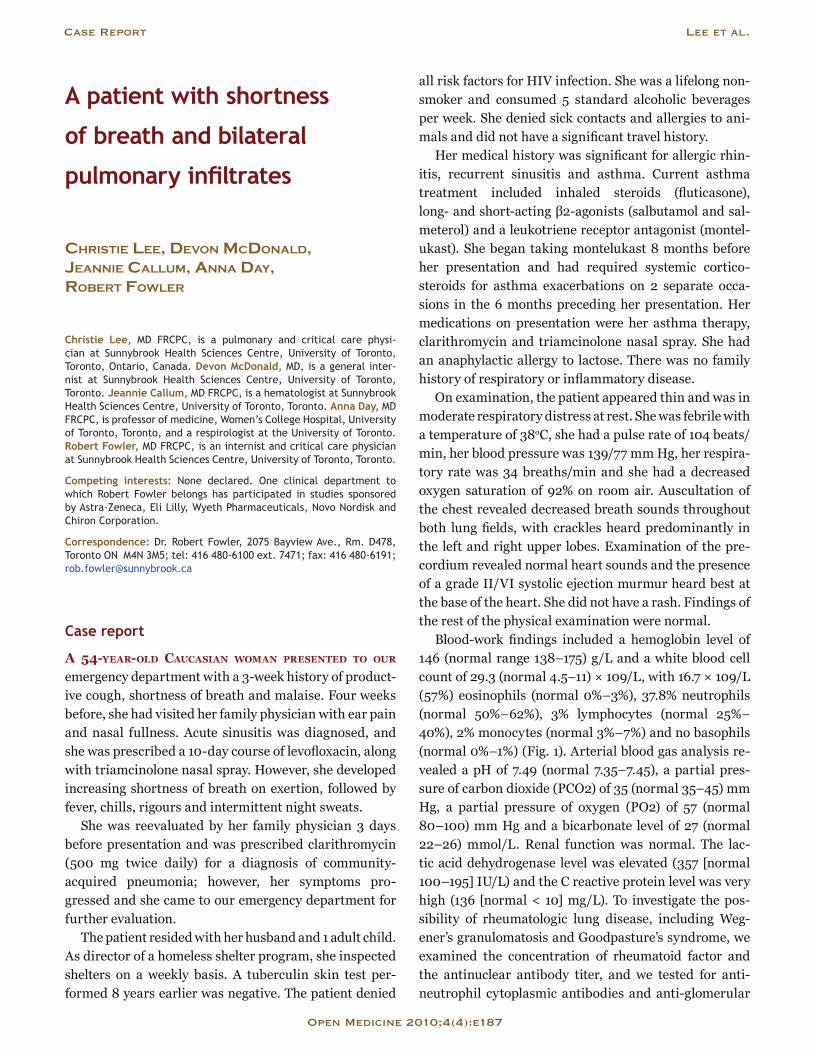
Blood-work findings included a hemoglobin level of 146 (normal range 138–175) g/L and a white blood cell count of 29.3 (normal 4.5–11) × 109/L, with 16.7 × 10⁹/L (57%) eosinophils (normal 0%–3%), 37.8% neutrophils (normal 50%–62%), 3% lymphocytes (normal 25%–40%), 2% monocytes (normal 3%–7%) and no basophils (normal 0%–1%) (Fig. 1). Arterial blood gas analysis re-vealed a pH of 7.49 (normal 7.35–7.45), a partial pres-sure of carbon dioxide (PCO2) of 35 (normal 35–45) mm Hg, a partial pressure of oxygen (PO2) of 57 (normal 80–100) mm Hg and a bicarbonate level of 27 (normal 22–26) mmol/L. Renal function was normal. The lac-tic acid dehydrogenase level was elevated (357 [normal 100–195] IU/L) and the C reactive protein level was very high (136 [normal < 10] mg/L). To investigate the pos-sibility of rheumatologic lung disease, including Weg-ener’s granulomatosis and Goodpasture’s syndrome, we examined the concentration of rheumatoid factor and the antinuclear antibody titer, and we tested for anti-neutrophil cytoplasmic antibodies and anti-glomerular
Case Report Lee et al.
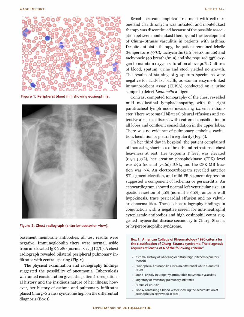
basement membrane antibodies; all test results were negative. Immunoglobulin titers were normal, aside from an elevated IgE (1280 [normal < 175] IU/L). A chest radiograph revealed bilateral peripheral pulmonary in-filtrates with central sparing (Fig. 2).
The physical examination and radiography findings suggested the possibility of pneumonia. Tuberculosis warranted consideration given the patient’s occupation-al history and the insidious nature of her illness; how-ever, her history of asthma and pulmonary infiltrates placed Churg–Strauss syndrome high on the differential diagnosis (Box 1).1
Broad-spectrum empirical treatment with ceftriax-one and clarithromycin was initiated, and montelukast therapy was discontinued because of the possible associ-ation between montelukast therapy and the development of Churg–Strauss vasculitis in patients with asthma. Despite antibiotic therapy, the patient remained febrile (temperature 39oC), tachycardic (110 beats/minute) and tachypneic (40 breaths/min) and she required 35% oxy-gen to maintain oxygen saturation above 90%. Cultures of blood, sputum, urine and stool yielded no growth. The results of staining of 3 sputum specimens were negative for acid-fast bacilli, as was an enzyme-linked immunosorbent assay (ELISA) conducted on a urine sample to detect Legionella antigen.
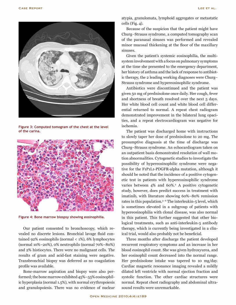
Contrast computed tomography of the chest revealed mild mediastinal lymphadenopathy, with the right paratracheal lymph nodes measuring 1.4 cm in diam-eter. There were small bilateral pleural effusions and ex-tensive air-space disease with scattered consolidation in all lobes and confluent consolidation in the upper lobes. There was no evidence of pulmonary embolus, cavita-tion, loculation or pleural irregularity (Fig. 3).
On her third day in hospital, the patient complained of increasing shortness of breath and retrosternal chest heaviness at rest. Her troponin T level was elevated (0.94 μg/L), her creatine phosphokinase (CPK) level was 290 (normal 5–160) IU/L, and the CPK MB frac-tion was 9%. An electrocardiogram revealed anterior ST segment elevation, and mild PR segment depression suggested a component of ischemia or pericarditis. An echocardiogram showed normal left ventricular size, an ejection fraction of 50% (normal > 60%), anterior wall hypokinesis, trace pericardial effusion and no valvul-ar abnormalities. These echocardiography findings in conjunction with a negative screen for anti-neutrophil cytoplasmic antibodies and high eosinophil count sug-gested myocardial disease secondary to Churg–Strauss or hypereosinophilic syndrome.
Open Medicine 2010;4(4):e188
Box 1: American College of Rheumatology 1990 criteria for the classifi cation of Churg–Strauss syndrome. The diagnosis requires at least 4 of 6 of the following criteria:1
• Asthma:Historyofwheezingordiffusehigh-pitchedexpiratoryrhonchi
• Eosinophilia:Eosinophilia>10%ondifferentialwhitebloodcellcount
• Mono-orpoly-neuropathyattributabletosystemicvasculitis
• Migratoryortransitorypulmonaryinfiltrates
• Paranasalsinusitis
• Biopsycontainingabloodvesselshowingtheaccumulationofeosinophilsinextravasculararea
Figure 1: Peripheral blood film showing eosinophilia.
Figure 2: Chest radiograph (anterior-posterior view).
Our patient consented to bronchoscopy, which re-vealed no discrete lesions. Bronchial lavage fluid con-tained 92% eosinophils (normal < 1%), 6% lymphocytes (normal 10%–20%), 0% neutrophils (normal 70%–80%) and 2% histiocytes. There were no malignant cells. The results of gram and acid-fast staining were negative. Transbronchial biopsy was deferred as no coagulation profile was available.
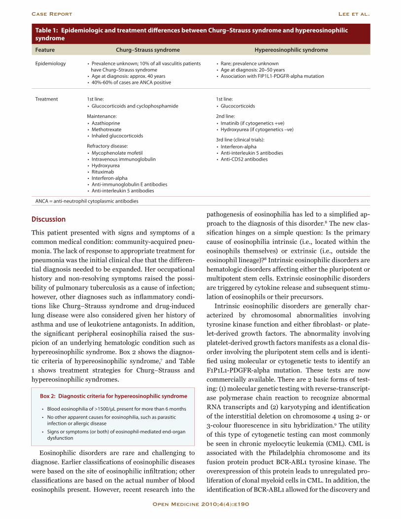
Bone-marrow aspiration and biopsy were also per-formed; the bone marrow exhibited 45%–55% eosinophil-ic hyperplasia (normal 1.5%), with normal erythropoiesis and granulopoiesis. There was no evidence of nuclear
atypia, granulomata, lymphoid aggregates or metastatic cells (Fig. 4).
Because of the suspicion that the patient might have Churg–Strauss syndrome, a computed tomography scan of the paranasal sinuses was performed and revealed minor mucosal thickening at the floor of the maxillary sinuses.
Given the patient’s systemic eosinophilia, the multi-system involvement with a focus on pulmonary symptoms at the time she presented to the emergency department, her history of asthma and the lack of response to antibiot-ic therapy, the 2 leading working diagnoses were Churg–Strauss syndrome and hypereosinophilic syndrome.
Antibiotics were discontinued and the patient was given 50 mg of prednisolone once daily. Her cough, fever and shortness of breath resolved over the next 5 days. Her white blood cell count and white blood cell differ-ential returned to normal. A repeat chest radiogram demonstrated improvement in the bilateral lung opaci-ties, and a repeat electrocardiogram was negative for ischemia.
The patient was discharged home with instructions to slowly taper her dose of prednisolone to 20 mg. The presumptive diagnosis at the time of discharge was Churg–Strauss syndrome. An echocardiogram taken on an outpatient basis demonstrated resolution of wall mo-tion abnormalities. Cytogenetic studies to investigate the possibility of hypereosinophilic syndrome were nega-tive for the F1P1L1-PDGFR-alpha mutation, although it should be noted that the incidence of a positive cytogen-etic test in patients with hypereosinophilic syndrome varies between 4% and 60%.2 A positive cytogenetic study, however, does predict success in treatment with imatinib, with literature showing 60%–80% remission rates in this population.3–6 The interleukin-5 level, which is sometimes elevated in a subgroup of patients with hypereosinophilia with clonal disease, was also normal in this patient. This further suggested that other bio-logical treatments, such as anti-interleukin-5 antibody therapy, which is currently being investigated in a clin-ical trial, would also probably not be beneficial.
Three months after discharge the patient developed recurrent respiratory symptoms and an increase in her blood eosinophil count. She was given hydroxyurea, and her eosinophil count decreased into the normal range. Her prednisolone intake was tapered to 10 mg/day. Cardiac magnetic resonance imaging revealed a mildly dilated left ventricle with normal ejection fraction and systolic function. The other cardiac structures were normal. Repeat chest radiography and abdominal ultra-sound results were unremarkable.
Case Report Lee et al.
Open Medicine 2010;4(4):e189
Figure 3: Computed tomogram of the chest at the level of the carina.
Figure 4: Bone marrow biospsy showing eosinophilia.
Discussion
This patient presented with signs and symptoms of a common medical condition: community-acquired pneu-monia. The lack of response to appropriate treatment for pneumonia was the initial clinical clue that the differen-tial diagnosis needed to be expanded. Her occupational history and non-resolving symptoms raised the possi-bility of pulmonary tuberculosis as a cause of infection; however, other diagnoses such as inflammatory condi-tions like Churg–Strauss syndrome and drug-induced lung disease were also considered given her history of asthma and use of leukotriene antagonists. In addition, the significant peripheral eosinophilia raised the sus-picion of an underlying hematologic condition such as hypereosinophilic syndrome. Box 2 shows the diagnos-tic criteria of hypereosinophilic syndrome,7 and Table 1 shows treatment strategies for Churg–Strauss and hypereosinophilic syndromes.
Eosinophilic disorders are rare and challenging to diagnose. Earlier classifications of eosinophilic diseases were based on the site of eosinophilic infiltration; other classifications are based on the actual number of blood eosinophils present. However, recent research into the
pathogenesis of eosinophilia has led to a simplified ap-proach to the diagnosis of this disorder.8 The new clas-sification hinges on a simple question: Is the primary cause of eosinophilia intrinsic (i.e., located within the eosinophils themselves) or extrinsic (i.e., outside the eosinophil lineage)?8 Intrinsic eosinophilic disorders are hematologic disorders affecting either the pluripotent or multipotent stem cells. Extrinsic eosinophilic disorders are triggered by cytokine release and subsequent stimu-lation of eosinophils or their precursors.
Intrinsic eosinophilic disorders are generally char-acterized by chromosomal abnormalities involving tyrosine kinase function and either fibroblast- or plate-let-derived growth factors. The abnormality involving platelet-derived growth factors manifests as a clonal dis-order involving the pluripotent stem cells and is identi-fied using molecular or cytogenetic tests to identify an F1P1L1-PDGFR-alpha mutation. These tests are now commercially available. There are 2 basic forms of test-ing: (1) molecular genetic testing with reverse-transcript-ase polymerase chain reaction to recognize abnormal RNA transcripts and (2) karyotyping and identification of the interstitial deletion on chromosome 4 using 2- or 3-colour fluorescence in situ hybridization.9 The utility of this type of cytogenetic testing can most commonly be seen in chronic myelocytic leukemia (CML). CML is associated with the Philadelphia chromosome and its fusion protein product BCR-ABL1 tyrosine kinase. The overexpression of this protein leads to unregulated pro-liferation of clonal myeloid cells in CML. In addition, the identification of BCR-ABL1 allowed for the discovery and
Case Report Lee et al.
Open Medicine 2010;4(4):e190
Table 1: Epidemiologic and treatment di� erences between Churg–Strauss syndrome and hypereosinophilic syndrome
Feature Churg–Strauss syndrome Hypereosinophilic syndrome
Epidemiology • Prevalenceunknown;10%ofallvasculitispatientshaveChurg–Strausssyndrome
• Ageatdiagnosis:approx.40years• 40%-60%ofcasesareANCApositive
• Rare;prevalenceunknown• Ageatdiagnosis:20–50years• AssociationwithFIP1L1-PDGFR-alphamutation
Treatment 1stline:• Glucocorticoidsandcyclophosphamide
Maintenance:• Azathioprine• Methotrexate• Inhaledglucocorticoids
Refractorydisease:• Mycophenolatemofetil• Intravenousimmunoglobulin• Hydroxyurea• Rituximab• Interferon-alpha• Anti-immunoglobulinEantibodies• Anti-interleukin5antibodies
1stline:• Glucocorticoids
2ndline:• Imatinib(ifcytogenetics+ve)• Hydroxyurea(ifcytogenetics–ve)
3rdline(clinicaltrials):• Interferon-alpha• Anti-interleukin5antibodies• Anti-CD52antibodies
ANCA=anti-neutrophilcytoplasmicantibodies
Box 2: Diagnostic criteria for hypereosinophilic syndrome
• Bloodeosinophiliaof>1500/μLpresentformorethan6months
• Nootherapparentcausesforeosinophilia,suchasparasiticinfectionorallergicdisease
• Signsorsymptoms(orboth)ofeosinophil-mediatedend-organdysfunction
successful use of small molecule inhibitors like imatinib mesylate to induce remission of CML.
Extrinsic eosinophilic disorders are numerous. They are subdivided into T-cell-mediated and tumour-cell-mediated eosinophilic disorders.8 T-cell-mediated eosin-ophilia can be due to allergic diseases like bronchial asthma or it can be due to autoimmune diseases such as Churg–Strauss syndrome. In both cases, interleukin-5 is responsible for mediating eosinophil differentia-tion, activation and survival.10 Eosinophils contribute to worsening inflammation, tissue damage and organ dysfunction. They have also been implicated in tissue remodeling following an acute inflammatory reaction.11 The negative cytogenetic testing in this patient’s case does not eliminate the possibility of an eosinophilic dis-order; rather, it pushes the clinician to consider other diagnoses.
Idiopathic hypereosinophilic syndrome is diagnosed on the basis of clinical and pathologic criteria and is characterized by blood eosinophilia that is neither sec-ondary nor clonal in nature.12 The diagnosis requires documentation of both target end-organ damage and an absolute eosinophil count of 1.5 × 10⁹/L or higher for at least 6 months.13 By definition, testing must demonstrate a lack of any known molecular or cytogenetic markers, and bone marrow histology must not display evidence of abnormal populations of mast cells, monocytosis, trilin-eage myeloproliferation or dysplasia.14 Specific therapy for hypereosinophilic syndrome includes high-dose ster-oids as first-line treatment, with hydroxyurea added if remission is not achieved.
Classical medical training pushed our diagnosis to-ward Churg–Strauss syndrome, in part because of the prominence of pulmonary involvement. However, as we assessed the spectrum of organs involved, an organ-fo-cused classification of our patient’s illness seemed less appropriate and we altered our diagnostic process to consider idiopathic hypereosinophilic syndrome.
The traditional method of categorizing diseases ac-cording to clinical features is in flux as we learn more about the genetic basis of many conditions. When gen-etic markers are present, they aid the clinician in making a diagnosis; when they are not, the clinician must rely on clinical and pathologic features to make a diagnosis. Our increasing understanding of underlying genetic ab-normalities has sparked a revolution in the diagnoses of many blood dyscrasias. For example, the discovery of the Philadelphia chromosome has dramatically changed the way we diagnose conditions involving neoplastic transformation, and research on JAK2 mutations has affected the diagnosis of myeloproliferative disorders.
Historically, blood disorders were classified using an operational system based on cell lines (i.e., myeloid v. lymphoid), cell morphology and the presence of blast cells (to delineate acute from chronic conditions). In the last decade, a classification system based on genetics and immunology has increasingly been used for blood disorders. Although physicians are trained to recognize syndromes on the basis of clinical and pathologic fea-tures, the reality is that many patients with blood dys-crasias are asymptomatic or, in rare cases, have normal peripheral blood films. In these cases, molecular and cytogenetic testing becomes the mainstay of diagnosis; these forms of testing also provide confirmation of the presence of a disease in patients who have a high clinical pre-test probability of having the disease.
Epigenetics is the study of heredity not related to the DNA sequence. It examines the relationship between genetic background, the environment and disease. Epi-genetic processes can modify or affect potentially dele-terious genes.15 The epigenetic modification of DNA through methylation and covalent modifications of nu-cleosome histone proteins underly much of the reclassifi-cation of blood dyscrasias. Unlike mutations in the DNA sequence, epigenetic changes are, by definition, revers-ible, which offers the exciting hope that they may be modified through therapy.15
For now, our patient’s condition will carry the label of idiopathic hypereosinophilic syndrome, unless further developments in molecular testing necessitate a change in diagnosis. We will continue to treat her with immuno-suppressive therapy as we await a deeper understanding of the true intrinsic or extrinsic trigger for her symptoms.
Contributors: Christie Lee and Robert Fowler wrote the original draft of the manuscript. All of the authors contributed substantially to the article conception, revised it critically for important intellectual con-tent and gave final approval of the version to be published. Robert Fowler is the guarantor.
References
1. Masi AT, Hunder GG, Lie JT, Michel BA, Bloch DA, Arend WP, et al. The American College of Rheumatology 1990 criteria for the classification of Churg-Strauss syndrome (allergic granulomato-sis and angiitis). Arthritis Rheum 1990;33:1094–1100.
2. Yoshiyuki Y, Rothenberg ME, Cancelas JA. Current concepts on the pathogenesis of the hypereosinophilic syndrome/chronic eosinophilic leukemia. Transl Oncogenomics 2006;1:53–63.
3. Ault P, Cortes J, Koller C, Kaled ES, Kantarjian H. Response of idiopathic hypereosinophilic syndrome to treatment with imatin-ib mesylate. Leuk Res 2002;26(9):881–884.
4. Cortes J, Ault P, Koller C, Thomas D, Ferrajoli A, Wierda W, et al. Efficacy of imatinib mesylate in the treatment of idiopathic hypereosinophilic syndrome. Blood 2003;101(12):4714–4716.
Case Report Lee et al.
Open Medicine 2010;4(4):e191
5. Gleich GJ, Leiferman KM, Pardanani A, Tefferi A, Butterfield JH. Treatment of hypereosinophilic syndrome with imatinib mesi-late. Lancet 2002;359(9317):1577–1578.
6. Pardanani A, Reeder T, Porrata LF, Li CY, Tazelaar HD, Baxter EJ, et al. Imatinib therapy for hypereosinophilic syndrome and other eosinophilic disorders. Blood 2003;101(9):3391–3397.
7. Chusid MJ, Dale DC, West BC, Wolff SM. The hypereosinophilic syndrome: analysis of fourteen cases with review of the litera-ture. Medicine (Baltimore) 1975;54(1):1–27.
8. Simon D, Simon H. Eosinophilic disorders. J Allergy Clin Immunol 2007;119:1291–1300; quiz 1301–1302.
9. Cools J, Stover EH, Wlodarska I, Marynen P, Gilliland DG. The FIP1L1-PDGFRalpha kinase in hypereosinophilic syndrome and chronic eosinophilic leukemia. Curr Opin Hematol 2004;11(1):51–57.
10. Sanderson CJ. Interleukin-5, eosinophils, and disease. Blood 1992;79(12):3101–3109.
11. Kay AB, Phipps S, Robinson DS. A role for eosinophils in airway remodelling in asthma. Trends Immunol 2004;25(9):477–482.
12. Tefferi A, Elliott MA, Pardanani A. Atypical myeloprolifera-tive disorders: diagnosis and management. Mayo Clin Proc 2006;81(4):553–563.
13. Bain B, Pierre R, Imbert M, Vardiman J, Brunning R, Dlandrin G. Chronic eosinophilic leukemia and hypereosinophilic syndrome.
In Jaffe ES, Harris NL, Stein H, Vardiman JW, editors. World Health Organization classification of tumours: pathology and genetics of tumours of haematopoietic and lymphoid tissues. Lyon: IARC Press; 2001. p. 29–31.
14. Bain BJ. Relationship between idiopathic hypereosinophilic syn-drome, eosinophilic leukemia, and systemic mastocytosis. Am J Hematol 2004;77(1):82–85.
15. Feinberg AP. Epigenetics at the epicenter of modern medicine. JAMA 2008;299(11):1345–1350.
Citation: Lee C, McDonald D, Callum J, Day A, Fowler R. A patient with shortness of breath and bilateral pulmonsy infiltrates. Open Med 2010;4(4):187–192.
Published: 2 November 2010
Copyright: This article is licenced under the Creative Commons Attibu-tion–ShareAlike 2.5 Canada License, which means that anyone is able to freely copy, download, reprint, reuse, distribute, display or perform this work and that the authors retain copyright of their work. Any derivative use of this work must be distributed only under a license identical to this one and must be attributed to the authors. Any of these conditions can be waived with permission from the copyright holder. These condi-tions do not negate or supersede Fair Use laws in any country. For further information see http://creativecommons.org/licenses/by-sa/2.5/ca.
Case Report Lee et al.
Open Medicine 2010;4(4):e192