a one-step enzyme immunoassay for human manganese superoxide dismutase with monoclonal antibodies
TRANSCRIPT

Free Radical Biology & Medicine, Vol. 8, pp. 25-31, 1990 0891-5849/90 $3.00 + .00 Printed in the USA. All rights reserved. © 1990 Pergamon Press plc
Original Contribution
A O N E - S T E P E N Z Y M E I M M U N O A S S A Y F O R H U M A N M A N G A N E S E
S U P E R O X I D E D I S M U T A S E W I T H M O N O C L O N A L A N T I B O D I E S
TETSUO ADACHI,*'t KAZUYUKI HIRANO,* KYOZO HAYASHI,* YASUTOSHI MUTO,~: and FUMITAKA OKUNO§
*Department of Pharmaceutics, Gifu Pharmaceutical University, 6-1, Mitahora-higashi 5 chome, Gifu 502, Japan; :[:First Department of Internal Medicine, Gifu University, School of Medicine, Gifu 500, Japan; §Department of Internal Medicine,
Gihoku Hospital, Gifu 501-21, Japan
(Received 19 April 1989; Revised 28 July 1989; Accepted 10 August 1989)
A b s t r a c t - - A one-step enzyme immunoassay for the determination of manganese superoxide dismutase in serum has been developed with two kinds of monoclonal antibodies. Proposed method had high sensitivity (assay range, 0.4-200 ng/ml), good recovery (recovery percentage, 102.9-106.2%) and reproducibility (intraassay, C.V. = 1.87-3.66%; interassay, C.V. = 3.03-10.4%). From these results, it is possible to apply this method to routine clinical analysis and biochemical research with various purposes.
Keywords--Manganese superoxide dismutase, Enzyme immunoassay, Monoclonal antibody, Clinical analysis, Horseradish peroxidase, Free radicals
INTRODUCTION
Superoxide anion, a free radical generated during the monovalent metabolic reduction of oxygen, is toxic to biological systems. There are several pathways in aero- bic cells that can lead to the production of superoxide anion. Mitochondria ~ and endoplasmic reticulum 2 have been shown to produce superoxide anion as a conse- quence of autoxidation of electron transport chain com- ponents. Another source of superoxide anion are cytosolic enzyme such as xanthine oxidase 3 and acti- vated neutrophils# Superoxide dismutase (SOD) is an enzyme that catalytically scavenges superoxide anion and thus provides a defense against oxygen toxici ty? Human SODs were classified into three isoenzymes, a cytoplasmic copper and zinc containing SOD (Cu,Zn- SOD), a mitochondrial manganese SOD (Mn-SOD) and extracellular-SOD (EC-SOD) of high molecular weight.
In order to clarify the behaviors of cellular SODs in diseases, Cu,Zn-SOD and Mn-SOD activities were often determined. Cu,Zn-SOD in erythrocytes has been confirmed to be higher in Down's syndrome (trisomy 21), 6 and lower in Fanconi's anemia, 7 hemolytic uremic syndrome s and insulin dependent diabetes mel-
tAuthor to whom correspondence should be addressed.
litus. 9 Total SOD and Mn-SOD activities in leukocytes have been reported to be higher in acute myelogenous leukemia and lower in acute lymphocytic leukemia and ring chromosome 6. m
There are few reports describing the serum SODs activities, ~'~z because Cu,Zn-SOD and Mn-SOD ac- tivities are very low in extracellular fluids such as serum and plasma. Furthermore, it is well known that some redox components and SOD like components which can scavenge superoxide anion exist in extra- cellular fluids. In our previous studies, we developed sandwich enzyme immunoassays (EIAs) for Cu,Zn- SOD and Mn-SOD involving polyclonal antibodies, and found that Cu,Zn-SOD and Mn-SOD levels in serum were high in patients with renal diseases and liver diseases, respectively.~3-~5 EIAs with polyclonal antibodies necessitate procedures of two separate im- munoreaction steps and are time consuming. In this paper, we describe a high sensitive, specific one-step EIA for human Mn-SOD using monoclohal antibodies.
MATERIALS AND METHODS
M a t e r i a l s
RPMI 1640 was purchased from Nissui Pharma- ceutical Company, Ltd. (Japan). Fetal bovine serum (FBS) was from JR Scientific, Inc. (U.S.A.). Poly-
25
26 T. ADACHI et al .
ethylene glycol 4000 was from Merck (West Ger- many). Hypoxanthine, aminopterin, thymidine and horseradish peroxidase (HRP, Type VI) were from Sigma Chemical Company (U.S.A.). 2,6,10,14-tetra- methylpentadecane (pristane) was from Aldrich Chem- ical Company, Inc. (U.S.A.). Alkaline phosphatase labeled rabbit anti-mouse IgG antibody was from Zymed Laboratories, Inc. (U.S.A.). Mouse typer kit was from Bio-Rad (U.S.A.). 96-well tissue culture plate, 24-well tissue culture plate were from Costar (U.S.A.). 96-well immunoplate and tissue culture flasks were from Nunc (Denmark).
Enzyme
Human Mn-SOD was purified as previously de- scribed. 16
Mice
BALB/c mice were purchased from Shizuoka Lab- oratory Animal Center (Japan).
Myeloma cell line
X63-Ag8-6,5,3 mouse myeloma cells were pur- chased from Dainippon Pharmaceutical Company, Ltd. (Japan). They were grown in RPMI 1640 supplemented with 15% FBS, 2 mM L-glutamine, 100 U/ml peni- cillin, 100/zg/ml streptomycin and 50/~M 2-mercap- toethanol (complete culture medium) at 37°C in humidified 5% CO2/95% air.
Immunization of mice
Six-week-old, female BALB/c mice were immu- nized intraperitoneally with 100/lg of purified human Mn-SOD emulsified in complete Freund's adjuvant. The mice were boosted with 30 ltg of antigen in com- plete Freund's adjuvant 2 and 4 weeks later. The final injection of Mn-SOD (50/~g) without the adjuvant was given intravenously, 2 weeks after the third injection.
tively, in 50 ml centrifuge tubes, and packed by cen- trifugation (1200 x g for 5 min). Supernatant fluid was decanted and the cell pellet was resuspended in residual fluid by tapping the bottom on the tube, re- sulting in a cell slurry. Cell fusion was effected by the addition of 1 ml of 50% polyethylene glycol 4000 dis- solved in FBS-free RPMI 1640 medium with gentle mixing, followed by stirring for 1 rain. Then the poly- ethylene glycol 4000 was slowly diluted by the drop- wise addition of 9 ml of FBS-free RPMI 1640 medium. Cells were centrifuged and the supernatant was dis- carded. Finally, cells were resuspended to a concen- tration of 1 x 10⁷ spleen cells/ml in complete culture medium. One hundred microliter of the cell suspension was seeded into each well of 96-well tissue culture plates and the plates were incubated at 37°C in hu- midified 5% CO2/95% air.
After 1 day, 0.1 ml of complete culture medium containing 100/tM hypoxanthine, 0.4/~M aminopterin and 16 /.tM thymidine (selective HAT medium) was added to each well. Then, half of the medium was removed and replaced with fresh HAT medium every other day. Hybridoma growth was monitored for 2 to 3 weeks by daily observation with an inverted micro- scope. Culture supernatant from each well displaying hybrids were tested by enzyme-linked immunosorbent assay (ELISA). Following the identification of positive hybridomas, some hybrid cells were subcloned by lim- iting dilution and grown in 96-well tissue culture plates containing feeder cells of thymocytes from normal non- immunized 4-week-old BALB/c mice. After 1 to 2 weeks, culture supernatants from the wells containing subclones were tested by ELISA. Selected subclones were then expanded and grown as ascitic tumors in pristane-primed BALB/c mice.
Purification of monoclonal antibody
Each monoclonal antibody was purified from as- cites fluid or culture medium by precipitation with 40% saturated (NH4)2SO4 and DE-32 column chromatog- raphy.
Production of hybridoma
Four days after the final booster, BALB/c mice were sacrificed by cervical dislocation, and their spleens were aseptically removed. Single-cell suspen- sion of splenocytes was prepared by passing the spleen tissue through a 150-mesh stainless-steel screen. Spleen cells prepared as described above were fused with X63-Ag8-6,5,3 murine myeloma cells. Myeloma and spleen cells were washed separately in FBS-free RPMI 1640 medium, mixed at a ratio 1:10, respec-
Preparation of the HRP-labeled monoclonal antibody
The HRP-labeled monoclonal antibody was pre- pared with glutaraldehyde as a crosslinking reagent.~7 Ten milligram of HRP was dissolved in 0.15 ml of 1.25% glutaraldehyde solution (25% glutaraldehyde was diluted with 0.1 M sodium phosphate buffer, pH 6.8) and allowed to stand for 18 hr at room tempera- ture. The activated HRP solution was applied to a col- umn (1.0 x 50 cm) of Sephadex G-25 equilibrated
One-step EIA for human Mn-SOD 27
with 0.15 M NaCI solution. The brown activated HRP fractions were pooled (1.5 ml) and mixed with 0.1 ml of 1 M carbonate buffer, pH 9.5 and 0.5 ml of mono- clonal antibody solution (10 mg/ml) dissolved in 0.15 M NaC1 solution. The reaction mixture was stirred at 4°C for 24 h, followed by addition of 50/~1 of 0.2 M lysine solution. After standing at 4°C for 2 h, the mix- ture was applied to a column (1.5 × 90 cm) of Se- pharose 6B equilibrated with 10 mM sodium phosphate buffer, pH 7.5 containing 0.15 M NaC1 and 0.05% merthiolate. The active fractions were pooled and stored at 4°C.
Preparation of monoclonal antibody coated immunoplate
A monoclonal antibody was coated onto immuno- plate by physical adsorption. A hundred microliter of monoclonal antibody solution (0.1 mg/ml dissolved in 50 mM carbonate buffer, pH 9.5 containing 0.02% sodium azide) was added into each well and allowed to stand at 37°C for 3 h followed by standing overnight at 4°C. Each well was washed three times with phos- phate-buffered saline (PBS) containing 0.05% Tween 20 and 0.05% merthiolate (PBS-Tween) and then blocked with 300 ~1 of PBS containing 1% bovine serum albumin (BSA), 0.05% Tween 20 and 0.05% merthiolate (BSA-PBS).
Standard assay procedure
Serum samples were diluted 10-fold with BSA-PBS. Equal volume of diluted serum and HRP-labeled mono- clonal antibody (MS1/B-11/D-4) diluted 100-fold with the same buffer were mixed and 70 /.tl of this mixture was added into each well of immunoplate pre- viously coated with monoclonal antibody (MS 1/A-4/ D-8). After incubated for 1.5 h at room temperature, each well was washed three times with PBS-Tween. The activity of HRP trapped was assayed by the fol- lowing spectrophotometric method. A hundred mi- croliter of 0.1 M Mcllvain buffer, pH 6.5 containing 0.02% H202 and 16.6 mM o-phenylenediamine dihy- drochloride was added to each well and incubated for 15 min with shaking at room temperature. The enzyme reaction was stopped by the addition of 100/tl of 4.5 N H2SO4 and the absorbance at 492 nm was measured using a Corona MTP-32 microplate reader.
Assay procedure of two-step enzyme immunoassay
Monoclonal antibody coated plate and HRP-labeled monoclonal antibody used were same to the standard method. Seventy microliter of serum diluted 10-fold
with BSA-PBS was added into each well of immuno- plate and allowed to stand for 1 h at room temperature. After washed with PBS-Tween, 85/~1 of diluted HRP- labeled monoclonal antibody was added in each well and incubated for 2 h at room temperature, followed by assay of HRP activity similar to the above standard method.
Method for epitope analysis
A hundred microliter of different concentrations of monoclonal antibody dissolved in BSA-PBS was added to separate wells of the immunoplate previously coated with 50/~g/mi of human Mn-SOD. After incubating for 2 h at room temperature, each well was washed three times with PBS-Tween. Then 100/11 of diluted HRP-labeled monoclonal antibody was added to each well and incubated for 2 h at room temperature, fol- lowed by assay of HRP activity similar to the method above mentioned.
RESULTS
Production and some properties of monoclonal antibody
Two kinds of monoclonal antibodies against human Mn-SOD, MS1/A-4/D-8 and MS1/B-I1/D-4, were prepared according to the method of K6hler and Mil- stein with X63-Ag8-6,5,3 as the myeloma.18 After the fusion step, 125 out of 227 wells showed hybridoma growth and 42 wells screened positively by ELISA. Twenty-four clones of these expansively cultured, but many were unstable and subsequently became nega- tive. Three cultures were cloned by limiting dilution on thymus cell-feeder layers. Two clones of these, MS1/A-4 and MSI /B- I1 , were selected for culture and recloned with the same finding. All MS1/A-4 clones and MS1/B-11 clones gave similarly positive values on screening, indicating a clonal origin. The clones MS1/A-4/D-8, MSI/A-4/G-5 , MS1/B-11/D- 4 and MS 1 / B- 11 / D-8 were selected for further culture and ascites production. For ascites production, mice were intraperitoneally injected with 0.5 ml of pristane, 10 and 3 days before the injection of hybridoma. Hy- bridoma cells (5 × l07 cells) in FBS and antibiotics- free RPMI 1640 medium were intraperitoneally in- jected to 5 mice. We obtained about 100 to 350 mg of IgG fraction (purified by (NH4)2SO4 precipitation and DE-32 column chromatography) from each of 4 clones. Two kinds of antibodies MS1/A-4/D-8 and MS1/B- 11/D-4 were selected and used for EIA, because we obtained these antibodies more than the others. The subclass of both antibodies was IgG1 k (rlr~KK).
A monoclonal antibody should recognize a single
28 T. ADACHI et al.
.5 t5 ~ B
0 I I I l * ;,~--I s 0 i J , J i ;,,_i 0 1.3 6.4 32 160 800 lxlO 0 1.3 6.4 32 160800 lx10 s
Unlabeled monoclonal antibody (ng)
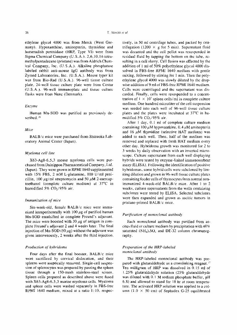
Fig. 1. Epitope analysis using solid-phase enzyme immunoassay. The wells of the immunoplate were coated with human Mn- SOD and incubated with indicated content of unlabeled monoclonal antibodies, MS1/A-4/D-8 (O) and MS I/B-11/D-4 (O). After washing each well, HRP-labeled monoclonal antibodies, MS I/A-4/D-8 (A) and MS I/B-11/D-4 (B) were incubated and followed by assaying HRP activity.
epitope on the antigen used for immunization. How- ever this generalization can be complicated by the oc- currence of multiple identical or cross-reactive epitope, and so we analyzed about the epitope on the Mn-SOD molecule which were recognized by each monoclonal antibody. The principle of the epitope assay is that HRP-labeled monoclonal antibody will complete for the binding site in an antigen as unlabeled monoclonal antibody. ~9 As shown in Figure 1, the reaction of HRP- labeled MS 1/A-4/D-8 with antigen was weakly inhib- ited by unlabeled MS1/A-4/D-8 and equivalently by unlabeled MS1/B-11/D-4, and vice versa. The inhi- bition rate was gradual and scarcely achieved to less than 60% in the presence of 100/,zg of unlabeled an- tibody.
Effect of reaction time
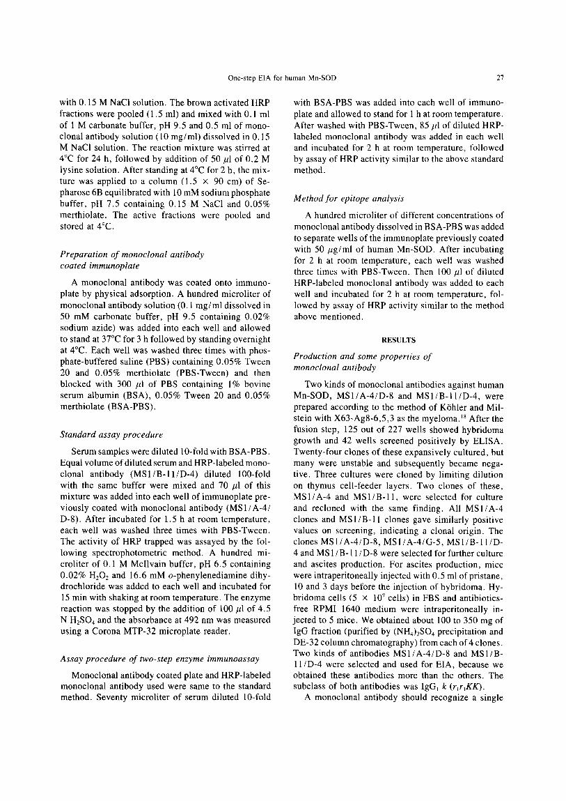
The incubation time required for immunoreaction was investigated with the standard Mn-SOD and var- ious sera. As shown in Figure 2, the immunoreaction proceeded rapidly up to 1 h. The data of Mn-SOD concentration in sera obtained from experiments with various incubation times (0.5, 1, 1.5, 2, and 3 h) were approximate quantity (C.V. = 2.50-5.74%). From these results, the incubation time for immunoreaction was settled to 1.5 h.
Calibration curve
A calibration curve was prepared according to the standard assay procedure using purified human Mn-
3 A 3h 6O0 B ~.~h Sarum l h 400 t " g l ~ F - . Q - - ' ~ 4 !
1 0.5h
001 . . . . . . I . . . . I . a , ~ ,
o~ 1 z 4 lo ~ 40 too o.s I ts z 3 Mn-SO0 (ng/ml) Time (hi
Fig. 2. Effect of incubation time required for immunoreaction. (A) Calibration curves obtained from experiments with indicated times for immunoreaction in the proposed method. (B) Mn-SOD concentrations in 7 sera which assayed by the method with indicated times for immunoreaction.

One-step EIA for human Mn-SOD 29
SOD. As shown in Figure 3, a range of 0 .4 -200 ng/ ml (0 .028-14 ng/well) of Mn-SOD could be deter- mined by the proposed method and it is sensitive enough to detect serum Mn-SOD levels.
Recovery of Mn-SOD from serum
Purified human Mn-SOD with 3 different amounts were added to 10 sera and the recoveries from the sera were investigated. The average recoveries were 102.9 ___ 4 .71 ,105.0 ± 4.73 and 106.2 ± 3.83%. The effect of dilution of sample was also investigated with 4 sera samples. As shown in Figure 4, the calculated Mn-SOD values showed linear relationships with di- lution factors and no inhibitory reaction of the serum components with antigens was observed.
Reproducibility
The coefficients of intraassay and interassay vari- ations were 1.87 to 3.66% (assayed 10 times with 6 sera) and 3.03 to 10.4% (assayed 7 days with 7 sera), respectively.
Correlation between proposed method and two-step enzyme immunoassay
Twenty kinds of sera were assayed by the standard assay method and two-steps EIA (in both methods, MSI/A-4/D-8 was coated on immunoplate and MS1/ B-11/D-4 was labeled with HRP) (Fig. 5). The data obtained from the standard method were good corre- lated with data from two-steps EIA (r = 0.997, y = 0.965x + 7.37; y:proposed method; x:two-steps EIA).
Sa'um 150 1
2
f f 3
4
0 1(1050 ZO 10 5
t, , I I I
20 10 5 2
Dilution factor
Fig. 4. Effect of dilution of serum on calculated Mn-SOD values.
DISCUSSION
Monoclonal antibodies have displaced polyclonal antibodies in many facets of immunology. As com- pared with polyclonal antibodies, some major immu- nodiagnostic advantages of monoclonal antibodies are evident. Firstly, a monoclonal antibody is homoge- neous immunoglobulin produced by a single hybri- doma which contains identical genetic material derived from a single spleen cell. Secondly, monoclonal an- tibody eliminated the problems, cross reactions and nonspecific reactions, that are frequently associated with polyclonal antibodies because a monoclonal an- tibody recognizes specifically only one epitope on antigen.
In the proposed method two kinds of monoclonal antibodies, MSI/A-4/D-8 and MS1/B-11/D-4 were
3
1
- 5O0
4OO
3OO
0 ,~ 100
, , I . . . . I _ _ , , ~ 1 , O01 04 1 2 4 10 20 40 1002Q0
Mn-$OD (nglml)
I I I I I o lOO 290 300 400 50o
Mn-SOD (ng/ml) T w o - s t e p s E I A
Fig. 3. Calibration curves for human Mn-SOD obtained by the pro- Fig. 5. Comparison of proposed method with two-steps enzyme posed method, immunoassay.

30 T. ADACHI et al.
used. These antibodies may recognize same or over- lapping epitope from the results of epitope analysis (Fig. 1). However, the inhibitions of immunoreactions between HRP-labeled antibodies and antigen by un- labeled antibodies were weak compared to experiments with Cu,Zn-SOD 2° or a reference. ~9 The proposed method correlated well with not only two-steps EIA but also one-step EIA with only one monoclonal an- tibody (used M S 1 / A - 4 / D - 8 coated immunoplate and HRP-labeled MS 1/A-4/D-8)(data not shown) in assays of 20 human sera. These results are supposed to be caused by that Mn-SOD is homotetramer and contains four identical epitopes. In principle, the assay of an- tigen which contains plural identical epitopes in the molecule is possible with a kind of monoclonal anti- body as above mentioned, if the plural epitopes are far away from each other enough to bind with antibodies.
Up to now, a lot of antibodies against various an- tigens have been produced and used as immunological reagents in immunoassay. However, few reports have been published about the applications of monoclonal antibodies against SODs. A clinical application of monoclonal antibodies against human Cu,Zn-SOD 21 was reported, while there are no reports about the EIA for Mn-SOD with monoclonal antibodies.
As it has been appeared that active oxygens take part in inflammation, z2 carcinogenesis, ~3 and cata- ract, 24 SOD and other radical scavengers are noted as drugs especially antiinflammatory agents 25 in addition to clinical diagnosis reagents. SOD synthesis are in- fluenced by the changes of environments, hyperoxia 26 or treatments of drugs, z7 Mn-SOD level tends to in- crease particularly as compared to Cu,Zn-SOD at above conditions, suggesting that the assay of Mn-SOD is useful for clinical diagnosis.
In conclusion, it can be said that the proposed method is specific to Mn-SOD and sensitive enough, and it allows not only the routine clinical analysis but also biochemical research. Work is now in progress to apply this method to the measurement of Mn-SOD in sera of patients with various diseases.
REFERENCES
1. Freeman, B. A.; Topolosky, M. K.; Crapo, J. D. Hyperoxia increases oxygen radical production in rat lung homogenates. Arch. Biochem. Biophys. 216:477-484; 1982.
2. Turrens, J. F.; Freeman, B. A.; Crapo, J. D. Hyperoxia in- creases H202 release by lung mitochondria and microsomes. Arch. Biochem. Biophys. 217:411-421; 1982.
3. Fridovich, I. Quantitative aspects of the production of super- oxide anion radical by milk xanthine oxidase. J. Biol. Chem. 245:4053-4057; 1970.
4. Babior, B. M.; Curnutte, J. T.; McMurrick, B. J. The particulate superoxide-forming system from human neutrophils: properties of the system and further evidence supporting its participation in the respiratory burst. J. Clin. Invest. 58:989-996; 1976.
5. Fridovich, I. Superoxide dismutases. Annu. Rev. Biochem. 44:147-159; 1975.
6. Feaster, W. W.; Kwok, L. W.: Epstein, C. J. Dosage effects for superoxide dismutase- 1 in nucleated cells aneuploid for chro- mosome 21. Am. J. Hum. Genet. 29:563-570; 1977.
7. Okahata, S.; Kobayashi, Y.; Usui, T. Erythrocyte superoxide dismutase activity in Fanconi's anemia. Clin. Sci. 58:173-175: 1980.
8. Kobayashi, Y. ; Okahata, S. ; Tanabe, K.; Tanaka, Y.; Ueda, K. ; Usui, T. Erythrocyte superoxide dismutase activity in hemolytic uremic syndrome. Hiroshima J. Med. Sci. 27:181-183; 1978.
9. Haggl6f, B.; Marklund, S. L.; Holmgren, G. CuZn superoxide dismutase, Mn superoxide dismutase, catalase and glutathione peroxidase in lymphocytes and erythrocytes in insulin-depen- dent diabetic children. Acta Endocrinologica 102:235-239; 1983.
10. Yoshimitsu, K.; Kobayashi, Y.; Usui, T. Superoxide dismutase activity in leukemic blasts of children with acute leukemia. Acta Paediatr. Scand. 73:92-96; 1984.
11. Marklund, S. L.; Heiskala, H.; Westermarck, T.; Santavuori, P. Superoxide dismutase isoenzymes in cerebrospinal fluid and plasma from patients with neuronal ceroid-lipofuscinoses. Clin. Sci. 71:57-60; 1986.
12. Ohman, M.; Marklund, S. L. Plasma extracellular superoxide dismutase and erythrocyte Cu,Zn-containing superoxide dis- mutase in alcoholics treated with disulfiram. Clin. Sci. 70:365- 369; 1986.
13. Adachi, T.; Hirano, K.; Sugiura, M.; Muto, Y.; Yamada, M.; Okuno, F.; Sugiura, K.; Ueda, H. Serum manganese-superoxide dismutase level in patients with liver diseases. Jap. J. Clin. Chem. 14:118-122; 1985.
14. Nishimura, N.; Ito, Y.; Adachi, T.; Hirano, K.; Sugiura, M.: Sawaki, S. Enzyme immunoassay for cuprozinc-superoxide dis- mutase in serum and urine. J. Pharm. Dyn. 5:394-402; 1982.
15. Nishimura, N.; Ito, Y.; Adacbi, T.; Hirano, K.; Sugiura, M.; Sawaki, S. Enzyme immunoassay for manganese-superoxide dismutase in serum and urine. J. Pharm. Dyn. 5:869-876; 1982.
16. Sugiura, M.; Adachi, T.; Inoue, H.; Ito, Y.; Hirano, K. Puri- fication and properties of two superoxide dismutases from hu- man placenta. J. Pharm. Dyn. 4:235-244; 1981.
17. Avrameas, S.; Guilbert, B. Enzyme immunoassay for the mea- surement of antigens using peroxidase conjugates. Biochimie 54:837-842; 1972.
18. K6hler, G.; Milstein, C. Continuous cultures of fused cells secreting antibody of predefined specificity. Nature 256:495- 497; 1975.
19. Shively, J. E.; Wagener, C.; Clark, B. R. Solution-phase R1A and solid-phase EIA using avidin-biotin systems for analysis of monoclonal antibody epitopes and affinity constants. In: Lon- gone, J. J. ; van Vunakis, H., eds. Methods in Enzymology. Vol. 121. New York: Academic Press; 1986:459-472.
20. Adachi, T.; Usami, Y.; Kishi, T.; Hirano, K.; Hayashi, K. An enzyme immunoassay for cuprozinc superoxide dismutase using monoclonal antibodies: application for pharmacokinetic study. J. lmmunol. Methods 109:93-101; 1988.
21. Ogino, K.; Oka, S.; Sakaida, I.; Ando, K.; Okita, K.; Take- moto, T.; Uda, T.; Noji, S. Characterization of a monoclonal antibody to copper-zinc superoxide dismutase. Jpn. J. lnflamm. 7:259-263; 1987.
22. McCord, J. M. Free radicals and inflammation: Protection of synovial fluid by superoxide dismutase. Science 185:528-531; 1974.
23. Slaga, T. J.; Bracken, W. M. The effects of antioxidants on skin tumor initiation and aryl hydrocarbon hydroxylase. Cancer Res. 37:1631-1635; 1977.
24. Bhuyan, K. C.; Bhuyan, D. K. Regulation of hydrogen peroxide in eye humors. Effect of 3-amino-lH-1,2,4-triazole on catalase and glutathione peroxidase of rabbit eye. Biochim. Biophys. Acta 497:641-651 ; 1977.
25. Adachi, T.; Fukuta, M.; Ito, Y.; Hirano, K.; Sugiura, M. ; Sug- iura, K. Effect of superoxide dismutase on glomerular nephritis. Biochem. Pharmacol. 35:341-345; 1986.

One-step EIA for human Mn-SOD 31
26. Frank, L.; Bucher, J. R.; Roberts, R. J. Oxygen toxicity in neonatal and adult animals of various species. J. Appl. Physiol. 45:699-704; 1979.
27. Andersen, R. A.; lngebrigtsen, K.; Nafstad, I.; Mikalsen, A. Biochemical characteristics of rat superoxide dismutase and the effect caused by paraquat injection on the enzyme activity in various tissues. Gen. Pharmacol. 15:205-210; 1984.
ABBREVIATIONS
SOD--superoxide dismutase Cu,Zn-SOD--copperzinc superoxide dismutase
Mn-SOD--manganese superoxide dismutase EC-SOD--extracellular-superoxide dismutase EIA--enzyme immunoassay FBS--fetal bovine serum HRP--horseradish peroxidase ELISA--enzyme-linked immunosorbent assay PBS--phosphate buffered saline BSA--bovine serum albumin HAT--culture medium containing hypoxanthine, ami- nopterin, and thymidine