a newly developed rotating bed bioreactor for bone …. kasper*, k. suck, f. anton, t. scheper, s....
TRANSCRIPT
C. Kasper*, K. Suck, F. Anton, T. Scheper, S. Kall, M. van Griensven
Summary
T he influence of a new 3D macroporous ceramic scaffold (Sponceram®) on the differentiation
process of preosteoblastic MC3T3-E1 cells into bone tissue was initially investigated under static
conditions in cell culture dishes. Moreover, cultivations of the cell-seeded scaffolds were performed
using a newly developed rotating bed bioreactor system in the presence or absence of bone
morphogenetic protein 2 (BMP-2). Von Kossa staining showed a significant calcification of the
extracellular matrix in the presence of BMP-2 but even in the absence of BMP-2 mineralization was
observed. This could also be confirmed by mRNA expression of collagen I, osteocalcin and bone
sialoprotein. Furthermore, the capability of this approach was verified by the outcome of a
cultivation of human primary osteoblasts on the Sponceram® discs in the rotating bed bioreactor for
26 days yielding in a dense layer of mineralized ECM layer.
Keywords: Rotating bed bioreactor, Sponceram®, bone tissue engineering, differentiation
A Newly Developed Rotating Bed Bioreactor for Bone Tissue
Engineering
IV
B
IO
RE
AC
TO
RS
C H A P T E R 7
Topics in Tissue Engineering, Vol. 3, 2007. Eds. N Ashammakhi, R Reis & E Chiellini © 2007.
*Correspondence to: Dr. Cornelia Kasper, Institut für Technische Chemie, Universität Hannover, Callinstr. 3, 30167 Hannover, Germany. Phone: +49-511-7622967. Fax: +49-511-7623004. E-mail: [email protected]
Kasper et al. A Newly Developed Rotating Bed Bioreactor for Bone Tissue Engineering
2Topics in Tissue Engineering, Vol. 3, 2007. Eds. N Ashammakhi, R Reis & E Chiellini © 2007.
Introduction
Tissue engineering is an emerging, interdisciplinary modern field in cell culture applications.
It aims at the development of biological tissue substitutes to restore, remain or improve
diseased or damaged tissues [1].
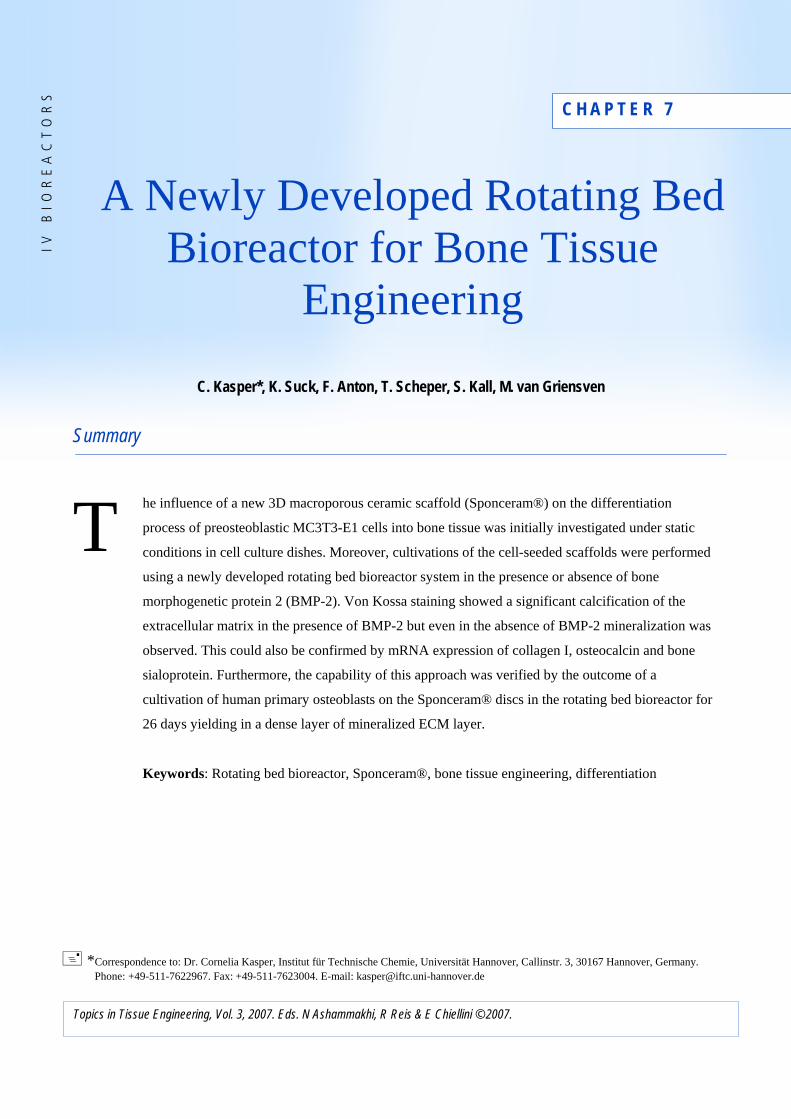
The essentials for tissue engineering are shown in figure 1. For the successful
generation of functional tissue, the different components (cells, medium and growth factors,
scaffold material) must be provided in a suitable environment (formation of cell-cell contacts,
mechanical loading, bioreactor).
Fig. 1. What is essential in Tissue Engineering?
Depending on the tissue that is supposed to be replaced, substrate materials have to be
provided for cell growth, which need to fulfil different requirements with regard to their
mechanical stability, biodegradability and porosity. These matrices have to be biocompatible,
should support cell attachment, growth and differentiation towards the desired phenotype.
Highly porous materials provide space for the bone tissue and allow an optimal cell growth
inside the scaffold. Many materials have been reported to fulfil these requirements including
natural/synthetic polymers, metals and ceramics [2-6]. Ceramics like hydroxyapatite,
calciumphosphates, aluminia, zirconia and composite materials etc. are widely used as
scaffold material [7-12]. Moreover, hydroxyapatites, calcium phosphates or composite
materials are known to enhance the osteogenic differentiation when seeded with progenitor
cells or preosteoblastic MC3T3-E1 cells [13, 14]. Furthermore, a controllable degradation of

Kasper et al. A Newly Developed Rotating Bed Bioreactor for Bone Tissue Engineering
3Topics in Tissue Engineering, Vol. 3, 2007. Eds. N Ashammakhi, R Reis & E Chiellini © 2007.
scaffolds is often desired, which should not cause any inflammatory reactions in vivo. After
transplantation, the scaffold is invaded by cells of the surrounding tissue.
Large bone defects caused by tumours, infectious diseases or trauma clearly result in
the medical need for bone regeneration. If possible an autologous bone graft (mostly derived
from the iliac crest) is carried out. However, the quantity of the obtained material is often not
sufficient in case of a large bone defect. Alternatively, allogeneic bone from donor (corpse)
patients (e.g. cryopreserved) is transplanted, which is critical with regards to infection risks
(AIDS, hepatitis). Therefore, alternative methods have been developed. One innovative
approach to bone tissue engineering is to seed osteoblasts or their precursor cells onto an
appropriate 3D matrix and to culture this scaffold in vitro before implantation into the defect
of a patient. During the generation of bone tissue different osteoblastic markers such as
alkaline phosphatase (AP), collagen I (COL I), osteocalcin (OC) and bone sialoprotein (BSP)
are chronologically expressed [15]. Finally, the cells are embedded in their extracellular
matrix and begin to mineralize by depositing mineral along and within the grooves of the
collagen fibrils [16].
The differentiation of cells into bone cells are mediated by growth factors. Specific
growth factors for bone differentiation are bone morphogenetic proteins (BMPs), members of
the TGF-β superfamily [17-19]. BMPs stimulate the differentiation of different cell types to
osteoblasts including undifferentiated mesenchymal cells, bone marrow stromal cells and
preosteoblasts [20]. For the in vitro promotion of the differentiation process into bone tissue
mainly BMP-7 and BMP-2 are used [20, 21], whereas BMP-2 increases the AP and OC level
in MC3T3-E1 cells [22]. This certifies the eminent influence BMP-2 and of BMPs in general
during the process of bone tissue formation and is thus considered as vital for bone tissue
engineering.
For an optimal supply of the cells with oxygen and nutrients within the scaffold the
cultivation is ideally performed in a bioreactor system, since static cultures are insufficient to
mimic the in vivo conditions. In a bioreactor oxygen, pH and the transport of nutrients and
metabolic waste in the tissue microenvironment can easily be controlled. The most
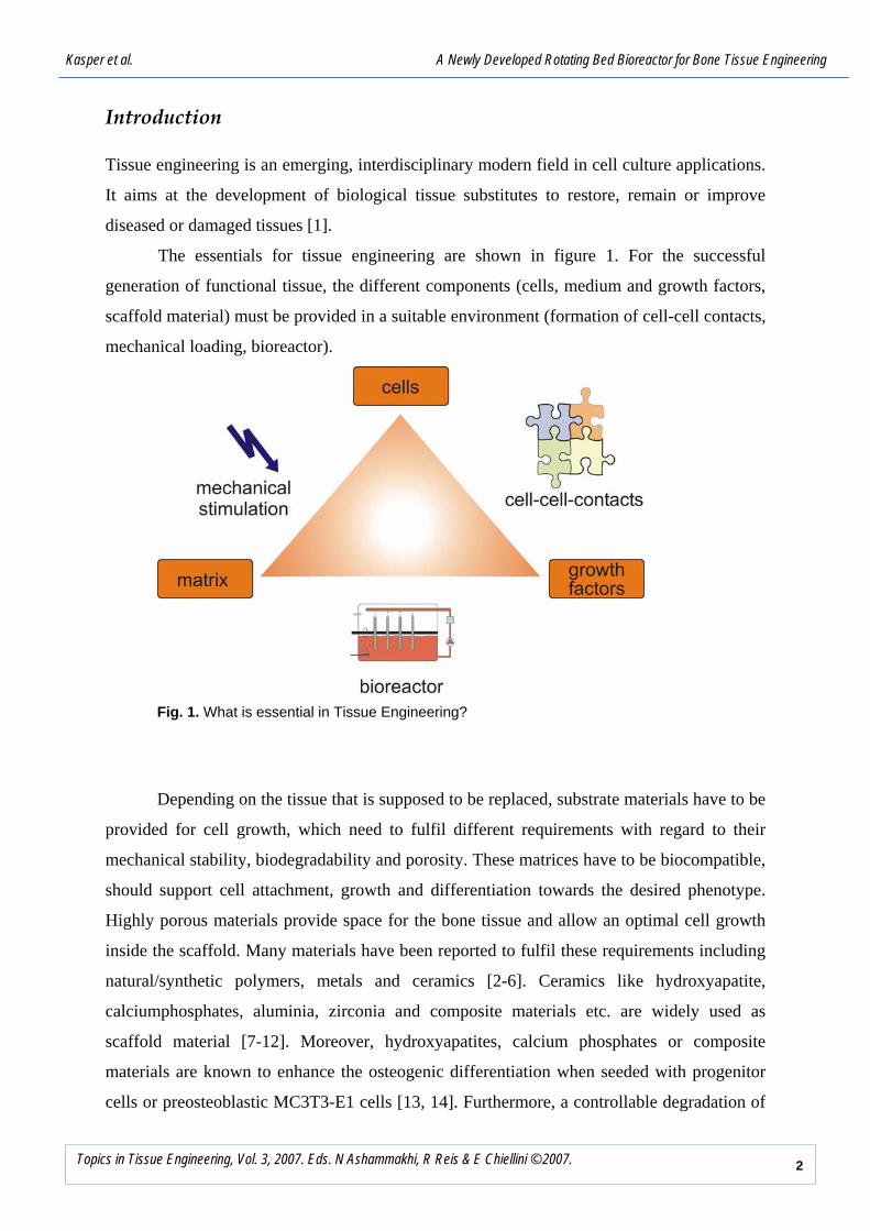
commonly used bioreactors for bone tissue engineering are the spinner flask, perfusion
culture system and the Rotating Wall Vessel Reactor (RWVR) (Fig. 2). In spinner flasks the
cells can be cultured either on scaffolds fixed on needles or seeded onto microcarriers [23,
24]. Perfusion culture systems provide a continuous exchange of medium which ensures the
removal of metabolic waste and supplementing essential nutrients throughout the cultivation.
Kasper et al. A Newly Developed Rotating Bed Bioreactor for Bone Tissue Engineering
4Topics in Tissue Engineering, Vol. 3, 2007. Eds. N Ashammakhi, R Reis & E Chiellini © 2007.
Perfusion systems are frequently applied for the cultivation of bone tissue [25-29]. Using the
RWVR, the scaffolds are cultured in a free fall manner. The RWVR has already been used
successfully for the cultivation of bone and cartilage cells [30].
perfusion rotating wallspinner flask
Fig. 2. Bioreactors for tissue engineering.
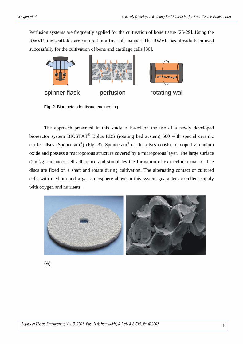
The approach presented in this study is based on the use of a newly developed
bioreactor system BIOSTAT® Bplus RBS (rotating bed system) 500 with special ceramic
carrier discs (Sponceram®) (Fig. 3). Sponceram® carrier discs consist of doped zirconium
oxide and possess a macroporous structure covered by a microporous layer. The large surface
(2 m2/g) enhances cell adherence and stimulates the formation of extracellular matrix. The
discs are fixed on a shaft and rotate during cultivation. The alternating contact of cultured
cells with medium and a gas atmosphere above in this system guarantees excellent supply
with oxygen and nutrients.
(A)
Kasper et al. A Newly Developed Rotating Bed Bioreactor for Bone Tissue Engineering
5Topics in Tissue Engineering, Vol. 3, 2007. Eds. N Ashammakhi, R Reis & E Chiellini © 2007.
(B)
Fig. 3. (A). Sponceram® disc and scanning electron microscopic picture of non-seeded macroporous structure of the material. The discs are fixed on a shaft and rotate with a speed of 2 rpm during cultivation (B). Scheme of the BIOSTAT® Bplus RBS 500 system setup.
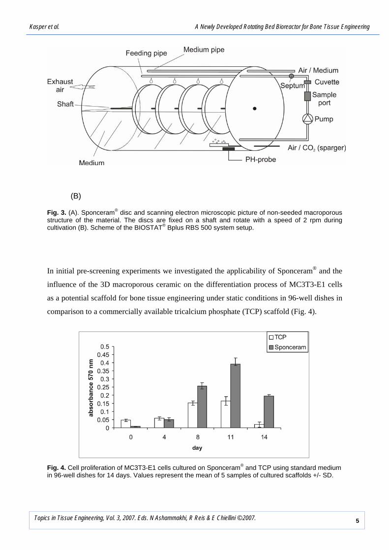
In initial pre-screening experiments we investigated the applicability of Sponceram® and the
influence of the 3D macroporous ceramic on the differentiation process of MC3T3-E1 cells
as a potential scaffold for bone tissue engineering under static conditions in 96-well dishes in
comparison to a commercially available tricalcium phosphate (TCP) scaffold (Fig. 4).
Fig. 4. Cell proliferation of MC3T3-E1 cells cultured on Sponceram® and TCP using standard medium in 96-well dishes for 14 days. Values represent the mean of 5 samples of cultured scaffolds +/- SD.

Kasper et al. A Newly Developed Rotating Bed Bioreactor for Bone Tissue Engineering
6Topics in Tissue Engineering, Vol. 3, 2007. Eds. N Ashammakhi, R Reis & E Chiellini © 2007.
Furthermore the rotating bioreactor system BIOSTAT® Bplus RBS 500 was used for
cultivation of MC3T3-E1 cells during a period of 3 weeks. The differentiation into the
osteoblastic phenotype with concomitant mineralization in the presence and absence of BMP-
2 under dynamic culture condition was investigated after cultivation (Fig. 5 and Fig. 6).
Fig. 5. Specific staining of mineralized ECM after cultivation of MC3T3-E1 cells in the BIOSTAT® Bplus RBS 500. (A) Von Kossa staining of non seeded Sponceram® (middle), cultured in standard medium (upper left) and in BMP-2 medium (upper right). (B) Alizarin red staining of non seeded Sponceram® (middle), cultured in standard medium (upper left) and in BMP-2 medium (upper right).
Fig. 6. RT-PCR analysis of alpha 1 (1) collagen, osteocalcin and bone sialoprotein mRNA of MC3T3-E1 cells after cultivation in the BIOSTAT® Bplus RBS 500. Cell culture was performed in standard medium (left) and in BMP-2 medium (right).
Kasper et al. A Newly Developed Rotating Bed Bioreactor for Bone Tissue Engineering
7Topics in Tissue Engineering, Vol. 3, 2007. Eds. N Ashammakhi, R Reis & E Chiellini © 2007.
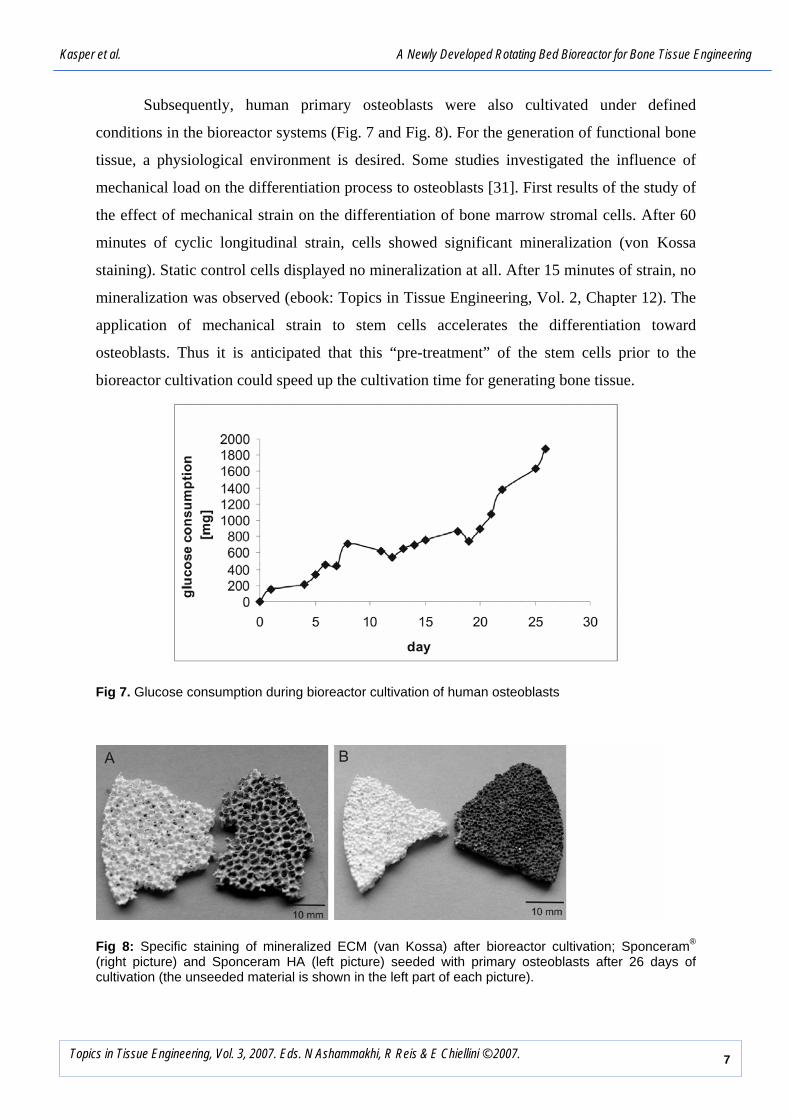
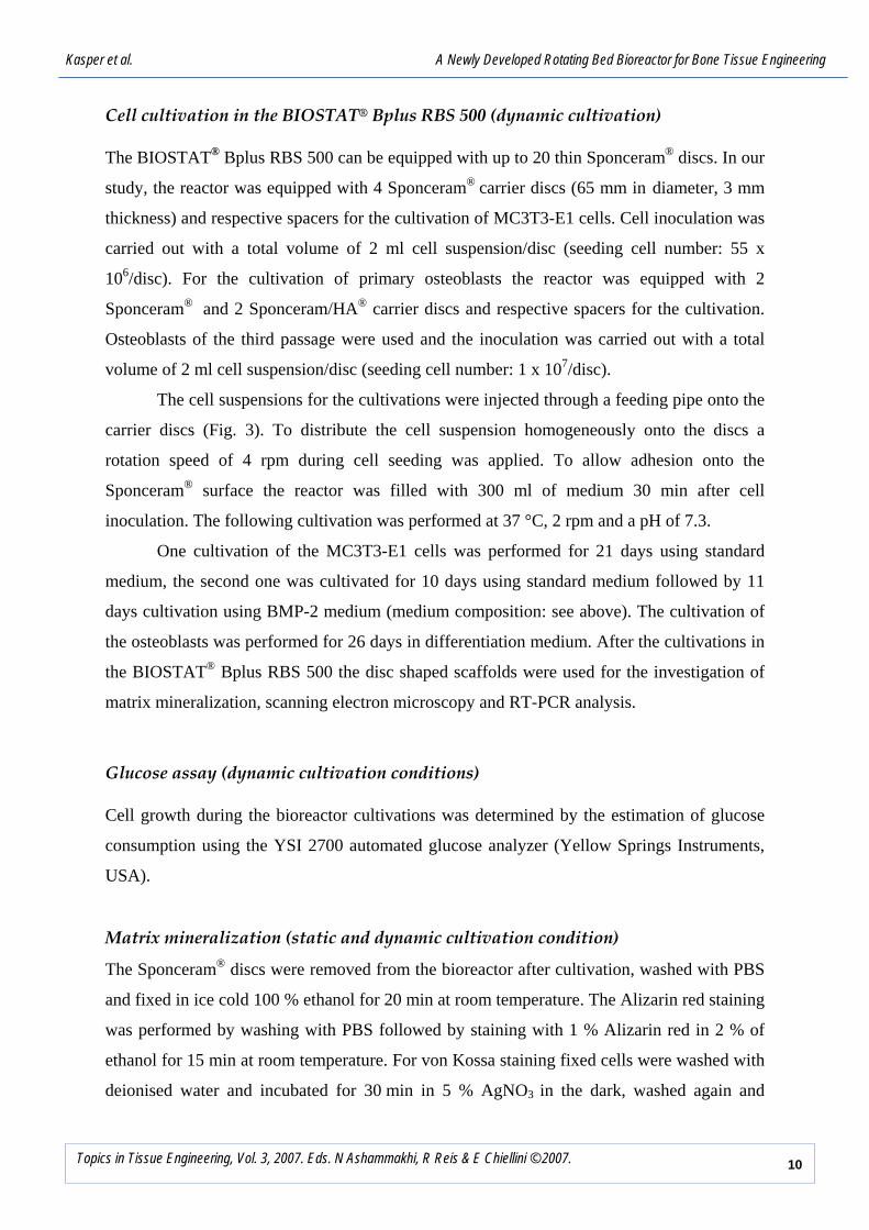
Subsequently, human primary osteoblasts were also cultivated under defined
conditions in the bioreactor systems (Fig. 7 and Fig. 8). For the generation of functional bone
tissue, a physiological environment is desired. Some studies investigated the influence of
mechanical load on the differentiation process to osteoblasts [31]. First results of the study of
the effect of mechanical strain on the differentiation of bone marrow stromal cells. After 60
minutes of cyclic longitudinal strain, cells showed significant mineralization (von Kossa
staining). Static control cells displayed no mineralization at all. After 15 minutes of strain, no
mineralization was observed (ebook: Topics in Tissue Engineering, Vol. 2, Chapter 12). The
application of mechanical strain to stem cells accelerates the differentiation toward
osteoblasts. Thus it is anticipated that this “pre-treatment” of the stem cells prior to the
bioreactor cultivation could speed up the cultivation time for generating bone tissue.
Fig 7. Glucose consumption during bioreactor cultivation of human osteoblasts
Fig 8: Specific staining of mineralized ECM (van Kossa) after bioreactor cultivation; Sponceram® (right picture) and Sponceram HA (left picture) seeded with primary osteoblasts after 26 days of cultivation (the unseeded material is shown in the left part of each picture).

Kasper et al. A Newly Developed Rotating Bed Bioreactor for Bone Tissue Engineering
8Topics in Tissue Engineering, Vol. 3, 2007. Eds. N Ashammakhi, R Reis & E Chiellini © 2007.
MATERIALS AND METHODS
All chemicals were purchased from Fluka/Sigma (Taufkirchen, Germany) and were of pro
analysi quality (if not stated otherwise). Deionized water (ARIUM, Sartorius AG,
Goettingen, Germany) was used for the preparation of media and buffers.
Sponceram®
Sponceram® is a ceramic support material consisting of doped zirconium oxide. The structure
of the material combines a unique mixture of macro- and micro pores. Technical data about
the surface area and pore size are summarized in table 1. For the cell cultivation under static
culture conditions in 96-well dishes samples of Sponceram® with a size of approximately 3
mm x 3 mm x 4 mm were used. The cultivation in the bioreactor system was performed with
identical Sponceram® (for MC3T3-E1 cells). This means that both Sponceram® (pore size:
900 µm) as well as Sponceram® with hydroxyapatite (HA) coating (pore size 600µm) (for
cultivation of primary osteoblasts) were used as carrier discs (65 mm in diameter, 3 mm
thickness) for the BIOSTAT® Bplus RBS 500.
Table 1. Surface characteristics of Sponceram
Sponceram®
sample
Sponceram®
disc
Pore size [µm] 600 600
Surface area (BET) [m2/g] 2 2
Total surface area per disc
[m2/disc]
- 14
Thickness [mm] 3 3
Diameter [mm] - 65
Density [g/ml] 0.7 0.7

Kasper et al. A Newly Developed Rotating Bed Bioreactor for Bone Tissue Engineering
9Topics in Tissue Engineering, Vol. 3, 2007. Eds. N Ashammakhi, R Reis & E Chiellini © 2007.
Cell cultivation under static conditions 96-well dishes
Samples of Sponceram®, Sponceram/HA® (Zellwerk GmbH, Oberkraemer, Germany) and
TCP (beta-tricalcium phophate, chronOSTM) (Mathys, Bettlach, Switzerland) (approximately
3 mm x 3 mm x 4 mm) were preconditioned in standard medium for 24 h in cell culture
medium at 37 °C/5 % CO2. Subsequently, 1.5 x 10⁴ MC3T3-E1 cells in 80 µl medium were
seeded on each scaffold in 96-well dishes for 30 min at gentle stirring at 37 °C/5 % CO2.
Non-attached cells were removed and the wells were filled up with 200 µl medium: 1.
Standard medium: DMEM (Sigma, Taufkirchen, Germany) containing 10 % FCS (PAA,
Coelbe, Germany), penicillin (50 U/ml), streptomycin (50 µg/ml) (PAA, Coelbe, Germany);
2. Differentiation medium: standard medium, 1 µM dexamethasone, 10 mM beta-
glycerolphosphate, 50 µg/ml ascorbic acid; 3. BMP-2 medium: differentiation medium + 10
ng/ml BMP-2. Before each analysis the scaffolds were placed into a new 96 well dish. Since
it is not possible to estimate the numbers of attached cells on the scaffolds directly the first
measurements were performed immediately after cell seeding (day 0). The cultivations were
performed in 96-well culture dishes with 200 µl of medium at 37 °C/5 % CO2. The medium
was changed every second to third day.
Cell proliferation assay (static cultivation conditions)
Cell proliferation was assayed using MTT (3-(4,5-dimethylthiazol-2-yl)-2,5-diphenyl-
tetrazolium-bromid (Sigma-Aldrich, Steinheim, Germany). Five replicates for each
experimental condition were placed in a 96-well culture dish. 100 µl of fresh medium and 10
µl of MTT solution (5 mg/ml in PBS) was added to each well and incubated for 4 h at 37
°C/5 % CO2. Subsequently, 100 µl of 10 % (weight/volume) SDS in 0.01 M HCl was added
and the samples were incubated for 16 h using the same conditions. The transmission signal
was measured at 570/630 nm using a Microplate Reader (Bio-Rad, Muenchen, Germany).
Statistical Analysis (static cultivation conditions)
The experiments were carried out twice with each time n=5 per sample. The data are
presented as mean values ± standard deviation.
Kasper et al. A Newly Developed Rotating Bed Bioreactor for Bone Tissue Engineering
10Topics in Tissue Engineering, Vol. 3, 2007. Eds. N Ashammakhi, R Reis & E Chiellini © 2007.
Cell cultivation in the BIOSTAT® Bplus RBS 500 (dynamic cultivation)
The BIOSTAT® Bplus RBS 500 can be equipped with up to 20 thin Sponceram® discs. In our
study, the reactor was equipped with 4 Sponceram® carrier discs (65 mm in diameter, 3 mm
thickness) and respective spacers for the cultivation of MC3T3-E1 cells. Cell inoculation was
carried out with a total volume of 2 ml cell suspension/disc (seeding cell number: 55 x
106/disc). For the cultivation of primary osteoblasts the reactor was equipped with 2
Sponceram® and 2 Sponceram/HA® carrier discs and respective spacers for the cultivation.
Osteoblasts of the third passage were used and the inoculation was carried out with a total
volume of 2 ml cell suspension/disc (seeding cell number: 1 x 10⁷/disc).
The cell suspensions for the cultivations were injected through a feeding pipe onto the
carrier discs (Fig. 3). To distribute the cell suspension homogeneously onto the discs a
rotation speed of 4 rpm during cell seeding was applied. To allow adhesion onto the
Sponceram® surface the reactor was filled with 300 ml of medium 30 min after cell
inoculation. The following cultivation was performed at 37 °C, 2 rpm and a pH of 7.3.
One cultivation of the MC3T3-E1 cells was performed for 21 days using standard
medium, the second one was cultivated for 10 days using standard medium followed by 11
days cultivation using BMP-2 medium (medium composition: see above). The cultivation of
the osteoblasts was performed for 26 days in differentiation medium. After the cultivations in
the BIOSTAT® Bplus RBS 500 the disc shaped scaffolds were used for the investigation of
matrix mineralization, scanning electron microscopy and RT-PCR analysis.
Glucose assay (dynamic cultivation conditions)
Cell growth during the bioreactor cultivations was determined by the estimation of glucose
consumption using the YSI 2700 automated glucose analyzer (Yellow Springs Instruments,
USA).
Matrix mineralization (static and dynamic cultivation condition)
The Sponceram® discs were removed from the bioreactor after cultivation, washed with PBS
and fixed in ice cold 100 % ethanol for 20 min at room temperature. The Alizarin red staining
was performed by washing with PBS followed by staining with 1 % Alizarin red in 2 % of
ethanol for 15 min at room temperature. For von Kossa staining fixed cells were washed with
deionised water and incubated for 30 min in 5 % AgNO3 in the dark, washed again and

Kasper et al. A Newly Developed Rotating Bed Bioreactor for Bone Tissue Engineering
11Topics in Tissue Engineering, Vol. 3, 2007. Eds. N Ashammakhi, R Reis & E Chiellini © 2007.
exposed to ultraviolet light for 2 min. Cells were fixed with 5 % sodium thiosulphate for
2 min and washed 3 times with deionised water.
Scanning electron microscopy (static and dynamic cultivation condition)
Cell grown Sponceram® discs were fixed in Karnovsky buffer at 4°C over night prior to
scanning electron microscopy. Samples were then dehydrated in solutions containing
increasing percentages of acetone (10 %, 30 %, 50 %, 70 %, 90 %, 100 %) and subsequently
imaged with a JEOL JSM-6700F scanning electron microscope.
Reverse transcriptase-polymerase chain reaction (RT-PCR) (dynamic cultivation
condition)
MC3T3-E1 cells were cultivated on scaffolds in the BIOSTAT® Bplus RBS 500 as described
above. Cells were removed from the disc by incubation in the enzyme mix ZW-DT-04
(Zellwerk GmbH, Oberkraemer, Germany) at 37 °C for 2 h and centrifuged at 400 ×g for 5
min. Cells were disrupted with RiboLyse tubes green (Hybaid, Heidelberg, Germany) for 40 s
at 6.0 Fast Prep FP 120 (Bio 101® Systems, Qbiogene, Heidelberg, Germany). The RNA was
isolated by the SV total RNA Isolation System (Promega, Mannheim, Germany).
RT was carried out with 2 µg RNA using the Superscript II system (Promega, Mannheim,
Germany) with oligo dT primer in a total volume of 40 µl. PCR was performed in a PCR-
Thermocycler (MWG Biotech, Ebersberg, Germany) using specific primers. The reaction
volume was 50 μl with an equivalent RNA concentration of 0.1 µg.
Amplification reactions were performed using the following primers (each 10 pmol)
and protocols (30 cycles): α1 (I) collagen: forward: 5`-TTC TCC TGG TAA AGA TGG
TGC-3`, reverse: 5`-GGA CCA GCA TCA CCT TTA ACA-3` (annealing 57°C, 255 bp
product) (Roth, Karlsruhe, Germany); Osteocalcin (OC): forward: 5`-ACA AGT CCC ACA
CAG CAG CTT-3`, reverse: 5`- GCC GGA GTC TGT TCA CTA CCT-3`(annealing 62°C,
187 bp product) (Roth, Karlsruhe, Germany); Bone sialoprotein (BSP): forward: 5´-CTG
TAG CAC CAT TCC ACA CT –3´, reverse: 5´-ATG GCC TGT GCT TTC TCG AT-
3´(annealing 56°C, 1055 bp product) (MWG Biotech, Ebersberg, Germany); GAPDH:
forward: 5`- GCC ACC CAG AAG ACT GTG GAT-3`, reverse: 5`- TGG TCC AGG GTT
TCT TAC TCC-3` (annealing 60°C, 455 bp product) (Roth, Karlsruhe, Germany).

Kasper et al. A Newly Developed Rotating Bed Bioreactor for Bone Tissue Engineering
12Topics in Tissue Engineering, Vol. 3, 2007. Eds. N Ashammakhi, R Reis & E Chiellini © 2007.
Results and Discussion Results
The results of the static pre-screening experiments for testing the applicability of Sponceram®
as a potential scaffold for bone tissue engineering were compared to the data obtained from
the employment of TCP after cultivation of the MC3T3-E1 cells in standard medium for 14
days. The results revealed that the proliferation of MC3T3-E1 cells was up to 60 % higher on
Sponceram® (Fig 4, day 11). Results of the cultivation of MC3T3-E1 cells cultured in the
BIOSTAT® Bplus RBS 500 bioreactor system on Sponceram® in standard medium and
BMP-2 medium respectively showed interestingly a clear mineralization by von Kossa and
Alizarin red staining when MC3T3-E1 cells were cultured in standard medium (Fig. 5 A and
B, upper left).
These finding were confirmed by RT-PCR. The results of mRNA levels showed that
collagen I, osteocalcin and bone sialoprotein were expressed independently of the presence of
BMP-2 in the medium (Fig. 6) but the intensity of the resulting protein bands was stronger
when the cultivation was performed in the presence of BMP-2 in the medium. Thus the
differentiation of the MC3T3-E1 cells to osteoblastic lineage [32] was demonstrated.
A cultivation of primary human osteoblasts was also performed in the BIOSTAT®
Bplus RBS 500. As an indicator for cell proliferation the glucose consumption was
determined during the cultivation in the BIOSTAT® Bplus RBS 500 (Fig. 7). The results
revealed that the glucose consumption was 14.05 g total over 26 days of cultivation in
differentiation medium. The final specific Alizarin Red and von Kossa stainings for
mineralization were clearly positive and the SEM pictures show articulate parallel structures
of collagen strands with mineralised ECM (Fig. 9).
Fig. 9: Scanning electron micrographs after the cultivation of primary osteoblasts for 26 days in the BIOSTAT® Bplus RBS 500 on Sponceram® (A, B) and Sponceram® HA (C, D).

Kasper et al. A Newly Developed Rotating Bed Bioreactor for Bone Tissue Engineering
13Topics in Tissue Engineering, Vol. 3, 2007. Eds. N Ashammakhi, R Reis & E Chiellini © 2007.
Conclusion
Within this study macroporous Sponceram® biomaterial based on zirconium dioxide was
tested towards its suitability as biomaterial for bone tissue engineering. The scaffold used for
tissue engineering has to provide all the necessary signals for the cells to grow, differentiate
and interact, forming the desired structure and thus mimic extra cellular matrix (ECM)
features. The results of the static cultivations revealed a limitation of the cultivation due to
confluent cell layer on the biomaterial pieces used. This lead to cell death after 11 days of
cultivation because the delivery of nutrients and oxygen as well as the elimination of
metabolic waste products is not efficient Thus a newly developed rotating bed bioreactor
systems was combined with the Sponceram® ceramic discs and successfully applied for
dynamic cultivation of MC3T3-E1 cells and primary human osteoblasts since an appropriate
dynamic culture system that mimics the in vivo environment (e.g. mechanical stimulation) is
needed for the generation of a functional bone tissue substitute.
The results obtained by RT-PCR, Alizarin and von Kossa staining as well as glucose
consumption and SEM pictures confirm that the scaffold itself is able to induce
differentiation of MC3T3-E1 cells in bone cells with concomitant mineralization when
cultured in the bioreactor system. This promotion can be due to the composition of the
Sponceram® scaffold and/or is related to its 3D structure. Additionally, the alternate contact
of cells to the medium and the oxygen atmosphere within the rotating bioreactor system
supported the proliferation and differentiation process of cells within the scaffold.
Outlook In future experiments, the application of mechanical strain to bone marrow and fat tissue
derived stem cells prior to the bioreactor cultivation will be studied in detail. The
differentiation process will be the focus of the work since the mechanical load accelerates to
differentiation in 2D cultures (ebook: Topics in Tissue Engineering, Vol. 2, Chapter 12),
therefore the conditions for the 3D culture will be optimized with regard to generation of
functional bone tissue.
Acknowledgement The authors would like to thank Prof. Walter Sebald (Biozentrum Würzburg, Germany) for kindly providing the BMP-2.
Kasper et al. A Newly Developed Rotating Bed Bioreactor for Bone Tissue Engineering
14Topics in Tissue Engineering, Vol. 3, 2007. Eds. N Ashammakhi, R Reis & E Chiellini © 2007.
References
1. Skalak R, Fox CF: Tissue engineering: proceedings of a workshop held at Granlibakken, Lake Tahoe, California, February 26-29, 1988. New York: Liss, 1988 UCLA symposia on molecular and cellular biology; new ser., v. 107). 2. Shin H, Jo S, Mikos AG: Biomimetic materials for tissue engineering. Biomaterials 2003; 24(24): 4353-64. 3. Sefton MV, Woodhouse KA: Tissue engineering. J Cutan Med Surg 1998; 3 Suppl 1: S1-18-23. 4. Langer R: Biomaterials in drug delivery and tissue engineering: one laboratory's experience. Acc Chem Res 2000; 33(2): 94-101. 5. Hutmacher DW, Goh JC, Teoh SH: An introduction to biodegradable materials for tissue engineering applications. Ann Acad Med Singapore 2001; 30(2): 183-91. 6. Hubbell JA: Biomaterials in tissue engineering. Biotechnology (N Y) 1995; 13(6): 565-76. 7. Zhao L, Chang J: Preparation and characterization of macroporous chitosan/wollastonite composite scaffolds for tissue engineering. J Mater Sci Mater Med 2004; 15(5): 625-9. 8. Hardouin P, Anselme K, Flautre B, Bianchi F, Bascoulenguet G, Bouxin B: Tissue engineering and skeletal diseases. Joint Bone Spine 2000; 67(5): 419-24. 9. Burg KJ, Porter S, Kellam JF: Biomaterial developments for bone tissue engineering. Biomaterials 2000; 21(23): 2347-59. 10. Bohner M: Calcium orthophosphates in medicine: from ceramics to calcium phosphate cements. Injury 2000; 31 Suppl 4: 37-47. 11. Arinzeh TL, Tran T, McAlary J, Daculsi G: A comparative study of biphasic calcium phosphate ceramics for human mesenchymal stem-cell-induced bone formation. Biomaterials 2005; 26(17): 3631-8. 12. Alam MI, Asahina I, Ohmamiuda K, Takahashi K, Yokota S, Enomoto S: Evaluation of ceramics composed of different hydroxyapatite to tricalcium phosphate ratios as carriers for rhBMP-2. Biomaterials 2001; 22(12): 1643-51. 13. Shu R, McMullen R, Baumann MJ, McCabe LR: Hydroxyapatite accelerates differentiation and suppresses growth of MC3T3-E1 osteoblasts. J Biomed Mater Res A 2003; 67(4): 1196-204. 14. Chou YF, Dunn JC, Wu BM: In vitro response of MC3T3-E1 preosteoblasts within three-dimensional apatite-coated PLGA scaffolds. J Biomed Mater Res B Appl Biomater 2005. 15. Owen TA, Aronow M, Shalhoub V, et al.: Progressive development of the rat osteoblast phenotype in vitro: reciprocal relationships in expression of genes associated with osteoblast proliferation and differentiation during formation of the bone extracellular matrix. J Cell Physiol 1990; 143(3): 420-30. 16. Lian JB, Stein GS: Development of the osteoblast phenotype: molecular mechanisms mediating osteoblast growth and differentiation. Iowa Orthop J 1995; 15: 118-40. 17. Reddi AH: Regulation of cartilage and bone differentiation by bone morphogenetic proteins. Curr Opin Cell Biol 1992; 4(5): 850-5. 18. Kawabata M, Imamura T, Miyazono K: Signal transduction by bone morphogenetic proteins. Cytokine Growth Factor Rev 1998; 9(1): 49-61. 19. Sebald W, Nickel J, Zhang JL, Mueller TD: Molecular recognition in bone morphogenetic protein (BMP)/receptor interaction. Biol Chem 2004; 385(8): 697-710. 20. Yamaguchi A, Komori T, Suda T: Regulation of osteoblast differentiation mediated by bone morphogenetic proteins, hedgehogs, and Cbfa1. Endocr Rev 2000; 21(4): 393-411. 21. Hollinger JO, Schmitt JM, Buck DC, et al.: Recombinant human bone morphogenetic protein-2 and collagen for bone regeneration. J Biomed Mater Res 1998; 43(4): 356-64.
Kasper et al. A Newly Developed Rotating Bed Bioreactor for Bone Tissue Engineering
15Topics in Tissue Engineering, Vol. 3, 2007. Eds. N Ashammakhi, R Reis & E Chiellini © 2007.
22. Takuwa Y, Ohse C, Wang EA, Wozney JM, Yamashita K: Bone morphogenetic protein-2 stimulates alkaline phosphatase activity and collagen synthesis in cultured osteoblastic cells, MC3T3-E1. Biochem Biophys Res Commun 1991; 174(1): 96-101. 23. Temenoff J, Mikos A: Injectable biodegradable materials for orthopedic tissue engineering. BIOMATERIALS 2000; 21(23): 2405-2412. 24. Sikavitsas VI, Bancroft GN, Mikos AG: Formation of three-dimensional cell/polymer constructs for bone tissue engineering in a spinner flask and a rotating wall vessel bioreactor. J Biomed Mater Res 2002; 62(1): 136-48. 25. Glowacki J, Mizuno S, Greenberger JS: Perfusion enhances functions of bone marrow stromal cells in three-dimensional culture. Cell Transplant 1998; 7(3): 319-26. 26. Mueller SM, Mizuno S, Gerstenfeld LC, Glowacki J: Medium perfusion enhances osteogenesis by murine osteosarcoma cells in three-dimensional collagen sponges. J Bone Miner Res 1999; 14(12): 2118-26. 27. Sikavitsas VI, Bancroft GN, Holtorf HL, Jansen JA, Mikos AG: Mineralized matrix deposition by marrow stromal osteoblasts in 3D perfusion culture increases with increasing fluid shear forces. Proc Natl Acad Sci U S A 2003; 100(25): 14683-8. 28. Wang Y, Uemura T, Dong J, Kojima H, Tanaka J, Tateishi T: Application of perfusion culture system improves in vitro and in vivo osteogenesis of bone marrow-derived osteoblastic cells in porous ceramic materials. Tissue Eng 2003; 9(6): 1205-14. 29. Yu X, Botchwey EA, Levine EM, Pollack SR, Laurencin CT: Bioreactor-based bone tissue engineering: the influence of dynamic flow on osteoblast phenotypic expression and matrix mineralization. Proc Natl Acad Sci U S A 2004; 101(31): 11203-8. 30. Qiu QQ, Ducheyne P, Ayyaswamy PS: Fabrication, characterization and evaluation of bioceramic hollow microspheres used as microcarriers for 3-D bone tissue formation in rotating bioreactors. Biomaterials 1999; 20(11): 989-1001. 31. Jagodzinski M, Drescher M, Zeichen J, et al.: Effects of cyclic longitudinal mechanical strain and dexamethasone on osteogenic differentiation of human bone marrow stromal cells. Eur Cell Mater 2004; 7: 35-41; discussion 41. 32. Aubin JE: Advances in the osteoblast lineage. Biochem Cell Biol 1998; 76(6): 899-910.