a necropsy procedure for sampling if ... - charles van...
TRANSCRIPT
Condor, 82235-98 0 The Cooper Ornithological Society 1980
A NECROPSY PROCEDURE FOR SAMPLING DISEASE IN WILD BIRDS
CHARLES VAN RIPER III
AND
SANDRA G. VAN RIPER
ABSTRACT.-This paper presents a necropsy procedure for examining small wild birds, designed to be used by investigators without experience in avian pathology. It gives instructions on how to conduct a postmortem examination, lists the needed equipment, tells what parts of a bird to save when a disorder in encountered, and lists possible diseases which each may signify. This procedure would enable ornithologists to include parasites and diseases in their investigations of avian populations.
Parasites and diseases should be considered in studying the demographic parameters of a population. Ornithologists often neglect these factors, however, because they lack training or facilities, and pathologists are not readily available for consultation. Our purpose here is to outline a procedure that will enable an ornithologist, without so- phisticated laboratory facilities, to conduct postmortem examinations of birds and to diagnose diseases.
For accurate disease diagnosis, it is first necessary to standardize the postmortem technique, so that each animal is examined in a similar manner and data are organized and easily retrievable. Most present-day avian necropsy techniques were developed for poultry (e.g., Hungerford 1969, Zander 1972). Those designed specifically for other species usually emphasize caged birds (Keymer 1961, Arnall and Keymer 1975), particularly the Canary (Serinus canaria), and the Budgerigar (Melopsittacus undu- htus; Stone 1969). Most of the other ne- cropsy procedures are intended for veteri- narians and are very general (Ensley et al. 1976, J. Carpenter, pers. comm.). The tech- nique to be presented here was developed for small passerine birds, but with slight modifications, it can be applied to most avi- an groups. It is based on described disor- ders present in poultry, caged birds, and wild birds, and should identify most dis- eases commonly encountered in wild birds.
We present a standardized necropsy pro- cedure, interspersed with dissection direc- tions and supplemented with instructions on which tissues to save and how to pre- serve them. Other sections (1) outline ma- terials and facilities necessary for a post- mortem analysis; (2) give general hints on necropsy procedures, especially those relat-
ed to the problems of dealing with smaller birds; (3) indicate what possible diseases each disorder listed on the necropsy proto- col might indicate; and (4) summarize dis- eases presently known to occur in birds, from what avian groups they have been re- ported, and indicate which tissue to collect.
MATERIALS
Certain basic equipment is required for this postmortem technique. Both a dissecting and a compound microscope are necessary. Necropsy of small birds is tedious, and un- less fine instruments are used, much infor- mation can be lost. Ophthalmic tools are ideal, and we have found iris microdissect- ing scissors, watchmaker’s and microdissec- tion forceps, and microprobes invaluable. Other essential equipment includes a small pane of glass for examining the gastrointes- tinal tract, clean microscope slides and cov- er slips, sterile swabs and syringes, sterile petri dishes and vials for collecting tissue samples, sterile plastic bags for freezing tis- sues, and an alcohol lamp. A dissecting tray or neoprene cutting board is useful for larg- er specimens.
Recommended chemicals and solutions for processing necropsy material include: 10% buffered formalin (add a pinch CaCO, per gallon); 70% ethyl alcohol; glycerine- alcohol (90 parts 70% ethyl alcohol, 10 parts glycerine); formalin-acetic-alcohol (F.A.A.; 50 parts 95% ethyl alcohol, 10 parts com- mercial formalin, 2 parts glacial acetic acid, 40 parts distilled water); absolute methyl alcohol; 2% potassium dichromate solution; sterile transport medium for fungi (e.g., Sa- bouraud’s or Mycobiotic agar available from Difco Laboratories, Detroit, Michigan 48201); sterile transport medium for bacte- ria (e.g., Stuart’s medium, a modified form
G351
86 CHARLES VAN RIPER III AND SANDRA G. VAN RIPER
packaged with a sterile swab called a Cul- turette, American Hospital Supply Corp., McGraw Park, Illinois 60085); and dry ice. Optional but useful supplies include Lu- gol’s solution (5 g iodine, 10 g potassium iodide, 100 ml distilled water; filter; dilute with 5 times the amount of distilled water before use), Hoyer’s mounting medium (30 g gum arabic, 50 ml distilled water, 20 ml glycerol, 200 g chloral hydrate; mix in order listed and filter through fine gauze), 10% solution of potassium hydroxide, sterile transport medium for viruses (available from Colab Laboratories, Chicago Heights, Illinois 60412).
Safety is an important consideration in doing any postmortem analysis because some avian disease organisms are pathogen- ic to man. The working area should have limited access so as to reduce bio-hazard. Use standard procedures in handling dis- eased tissue and liberal amounts of a strong detergent and disinfectant (e.g., Tincture Green Soap, Phenol, or Pine Oil).
PROCEDURE
GENERAL HANDLING OF A BIRD
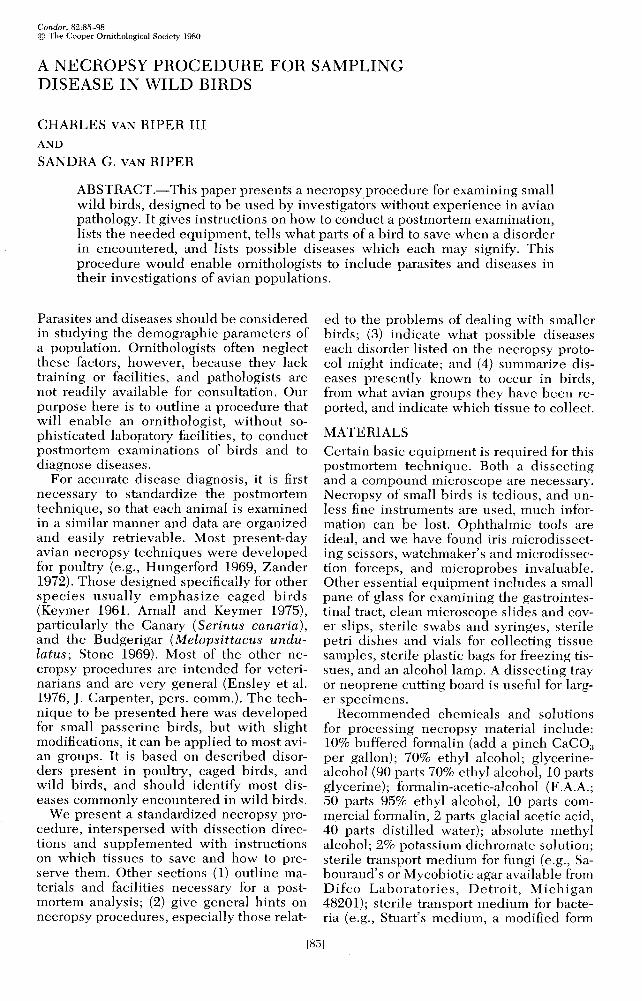
Using the necropsy form (Fig. 1) as a guide, perform the necropsy as soon as the bird is received because internal organs decom- pose rapidly and postmortem migration of parasites and bacterial invasion might oc- cur. Immediately take measurements be- cause weight, in particular, will change. Measurements are important for aging pur- poses and may later prove useful as indi- cators of specific diseases within the host population. Feather wear, cloaca1 protuber- ance, and brood patch will help define the breeding condition of the specimen. Tag the bird, and label each sample taken from it with the same necropsy number. In- dicate if samples are “sterile” or “non-ster- ile” and the type of medium in which they are preserved. Obtain a detailed history of the specimen as this often gives clues as to what might be wrong. When the necropsy is completed, fill in the preliminary findings under “necropsy summary,” but complete the “diagnosis” section only after the labo- ratory results are returned.
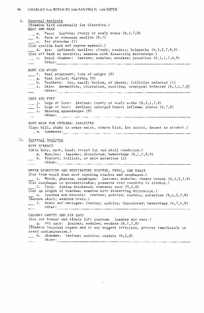
The actual examination is accomplished by following the directions given in paren- theses until a particular disorder is seen. Check the appropriate space, circle the term which best describes the disorder, and then, using the numbers at the end of the line as a guide, consult the Appendix for instruc- tions on how to handle the diseased tissue.
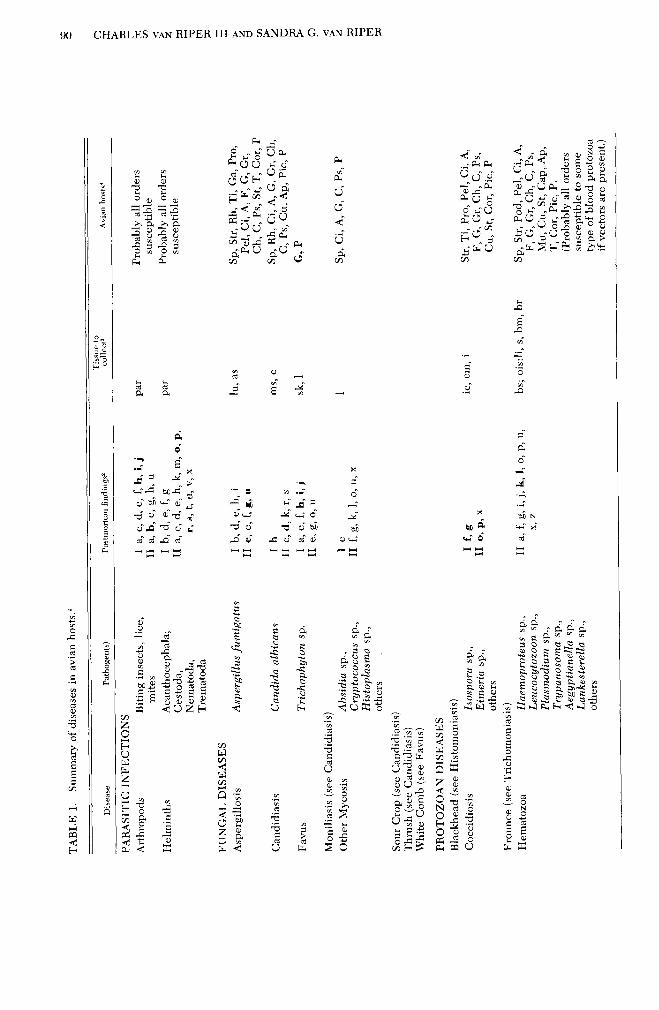
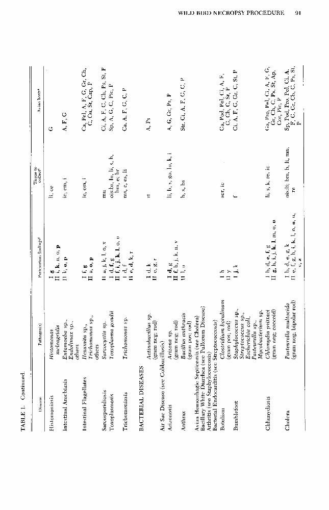
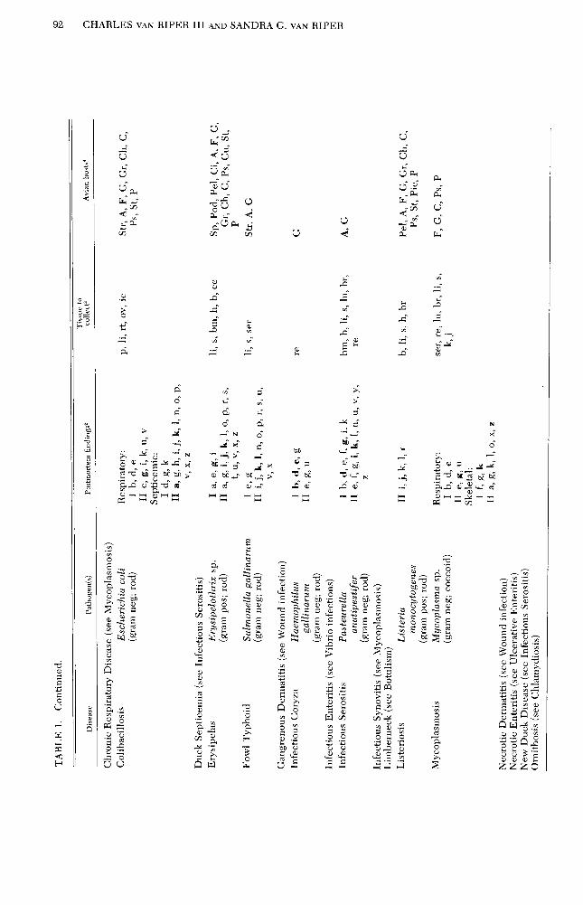
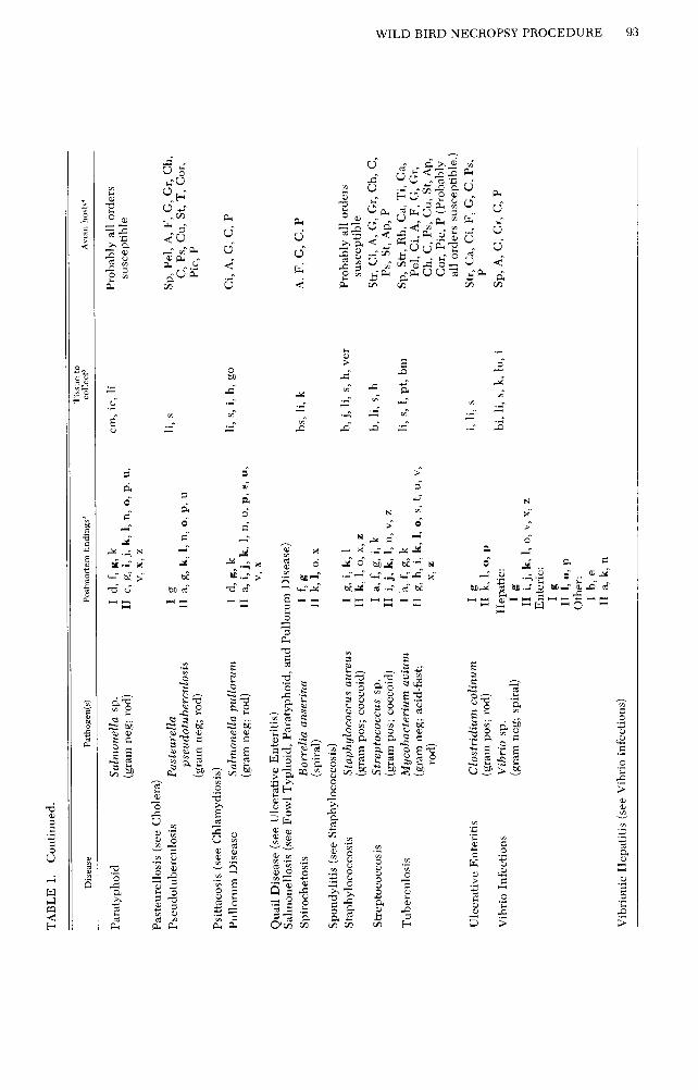
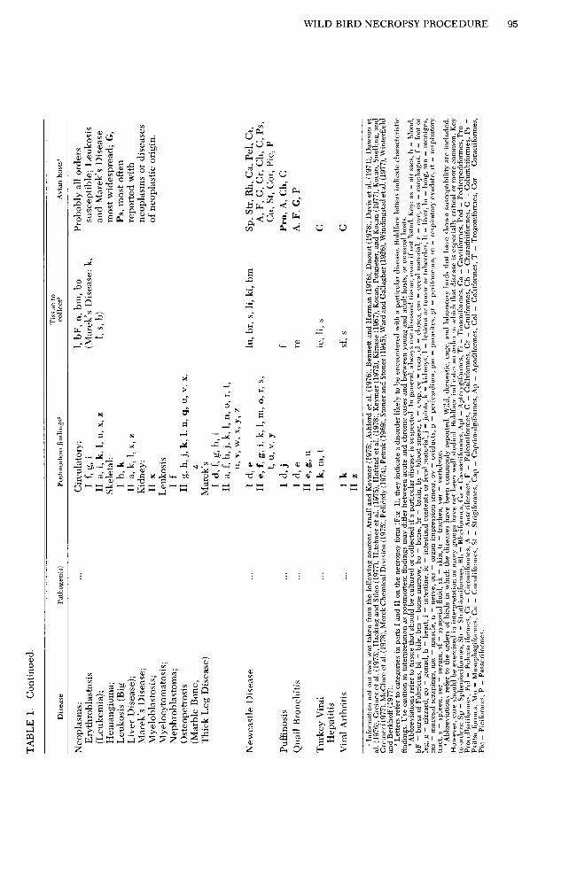
Each disorder can also be cross-referenced to a summary of avian diseases (Table 1) so that the investigator has some idea of pos- sible diseases with which he may be deal- ing. Since the presence of an organism does not necessarily imply a pathogenic condi- tion, careful notes concerning disorders are important.
Example. A bird is collected with exten- sive lesions on one leg. On the necropsy form (Fig. l), body measurements, condi- tion of plumage, and a complete history of the specimen are recorded. Section “I. Ex- ternal Analysis” is followed until the sub- section on “Legs and Feet” is reached. Line “j” is checked, the word “lesions” is cir- cled, and the numbers at the right of the line (6, 3, 1, 7, 8) are referred to in the Ap- pendix for further directions on handling of tissue. First a smear of any exudate is pre- pared for gram stain or a bacterial culture (6), then a portion of the lesion is cultured on mycotic medium or examined for fungal hyphae (3). Next, a wet smear of the infect- ed tissue is examined under a compound microscope for the presence of ectoparasites (l), and finally the remainder of the infected tissue is divided in half and placed in 10% buffered formalin and frozen (7,8). Possible causes of this disorder are cross-referenced to all Ij diseases in Table 1, and include Bumblefoot, Favus, Hematozoa, arthropods (mites), or Avian Pox; Bumblefoot, mites, and Pox are most probable. After the section is completed in the above manner, the in- vestigator continues the necropsy until each disorder is accounted for.
NECROPSY HINTS APPLICABLE TO SMALL
BIRDS
Because of their size, smaller birds pose special problems during a necropsy. We have found the following techniques (pref- aced by their corresponding sections in Fig. I) useful in working with passerines.
I. Body wash for external parasites. Following the external analysis of a bird, the beak should be taped shut and the specimen washed in a jar with detergent water. This is not only a good way to collect ectoparasites (Watson and Amerson 1967), but also it minimizes flying feathers during the remainder of the necropsy.
II. Celomic cavity and air sacs. Care- ful examination of these structures is crit- ical, especially for the identification of bacteria. To remove the keel, lift the ab- dominal wall with a forceps and cut trans- versely; grasp the tip of the keel, lift care-
WILD BIRD NECROPSY PROCEDURE 87
SPECIES : FIELD #: NECROPSY # : MUSEUM #:
Area collected: Collector: Date collected: Date examined: Examiner: Age: Sex: Fat: Skull: Weight: g P.M. state: Preserved in: Other:
History of bird:
Body measurements-length of: Body:_ mm Wing: mm Tail: mm Beak: mm Tarsus: mm Other: mm
Condition of plumage: Body molt: Wing molt: Tail molt: Head molt: Worn plumage: Area:
Breeding condition: Brood patch: Clo. P.: Gonad meas.: mm
MATERIAL TO LABORATORY Smears: peripheral
blood brain fecal
heart lung
liver kidney
spleen bone marrow
Tissue: entire bird leg feet eye esophagus crop trachea brain muscle blood serum heart liver gall bladder spleen pancreas bursa intestine proventriculus gizzard lung gonad kidney nerves endocrine glands
other: Body wash: Crop contents: Parasites: helminths:
arthropods: other:
Cultures:
Other:
NECROPSY SUMMARY:
LABORATORY RESULTS:
DIAGNOSIS:
FIGURE 1. A detailed avian necropsy protocol, keyed for tissue preservation and a preliminary disease diag- nosis.
88 CHARLES VAN RIPER III AND SANDRA G. VAN RIPER
I. External Analysis (Examine bird externally for disorders.) HEAD AND BEAK
-_ ,": Face: lesions; crusty or scaiy scabs (6,1,7,8) Face or sinsuses swollen (6,7)
__ c. Ear disorder (1) (Cut eyelids back and expose eyeball.) _ d. Eye: inflamed; swollen; cloudy; exudate; helminths (6,3,2,7,8,9) (Cut off beak at nostrils; examine with dissecting microscope.) ~ e. Nasal chamber: lesions; nodules; exudate; parasites (6,3,1,7,8,9)
Other:
BODY AND WINGS __ f. Keel prominent; loss of weight (9)
_ ,": Vent soiled; diarrhea (9) Feathers: dry, easily broken, or absent; follicles infected (1)
1. ’ Skin: dermatitis, ulceration, swelling; uropygial infected (6,3,1,7,8,) Other:
LEGS AND FEET ’ Legs or feet:
_ ;: lesions; crusty or scaly scabs (6,3,1,7,8)
Legs or feet: swollen; enlarged bones; inflamed joints (6,7,8) __ 1. Missing appendanges (9)
Other:
BODY WASH FOR EXTERNAL PARASITES (Tape bill, shake in soapy water, remove bird, let settle, decant to alcohol.)
__ m. Comments:
II. Internal Analysis
BODY SURFACE (Skin body, neck, head; record fat and skull condition.)
a. Muscles: lesions; discolored; hemorrhage (6,1,2,8,9) b. Feather, follicle, or skin parasites (1)
Other:
UPPER DIGESTIVE AND RESPIRATORY SYSTEMS, SKULL, AND BRAIN (Cut from mouth down neck exposing trachea and esophagus.)
Mouth, pharynx, esophagus: (Cut'esophagus to proventriculus
lesions; nodules; cheesy masses (6,3,5,7,8) ; preserve crop contents in alcohol.)
d. Crop: lining thickened; contents sour (5,2,8) (Cut up length of trachea; examine with dissecting microscope.)
e. Trachea and bronchi: lesions; nodules; exudate; parasites (6,1,2,7,8) (Remove skull; examine brain.)
f. Brain and meninges: lesions; nodules; discolored; hemorrhage (6,7,4,8) Other:
CELOMIC CAVITY AND AIR SACS (Cut out breast and slowly lift sternum. Examine air sacs.)
- g. Air sacs: lesions; nodules; exudate (6,1,7,8) (Examine internal organs and if any suggest infection, process immediately to avoid contamination.)
h. Abdomen: lesions; nodules; exudate (6,1,8) Other:_
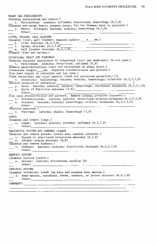
WILDBIRDNECROPSYPROCEDURE 89
HEART AND PERICARDIUM (Examine pericardium and remove.)
1. . Pericardium: exudate; inflamed; discolored; hemorrhage (6,7,8) (Examine and weigh heart; prepare smear; fix for Giemsa; wait to preserve.)
_ j. Heart: enlarged; lesions; nodules; hemorrhage (6,7,8) Other:
LIVER, SPLEEN, GALL BLADDER (Examine liver, gall bladder; measure spleen: x mm.) ~-
k. Liver disorder (6,4,7,8) 1. Spleen disorder (6,4,7,8) m. Gall bladder disorder (6,2,7,8)
(Remove liver and spleen.)
INTESTINAL TRACT AND PANCREAS (Examine external appearance of intestinal tract and membranes; do not open.)
n. Peritoneum: nodules; discolored; inflamed (6,8) (Remove gastrointestinal tract and straighten on glass plate.) (Intestine length: Separate proventriculus and gizzard.) (Cut down length of in:stine and lay open.) (Take intestinal and cecal smears; check for protozoan parasites.) (5)
0. Intestine: ballooning; lesions; nodules; hemorrhage; helminths (6,2,5,7,8) (Record location of parasites: )
_ p. Ceca: lesions; nodules; exudate; hemorrhage; thickened; helminths (6,2,5,7,8)
~ q* Bursa of Fabricius abnormal (7,8): Other:
(Cut open proventriculus and gizzard. Remove lining; preserve contents.) r. Proventriculus: lesions; nodules; hemorrhage; erosion; helminths (6,3,2,5,8) S. Gizzard: lesions; nodules; hemarrhage; erosion; helminths (6,3,2,5,8)
Other: (Examine pancreas.)
t. Pancreas: lesions; chalky; hemorrhage (7,8)
LUNGS (Examine and remove lungs.)
U. Lungs: lesions; nodules; exudate; inflamed (6,3,7,8) Other:
UROGENITAL SYSTEM AND ADRENAL GLANDS (Measure and remove gonads; record sex; examine adrenals.)
v. Gonads or associated structures abnormal (6,7,8) w. Adrenal glands abnormal (8,9)
(Examine and remove kidneys.) X. Kidneys: lesions; nodules; discolored; enlarged (6,5,2,7,8)
Other:
NERVOUS SYSTEM (Examine nervous plexus.)
- y* Nerves: lesions; discolored; swollen (8) Other:
SKELETAL SYSTEM (Examine vertebrae; break leg bone and examine bone marrow.)
2. Bone marrow, vertebrae, bones, tendons, or joints infected (6,4,7,8) Other:
COMMENTS:

fully, and cut anteriorly across the ribs with over, diseases may share common signs, es- a scissors (for larger birds bone shears will be necessary). Do not touch the surface of
pecially those that undergo a septicemic phase. Therefore it is usually only by a thor-
any internal organs lest they become con- ough necropsy analysis, supplemented with taminated. Cut through the coracoids and laboratory tests, that a particular disease can clavicles to complete removal of the ster- be positively identified. num. If any of the internal organs indicate bacterial infection, make cultures now, be-
Problems may arise in trying to find a lab-
fore their surfaces become contaminated. oratory that will process tissues and identify pathogens. Several agencies have well-es-
11.1. Spleen disorder. The spleen is an im- tablished laboratories specializing in avian portant indicator organ, but is sometimes disease (e.g., U.S. Fish and Wildlife Ser- difficult to find in small birds. It is usually vice), but they are usually reluctant to ac- reddish-brown, either spherical or slightly cept material from outside researchers due oblongate, and lies on the crania-dorso side to lack of time or personnel. Instead, try of the gizzard. It can be seen by rotating the state departments of agriculture or health, gizzard 180” to the left (counter-clockwise) veterinary pathology laboratories, universi- with the forceps. ty medical laboratories, or veterinary sci-
II. Intestinal tract and pancreas. In ence departments. The U.S. Department of order to avoid contaminating the remain- Agriculture Veterinary Services Diagnostic ing organs, remove the gastrointestinal Laboratory at Ames, Iowa, might also be tract as an entire unit. Cut the abdominal consulted. wall around the cloaca, and remove the tract whole, cutting as far anterior of the pro- ACKNOWLEDGMENTS
ventriculus as possible. Stretch the intes- We thank J. Carpenter, A. Miyahara, S. Perry, and T.
tine on a dry glass plate; separate the pro- Sawa for suggestions on the necropsy protocol, and G.
ventriculus-gizzard and set them aside. E. Duke, D. J. Forrester, H. W. Kale II, G. N. Stem-
Measure the length of the intestine so that mermann, C. E. Whiteman, and B. Williams for com-
the location of all parasites can be pinpoint- ments on the paper. Financial support was provided bv The Center for Field Research and Earthwatch of
ed. A 45” angle iridectomy scissor is a useful Belmont, Massachusetts, and Contract CX 8000 7 0009
tool for opening the small intestine. Some from the U.S. National Park Service to the Cooperative
intestinal helminths of small birds are mi- National Park Resources Studies Unit at the University
nute and need to be manipulated with in- of Hawaii.
struments as small as microprobes. These probes can be cheaply made by inserting or
LITERATURE CITED
melting an insect pin into a swizzle-stick. AR~ALL, L., AND I. F. KEYMER. 1975. Bird diseases.
1I.r. and s. Proventriculus-Gizzard. Ex- T.F.H. Publ., Neptune City, N.J.
amine the proventriculus and gizzard after ASHFORD, R. W., T. T. PALMER, J. S. ASH, AND R. S.
the intestinal tract as they dry out less rap- BRAY. 1976. Blood parasites of Ethiopian birds. I. General survey. J. Wildl. Dis. 12:409-426.
idly. Be sure to carefully tease the gizzard BENNETT, G. F., AND C. M. HERMAN. 1976. Blood
lining apart from the muscle, because many parasites of some birds from Kenya, Tanzania and
parasitic helminths occur there. Zaire. I. Wildl. Dis. 12:59-65.
II. Nervous system. Remove all inter- DAOUST, P.-Y. 1978. Osteomyelitis and arthritis
caused by Salmonella typhimurium in a crow. J.
nal organs so that the nerve plexes can be Wildl. Dis. 14:483485.
seen crossing over the various bones. DAVIS, J. W., R. C. ANDERSON, L. KARSTAD, AND D. 0. TRAINER [Eds.]. 1971. Infectious and parasitic
DISCUSSION diseases of wild birds. Iowa State Univ. Press, Ames.
Research is needed on disease in wild DAWSON, C. O., E. B. WHEELDON, AND P. E. Mc-
birds, especially to delimit the parasite fau- NEIL. 1976. Air sac renal mucormycosis in an Af-
na present within a host. Recording the rican Gray Parrot (Psittacus erithacus). Avian Dis.
levels of a single pathogen cannot possibly 20:593-660.
determine the impact of diseases upon wild ENSLEY, P. K., R. J. MONTALI, AND E. E. SMITH.
1976. A necronsv procedure for exotic birds. D.
bird populations. A comprehensive ap- 131-144. Ann;. P&c. Am. Assoc. Zoo Vet.,‘St.
proach is necessary in order to reveal how Louis, MO.
diseases are interrelated. We hope that our GREINER, E. C., G. F. BENNETT, E. M. WHITE, AND R.
postmortem technique will assist ornithol- F. COOMBS. 1975. Distribution of the avian he- matozoa of North America. Can. J. Zool. 53:1762-
ogists to attempt such multi-disease studies. 1787.
Investigators should remember, however, HACKING, M. A., AND L. SILEO. 1977. Isolation of a
that disease categories have been largely hemolytic Actinobacillus from waterfowl. J. Wildl.
determined from poultry disorders, and Dis. 13:69-73.
_ ^^ they may differ in other avian groups. More-
HITCHNER, S. B., C. H. DOMERMUTH, H. G. PUR- CHASE, AND J. E. WILLIAMS [Eds.]. 1975. Isola-
96 CHARLES VAN RIPER III AND SANDRA G. VAN RIPER
WILD BIRD NECROPSY PROCEDURE 97
tion and identification of avian pathogens. Am. Assoc. Avian Pathologists. Arnold Printing Co., Ithaca, N.Y.
HOFSTAU, M. S., B. W. CALNEK, C. F. HELMBOLDT, W. M. REID, AND H. W. YODER, JR. [Eds.]. 1978. Diseases of poultry. Iowa State Univ. Press, Ames.
HUNGERFOHD, T. G. 1969. Diseases of poultry in- clnding cage birds and pigeons. Angus and Roh- ertson, Sydney, Australia.
KEYMER, I. F. 1961. Postmortem examinations of pet birds. Mod. Vet. Pratt. 42(23):35-38,(24):47-511
KEYMER, I. F. 1972. Diseases of birds of prey. Vet. Rec. 90:579-594.
KIRILISE, P. 1967. Pox in wild birds: an annotated hib- IiograDhv. Wildl. Dis. 49:1-10.
KCXAN;’ A: A., L. N. D. POTGIETER, AND K. M. Ko- CAN. 1977. Inclusion body disease of falcons (her- pesvirns infection) in an American Kestrel. J. Wildl. Dis. 13:199-201.
KOCAN, A. A., J. SNELLING, AND E. C. GREINER. 1977. Some infections and parasitic diseases in Oklahoma raptors. J. Wildl. Dis. 13:304-306.
MCCLURE, H. E., P. POO~~SWAD, E. C. GREINER, AND M. LAIHD. 1978. Haematozoa in the birds of east- ern and southern Asia. Memorial Univ. of New- fonndland, St. John’s, Newfoundland, Canada.
MEHCK CIIE~~ICAL Drvrsrolv. 1975. The Merck poul- try serviceman’s manual. Merck and Co., Rahway, N.J.
PELL~RDY, L. P. 1974. Coccidia and coccidiosis. Ver- lag Pan1 Parey, Berlin.
PETRAK, M. L. [Ed.]. 1969. Diseases of cage and avi- ary birds. Lea and Fehiger, Philadelphia.
STONE, R. M. 1969. Clinical examination and methods of treatment, p. 177-187. In M. L. Petrak [Ed.], Diseases of cage and aviary birds. Lea and Fehi- ger, Philadelphia.
STONER, D., AND L. C. STONER. 1945. An example of bumblefoot in the Great Horned Owl. Auk 62:405- 408.
WARD, A. R., AND B. A. GALLAGHER. 1926. Diseases of domesticated birds. MacMillan Co., N.Y.
WATSON. G. E.. AND A. B. AILIERSON. 1~. 1967. In- structions fir collecting bird parasites. Mus. Nat. Hist. Info. Leaflet No. 477.
WINDINGSTAD, R. M., D. 0. TRAINER, AND R. DUN- CAN. 1977. Salmonella enteritidis and Arizona hinshawii isolated from wild Sandhill Cranes. Avian Dis. 21:704-707.
WINTERFIELD, R. W., AND G. A. BERKHOFF. 1977. Ulcerative enteritis in robins. Avian Dis. 21:328- 330.
ZANDER, D. V. 1972. Principles of disease prevention: diagnosis and control, p. 349. In M. S. Hofstad, B. W. Calneck, C. F. Helmholdt, W. M. Reid, and H. W. Yoder, Jr. [Eds.], Diseases of poultry. Iowa State Univ. Press. Ames.
APPENDIX
The following section is an outline of how to handle diseased tissue for diagnosis or shipment to a diagnostic laboratory. Be sure to check with the laboratory for its preferred procedure before sending specimens. Num- bers preceding each paragraph correspond to the numbers at the ends of the lines in the necropsy form (Fig. 1).
Zf you suspect u PARASITIC infection:
(1) Arthropod Parasites. Examine suspect area (e.g., nasal chamber, inner surface of skin, feather vane or shaft, inner feather shaft, follicles) with dissecting microscope for parasites. Scrape lesions and suspect tis- sue (e.g., irritated areas, crusty scales or scabs, dry skin, nodules) onto a slide with mineral oil or water and make a wet mount; examine with a compound microscope. Pre- serve mites, lice, and small insects in 70% alcohol. For permanent mounts of ectopar- asites drop the specimen directly into Hoy- er’s mounting medium, pass over a flame to relax the specimen, then place on a cover slip; store in a horizontal position until dry.
(2) Helminth Parasites. Examine appropri- ate area (e.g., under gizzard lining, under nic- titating membrane of eye, inside trachea, in bile ducts, along intestine and ceca) with compound microscope. Nematodes may be fixed directly in glycerine-alcohol solution and shipped. Cestodes, trematodes, and acanthocephalans should be placed in F.A.A. for 24 h and then transferred to 70% alcohol for shipment. To examine fecal ma- terial for helminth ova, follow procedure outlined under digestive tract protozoa.
Zf you suspect a FUNGAL infection:
(3) Culture suspect tissue on mycotic medi- um. Sear the surface with a hot spatula if tissue has been contaminated, incise tissue, and sample cut surface. If no medium is available, place sample in 10% formalin. Fungal hyphae may be observed using a wet mount (Lugol’s solution will make them more visible but is not necessary); they may be permanently mounted in 10% potassium hydroxide, relaxed and cleared with gentle heat.
Zf you suspect a PROTOZOAN infection:
(4) Blood Protozoa. Prepare a smear for Giemsa stain using heart blood, bone mar- row, organ impressions (touch cut surface of organ to clean slide) of liver, spleen, and brain. Fix in absolute methyl alcohol for 30 s, and stain according to standard tech- nique. Examine using oil immersion lens.
(5) Digestive Tract Protozoa. Prepare a wet smear from appropriate material (e.g., mouth lesions, intestinal and cecal con- tents) and examine under compound micro- scope. Lugol’s solution will be helpful but is not necessary. If coccidia are present and sporulation is desired, place sample in 2% potassium dichromate and examine again in four days.

98 CHARLES VAN RIPER III AND SANDRA G. VAN RIPER
Zf you suspect a BACTERZAL infection:
(6) Most bacterial infections cannot be posi- tively diagnosed without identification of the pathogen, and for this reason it is best to collect a sample for bacterial culture. If dn organ has been collected under nonster- ile conditions, sear the surface with a hot spatula, incise the tissue, and sample the cut surface. Sample moist membranes and soft organs with sterile swabs; place the en- tire swab directly in medium for shipment and refrigerate. Collect joint or nasal exu- date with sterile hypodermic needle or swab. If solid agar is used, place the tissue firmly against agar or embed several small pieces. Use appropriate material from le- sions, exudate, or abnormal tissue. If certain diseases are suspected, one tissue will be preferred over others (see Table 1). Other- wise the liver, spleen, heart and any dis- eased tissue are usually good choices.
Sometimes a presumptive diagnosis can be made on the basis of slides showing the pathogen. To prepare a smear for gram stain, use appropriate material (e.g., exu- date, necrotic lesions, organ impression smears, heart blood, bone marrow, joint fluid collected with a sterile syringe). Fix by drying over heat and stain according to standard methods. Use the same procedure for staining with Giemsa, except fix in ab- solute methyl alcohol for 30 s without heat. Ziehl-Neelsen stain should be used if acid- fast bacteria are suspected or tubercles are present. Squash a small (2 mm) piece of tis- sue between two slides and fix over heat.
For serological testing, collect blood aseptically from the heart and centrifuge immediateIy. Transfer serum to sterile vials and freeze for shipment. If little blood is available, sealed capillary tubes are usually sufficient.
Zf you suspect a VZRAL infection:
(7) Most viral infections cannot be diagnosed without isolating the pathogen. Viral cul- ture medium is acceptable, but freezing is usually preferred. Freeze the sample, using dry ice to rapidly lower the temperature he- low -60°C (glass often shatters so plastic bags may be used). Use material from in- fected tissue, exudate, or joint fluid (collect with a sterile syringe). If one section of the respiratory system (e.g., air sacs) is in-
volved, it is good to freeze tissue from other respiratory areas (e.g., sinus, trachea, lungs), and cloaca, even though the tissue may ap- pear normal. Other suggested representa- tive tissue to collect includes liver, spleen, bone marrow, blood or serum, and bursa of Fabricius. See Table 1 if a particular disease is suspected. Collect blood for serological testing as described under section 5 above; do not freeze whole blood.
Zf tissue is to be for GENERAL HZSTO- PATHOLOGY:
(8) Preserve infected tissue in 10% buffered formalin (some laboratories prefer other me- dia such as Zenker’s). When nerves are dis- eased, check the brain carefully for abnor- malities and preserve in 10% formalin, even if it appears normal. With any disease, take representative sections of the body; sec- tions of heart, lung, respiratory system, gas- trointestinal tract, kidney, liver, brain (es- pecially if nerves are diseased), spleen, gonads, muscle, and skeleton are recom- mended. Cut tissue samples in pieces no larger than 1.0 x 2.0 x 0.5 cm and place in 10 times the amount of formalin. After 24 h the tissue may be packed with less formalin or left as is.
Zf SECONDARY SYMPTOMS and NER- VOUS SZGNS are present before death:
(9) Many signs and symptoms are of a sec- ondary nature, and when they are encoun- tered the investigator should be aware of other abnormalities. Especially note the condition of the respiratory or digestive sys- tems. Earlier trauma (e.g., missing toes due to freezing or pox infections) or shock signs should be noted in history of the bird. If nervous signs were noticed before the bird was collected or died, preserve the brain and nerve tissue at end of necropsy on dry ice and submit for viral and histopathology analysis. If poisoning is suspected, collect and freeze a sample of the body fiat.
Avian Di.sease Laboratory, Hawaii Field Research Center, P.O. Box 54, Hawaii Volcanoes National Park, Hawaii 96718. Present uddress of jir,yt author: De- partment of Zoology und Cooperatizje National Park Resources Unit, University of California ut Duais, Da&s, California 95616. Accepted for publication 19 September 1979.