a mutant isoform of obge causes cell death by interfering ... filefmicb-08-01193 june 27, 2017 time:...
TRANSCRIPT

fmicb-08-01193 June 27, 2017 Time: 14:25 # 1
ORIGINAL RESEARCHpublished: 28 June 2017
doi: 10.3389/fmicb.2017.01193
Edited by:Marc Bramkamp,
Ludwig-Maximilians-UniversitätMünchen, Germany
Reviewed by:Susan Schlimpert,
John Innes Centre (BBSRC),United KingdomDaniel Haeusser,
Canisius College, United States
*Correspondence:Jan Michiels
†These authors are joint seniorauthors.
Specialty section:This article was submitted to
Microbial Physiology and Metabolism,a section of the journal
Frontiers in Microbiology
Received: 03 May 2017Accepted: 12 June 2017Published: 28 June 2017
Citation:Dewachter L, Verstraeten N,
Jennes M, Verbeelen T, Biboy J,Monteyne D, Pérez-Morga D,
Verstrepen KJ, Vollmer W, Fauvart Mand Michiels J (2017) A Mutant
Isoform of ObgE Causes Cell Deathby Interfering with Cell Division.
Front. Microbiol. 8:1193.doi: 10.3389/fmicb.2017.01193
A Mutant Isoform of ObgE CausesCell Death by Interfering with CellDivisionLiselot Dewachter1, Natalie Verstraeten1, Michiel Jennes1, Tom Verbeelen1,Jacob Biboy2, Daniel Monteyne3, David Pérez-Morga3,4, Kevin J. Verstrepen1,5,Waldemar Vollmer2, Maarten Fauvart1,6† and Jan Michiels1*†
1 Centre of Microbial and Plant Genetics, KU Leuven – University of Leuven, Leuven, Belgium, 2 Centre for Bacterial CellBiology, Institute for Cell and Molecular Biosciences, Newcastle University, Newcastle upon Tyne, United Kingdom,3 Laboratory of Molecular Parasitology, Institut de Biologie et de Médecine Moléculaires, Université Libre de Bruxelles,Gosselies, Belgium, 4 Center for Microscopy and Molecular Imaging, Université Libre de Bruxelles, Gosselies, Belgium,5 Systems Biology Laboratory, VIB Center for Microbiology, Leuven, Belgium, 6 Department of Life Sciences and Imaging,Smart Electronics Unit, Imec, Leuven, Belgium
Cell division is a vital part of the cell cycle that is fundamental to all life. Despite decadesof intense investigation, this process is still incompletely understood. Previously, theessential GTPase ObgE, which plays a role in a myriad of basic cellular processes (suchas initiation of DNA replication, chromosome segregation, and ribosome assembly), wasproposed to act as a cell cycle checkpoint in Escherichia coli by licensing chromosomesegregation. We here describe the effect of a mutant isoform of ObgE (ObgE∗) thatcauses cell death by irreversible arrest of the cell cycle at the stage of cell division.Notably, chromosome segregation is allowed to proceed normally in the presence ofObgE∗, after which cell division is blocked. Under conditions of rapid growth, ongoingcell cycles are completed before cell cycle arrest by ObgE∗ becomes effective. However,cell division defects caused by ObgE∗ then elicit lysis through formation of membraneblebs at aberrant division sites. Based on our results, and because ObgE was previouslyimplicated in cell cycle regulation, we hypothesize that the mutation in ObgE∗ disruptsthe normal role of ObgE in cell division. We discuss how ObgE∗ could reveal more aboutthe intricate role of wild-type ObgE in division and cell cycle control. Moreover, since Obgis widely conserved and essential for viability, also in eukaryotes, our findings might beapplicable to other organisms as well.
Keywords: Obg, ObgE, cell division, cell cycle, cell cycle checkpoint, lysis, cell separation
INTRODUCTION
The bacterial cell cycle can be divided into three stages. The first stage, the B period, extendsfrom cell birth until the initiation of DNA replication and is followed by the C period in whichreplication proceeds. Finally, after termination of replication, cell division occurs during the Dperiod (Wang and Levin, 2009). Division starts with the formation of the proto-ring at futuredivision sites (Lutkenhaus et al., 2012; Haeusser and Margolin, 2016). In Escherichia coli, theproto-ring consists of cytoplasmic FtsZ and its membrane tethers FtsA and ZipA, all of whichare essential for cell division (Pichoff and Lutkenhaus, 2002, 2005; Adams and Errington, 2009).
Frontiers in Microbiology | www.frontiersin.org 1 June 2017 | Volume 8 | Article 1193

fmicb-08-01193 June 27, 2017 Time: 14:25 # 2
Dewachter et al. ObgE∗ Causes Cell Cycle Arrest
With the aid of FtsA and ZipA, the tubulin homologFtsZ assembles into a dynamic ring-like polymer structureunderneath the cytoplasmic membrane (Adams and Errington,2009; Erickson et al., 2010; Lutkenhaus et al., 2012; Meierand Goley, 2014; Haeusser and Margolin, 2016). After acertain lag period, the proto-ring acts as a scaffold for theassembly of the divisome by recruiting the remaining celldivision proteins to the division site (Chen and Beckwith,2001; Aarsman et al., 2005; Lutkenhaus et al., 2012; Haeusserand Margolin, 2016). The precise function of many of theseproteins remains unknown, but many of them appear tobe involved in peptidoglycan metabolism. Recruitment of thefinal essential division protein, FtsN, triggers the activation ofthe divisome after which the septum is formed (Chen andBeckwith, 2001; Aarsman et al., 2005; Gerding et al., 2009).Septum formation is carried out by the divisome proteinPBP3, a peptidoglycan transpeptidase also known as FtsI,in concert with at least one peptidoglycan transglycosylase,such as PBP1B (Bertsche et al., 2006; Typas et al., 2011;Lutkenhaus et al., 2012; Egan and Vollmer, 2013). During thisprocess, invagination of both inner and outer membrane iscoordinated with peptidoglycan synthesis to maintain a closeassociation between these three cell envelope layers (Grayet al., 2015). Invagination of the inner membrane is drivenby either peptidoglycan synthesis, Z-ring contraction or acombination of both (Meier and Goley, 2014), and treadmillingof FtsZ drives peptidoglycan synthesis (Bisson-Filho et al.,2017). Invagination of the outer membrane is promoted by theTol-Pal system, a protein complex that may span the entirecell envelope. This complex uses proton motive force to aidin constriction and thus maintain a constant small distancebetween the outer membrane and the peptidoglycan layer(Cascales et al., 2001; Gerding et al., 2007). Moreover, the Tol-Pal system can interact with PBP1B through the periplasmicprotein CpoB and modulate its activity, thus coupling outermembrane invagination with septal peptidoglycan synthesis(Gray et al., 2015). While constriction proceeds, the initiallyshared peptidoglycan layer of the septum is cleaved by thecoordinated effort of several partially redundant peptidoglycanhydrolases. The peptidoglycan amidases (AmiA, AmiB, andAmiC) have the most prominent role in cell separation (Heidrichet al., 2002; Vollmer et al., 2008; Typas et al., 2011). Deletionof these amidases severely impedes the separation of daughtercells and results in the formation of long cell chains (Heidrichet al., 2001). Ultimately, after cytokinesis has completed andshared peptidoglycan is split, two separate daughter cellsemerge.
Obg is a widely conserved GTPase that is essential for bacterialviability. By binding to GTP, GDP or ppGpp, the mediator ofthe stringent response, Obg can sense the cell’s energy statusand act accordingly (Lin et al., 1999; Buglino et al., 2002; Woutet al., 2004; Persky et al., 2009). Obg depletion studies in E. colihave shown that when ObgE (Obg of E. coli) levels decline,cells become elongated and cease to divide (Kobayashi et al.,2001; Foti et al., 2007). Similar phenotypes are found uponoverexpression of ObgE (Kobayashi et al., 2001; Dutkiewiczet al., 2002). Filamentation upon perturbation of ObgE levels was
proposed to be caused by a defect in chromosome segregation(Kobayashi et al., 2001; Foti et al., 2007). This hypothesisis supported by the observation that aberrant chromosomesegregation upon ObgE depletion is associated with inefficientFtsZ-ring formation (Foti et al., 2007). Apart from its effecton chromosome segregation, Obg plays a role in many otherimportant cellular processes. For example, ObgE is involvedin the initiation of DNA replication since a temperature-sensitive ObgE mutant failed to initiate replication at the non-permissive temperature. Under these conditions, the cellularconcentration of the initiator protein DnaA is lowered, providingan explanation for the observed phenotype (Ulanowska et al.,2003; Sikora et al., 2006). Additionally, overexpression ofObgE results in overreplicated chromosomes and asynchronousinitiation of replication, the latter of which was also detectedupon ObgE depletion (Dutkiewicz et al., 2002; Foti et al., 2005,2007). Depletion of ObgE, however, did not impede replicationinitiation and thus failed to reproduce the temperature-sensitivephenotype (Foti et al., 2007). Other processes in which ObgE isinvolved include replication fork stability, ribosome assembly,the stringent response and antibiotic tolerance (Wout et al.,2004; Foti et al., 2005; Jiang et al., 2006; Persky et al., 2009;Feng et al., 2014; Verstraeten et al., 2015). Based on its role inDNA metabolism, Obg was previously proposed to act as a cellcycle checkpoint capable of halting progression through the cellcycle and blocking cell division (Datta et al., 2004; Foti et al.,2005).
We previously identified ObgEK268I (referred to as ObgE∗),a toxic isoform of the essential GTPase ObgE (Dewachteret al., 2015, 2016a). The K268I amino acid substitution islocated in the G domain of ObgE which is responsible fornucleotide binding and hydrolysis. However, the K268 residueis not immediately involved in interactions with GTP orGDP (Gkekas et al., 2017). Since this residue is locatedat the surface of the protein, it might be involved in theinteraction with effector molecules rather than influencingthe nucleotide binding state of ObgE. When expressed inE. coli, ObgE∗ causes rapid cell death. Previous efforts toidentify the pathway triggered by ObgE∗ have allowed us toexclude several known bacterial cell death pathways (Dewachteret al., 2015). However, the mechanism underlying ObgE∗-mediated cell death has remained elusive. We here show thatObgE∗ causes cell death by irreversibly halting the cell cyclethrough inhibition of cell division. Depending on conditionsat the time of ObgE∗ expression, cell cycle arrest occursinstantaneously or is activated after one or two rounds ofdefective cell division in which the separation of daughtercells is prevented. Because of the previously proposed role ofObgE in the cell cycle (Kobayashi et al., 2001; Dutkiewiczet al., 2002; Foti et al., 2005, 2007), we postulate that wild-type ObgE is involved in the regulation of cell division andthat this functionality is severely disturbed by the aminoacid permutation present in ObgE∗. Investigation of themechanism by which ObgE∗ interferes with these processes istherefore likely to reveal the role of ObgE in the regulationof the cell cycle and more specifically, its role in celldivision.
Frontiers in Microbiology | www.frontiersin.org 2 June 2017 | Volume 8 | Article 1193

fmicb-08-01193 June 27, 2017 Time: 14:25 # 3
Dewachter et al. ObgE∗ Causes Cell Cycle Arrest
RESULTS
ObgE∗ Causes Loss of Viability and LysisWe previously discovered a dominant-negative isoform of ObgEthat causes cell death in E. coli. This mutant isoform containsa single amino acid substitution, K268I, and is called ObgE∗(Dewachter et al., 2015, 2016a,b). When ObgE∗ is expressed,it causes a drastic reduction in the number of viable cells,as measured by colony forming units (CFUs). In contrast,expression of wild-type ObgE does not influence viability incomparison to the vector control (Figure 1A). The drop inviability caused by ObgE∗ is accompanied by a loss of membraneintegrity, which can be detected by staining with propidiumiodide (PI), a red fluorescent dye that can only enter cells withcompromised membranes. To reconstruct in detail the order ofevents occurring upon ObgE∗ expression, we carried out timelapse experiments of E. coli expressing ObgE∗ in the presenceof PI (Figure 1B). First, ObgE∗ very rapidly causes a defectin cell separation; newly formed daughter cells fail to separateand instead remain together in a cell chain. After one or tworounds of defective cell division, cells cease to grow and divideand start staining PI-positive, indicating that their membraneintegrity is lost. Remarkably, not all cells in one chain stainPI-positive at the same time, indicating that at least in somecases constriction has proceeded normally and has separated thecytoplasm of the daughter cells. Cells that stain PI-positive areable to maintain this PI staining over several hours. However,over a time course of approximately 10–12 h, cytoplasmic contenttogether with PI is lost from the cell, indicating that ObgE∗causes stepwise, slowly progressing cell lysis. Since all PI-positivecells eventually lyse and PI-negative cells remain intact, we canquantify lysis by PI staining, as was done previously (Packardet al., 2013). Because individual cells in a chain were never ableto remain intact when parts of the chain stained PI-positive,the entire chain was considered to be compromised if at leastone cell lost its membrane integrity. This analysis shows thatObgE∗ triggers lysis in the majority of the population, whilevirtually all cells remain intact upon expression of wild-typeObgE (Figure 1C).
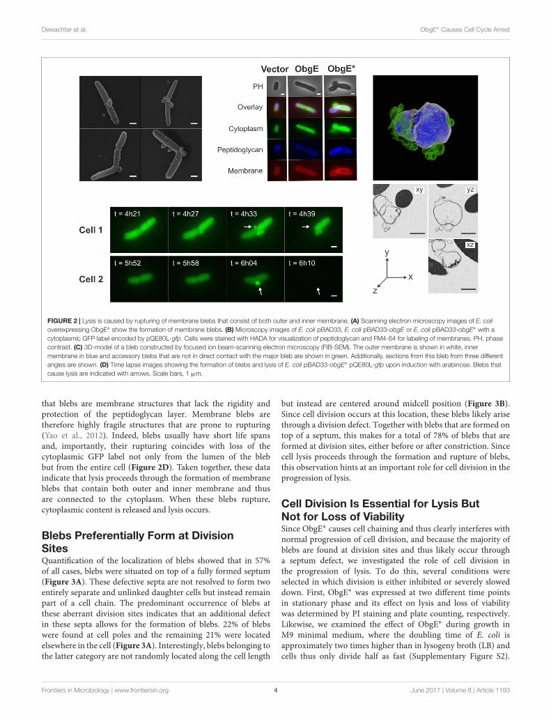
Lysis Proceeds through Formation ofMembrane BlebsA detailed study of E. coli morphology by scanning electronmicroscopy revealed that ObgE∗ expression leads to theformation of membrane protrusions, termed blebs (Figure 2A).Similar membrane structures were previously associated with celllysis (Yao et al., 2012; Sutterlin et al., 2016). The excess amount ofmembrane that forms blebs points to disturbance of membranehomeostasis by ObgE∗. To gain further structural insight intothe nature of these blebs, the cytoplasm, membranes andpeptidoglycan of E. coli expressing ObgE∗ were simultaneouslylabeled (Figure 2B). Cytoplasm was visualized by the expressionof a cytoplasmic GFP label, membranes were stained with thered lipophilic dye FM4-64, and peptidoglycan was visualizedusing HADA [HCC-amino-D-alanine, a fluorescently labeled D-amino acid that is readily incorporated into the peptides of
FIGURE 1 | Characterization of ObgE∗-mediated cell death.(A) Exponential-phase cultures of Escherichia coli pBAD33, E. colipBAD33-obgE or E. coli pBAD33-obgE∗ were induced at time 0. At severaltime points before and after induction, the number of viable cells wasdetermined by plate counting. Error bars represent the standard error of themean, n ≥ 3. (B) Time lapse observations of E. coli pBAD33-obgE∗ seededon a lysogeny broth (LB) agar pad containing the inducer of ObgE∗ expressionand propidium iodide (PI). Pictures were taken over a time course of 12 h.Scale bar, 1 µm. (C) Exponential-phase cultures of E. coli pBAD33, E. colipBAD33-obgE or E. coli pBAD33-obgE∗ were induced at time 0. At severaltime points after induction, cultures were stained with PI and the percentageof PI-negative and thus intact cells in the population was measured by flowcytometry. Data are represented as mean ± SEM, n ≥ 3. In every repeat100,000 cells were collected.
peptidoglycan (Kuru et al., 2015)]. No membrane blebs werefound in the vector control or E. coli expressing wild-type ObgE,although the latter did influence cell morphology by increasingcell length, in accordance with literature (Kobayashi et al., 2001;Dutkiewicz et al., 2002). Expression of ObgE∗ leads to theformation of membrane blebs that contain the cytoplasmic GFPlabel. The lumen of these blebs is therefore in direct contact withthe cytoplasm. Because of this continuum between cytoplasm andblebs, it is likely that they are lined by inner as well as outermembrane. The presence of inner membrane inside blebs wasconfirmed by construction of a 3D-image of blebs by focusedion beam-scanning electron microscopy (FIB-SEM), a techniquethat allows for high resolution imaging of a desired volume inthree dimensions by electron microcopy (Kizilyaprak et al., 2014)(Figure 2C). However, although blebs contain inner membrane,there is no clear defect in the underlying peptidoglycan layersince HADA labeling is uniform and uninterrupted at the siteof bleb formation. Any potential peptidoglycan defect allowingfor the protrusion of inner membrane should therefore berather small. Additionally, the composition of peptidoglycanremains unaltered in the presence of ObgE∗, arguing againstmajor rearrangements or disturbance of peptidoglycan structure(Supplementary Figure S1). FM4-64 and HADA staining revealed
Frontiers in Microbiology | www.frontiersin.org 3 June 2017 | Volume 8 | Article 1193
fmicb-08-01193 June 27, 2017 Time: 14:25 # 4
Dewachter et al. ObgE∗ Causes Cell Cycle Arrest
FIGURE 2 | Lysis is caused by rupturing of membrane blebs that consist of both outer and inner membrane. (A) Scanning electron microscopy images of E. colioverexpressing ObgE∗ show the formation of membrane blebs. (B) Microscopy images of E. coli pBAD33, E. coli pBAD33-obgE or E. coli pBAD33-obgE∗ with acytoplasmic GFP label encoded by pQE80L-gfp. Cells were stained with HADA for visualization of peptidoglycan and FM4-64 for labeling of membranes. PH, phasecontrast. (C) 3D-model of a bleb constructed by focused ion beam-scanning electron microscopy (FIB-SEM). The outer membrane is shown in white, innermembrane in blue and accessory blebs that are not in direct contact with the major bleb are shown in green. Additionally, sections from this bleb from three differentangles are shown. (D) Time lapse images showing the formation of blebs and lysis of E. coli pBAD33-obgE∗ pQE80L-gfp upon induction with arabinose. Blebs thatcause lysis are indicated with arrows. Scale bars, 1 µm.
that blebs are membrane structures that lack the rigidity andprotection of the peptidoglycan layer. Membrane blebs aretherefore highly fragile structures that are prone to rupturing(Yao et al., 2012). Indeed, blebs usually have short life spansand, importantly, their rupturing coincides with loss of thecytoplasmic GFP label not only from the lumen of the blebbut from the entire cell (Figure 2D). Taken together, these dataindicate that lysis proceeds through the formation of membraneblebs that contain both outer and inner membrane and thusare connected to the cytoplasm. When these blebs rupture,cytoplasmic content is released and lysis occurs.
Blebs Preferentially Form at DivisionSitesQuantification of the localization of blebs showed that in 57%of all cases, blebs were situated on top of a fully formed septum(Figure 3A). These defective septa are not resolved to form twoentirely separate and unlinked daughter cells but instead remainpart of a cell chain. The predominant occurrence of blebs atthese aberrant division sites indicates that an additional defectin these septa allows for the formation of blebs. 22% of blebswere found at cell poles and the remaining 21% were locatedelsewhere in the cell (Figure 3A). Interestingly, blebs belonging tothe latter category are not randomly located along the cell length
but instead are centered around midcell position (Figure 3B).Since cell division occurs at this location, these blebs likely arisethrough a division defect. Together with blebs that are formed ontop of a septum, this makes for a total of 78% of blebs that areformed at division sites, either before or after constriction. Sincecell lysis proceeds through the formation and rupture of blebs,this observation hints at an important role for cell division in theprogression of lysis.
Cell Division Is Essential for Lysis ButNot for Loss of ViabilitySince ObgE∗ causes cell chaining and thus clearly interferes withnormal progression of cell division, and because the majority ofblebs are found at division sites and thus likely occur througha septum defect, we investigated the role of cell division inthe progression of lysis. To do this, several conditions wereselected in which division is either inhibited or severely sloweddown. First, ObgE∗ was expressed at two different time pointsin stationary phase and its effect on lysis and loss of viabilitywas determined by PI staining and plate counting, respectively.Likewise, we examined the effect of ObgE∗ during growth inM9 minimal medium, where the doubling time of E. coli isapproximately two times higher than in lysogeny broth (LB) andcells thus only divide half as fast (Supplementary Figure S2).
Frontiers in Microbiology | www.frontiersin.org 4 June 2017 | Volume 8 | Article 1193

fmicb-08-01193 June 27, 2017 Time: 14:25 # 5
Dewachter et al. ObgE∗ Causes Cell Cycle Arrest
FIGURE 3 | Blebs are located at sites of cell division. (A) Quantitativelocalization of blebs in E. coli pBAD33-obgE∗. Cultures were induced in thepresence of 100 mM MgSO4 to increase bleb size and lifetime and thusimprove visibility under the microscope. Membranes were stained withFM4-64 before visualization. Data are represented as mean ± SEM, n = 3. Inevery repeat ± 200 blebs were counted. (B) Violin plot of the distribution ofblebs from the category ‘Other’ within the cell. To account for the randomdistribution of bleb formation in either the left hand or right hand side of thecell, all data points were duplicated and mirrored around midcell position.Scale bars, 1 µm.
However, since both conditions also have a large effect on generalmetabolism and other aspects of cell growth, the role of celldivision was also investigated more directly by expressing ObgE∗in the presence of either aztreonam or cephalexin, two antibioticsthat specifically inhibit division. They do so by mainly targetingPBP3 and thus inhibit the synthesis of septal peptidoglycan whilehaving no or very limited effects on cell elongation (Satta et al.,1995; Yao et al., 2012). It is likely that some of these conditionsaffect the expression level of ObgE∗ inside the cell. To accountfor this, lysis and loss of viability were first quantified understandard conditions at 25 different ObgE∗ concentrations. Thesedata were used to construct a correlation curve that reflects theextent of lysis and loss of viability at every possible concentrationof ObgE∗ within a certain range (Supplementary Figure S3). Inconditions that slow down or inhibit cell division, the percentageof intact cells is greatly increased and is much higher than whatis expected based on the expression level (Figure 4A). In fact,in all conditions where cell division is inhibited (i.e., ObgE∗expression in stationary phase or in the presence of aztreonamor cephalexin), the percentage of intact cells approaches 100%,indicating that virtually no lysis occurs anymore. Cell divisiontherefore appears to be essential for the progression of ObgE∗-mediated lysis, more specifically we postulate that a defect inseptal peptidoglycan metabolism is responsible for lysis. WhenObgE∗ is expressed in M9 minimal medium there is still adetectable amount of lysis. This is to be expected since celldivision and septal peptidoglycan synthesis still occur in minimalmedium, albeit with a lower frequency. To validate that itis division and/or septal peptidoglycan synthesis that causeslysis and not merely peptidoglycan synthesis in general, theamount of lysis was also measured in the presence of mecillinam.Mecillinam is an antibiotic that targets PBP2 and thus specificallyinhibits peptidoglycan synthesis for cell elongation while havingno effect on septal peptidoglycan synthesis (Satta et al., 1995).In the presence of this antibiotic, the percentage of intact cells is
exactly what is expected based on the expression level, confirmingthat it is indeed cell division that is necessary for lysis to occur(Supplementary Figure S4).
In addition to estimating lysis by PI staining, ObgE∗-mediatedtoxicity was also quantified by CFU counting. Since the latterassay is based on cell growth, it determines the viability of cellsinstead of the integrity of the membrane as PI staining does. Asshown in Figure 4B, expression of ObgE∗ in stationary phase,in M9 minimal medium or in the presence of either aztreonamor cephalexin has no effect on ObgE∗-mediated loss of viability.Measured values are located within the 99% prediction bandsand thus do not significantly differ from the expected value. Wetherefore conclude that, while cell division is essential for theprogression of lysis, it does not affect ObgE∗-mediated loss ofviability.
Lysis Is Not Caused by Amidases or theTol-Pal SystemThe cell chaining phenotype caused by ObgE∗ points to a defectin the separation of newly formed daughter cells. Moreover,at these unresolved division sites, the cytoplasmic membraneis able to penetrate the peptidoglycan layer to form blebsthat cause lysis. Both phenotypes could be caused by a faultyregulation of the amidases AmiA, AmiB, and AmiC. Althoughmany enzymes cooperate to achieve cell separation, they areseen as the main executioners of this final stage of cell division(Heidrich et al., 2002; Vollmer et al., 2008; Typas et al., 2011).In their absence, E. coli forms chains of cells up to 20 cellslong that are unable to separate (Heidrich et al., 2002). A lackof amidase activity thus causes cell chaining. Moreover, sinceamidases are peptidoglycan hydrolases, uncontrolled activitycould also cause excessive degradation of peptidoglycan, leadingto cell lysis. Another system of interest is the Tol-Pal proteincomplex that helps maintain a fixed distance between allthree envelope layers (Gerding et al., 2007; Gray et al., 2015).Disruption of this system leads to a moderate cell chainingphenotype in medium of low osmolarity. Moreover, in theabsence of a functional Tol-Pal complex, cells form outermembrane vesicles at division sites and cell poles (Gerdinget al., 2007). Because of the obvious similarities with phenotypesassociated with ObgE∗-mediated lysis, we quantified lysis in anE. coli triple amidase knock-out strain (E. coli 1amiA 1amiB1amiC) and in single-gene knock-outs of the tolQ or tolRgenes (Figures 5A,B). In all three strains, membrane integrityis decreased under control conditions; whereas 99.6% of awild-type population is intact, this value drops to 63.6% bydeletion of amiA, amiB, and amiC, and is lowered to 93.8 and97.1% in the 1tolQ and 1tolR strain, respectively (Figure 5A,Vector). Surprisingly, overexpression of the essential ObgEprotein further decreases membrane integrity of these knock-outstrains. Upon expression of ObgE, integrity decreases by 5.1%in the triple amidase knock-out strain (p = 0.0322), 5.7% inthe 1tolQ strain (p = 0.0471), and 6.2% in the 1tolR strain(p = 0.0305). Since ObgE has no effect on the integrity of awild-type strain (p = 0.999), these remarkable results hint ata functional link between ObgE, the amidases and the Tol-Palcomplex.
Frontiers in Microbiology | www.frontiersin.org 5 June 2017 | Volume 8 | Article 1193

fmicb-08-01193 June 27, 2017 Time: 14:25 # 6
Dewachter et al. ObgE∗ Causes Cell Cycle Arrest
FIGURE 4 | Cell division is necessary for lysis but does not affect loss of viability. (A) Correlation curve showing the expected fraction of intact cells of E. colipBAD33-obgE∗ in function of the intracellular ObgE∗ concentration. (B) Correlation curve showing the expected level of viability of E. coli pBAD33-obgE∗ incomparison to E. coli pBAD33-obgE in function of the intracellular ObgE∗ concentration. Colored data points were collected from conditions that inhibit or slowdown cell division. ObgE∗ concentration was determined by measuring fluorescence of an ObgE∗-Venus fusion by flow cytometry. Gray bands around the expectedvalue represent 99% prediction intervals. Data are represented as mean ± SEM, n ≥ 3, error bars are mostly too small to be visible.
ObgE∗ expression still has a very strong negative effect on cellintegrity in all three deletion mutants. The amount of ObgE∗-mediated lysis varies slightly in the different strains, owing to thefact that the baseline of membrane integrity is affected by thegenomic deletions. When the amount of lysis caused by ObgE∗is normalized to the amount of lysis detected when wild-typeObgE is expressed, no significant differences in the percentageof intact cells were found among the three deletion strains(Figure 5B). Since lysis upon ObgE∗ expression does not decreasein the absence of AmiA, AmiB, and AmiC or in the absence ofa functional Tol-Pal complex, we conclude that none of theseproteins directly cause ObgE∗-mediated lysis.
ObgE∗ Causes Loss of Viability byIrreversible Inhibition of the Cell CycleWhen lysis is prevented, ObgE∗ expression still decreases cellviability (Figure 4). To determine how ObgE∗ does so, theresumption of growth after ObgE∗ expression was monitoredby time lapse microscopy. To separate the lysis and loss ofviability phenotypes, ObgE∗ was expressed in stationary phasewhere lysis is prevented. Afterward, cells were seeded on anLB agarose pad lacking the inducer of ObgE∗ expression andincubated to allow cell growth. These conditions thus mimicthe plate counting experiments in which a loss of viability isdetected. In the control experiment where wild-type ObgE wasexpressed during stationary phase, cells rapidly resume growthafter they are transferred to fresh medium. They quickly startto divide and form colonies (Figure 6A). When ObgE∗ wasexpressed during stationary phase, plate counting experimentsdemonstrated that there is a drop of 6 log units in colonyformation (Figure 1A). This time lapse experiment showed thatcolonies fail to develop because cell division cannot be restoredwhen cells are allowed to recover on fresh medium (Figure 6A).Even after 48 h, cells have not divided (data not shown). However,bacteria do appear to resume cell growth briefly (Figure 6A,inset). Quantification of cell length at the start of the time lapseand 6 h later revealed that, during this time, cell length increases
to a limited extent (Figure 6B). ObgE∗ thus causes loss of viabilityby blocking progression through the cell cycle after allowing alimited amount of cell elongation but before cell division occurs.Importantly, this cell cycle arrest is not specific to exit fromstationary phase and/or elevated (p)ppGpp levels. When ObgE∗was expressed in exponential phase in the presence of aztreonamto prevent cell lysis, cell division could not be restored once cellswere washed and resuspended in fresh medium (SupplementaryFigure S5). Eventually, the arrested cells lose membrane integrity,as evidenced by gradually progressive PI staining. No membraneblebs were observed (data not shown).
Because of the role of ObgE in licensing chromosomesegregation and subsequent cell division (Kobayashi et al.,2001; Foti et al., 2007), we checked whether ObgE∗ causes cellcycle arrest at the stage of chromosome segregation. To thisend, the distribution of DNA inside the cell was monitoredby a fluorescent fusion of venus to the hupA subunit of thegeneral nucleoid-associated HU protein. Results indicate thatchromosome segregation is not prevented by ObgE∗ (Figure 6C).One hour after transfer from stationary phase to fresh medium,chromosome segregation has occurred in approximately 42%of all cells. After this point, segregated nucleoids slowlystart to reunite and spread throughout the cell (Figure 6D).However, even cells with segregated nucleoids fail to divide. Thisexperiment demonstrates that it is not a defect in chromosomesegregation per se that inhibits further progression through thecell cycle. We thus conclude that ObgE∗ interferes with anotherstage of the cell cycle. This point of interference is located aftercompletion of chromosome segregation and inhibits the onset ofcell division.
DISCUSSION
In this study, we describe the effect of a dominant-negativemutation in the essential GTPase ObgE on E. coli. Expressionof the mutant ObgE∗ protein halts the cell cycle. This cell cycle
Frontiers in Microbiology | www.frontiersin.org 6 June 2017 | Volume 8 | Article 1193

fmicb-08-01193 June 27, 2017 Time: 14:25 # 7
Dewachter et al. ObgE∗ Causes Cell Cycle Arrest
FIGURE 5 | Amidases and the Tol-Pal system do not directly causeObgE∗-mediated lysis. (A) Induced cultures of E. coli (wt), E. coli 1amiA1amiB 1amiC (1amiABC), E. coli 1tolQ and E. coli1tolR with plasmidspBAD33, pBAD33-obgE or pBAD33-obgE∗ were stained with PI and thepercentage of PI-negative and thus intact cells in the population wasmeasured by flow cytometry. (B) The percentage of intact cells upon ObgE∗
expression was divided by the fraction of intact cells upon expression of ObgEto correct for the differences in baseline levels of membrane integrity indifferent strains. Data are represented as mean ± SEM, n ≥ 3. In every repeat100,000 cells were collected (one-way ANOVA, Bonferroni correction:∗p < 0.05, ∗∗∗∗p < 0.0001).
arrest occurs after completion of chromosome segregation butbefore the onset of constriction for cell division. It is irreversibleand therefore leads to cell death. Under conditions of rapid celldivision at the time of ObgE∗ expression, cell death is associatedwith cell chain formation and lysis.
Since lysis can be prevented without affecting the loss ofviability caused by ObgE∗, it is not a prerequisite for ObgE∗-mediated cell death. The underlying cause of cell death ratherstems from ObgE∗’s capability to irreversibly halt the cellcycle and thus ultimately prevent cell division and colonyformation. However, it is currently unclear at which pointObgE∗ halts the cell cycle to prevent the formation of daughtercells. Results indicate that ObgE∗ does not merely inhibitchromosome segregation, since a considerable fraction of thepopulation displayed separated nucleoids without initiatingcell division. Previously, filamentation by either depletion oroverexpression of ObgE was suggested to be coupled to a defectin chromosome segregation (Kobayashi et al., 2001; Foti et al.,2007). However, our data show that cell division is prevented evenwhen chromosomes successfully segregate, suggesting that the
observed ObgE-related phenotypes, filamentation and aberrantchromosome segregation, might not be as tightly linked aspreviously thought. Neither does ObgE∗ inhibit cell growthin general, since limited elongation could be observed afterObgE∗ expression. However, it also seems unlikely that ObgE∗inhibits cell division by directly affecting divisome activity, sincethe amount of elongation that occurs is not consistent withthis possibility. When cell growth is halted by ObgE∗, mediancell length has increased by 0.36 µm, which is only ±16%of the median cell length. At this point, a mature divisomemost likely has not assembled since cell size approximatelydoubles before constriction occurs (Chien et al., 2012). Moreover,specific inhibition of divisome activity, for example by treatmentwith aztreonam or cephalexin, allows for more extensive cellelongation resulting in filaments that are much longer than theones formed by cells that have expressed ObgE∗ (Vollmer andBertsche, 2008; Yao et al., 2012). At which point does ObgE∗then block the cell cycle? Interference must occur betweenthe completion of chromosome segregation and the onset ofconstriction. A vital process that occurs during this time is theformation of the proto-ring, and thus the localization of FtsZ,at midcell. Interestingly, FtsZ localization was previously shownto be influenced by ObgE (Foti et al., 2007). Upon recruitmentof FtsZ to the site of cell division, the mode of peptidoglycangrowth switches from dispersed growth to preseptal elongation(Aarsman et al., 2005; Aaron et al., 2007; Vollmer and Bertsche,2008; Olrichs et al., 2011; Typas et al., 2011). During the former,cells elongate through insertion of peptidoglycan precursorsalong the sidewalls. This insertion is directed by MreB andmediated by PBP1A and PBP2 (Vollmer and Bertsche, 2008;Typas et al., 2011). When FtsZ and ZipA localize at midcell,however, insertion of new peptidoglycan is redirected to midcellposition (de Pedro et al., 1997; Aaron et al., 2007; Olrichset al., 2011; Potluri et al., 2012). This preseptal peptidoglycansynthesis, also known as PIPS (PBP3-independent peptidoglycansynthesis), forms the transition between elongation and divisionand is independent of MreB, PBP1A and other elongation-related proteins (Potluri et al., 2012). Since elongation canoccur in the presence of ObgE∗, although much more limitedthan expected, it is possible that ObgE∗ interferes with onlyone of both independent elongation modes while allowing theother to proceed normally. Because of the FtsZ dependency ofpreseptal growth and the influence of ObgE on FtsZ localization(de Pedro et al., 1997; Foti et al., 2007; Olrichs et al., 2011;Potluri et al., 2012), it appears more likely that ObgE∗ interfereswith this phase of cell elongation. However, it should benoted that although preseptal elongation greatly contributesto cell elongation in Caulobacter crescentus, it only has alimited effect on E. coli cell length (de Pedro et al., 1997;Aaron et al., 2007; Olrichs et al., 2011; Potluri et al., 2012).Regardless of which step is affected, it is clear that cell divisionis ultimately inhibited. This inhibition is irreversible and canconsequently explain the loss of viability observed in the presenceof ObgE∗.
Whether or not ObgE∗-mediated cell cycle arrest is associatedwith cell chain formation and lysis, depends on growthconditions at the time of ObgE∗ expression. When ObgE∗ is
Frontiers in Microbiology | www.frontiersin.org 7 June 2017 | Volume 8 | Article 1193

fmicb-08-01193 June 27, 2017 Time: 14:25 # 8
Dewachter et al. ObgE∗ Causes Cell Cycle Arrest
FIGURE 6 | ObgE∗ causes irreversible cell cycle arrest. (A) After ObgE or ObgE∗ was expressed in stationary phase, cells were seeded on an agarose pad withoutthe inducer of expression. Resumption of growth on the pad was monitored by time lapse microscopy. Insets are enlargements indicated by red lines. (B) Violin plotand box plot of the cell length of E. coli pBAD33-obgE∗ at the start of the time lapse experiment and 6 h later. Cell length was quantified in ±220 cells spread overthree independent repeats. (C) After ObgE∗ was expressed in E. coli hupA-venus in stationary phase, cells were seeded on an agarose pad without the inducer ofexpression. Chromosome segregation was monitored by time lapse microscopy. (D) Quantitative analysis of chromosome segregation in E. coli hupA-venuspBAD33Gm-obgE∗ after induction in stationary phase. Data are represented as mean ± SEM, n = 4. In every repeat ± 100 cells were analyzed. Scale bars, 10 µm.
expressed during exponential-phase growth in rich medium, cellchaining occurs through a defect in cell separation. Usually,these cell chains consist of 2–4 cells (data not shown), indicatingthat 1 or 2 rounds of defective cell division have occurredbefore complete cell cycle arrest by ObgE∗ is activated. Whydo these exponential-phase cells not immediately halt their cellcycle? We hypothesize that, depending on the stage of the cellcycle at the time of ObgE∗ expression, cells may already becommitted to executing cell division even in the presence ofthe cell cycle inhibitor ObgE∗. Although no research has beendone as to the nature of this decision point in the cell cycle,initiation of DNA replication is an attractive candidate. Notonly is ObgE involved in the regulation of replication initiation(Ulanowska et al., 2003; Foti et al., 2005; Sikora et al., 2006),but the number of cells found in cell chains also correspondsto one or two rounds of replication being active at the timeof ObgE∗ expression, which is the case in exponential-phasecells grown in rich medium (Foti et al., 2005; Nielsen et al.,2007). In eukaryotic cells, such early commitment to cell divisionindeed exists and is referred to as the restriction point. If, at
the restriction point, conditions are favorable, eukaryotic cellswill initiate a new cell cycle, proceed to S phase and replicatetheir genomes. These cells are subsequently obliged to divideeven if favorable conditions come to an end (Rhind and Russell,2012).
The lethal commitment to cell division during cell cyclearrest imposed by ObgE∗ results in cell lysis. Lysis proceedsthrough the formation and rupturing of membrane blebs atdivision sites, implicating the presence of excess membrane uponObgE∗ expression. These membrane structures are filled withcytoplasmic content and are lined by both outer and innermembrane. The inner membrane therefore is able to penetratethe peptidoglycan layer, hinting at a defect in this part of thecell envelope. ObgE∗ thus appears to lead to peptidoglycandefects and excess membrane. Interestingly, both conditionscan induce the transition from rod-shaped bacteria to L-forms,wall-less bacteria able to proliferate independently of the FtsZ-based division machinery (Mercier et al., 2013, 2014). Thenature of the peptidoglycan defect caused by ObgE∗ is currentlyunknown. Fluorescent HADA labeling did not reveal an apparent
Frontiers in Microbiology | www.frontiersin.org 8 June 2017 | Volume 8 | Article 1193
fmicb-08-01193 June 27, 2017 Time: 14:25 # 9
Dewachter et al. ObgE∗ Causes Cell Cycle Arrest
breach in peptidoglycan at the site of bleb formation. Moreover,peptidoglycan composition remains unaltered in the presenceof ObgE∗. These data argue against large-scale peptidoglycandegradation but cannot exclude small and localized effects. Thepossibility of ObgE∗ causing localized peptidoglycan defects iscorroborated by the fact that blebs do not form randomly,but instead are preferentially localized at sites of cell division.Conclusive evidence demonstrating the tight link between celldivision and formation of blebs that cause lysis was providedby monitoring lysis in the absence of cell division. Underthese conditions, lysis is prevented completely. Even whendivision is blocked after divisome assembly by addition of theFtsI-inhibitors, aztreonam or cephalexin, lysis does not occur.We can therefore trace back the timing of bleb formationand lysis to the third and final stage of cell division, whichconsists of active constriction, septal peptidoglycan synthesisand cell separation. Overall, our results indicate that cells thatare committed to executing cell division in the presence ofObgE∗ experience a defect in correct peptidoglycan metabolismduring cell constriction. The integrity of the septum is therebydisrupted, causing bleb formation and subsequent lysis. As aside note, blocking cell division rescues all cells from undergoingcell lysis, while approximately 80% of blebs are found atdivision sites. Since the remaining 20% of blebs is localizedat cell poles, we speculate that these are new poles that wereformed very early after induction of ObgE∗ expression. Atthis time, cells are presumably still able to separate, althougha peptidoglycan defect that causes bleb formation has alreadyoccurred during septum formation. The newly formed cell polesare therefore fragile and will allow bleb formation and subsequentlysis.
In search of systems that function during division andthat can potentially contribute to cell lysis, we turned to theamidases AmiA, AmiB, and AmiC and the Tol-Pal system byinvestigating lysis in selected deletion mutants. Intriguingly, forthe first time, a link was observed between wild-type ObgEand the amidases and between wild-type ObgE and the Tol-Pal system. In the absence of amidases or upon disturbanceof the Tol-Pal complex, cells become sensitive toward ObgEexpression and lose their membrane integrity. Apart from thepreviously published effect on FtsZ (Foti et al., 2007), we thusreport two additional links between ObgE and the divisionmachinery. However, the amount of lysis upon ObgE∗ expressionis not altered in the selected deletion strains, indicating thatObgE∗ does not cause lysis by disturbing the link that existsbetween wild-type ObgE and amidases or Tol-Pal. This alsomeans that although AmiA, AmiB, and AmiC and the Tol-Pal system are attractive candidates to mediate lysis during celldivision, they are not the main executioners of lysis caused byObgE∗.
In conclusion, we here present evidence for the involvement ofObgE in the regulation of the E. coli cell cycle, more specificallyat the stage of cell division. Apart from the direct link wefound between ObgE and the amidases AmiA, AmiB, and AmiCand between ObgE and the Tol-Pal complex, we additionallyobserved irreversible cell cycle arrest upon expression of amutant ObgE isoform. Cell cycle arrest occurs after completion
of chromosome segregation but before the onset of constrictionand thus likely affects an early event in the preparation forcell division. This phenotype highly resembles that of an Eramutant (Britton et al., 1998). Like Obg, Era is an essentialand widely conserved GTPase, which was suggested to act as agrowth-rate regulated checkpoint of the E. coli cell cycle (Brittonet al., 1998). Likewise, it was previously suggested that wild-type ObgE serves as a bacterial cell cycle checkpoint that canblock or allow further progression through the cell cycle basedon its nucleotide occupancy (Datta et al., 2004; Foti et al., 2005;Dewachter et al., 2016b). Because of the central position ofObgE in cellular metabolism and its previously observed effecton cell division, ObgE is an attractive candidate to monitorseveral different cellular processes and adjust the progressionof the cell cycle accordingly. The presence of the mutation inObgE∗ blocks the cell cycle indefinitely, regardless of any cellularinput, and thus appears to disturb the function of wild-typeObgE in this process. If true, this mutant can be used as avaluable tool to gain more insight into the bacterial cell cycleand its regulation. Importantly, since Obg is widely conserved,including in eukaryotes, our findings might be applicable to otherorganisms as well.
MATERIALS AND METHODS
Strains, Plasmids and Growth ConditionsExperiments were performed with E. coli K-12 BW25113,except for data shown in Supplementary Figure S3. Single-gene knock-out strains were obtained from the Keio collection(Baba et al., 2006). The E. coli BW25113 1amiABC strainwas constructed by sequentially replacing the amiA, amiB, andamiC genes with a kanamycin resistance cassette by phagetransduction from HSC085, HSC078, and HSC071, respectively(Chung et al., 2009). After replacement of the amiA and amiBgenes, the resistance cassette was removed by FLP-mediatedrecombination prior to the subsequent round of gene deletion.Knock-out of the correct genes was confirmed by PCR. E. coliBW25113 hupA-venus was constructed by phage transductionstarting from E. coli MG1655 hupA-venus-CmR (Abram Aertsen,KU Leuven, personal communication). The chloramphenicolresistance cassette was excised by FLP-mediated recombination.
For all tests, unless indicated otherwise, overnight cultureswere diluted 100 times in LB containing the appropriateantibiotics (ampicillin 100 µg/ml, chloramphenicol 35 µg/ml,gentamicin 25 µg/ml, and kanamycin 40 µg/ml) and incubatedat 37◦C with continuous shaking at 200 rpm. When theOD595 nm reached 0.4, expression from pBAD/His A (Figure 2A),pBAD33Gm (Figures 6C,D, for construction see supplementalexperimental procedures) or pBAD33 (all other figures) wasinduced with arabinose (0.2% w/v). Expression of GFP frompQE80L-gfp (Orman and Brynildsen, 2015) was induced byIPTG (1 mM) at the same time. An alternative protocol wasfollowed to investigate the effect of cell division on toxicity;induction was either postponed until one of two possiblepoints in stationary phase (10 or 16 h after dilution of theovernight culture), carried out in M9 medium with casamino
Frontiers in Microbiology | www.frontiersin.org 9 June 2017 | Volume 8 | Article 1193

fmicb-08-01193 June 27, 2017 Time: 14:25 # 10
Dewachter et al. ObgE∗ Causes Cell Cycle Arrest
acids (1% w/v) as carbon source, or simultaneously aztreonam(0.2 µg/ml), cephalexin (50 µg/ml) or mecillinam (0.64 µg/ml)were added to the cultures. The concentration of these antibioticsis situated between the minimal inhibitory concentration (MIC)and minimal bactericidal concentration (MBC) (SupplementaryTable S1), so as to inhibit cell growth without causing cell death.
Cell Viability AssaySerial dilutions were prepared in 10 mM MgSO4 and platedon medium containing 1.5 % agar. After overnight incubationat 37◦C, CFUs were determined and, if needed, the percentageviability was calculated by dividing the number of CFUs per mlobtained after ObgE∗ expression by the number of CFUs per mlafter ObgE expression.
Lysis AssayAt indicated time points or – when no time point was specified –6 h after induction with arabinose, cultures were diluted 10(exponential-phase cultures) or 100 (stationary-phase cultures)times in PBS containing 0.03 µM PI and incubated in the darkat room temperature for 15 min. The fraction of the populationthat stained PI-positive was measured by flow cytometry with aBD Influx cell sorter equipped with 488 and 561 nm lasers andstandard filter sets. Flow cytometry experiments were repeated ntimes. In each repeat, 100,000 cells were collected.
ObgE∗ ConcentrationTo determine the intracellular ObgE∗ concentration in severalconditions, a translational ObgE∗-Venus fusion (Dewachter et al.,2016a) was expressed and Venus fluorescence was measured byflow cytometry. Presence of the Venus tag did not influencetoxicity in any way (data not shown).
MicroscopyTo simultaneously visualize blebs, cytoplasm, membranes andpeptidoglycan (Figure 2B), 0.5 mM HADA was added to thecultures at the time of induction with arabinose. At this timeGFP expression was also induced by adding 1 mM IPTG. Twohours later, cultures were washed twice with PBS to removeunincorporated HADA and pellets were dissolved in LB mediumcontaining the appropriate antibiotics, 0.2% arabinose, 1 mMIPTG, and 100 mM MgSO4 to stabilize blebs. Cultures wereincubated for another 2 h to allow bleb formation and were thenstained with 10 µg/ml FM4-64. After 10 min incubation in thedark at room temperature cells were visualized on a poly-L-lysinecoated glass slide.
Visualization of membrane blebs in E. coli pBAD33-obgE∗ wasperformed by adding 100 mM MgSO4 at the time of inductionand staining cells with 10 µg/ml FM4-64 4 h later (Figure 3A).MgSO4 was added to stabilize blebs and thus increase theirlifetime, as was done previously (Yao et al., 2012).
For the time lapse microscopy experiment showing PI staining(Figure 1B), an overnight culture of E. coli pBAD33-obgE∗ wasseeded on an LB agarose pad (2% w/v) containing the appropriateantibiotic, 0.2% arabinose and 0.06 µM PI. Cells were incubatedat 37◦C and growth was monitored for 12 h.
The time lapse microscopy experiment with a cytoplasmicGFP label (Figure 2D) was performed with E. coli pBAD33-obgE∗pQE80L-gfp. An overnight culture of this strain was seeded onan LB agarose pad containing the appropriate antibiotics, 0.2%arabinose and 1 mM IPTG. Cells were incubated at 37◦C andgrowth was monitored for 12 h.
Resumption of growth and chromosome segregation afterObgE or ObgE∗ expression in stationary phase (Figures 6A,C)was monitored in a time lapse experiment where overnightcultures of E. coli pBAD33-obgE and E. coli pBAD33-obgE∗or E. coli hupA-venus pBAD33Gm-obgE∗ were diluted andgrown for 16 h into stationary phase. Expression was theninduced by adding 0.2% arabinose for 2 h after which cells wereseeded on LB agarose pads containing the appropriate antibioticswithout arabinose. Cells were incubated at 37◦C and growth wasmonitored for 12–48 h. Cell length or segregation of fluorescentfoci were quantified by MicrobeJ (Ducret et al., 2016).
For all microscopy experiments, cells were imaged by aNikon Ti-E inverted microscope with Qi2 CMOS camera andtemperature controlled cage incubator.
Scanning Electron MicroscopyScanning electron microscopy of E. coli WM2949 1recApBAD/His A-obgE∗ was performed as described previously(Dewachter et al., 2015).
Focused Ion Beam-Scanning ElectronMicroscopy (FIB-SEM)Focused ion beam-scanning electron microscopy was performedas described previously (Vanwalleghem et al., 2015).
Statistical AnalysisAll statistical analyses were performed with GraphPad Prism 6.Normality of representative data from CFU counts (n = 30) andPI staining (n = 15) was verified by D’Agostino and Pearsonomnibus normality test. The different samples were comparedusing one-way ANOVA with p-values obtained from Bonferroni’smultiple comparisons test.
AUTHOR CONTRIBUTIONS
Conceptualization, LD, NV, DP-M, WV, MF, and JM;Methodology, LD, NV, MF, and JM; Formal analysis, LD;Investigation, LD, MJ, TV, JB, DM; Writing – original draft, LD;Writing – Review and Editing, LD, NV, DP-M, KV, WV, MF, andJM; Visualization, LD; Supervision, NV, MF, and JM.
FUNDING
LD received a fellowship from the Fund for Scientific Research,Flanders (FWO). This work was supported by grants fromthe FWO (G.0471.12N, G.0B25.15N, 1522214N), KUL-BOF(CREA/13/019) and the Interuniversity Attraction Poles-BelgianScience Policy Office IAP-BELSPO (IAP P7/28). WV wassupported by the Wellcome Trust (101824/Z/13/Z).
Frontiers in Microbiology | www.frontiersin.org 10 June 2017 | Volume 8 | Article 1193

fmicb-08-01193 June 27, 2017 Time: 14:25 # 11
Dewachter et al. ObgE∗ Causes Cell Cycle Arrest
ACKNOWLEDGMENTS
We thank Daniel Kahne (Harvard University) for providingus with the HSC085, HSC078, and HSC071 strains; AbramAertsen (KU Leuven) for the E. coli MG1655 hupA-venus-CmR strain; and Michael VanNieuwenhze and Yves V.Brun (Indiana University) for the HADA dye. FIB-SEMwas performed by Anneke Kremer at the Bio ImagingCore facility of UGhent, Belgium and Frédéric Fontaine
helped us with image treatment, segmentation and 3Dreconstruction.
SUPPLEMENTARY MATERIAL
The Supplementary Material for this article can be foundonline at: http://journal.frontiersin.org/article/10.3389/fmicb.2017.01193/full#supplementary-material
REFERENCESAaron, M., Charbon, G., Lam, H., Schwarz, H., Vollmer, W., and Jacobs-Wagner, C.
(2007). The tubulin homologue FtsZ contributes to cell elongation by guidingcell wall precursor synthesis in Caulobacter crescentus. Mol. Microbiol. 64,938–952. doi: 10.1111/j.1365-2958.2007.05720.x
Aarsman, M. E., Piette, A., Fraipont, C., Vinkenvleugel, T. M., Nguyen-Disteche, M., and Den Blaauwen, T. (2005). Maturation of the Escherichia colidivisome occurs in two steps. Mol. Microbiol. 55, 1631–1645. doi: 10.1111/j.1365-2958.2005.04502.x
Adams, D. W., and Errington, J. (2009). Bacterial cell division: assembly,maintenance and disassembly of the Z ring. Nat. Rev. Microbiol. 7, 642–653.doi: 10.1038/nrmicro2198
Baba, T., Ara, T., Hasegawa, M., Takai, Y., Okumura, Y., Baba, M., et al. (2006).Construction of Escherichia coli K-12 in-frame, single-gene knockout mutants:the Keio collection. Mol. Syst. Biol. 2:2006.0008. doi: 10.1038/msb4100050
Bertsche, U., Kast, T., Wolf, B., Fraipont, C., Aarsman, M. E., Kannenberg, K., et al.(2006). Interaction between two murein (peptidoglycan) synthases, PBP3 andPBP1B, in Escherichia coli. Mol. Microbiol. 61, 675–690. doi: 10.1111/j.1365-2958.2006.05280.x
Bisson-Filho, A. W., Hsu, Y. P., Squyres, G. R., Kuru, E., Wu, F., Jukes, C.,et al. (2017). Treadmilling by FtsZ filaments drives peptidoglycan synthesis andbacterial cell division. Science 355, 739–743. doi: 10.1126/science.aak9973
Britton, R. A., Powell, B. S., Dasgupta, S., Sun, Q., Margolin, W., Lupski, J. R.,et al. (1998). Cell cycle arrest in Era GTPase mutants: a potential growth rate-regulated checkpoint in Escherichia coli. Mol. Microbiol. 27, 739–750. doi: 10.1046/j.1365-2958.1998.00719.x
Buglino, J., Shen, V., Hakimian, P., and Lima, C. D. (2002). Structural andbiochemical analysis of the Obg GTP binding protein. Structure 10, 1581–1592.doi: 10.1016/S0969-2126(02)00882-1
Cascales, E., Lloubes, R., and Sturgis, J. N. (2001). The TolQ-TolR proteins energizeTolA and share homologies with the flagellar motor proteins MotA-MotB. Mol.Microbiol. 42, 795–807. doi: 10.1046/j.1365-2958.2001.02673.x
Chen, J. C., and Beckwith, J. (2001). FtsQ, FtsL and FtsI require FtsK, but notFtsN, for co-localization with FtsZ during Escherichia coli cell division. Mol.Microbiol. 42, 395–413. doi: 10.1046/j.1365-2958.2001.02640.x
Chien, A. C., Hill, N. S., and Levin, P. A. (2012). Cell size control in bacteria. Curr.Biol. 22, R340–R349. doi: 10.1016/j.cub.2012.02.032
Chung, H. S., Yao, Z., Goehring, N. W., Kishony, R., Beckwith, J., and Kahne, D.(2009). Rapid beta-lactam-induced lysis requires successful assembly of thecell division machinery. Proc. Natl. Acad. Sci. U.S.A. 106, 21872–21877.doi: 10.1073/pnas.0911674106
Datta, K., Skidmore, J. M., Pu, K., and Maddock, J. R. (2004). The Caulobactercrescentus GTPase CgtAC is required for progression through the cell cycle andfor maintaining 50S ribosomal subunit levels. Mol. Microbiol. 54, 1379–1392.doi: 10.1111/j.1365-2958.2004.04354.x
de Pedro, M. A., Quintela, J. C., Holtje, J. V., and Schwarz, H. (1997). Mureinsegregation in Escherichia coli. J. Bacteriol. 179, 2823–2834. doi: 10.1128/jb.179.9.2823-2834.1997
Dewachter, L., Herpels, P., Verstraeten, N., Fauvart, M., and Michiels, J. (2016a).Reactive oxygen species do not contribute to ObgE∗-mediated programmed celldeath. Sci. Rep. 6:33723. doi: 10.1038/srep33723
Dewachter, L., Verstraeten, N., Fauvart, M., and Michiels, J. (2016b). The bacterialcell cycle checkpoint Obg and its role in programmed cell death. Microb. Cell 3,255–256. doi: 10.15698/mic2016.06.507
Dewachter, L., Verstraeten, N., Monteyne, D., Kint, C. I., Versées, W., Morga,D. P., et al. (2015). A single-amino-acid substitution in Obg activates anew programmed cell death pathway in Escherichia coli. mBio 6:e01935-15.doi: 10.1128/mBio.01935-15
Ducret, A., Quardokus, E. M., and Brun, Y. V. (2016). MicrobeJ, a tool for highthroughput bacterial cell detection and quantitative analysis. Nat. Microbiol.1:16077. doi: 10.1038/nmicrobiol.2016.77
Dutkiewicz, R., Slominska, M., Wegrzyn, G., and Czyz, A. (2002). Overexpressionof the cgtA (yhbZ, obgE) gene, coding for an essential GTP-binding protein,impairs the regulation of chromosomal functions in Escherichia coli. Curr.Microbiol. 45, 440–445. doi: 10.1007/s00284-002-3713-x
Egan, A. J., and Vollmer, W. (2013). The physiology of bacterial cell division. Ann.N. Y. Acad. Sci. 1277, 8–28. doi: 10.1111/j.1749-6632.2012.06818.x
Erickson, H. P., Anderson, D. E., and Osawa, M. (2010). FtsZ in bacterialcytokinesis: cytoskeleton and force generator all in one. Microbiol. Mol. Biol.Rev. 74, 504–528. doi: 10.1128/MMBR.00021-10
Feng, B., Mandava, C. S., Guo, Q., Wang, J., Cao, W., Li, N., et al. (2014). Structuraland functional insights into the mode of action of a universally conserved ObgGTPase. PLoS Biol. 12:e1001866. doi: 10.1371/journal.pbio.1001866
Foti, J. J., Persky, N. S., Ferullo, D. J., and Lovett, S. T. (2007). Chromosomesegregation control by Escherichia coli ObgE GTPase. Mol. Microbiol. 65,569–581. doi: 10.1111/j.1365-2958.2007.05811.x
Foti, J. J., Schienda, J., Sutera, V. A. Jr., and Lovett, S. T. (2005). A bacterialG protein-mediated response to replication arrest. Mol. Cell 17, 549–560.doi: 10.1016/j.molcel.2005.01.012
Gerding, M. A., Liu, B., Bendezu, F. O., Hale, C. A., Bernhardt, T. G., and De Boer,P. A. (2009). Self-enhanced accumulation of FtsN at division sites and roles forother proteins with a SPOR domain (DamX, DedD, and RlpA) in Escherichiacoli cell constriction. J. Bacteriol. 191, 7383–7401. doi: 10.1128/JB.00811-09
Gerding, M. A., Ogata, Y., Pecora, N. D., Niki, H., and De Boer, P. A. (2007).The trans-envelope Tol-Pal complex is part of the cell division machinery andrequired for proper outer-membrane invagination during cell constriction inE. coli. Mol. Microbiol. 63, 1008–1025. doi: 10.1111/j.1365-2958.2006.05571.x
Gkekas, S., Singh, R. K., Shkumatov, A. V., Messens, J., Fauvart, M., Verstraeten, N.,et al. (2017). Structural and biochemical analysis of Escherichia coli ObgE,a central regulator of bacterial persistence. J. Biol. Chem. 292, 5871–5883.doi: 10.1074/jbc.M116.761809
Gray, A. N., Egan, A. J., Van’t Veer, I. L., Verheul, J., Colavin, A., Koumoutsi, A.,et al. (2015). Coordination of peptidoglycan synthesis and outer membraneconstriction during Escherichia coli cell division. eLife 4:e07118. doi: 10.7554/eLife.07118
Haeusser, D. P., and Margolin, W. (2016). Splitsville: structural and functionalinsights into the dynamic bacterial Z ring. Nat. Rev. Microbiol. 14, 305–319.doi: 10.1038/nrmicro.2016.26
Heidrich, C., Templin, M. F., Ursinus, A., Merdanovic, M., Berger, J., Schwarz, H.,et al. (2001). Involvement of N-acetylmuramyl-L-alanine amidases in cellseparation and antibiotic-induced autolysis of Escherichia coli. Mol. Microbiol.41, 167–178. doi: 10.1046/j.1365-2958.2001.02499.x
Heidrich, C., Ursinus, A., Berger, J., Schwarz, H., and Holtje, J. V. (2002). Effectsof multiple deletions of murein hydrolases on viability, septum cleavage,and sensitivity to large toxic molecules in Escherichia coli. J. Bacteriol. 184,6093–6099. doi: 10.1128/JB.184.22.6093-6099.2002
Jiang, M., Datta, K., Walker, A., Strahler, J., Bagamasbad, P., Andrews, P. C., et al.(2006). The Escherichia coli GTPase CgtAE is involved in late steps of largeribosome assembly. J. Bacteriol. 188, 6757–6770. doi: 10.1128/JB.00444-06
Frontiers in Microbiology | www.frontiersin.org 11 June 2017 | Volume 8 | Article 1193
fmicb-08-01193 June 27, 2017 Time: 14:25 # 12
Dewachter et al. ObgE∗ Causes Cell Cycle Arrest
Kizilyaprak, C., Bittermann, A. G., Daraspe, J., and Humbel, B. M. (2014). FIB-SEMtomography in biology. Methods Mol. Biol. 1117, 541–558. doi: 10.1007/978-1-62703-776-1_24
Kobayashi, G., Moriya, S., and Wada, C. (2001). Deficiency of essential GTP-binding protein ObgE in Escherichia coli inhibits chromosome partition. Mol.Microbiol. 41, 1037–1051. doi: 10.1046/j.1365-2958.2001.02574.x
Kuru, E., Tekkam, S., Hall, E., Brun, Y. V., and Van Nieuwenhze, M. S. (2015).Synthesis of fluorescent D-amino acids and their use for probing peptidoglycansynthesis and bacterial growth in situ. Nat. Protoc. 10, 33–52. doi: 10.1038/nprot.2014.197
Lin, B., Covalle, K. L., and Maddock, J. R. (1999). The Caulobacter crescentus CgtAprotein displays unusual guanine nucleotide binding and exchange properties.J. Bacteriol. 181, 5825–5832.
Lutkenhaus, J., Pichoff, S., and Du, S. (2012). Bacterial cytokinesis: from Z ring todivisome. Cytoskeleton 69, 778–790. doi: 10.1002/cm.21054
Meier, E. L., and Goley, E. D. (2014). Form and function of the bacterialcytokinetic ring. Curr. Opin. Cell Biol. 26, 19–27. doi: 10.1016/j.ceb.2013.08.006
Mercier, R., Kawai, Y., and Errington, J. (2013). Excess membrane synthesis drivesa primitive mode of cell proliferation. Cell 152, 997–1007. doi: 10.1016/j.cell.2013.01.043
Mercier, R., Kawai, Y., and Errington, J. (2014). General principles for theformation and proliferation of a wall-free (L-form) state in bacteria. eLife3:e04629. doi: 10.7554/eLife.04629
Nielsen, H. J., Youngren, B., Hansen, F. G., and Austin, S. (2007). Dynamicsof Escherichia coli chromosome segregation during multifork replication.J. Bacteriol. 189, 8660–8666. doi: 10.1128/JB.01212-07
Olrichs, N. K., Aarsman, M. E., Verheul, J., Arnusch, C. J., Martin, N. I., Hervé,M., et al. (2011). A novel in vivo cell-wall labeling approach sheds new lighton peptidoglycan synthesis in Escherichia coli. Chembiochem 12, 1124–1133.doi: 10.1002/cbic.201000552
Orman, M. A., and Brynildsen, M. P. (2015). Inhibition of stationary phaserespiration impairs persister formation in E. coli. Nat. Commun. 6:7983.doi: 10.1038/ncomms8983
Packard, M. M., Wheeler, E. K., Alocilja, E. C., and Shusteff, M. (2013).Performance evaluation of fast microfluidic thermal lysis of bacteriafor diagnostic sample preparation. Diagnostics 3, 105–116. doi: 10.3390/diagnostics3010105
Persky, N. S., Ferullo, D. J., Cooper, D. L., Moore, H. R., and Lovett, S. T.(2009). The ObgE/CgtA GTPase influences the stringent response to amino acidstarvation in Escherichia coli. Mol. Microbiol. 73, 253–266. doi: 10.1111/j.1365-2958.2009.06767.x
Pichoff, S., and Lutkenhaus, J. (2002). Unique and overlapping roles for ZipAand FtsA in septal ring assembly in Escherichia coli. EMBO J. 21, 685–693.doi: 10.1093/emboj/21.4.685
Pichoff, S., and Lutkenhaus, J. (2005). Tethering the Z ring to the membranethrough a conserved membrane targeting sequence in FtsA. Mol. Microbiol. 55,1722–1734. doi: 10.1111/j.1365-2958.2005.04522.x
Potluri, L. P., Kannan, S., and Young, K. D. (2012). ZipA is required for FtsZ-dependent preseptal peptidoglycan synthesis prior to invagination during celldivision. J. Bacteriol. 194, 5334–5342. doi: 10.1128/JB.00859-12
Rhind, N., and Russell, P. (2012). Signaling pathways that regulate cell division.Cold Spring Harb. Perspect. Biol. 4:a005942. doi: 10.1101/cshperspect.a005942
Satta, G., Cornaglia, G., Mazzariol, A., Golini, G., Valisena, S., and Fontana, R.(1995). Target for bacteriostatic and bactericidal activities of beta-lactamantibiotics against Escherichia coli resides in different penicillin-bindingproteins. Antimicrob. Agents Chemother. 39, 812–818. doi: 10.1128/AAC.39.4.812
Sikora, A. E., Zielke, R., Wegrzyn, A., and Wegrzyn, G. (2006). DNA replicationdefect in the Escherichia coli cgtA(ts) mutant arising from reduced DnaA levels.Arch. Microbiol. 185, 340–347. doi: 10.1007/s00203-006-0099-3
Sutterlin, H. A., Shi, H., May, K. L., Miguel, A., Khare, S., Huang, K. C., et al. (2016).Disruption of lipid homeostasis in the Gram-negative cell envelope activatesa novel cell death pathway. Proc. Natl. Acad. Sci. U.S.A. 113, E1565–E1574.doi: 10.1073/pnas.1601375113
Typas, A., Banzhaf, M., Gross, C. A., and Vollmer, W. (2011). From the regulationof peptidoglycan synthesis to bacterial growth and morphology. Nat. Rev.Microbiol. 10, 123–136. doi: 10.1038/nrmicro2677
Ulanowska, K., Sikora, A., Wegrzyn, G., and Czyz, A. (2003). Role of the cgtA genefunction in DNA replication of extrachromosomal elements in Escherichia coli.Plasmid 50, 45–52. doi: 10.1016/S0147-619X(03)00021-0
Vanwalleghem, G., Fontaine, F., Lecordier, L., Tebabi, P., Klewe, K., Nolan,D. P., et al. (2015). Coupling of lysosomal and mitochondrial membranepermeabilization in trypanolysis by APOL1. Nat. Commun. 6:8078. doi: 10.1038/ncomms9078
Verstraeten, N., Knapen, W. J., Kint, C. I., Liebens, V., Van den Bergh, B.,Dewachter, L., et al. (2015). Obg and membrane depolarization are part of amicrobial bet-hedging strategy that leads to antibiotic tolerance. Mol. Cell 59,9–21. doi: 10.1016/j.molcel.2015.05.011
Vollmer, W., and Bertsche, U. (2008). Murein (peptidoglycan) structure,architecture and biosynthesis in Escherichia coli. Biochim. Biophys. Acta 1778,1714–1734. doi: 10.1016/j.bbamem.2007.06.007
Vollmer, W., Joris, B., Charlier, P., and Foster, S. (2008). Bacterial peptidoglycan(murein) hydrolases. FEMS Microbiol. Rev. 32, 259–286. doi: 10.1111/j.1574-6976.2007.00099.x
Wang, J. D., and Levin, P. A. (2009). Metabolism, cell growth and the bacterial cellcycle. Nat. Rev. Microbiol. 7, 822–827. doi: 10.1038/nrmicro2202
Wout, P., Pu, K., Sullivan, S. M., Reese, V., Zhou, S., Lin, B., et al. (2004).The Escherichia coli GTPase CgtAE cofractionates with the 50S ribosomalsubunit and interacts with SpoT, a ppGpp synthetase/hydrolase. J. Bacteriol.186, 5249–5257. doi: 10.1128/JB.186.16.5249-5257.2004
Yao, Z., Kahne, D., and Kishony, R. (2012). Distinct single-cell morphologicaldynamics under beta-lactam antibiotics. Mol. Cell 48, 705–712. doi: 10.1016/j.molcel.2012.09.016
Conflict of Interest Statement: The authors declare that the research wasconducted in the absence of any commercial or financial relationships that couldbe construed as a potential conflict of interest.
Copyright © 2017 Dewachter, Verstraeten, Jennes, Verbeelen, Biboy, Monteyne,Pérez-Morga, Verstrepen, Vollmer, Fauvart and Michiels. This is an open-accessarticle distributed under the terms of the Creative Commons Attribution License(CC BY). The use, distribution or reproduction in other forums is permitted, providedthe original author(s) or licensor are credited and that the original publication in thisjournal is cited, in accordance with accepted academic practice. No use, distributionor reproduction is permitted which does not comply with these terms.
Frontiers in Microbiology | www.frontiersin.org 12 June 2017 | Volume 8 | Article 1193