a meta-analysis of heart rate variability and neuroimaging ... · heart rate of a healthy heart...
TRANSCRIPT

R
Af
Ja
b
c
d
e
a
ARRA
KHNSH
C
1f
0d
Neuroscience and Biobehavioral Reviews 36 (2012) 747–756
Contents lists available at SciVerse ScienceDirect
Neuroscience and Biobehavioral Reviews
jou rna l h omepa ge: www.elsev ier .com/ locate /neubiorev
eview
meta-analysis of heart rate variability and neuroimaging studies: Implicationsor heart rate variability as a marker of stress and health
ulian F. Thayera,b,∗, Fredrik Åhsc, Mats Fredriksonc, John J. Sollers III d, Tor D. Wagere
Department of Psychology, The Ohio State University, Columbus, OH, USAThe Mannheim Institute of Public Health, University of Heidelberg, Mannheim, GermanyDepartment of Psychology, Uppsala University, Uppsala, SwedenDepartment of Psychological Medicine, School of Medicine, University of Auckland, New ZealandDepartment of Psychology and Neuroscience, University of Colorado, Boulder, USA
r t i c l e i n f o
rticle history:eceived 16 May 2011eceived in revised form 9 November 2011ccepted 30 November 2011
eywords:eart rate variabilityeuroimaging
a b s t r a c t
The intimate connection between the brain and the heart was enunciated by Claude Bernard over 150years ago. In our neurovisceral integration model we have tried to build on this pioneering work. Inthe present paper we further elaborate our model and update it with recent results. Specifically, we per-formed a meta-analysis of recent neuroimaging studies on the relationship between heart rate variabilityand regional cerebral blood flow. We identified a number of regions, including the amygdala and ventro-medial prefrontal cortex, in which significant associations across studies were found. We further proposethat the default response to uncertainty is the threat response and may be related to the well known neg-
tressealth
ativity bias. Heart rate variability may provide an index of how strongly ‘top–down’ appraisals, mediatedby cortical-subcortical pathways, shape brainstem activity and autonomic responses in the body. If thedefault response to uncertainty is the threat response, as we propose here, contextual information repre-sented in ‘appraisal’ systems may be necessary to overcome this bias during daily life. Thus, HRV may serveas a proxy for ‘vertical integration’ of the brain mechanisms that guide flexible control over behavior withperipheral physiology, and as such provides an important window into understanding stress and health.
© 2011 Elsevier Ltd. All rights reserved.
ontents
1. Heart rate variability . . . . . . . . . . . . . . . . . . . . . . . . . . . . . . . . . . . . . . . . . . . . . . . . . . . . . . . . . . . . . . . . . . . . . . . . . . . . . . . . . . . . . . . . . . . . . . . . . . . . . . . . . . . . . . . . . . . . . . . . . . . . . . . . 7482. Perceptions of threat and safety—the roles of the amygdala and the prefrontal cortex . . . . . . . . . . . . . . . . . . . . . . . . . . . . . . . . . . . . . . . . . . . . . . . . . . . . . . . . . . . 7493. HRV and emotional regulation . . . . . . . . . . . . . . . . . . . . . . . . . . . . . . . . . . . . . . . . . . . . . . . . . . . . . . . . . . . . . . . . . . . . . . . . . . . . . . . . . . . . . . . . . . . . . . . . . . . . . . . . . . . . . . . . . . . . . . 7504. Meta-analysis of neuroimaging studies of HRV . . . . . . . . . . . . . . . . . . . . . . . . . . . . . . . . . . . . . . . . . . . . . . . . . . . . . . . . . . . . . . . . . . . . . . . . . . . . . . . . . . . . . . . . . . . . . . . . . . . . 7515. Neural structures associated with HRV . . . . . . . . . . . . . . . . . . . . . . . . . . . . . . . . . . . . . . . . . . . . . . . . . . . . . . . . . . . . . . . . . . . . . . . . . . . . . . . . . . . . . . . . . . . . . . . . . . . . . . . . . . . . . 7516. Functional divisions of the medial PFC . . . . . . . . . . . . . . . . . . . . . . . . . . . . . . . . . . . . . . . . . . . . . . . . . . . . . . . . . . . . . . . . . . . . . . . . . . . . . . . . . . . . . . . . . . . . . . . . . . . . . . . . . . . . . 7527. Summary and conclusion . . . . . . . . . . . . . . . . . . . . . . . . . . . . . . . . . . . . . . . . . . . . . . . . . . . . . . . . . . . . . . . . . . . . . . . . . . . . . . . . . . . . . . . . . . . . . . . . . . . . . . . . . . . . . . . . . . . . . . . . . . . 754
References . . . . . . . . . . . . . . . . . . . . . . . . . . . . . . . . . . . . . . . . . . . . . . . . . . . . . . . . . . . . . . . . . . . . . . . . . . . . . . . . . . . . . . . . . . . . . . . . . . . . . . . . . . . . . . . . . . . . . . . . . . . . . . . . . . . . . . . . . . . 754
“According to Darwin’s Origin of Species, it is not the most intel-lectual of the species that survives; it is not the strongest that
The search for biomarkers of stress and health remains a chal-lenging task for researchers and clinicians alike. Several obstacles
survives; but the species that survives is the one that is bestable to adapt and adjust to the changing environment in whichit finds itself.” Megginson (p. 4, 1963)
∗ Corresponding author at: Department of Psychology, The Ohio State University,835 Neil Avenue, Columbus, OH 43210, USA. Tel.: +1 614 688 3450;ax: +1 614 688 8261.
E-mail address: [email protected] (J.F. Thayer).
149-7634/$ – see front matter © 2011 Elsevier Ltd. All rights reserved.oi:10.1016/j.neubiorev.2011.11.009
exist in this search. One is a lack of consensus on the mean-ing and operationalization of the concept of stress. Another isthe lack of a comprehensive framework in which to investigatethe way in which organisms function and adapt in a constantlychanging environment. To help organize research on the diversetypes of stressors and adaptive responses to them we have pro-posed a model of Neurovisceral Integration (Thayer and Lane, 2000,
2009). In this model, adaptations to environmental challenges areshaped by influences from many sources: physiological, behav-ioral, affective, cognitive, social, and environmental. Despite this
7 iobeh
dterscbaIrta
imlptflb2vspcsbccttlir“hhu
hdlspe
triaac
otpotbatroine
48 J.F. Thayer et al. / Neuroscience and B
iversity, or perhaps because of it, a hallmark of successful adap-ation is flexibility in the face of changing physiological andnvironmental demands. We have proposed that a core set of neu-al structures provides an organism with the ability to integrateignals from inside and outside the body and adaptively regulateognition, perception, action, and physiology. This system functionsoth to continuously assess the environment for signs of threatnd safety and to prepare the organism for appropriate action.n addition, it monitors the match between the external envi-onment and the body’s internal homeostatic processes in ordero generate motivational drive states and adaptive physiologicaldjustments.
This system essentially operates as a “super-system” thatntegrates the activity in perceptual, motor, interoceptive, and
emory systems into gestalt representations of situations andikely adaptive responses. Thus, it is undoubtedly extremely com-lex. However, it is still possible that physiological measures existhat can serve as indices of the degree to which this system providesexible, adaptive regulation of its component systems. In a num-er of papers (Thayer and Brosschot, 2005; Thayer and Lane, 2000,009), we have proposed that heart rate variability (HRV) may pro-ide just such an index. The motivation behind this comes fromtudies of complex dynamical systems—systems in which multi-le processes each influence the others. When processes mutuallyonstrain one another, the system as a whole tends to oscillatepontaneously within a range of states. The various processes arealanced in their control of the whole system, and thus the systeman respond flexibly to a range of inputs. However, such systemsan also become unbalanced, and a particular process can comeo dominate the system’s behavior, rendering it unresponsive tohe normal range of inputs. In the context of physiological regu-ation, and regulation of the heart specifically, a balanced systems healthy, because the system can respond to physical and envi-onmental demands (Thayer and Sternberg, 2006). A system that islocked in” to a particular pattern is dysregulated. This is why theeart rate of a healthy heart oscillates spontaneously (i.e., showsigh HRV), whereas a diseased heart shows almost no variabilitynder certain conditions.
A critical idea is that HRV may be more than just an index ofealthy heart function, and may in fact provide an index of theegree to which the brain’s “integrative” system for adaptive regu-
ation provides flexible control over the periphery. Thus, HRV mayerve as an easily measured output of this neural network that mayrovide valuable information about the capacity of the organism toffectively function in a complex environment.
In spite of the tremendous amount of work on brain responses tohreat and on HRV, the literatures on these topics are largely sepa-ate and few studies address the neuroanatomical basis of the “corentegration” systems directly. The present paper was intended toddress this issue, both by reviewing existing studies and providing
new, quantitative meta-analysis of the brain regions consistentlyorrelated with HRV across studies and laboratories.
We will first provide a brief introduction to HRV. We then give anverview of the critical neural structures involved in perceptions ofhreat and safety. Here we emphasize the amygdala and the medialrefrontal cortex (mPFC). The mPFC is a particularly important partf the “core integration” system because it plays a critical role inhe representation of both internal and external context in therain and the use of both kinds of information to regulate behaviornd peripheral physiology. Its role in cognition is centered aroundhe construction of context, including autobiographical memoryetrieval (McDermott et al., 2009) and expectations about future
utcomes (Schoenbaum et al., 2009; Summerfield et al., 2006). Its also considered to be a key area for the representation of eco-omic value (Hare et al., 2010; McClure et al., 2004; Plassmannt al., 2008), the sense of the self (Kelley et al., 2002; Northoffavioral Reviews 36 (2012) 747–756
et al., 2006), and emotional appraisal (Urry et al., 2006; Wageret al., 2008c). Finally, it also plays a critical role in the regulationof both behavioral and physiological responses, including regula-tion of “fear responses” (Delgado et al., 2008; Milad et al., 2007;Schiller et al., 2008), heart-rate changes related to social threat(Wager et al., 2009c), and a variety of other peripheral responsesto stressors (Lane and Wager, 2009) through connectivity withthe brainstem (Keay and Bandler, 2001; Saper, 2002; Wager et al.,2008a, 2009b). Finally we report the results of a meta-analysis ofstudies that have recorded cerebral blood flow and HRV. Over-all, the meta-analysis provides support for the idea that HRV mayindex the degree to which a mPFC-guided “core integration” systemis integrated with the brainstem nuclei that directly regulate theheart.
1. Heart rate variability
Like many organs in the body, the heart is dually innervated.Although a wide range of physiologic factors determine cardiacfunctions such as heart rate (HR), the autonomic nervous system(ANS) is the most prominent. Importantly, when both cardiac vagal(the primary parasympathetic nerve) and sympathetic inputs areblocked pharmacologically (for example, with atropine plus pro-pranolol, the so-called double blockade), intrinsic HR is higher thanthe normal resting HR (Jose and Collison, 1970). This fact supportsthe idea that the heart is under tonic inhibitory control by parasym-pathetic influences. Thus, resting cardiac autonomic balance favorsenergy conservation by way of parasympathetic dominance oversympathetic influences. In addition, the HR time series is charac-terized by beat-to-beat variability over a wide range, which alsoimplicates vagal dominance as the sympathetic influence on theheart is too slow to produce beat to beat changes. There is anincreasing interest in the study of heart rate variability amongresearchers from diverse fields. Low heart rate variability (HRV) isassociated with increased risk of all-cause mortality, and low HRVhas been proposed as a marker for disease (Thayer and Lane, 2007;Thayer et al., 2010b).
The basic data for the calculation of all the measures of HRVis the sequence of time intervals between heart beats. This inter-beat interval time series is used to calculate the variability in thetiming of the heart beat. As mentioned earlier the heart is duallyinnervated by the autonomic nervous system such that relativeincreases in sympathetic activity are associated with heart rateincreases and relative increases in parasympathetic activity areassociated with heart rate decreases. Thus relative sympatheticincreases cause the time between heart beats (the interbeat inter-val) to become shorter and relative parasympathetic increasescause the interbeat interval to become longer. The parasympa-thetic influences are pervasive over the frequency range of theheart rate power spectrum whereas the sympathetic influences‘roll-off’ at about 0.15 Hz (Saul, 1990). Therefore high frequencyHRV represents primarily parasympathetic influences with lowerfrequencies (below about 0.15 Hz) having a mixture of sympatheticand parasympathetic autonomic influences. The differential effectsof the ANS on the sinoatrial node, and thus the timing of the heartbeats, are due to the differential effects of the neurotransmittersfor the sympathetic (norepinephrine) and parasympathetic (acetyl-choline) nervous systems. The sympathetic effects are slow, on thetime scale of seconds, whereas the parasympathetic effects are fast,on the time scale of milliseconds. Therefore the parasympatheticinfluences are the only ones capable of producing rapid changes in
the beat to beat timing of the heart.A variety of measures have been used use to operationalizeHRV. Long-term measures like the standard deviation of allinterbeat intervals in 24 h, short-term measures like the standard

iobeh
drbatIsohrtca
H0eotRAsrv
tpntItTtfaime
etTpa
tflmtitpetitsrrt
davmm
J.F. Thayer et al. / Neuroscience and B
eviation of 5 min intervals and beat-to-beat measures like theoot mean square of successive RR differences (RMSSD) have alleen used. Respiratory sinus arrhythmia (RSA) is another measurend is defined as the change in heart period corresponding withhe inspiratory and expiratory phases of the respiratory cycle.n addition, power spectral analysis of interbeat interval timeeries is frequently used to quantify HRV. The power spectrumf short-term time series contains two major components, aigh (0.15–0.40 Hz) and low (0.01–0.15 Hz) frequency componenteflecting cardiac vagal tone and a mixture of vagal and sympa-hetic influences, respectively. RSA, RMSSD and the high frequencyomponent of the power spectrum (HF power) are closely related,nd all reflect vagal cardiac influence.
HF power is primarily parasympathetically mediated. TheF band primarily reflects the respiration-mediated HRV at.15–0.4 Hz. The defined frequency band for this parameter usuallyncompasses the frequency range corresponding to the frequencyf normal respiration. This index of vagally mediated cardiac con-rol correlates highly with the time-domain based measure ofMSSD. The vagally mediated HRV is the topic of the present review.
detailed accounting of the other frequency bands is beyond thecope of the present paper (but see Thayer et al., 2010a for a recenteview). Thus, consistent with Claude Bernard, we will focus on theagal link between the brain and the heart.
An extensive body of research has been directed at identifyinghe pathways by which this neural control is achieved. For exam-le, Benarroch (1993, 1997) has described the central autonomicetwork (CAN). The output of the CAN has connections to the sinoa-rial node of the heart via the stellate ganglia and the vagus nerve.mportantly, the output of the CAN is under tonic inhibitory con-rol via GABAergic neurons in the nucleus of the solitary tract (NTS).he NTS has direct connections to the nucleus ambiguus (NA) andhe dorsal vagal motor nucleus (DVN) (see Thayer and Lane, 2009or a complete description of these pathways). These connectionsre via interneurons between the NTS, NA, and DVN traversing thentermediate reticular zone and provide input to the cardiovagal
otor neurons. In addition the NTS is a site where the afferent andfferent vagus meet.
Traffic in the vagus nerve flows in both directions. Vagal effer-nts are important for the control of a number of organs includinghe heart, lungs, kidneys, and liver (Thayer and Fischer, in press;hayer et al., 2011). Vagal afferents are important for inflammation,ain, and the control of blood pressure via the baroreflex (Thayernd Sternberg, 2009).
A primary function of the cardiovascular system is to main-ain optimal arterial blood pressure and to provide adequate bloodow to the brain and other vital organs. In response to environ-ental demands blood pressure and the distribution of blood flow
hroughout the body are finely tuned by an intricate system thatncludes the arterial baroreflex. Baroreceptors mainly located inhe central vascular tree and the heart sense changes in bloodressure via stretch receptors. These baroreceptors send affer-nt signals to the brain which reflexively adjust efferent outputso regulate the changes in blood pressure. When blood pressurencreases it elicits reflex decreases in heart rate, cardiac contrac-ility, and vascular resistance via parasympathetic activation andympathetic inhibition. Similarly, decreases in blood pressure eliciteflex increases in heart rate, cardiac contractility, and vascularesistance via parasympathetic inhibition and sympathetic activa-ion (Amerena and Julius, 1995).
In summary, the heart and the brain are connected bi-irectionally. Efferent outflow from the brain affects the heart and
fferent outflow from the heart affects the brain. Importantly, theagus is an integral part of this heart–brain system and vagallyediated HRV appears to be capable of providing valuable infor-ation about the functioning of this system.avioral Reviews 36 (2012) 747–756 749
2. Perceptions of threat and safety—the roles of theamygdala and the prefrontal cortex
What is stress? While researchers have debated the definitionof stress for decades, perceptions of threat and safety appear to be acommon, core element in “stressors” that are generated by mentalevents. Therefore, if HRV is to be considered as a potential markerof stress it needs to be tied to perceptions of threat and safety.These perceptions, and the associated actions that follow them, areimportant for the survival of the individual organism and ultimatelyof the species. As we argue below, HRV may be associated with neu-ral structures that are involved in the appraisal (whether consciousor unconscious) of threat and safety. To the extent that HRV can befunctionally and structurally linked to these processes, HRV mayprovide a useful index of stress.
To illustrate the fundamental importance of threat appraisal,imagine one of our ancestors walking in the woods. She seessomething coiled on the path ahead—it could be a harmless vineor it could be a deadly snake. What is the appropriate, adaptiveresponse? Our protagonist may assume that the path is safe andproceeds ahead, but if she is wrong, it may be the last choice shemakes. On the other hand, if she assumes the amorphous shape isa threat, she will surely live to walk another day and perhaps pro-create, passing on her genes to future generations. Thus, both theshort-term and long-term adaptive response is to assume that thecoiled object is a threat, and such appraisals can be made rapidlyand without much deliberation.
LeDoux (1996) has described in detail the neural circuitry associ-ated with such rapid emotional appraisals as well as more elaborateappraisals that unfold more slowly in time. He, and others, havesuggested that the amygdala may serve as a rapid, “quick anddirty” detector of potential threats, and a mediator of adaptive“fear” responses. Others have shown that amygdala circuits fig-ure prominently in the detection of biologically relevant stimulimore generally, be they aversive or appetitive (Belova et al., 2007;Holland and Gallagher, 2004; Johnson et al., 2009; Ruiz-Padial et al.,2011; Whalen et al., 2004; Whalen and Phelps, 2009). However,though there are amygdala neurons that encode both positive andnegative emotional outcomes, a predominance of single neuronsencode negative outcomes (Paton et al., 2006). Likewise, amygdalaactivation in human studies of emotion shows a bias toward nega-tive information (Cunningham et al., 2008; Wager et al., 2008a).
Given the evolutionary advantage associated with the assump-tion of threat, the view that we and others have proposed is that the“default” response to uncertainty, novelty, and threat is the sympa-thoexcitatory preparation for action commonly known as the fightor flight response (Thayer and Lane, 2009; Herry et al., 2007). Thisdefault threat response may be related to the well-known ‘nega-tivity bias,’ a phenomenon that describes the tendency to prioritizenegative information over positive (Cacioppo et al., 1999). Froman evolutionary perspective this represents a system that errs onthe side of caution—when in doubt prepare for the worst—thusmaximizing survival and adaptive responses (LeDoux, 1996).
However, in typical daily life in modern society, continualperception of threat is maladaptive, as it is associated with dysreg-ulation in hippocampal circuits, endocrine and autonomic output,and cognitive and general health decline (Chrousos and Kino,2005; McEwen, 2001; McEwen and Sapolsky, 1995; Sapolsky, 1996;Seeman et al., 2001). If an organism is to avoid living under a chronicstate of threat, it is imperative to determine if and when threatappraisals are appropriate depending on the context. The prefrontalcortex and the mPFC in particular, appear to be important in this
process.In safe contexts, ‘fear’ or threat representations in the amyg-dala appear to be inhibited by the prefrontal cortex and thevmPFC in particular. A variety of manipulations of vmPFC, including

7 iobeh
pcr2haedadard
cvmatoorontniTidcotaeibq(
tidcc“nlTstwi
aadotaawwpws
50 J.F. Thayer et al. / Neuroscience and B
harmacological or electrical stimulation of the vmPFC, inhibit sub-ortical threat circuits under some conditions and reduce ‘stress’esponses and ‘fear’ behavior (Amat et al., 2008; Milad and Quirk,002; Milad et al., 2004; Quirk and Beer, 2006). Studies in healthyumans suggest reciprocal inhibition between regions of the PFCnd the amygdala (Delgado et al., 2008; Milad et al., 2007; Schillert al., 2008). Clinically, patients with several types of anxietyisorders, including post-traumatic stress disorder (PTSD), socialnxiety, and specific phobias, share a common feature of amyg-ala hyper-responsiveness to a variety of affective challenges (Etkinnd Wager, 2007). In addition, PTSD specifically is associated witheduced activity in ventral mPFC systems implicated in the context-ependent inhibition of the amygdala (Etkin and Wager, 2007).
Interestingly, however, this relationship is likely to be moreomplicated than an automatic inhibition of the amygdala by themPFC, for several reasons. First, vmPFC stimulation does not auto-atically reduce fear or potentiate fear extinction; rather, in both
nimal and human studies cited above, it appears to play a role inhe consolidation and retrieval of safety context memories. Sec-nd, it is associated with higher-level appraisal processes thatperate under certain contexts, under the guidance of informationetrieved from long-term memory. For example, in an elegant seriesf studies, Maier and colleagues demonstrate that vmPFC activity isecessary and sufficient for the protective effects of behavioral con-rol on stress responses (for a review see Maier et al., 2006). Theyote that the common assumption has been that uncontrollabil-
ty is the “active ingredient” that potentiates the stress response.heir studies offer an alternative view: control may be the activengredient. Thus, the vmPFC may inhibit threat circuits that are byefault ‘on’ in a manner that depends on integrating the externalontext (environmental threat) with the internal one (perceptionsf control over the threat). This idea is consistent with other studieshat show that the vmPFC plays a protective role when cognitiveppraisals are specifically engaged to regulate emotion (Eippertt al., 2007; Urry et al., 2006; Wager et al., 2008c). Other researchs consistent with the idea that the amygdala responds rapidly toiologically relevant positive or negative stimuli but may be subse-uently inhibited if the stimuli are appraised to be safe or innocuousThayer and Siegle, 2002).
The tonic inhibition of the amygdala by the PFC is consis-ent with the so-called Hughlings Jackson principle of hierarchicalntegration through inhibition (Jackson, 1884). Thus, under con-itions of uncertainty and threat, critical areas of the prefrontalortex become hypoactive. Importantly the Hughlings Jackson prin-iple implies that the removal of inhibition “permits” rather thancauses” an increase in physiological activity (disinhibition). Asoted by Hughlings Jackson (Jackson, 1884), “In other words, the
ower level of evolution is not ‘goaded into activity,’ but is ‘let go’.”his prefrontal hypoactive state is associated with disinhibition ofympathoexcitatory circuits that are essential for energy mobiliza-ion. However, when this state is prolonged, it produces the excessear and tear on the system components that has been character-
zed by McEwen as allostatic load (McEwen, 1998).Generally speaking, the research discussed above suggests that
predisposition to chronic threat perception, amygdala hyper-ctivation, and a large negativity bias should be associated withysregulated brain-peripheral integration, and thus reduced levelsf complex neurogenic rhythms and lower HRV. Consistent withhis hypothesis, we have shown that greater resting HRV is associ-ted with a smaller negativity bias and with greater willingness topproach positive novel objects (Shook et al., 2007a). In addition,e have recently reported that greater resting HRV is associated
ith more rapid extinction in an interoceptive fear conditioningaradigm (Smets et al., 2011). Thus, HRV may index the degree tohich the brain’s threat-detection systems produce chronic allo-tatic load.
avioral Reviews 36 (2012) 747–756
The characterization above represents a new view of the stressresponse. For example, when a patient walks into the therapist’soffice and states that they are tense, and nervous, and can’t relaxthe question is not what is causing these reactions but why arethey not inhibited in a world that is relatively safe. As noted byMaier, Hoehn-Saric and ourselves, this appears to represent a fail-ure to recognize safety signals as these patients do not necessarilyshow exaggerated responses to threat as much as they show threatresponses to neutral or harmless stimuli (c.f., Ruiz-Padial et al.,2003; Thayer and Friedman, 2002). Consistent with this view, agrowing body of research on anxiety disorders focuses on problemswith the context-dependent regulation of anxiety rather than exag-gerated threat responses (Bishop et al., 2004; Lissek et al., 2005).
It is also important to note that psychopathological statessuch as anxiety, depression, post-traumatic stress disorder, andschizophrenia are associated with prefrontal hypoactivity and alack of inhibitory neural processes as reflected in poor habitua-tion to novel neutral stimuli and therefore a failure to recognizesafety signals, a pre-attentive bias for threat information includ-ing an increased negativity bias, deficits in working memory andexecutive function, and poor affective information processing andregulation (Shook et al., 2007b; Thayer and Friedman, 2002). In fur-ther support of these ideas, we have recently reported that patientswith damage to the medial prefrontal cortex perceived a challeng-ing social situation as more threatening compared to those withdamage to another brain region or non-brain damaged controls(Buchanan et al., 2010). Therefore proper functioning of the pre-frontal cortex, and vmPFC in particular, is vital to the detection ofthreat and safety, preservation of the integrity of the system, andtherefore is vital to health. Reduced HRV has been shown to beassociated with a range of risk factors for mortality and cardio-vascular morbidity including psychosocial stress (Thayer and Lane,2007; Thayer et al., 2010a,b). Importantly for our discussion, theseinhibitory prefrontal processes can be indexed by measures of vagalfunction such as HRV.
Thus, if we could show that HRV is associated with the structuresand functions of this neural network including the amygdala andthe mPFC then HRV might be useful as an index of perceptionsor appraisals of threat, safety, and therefore of stress. We start byreviewing some of the evidence linking HRV to important functionsassociated with the mPFC and the amygdala. We then present theresults of the meta-analysis providing evidence structurally linkingHRV to these neural structures.
3. HRV and emotional regulation
In addition to being linked to vmPFC and amygdala modula-tion, emotion regulation is linked to HRV (Appelhans and Luecken,2006; Thayer and Brosschot, 2005). Individuals with greater emo-tion regulation ability have been shown to have greater levels ofresting HRV (Appelhans and Luecken, 2006; Thayer and Lane, 2009).In addition, during successful performance on emotion regulationtasks HRV appears to be increased (Butler et al., 2006; Ingjaldssonet al., 2003; Smith et al., 2011).
The ability to regulate emotion is closely related to the abil-ity to flexibly shape perceptual and affective brain processes inresponse to changing contexts. (By flexibly, we mean up- anddown-regulating both negative and positive affect, as appropri-ate; a lack of negative emotion can also be pathological.) Emotionsrepresent a distillation of an individual’s perception of personallyrelevant environmental interactions, including not only challenges
and threats but also the ability to respond to them (Frijda, 1986).Viewed as such, emotions reflect the status of one’s ongoing adjust-ment to constantly changing environmental demands. In anothersense, an adequate emotional response represents a selection of
iobeh
atb
togodelebriRwschact
Hdptehiliac(Hcietudltith
naipsaechr
aavacb
J.F. Thayer et al. / Neuroscience and B
n optimal integrated response (and the inhibition of less func-ional ones) from a broad behavioral repertoire, in such a way thatehavior and energy use is matched to fit situational requirements.
Resting HRV, in our view, is a marker for flexible dynamic regula-ion of autonomic activity; thus, higher HRV signals the availabilityf context- and goal-based control of emotions. We have investi-ated the role of HRV in emotional regulation at two different levelsf analysis. One level is at the trait or tonic level where individualifferences in resting HRV have been associated with differences inmotional regulation. We have shown that individuals with higherevels of resting HRV, compared to those with lower resting lev-ls, produce context appropriate emotional responses as indexedy emotion-modulated startle responses, fear-potentiated startleesponses, and phasic heart rate responses in addition to behav-oral and self-reported emotional responses (Melzig et al., 2009;uiz-Padial et al., 2003; Thayer and Brosschot, 2005). In addition,e have recently shown that individuals with low resting HRV
how delayed recovery from psychological stressors of cardiovas-ular, endocrine, and immune responses compared to those withigher levels of resting HRV (Weber et al., 2010). Thus, individu-ls with higher resting levels of HRV appear more able to produceontext appropriate responses including appropriate recovery afterhe stressor has ended.
Another level of analysis is at the state or phasic level whereRV values increase during the successful regulation of emotionuring emotion regulation tasks. Thus, it has been shown thathasic increases in HRV in response to situations that require emo-ional regulation facilitate effective emotional regulation. In anarly study, we showed that HRV increased in recovering alco-olics in response to alcohol cues but only if they later reported an
ncreased ability to resist a drink. Those recovering alcoholics thatater reported an urge to drink did not exhibit increased HRV dur-ng the alcohol cues (Ingjaldsson et al., 2003). A recent replicationnd extension of this work reported increased HRV during the suc-essful regulation of emotion by either reappraisal or suppressionButler et al., 2006). We have recently shown that the increase inRV associated with emotional regulation is accompanied by con-omitant cerebral blood flow changes in areas identified as beingmportant in emotional regulation and inhibitory processes (Lanet al., 2009). Taken together these findings suggest that HRV func-ions at both the trait and state levels as a resource that can betilized in the service of emotional regulation. Future research isirected at assessing if this resource can be depleted and thus
ead to subsequent failures of emotional regulation. Clearly thenhe relationship between HRV and emotional regulation will havemportant implications for those that study the link between emo-ional states and dispositions such as depression, anxiety, anger andostility, alexithymia, and physical health.
As outlined above, the amygdala, which has outputs to auto-omic, endocrine, and other physiological regulation systems,nd becomes active during threat and uncertainty, is under tonicnhibitory control via GABAergic mediated projections from therefrontal cortex (Davidson, 2000; Thayer, 2006). Importantly,ympathoexcitatory, cardioacceleratory subcortical threat circuitsre under tonic inhibitory control by the prefrontal cortex (Amatt al., 2005; Thayer, 2006). That HRV might be related to this neuralircuitry, associated with perceptions of threat and safety, wouldave important implications for HRV as an index of stress andesilience if supported by empirical data.
In summary, the neurovisceral integration model has identified flexible neural network associated with self-regulation and adapt-bility that might provide a unifying framework within which to
iew the diversity of observed responses across domains. Thayernd Lane (2000) suggested that a common reciprocal inhibitoryortico-subcortical neural circuit serves as the structural linketween psychological processes like emotion and cognition, andavioral Reviews 36 (2012) 747–756 751
health-related physiological processes, and that this circuit canbe indexed with HRV. Thus, because of these reciprocally inter-connected neural structures that allow prefrontal cortex to exertan inhibitory influence on sub-cortical structures, the organism isable to respond to demands from the environment, and organizetheir behavior effectively. In the next section we briefly reviewthe evidence for the relationship of HRV to this network of neu-ral structures and further specify the prefrontal regions involved inthe inhibitory control of the heart.
4. Meta-analysis of neuroimaging studies of HRV
One of the basic ideas of the Neurovisceral Integration Model isthat HRV is important not so much for what it tells us about thestate of the heart as much as it is important for what it tells usabout the state of the brain. Thus the extent to which HRV reflectsimportant aspects of neural functioning is an empirically testablehypothesis associated with the model with critical implications forHRV as a marker of stress and resilience. Whereas a number ofindividual studies including neuroimaging studies have suggestedassociations between HRV and specific brain regions, the problemsassociated with such individual studies, including small samplesizes and differing methodologies, are an obstacle that needs tobe confronted to establish firm links between HRV and neuralfunction. One approach to dealing with the problems associatedwith individual neuroimaging studies is the use of meta-analysisto aggregate the effects of these studies and to assess the consis-tency of the findings across studies. To this end, a major goal of thepresent paper is to provide a meta-analytic review of the extantneuroimaging studies linking HRV to regional cerebral blood flowactivity and thus to try to put this basic notion of the NeurovisceralIntegration Model on more firm footing.
5. Neural structures associated with HRV
As part of the exposition of the Neurovisceral Integration Modelwe have previously described at set of neural structures associatedwith HRV (Thayer and Lane, 2000, 2009). The various aspects of thisnetwork of neural structures have been gleaned from numeroussources including animal studies, human lesion studies, pharma-cological blockade studies, and a few neuroimaging studies. Overthe past several years however a number of human neuroimagingstudies have appeared in which researchers have explicitly exam-ined the brain structures associated with HRV. In the present paperwe provide a meta-analysis of eight published studies in which HRVhas been related to functional brain activity using either PET or fMRI(see Table 1). These studies represent the data from 191 participants(97 females and 94 males). Whereas other studies have reportedresults on this relationship, these studies were chosen because theyprimarily and explicitly examined the association between HRVand cerebral blood flow. Thus this meta-analysis is illustrative andnot exhaustive. Furthermore important gender and age-related dif-ferences in the neural control of the heart exist but are beyond thescope of the present review (e.g., Nugent et al., 2011; Thayer et al.,2009).
The goal of this meta-analysis was to identify areas that wereconsistently associated with HRV across the ten contrasts in ourdataset, and subsequently, to identify areas in which HRV was moreclosely associated with emotional versus cognitive/motor tasks. Weaddressed these goals using Multi-level Kernel Density Analysis(MKDA), which analyzes the distribution of peak coordinates frompublished studies across the brain. Unlike some methods based only
on reported activation coordinates, MKDA treats contrast maps (notpeaks) as the unit of analysis, and therefore is suitable for evaluat-ing the consistency of activation across studies (Kober et al., 2008;Wager et al., 2007; Wager et al., 2009a).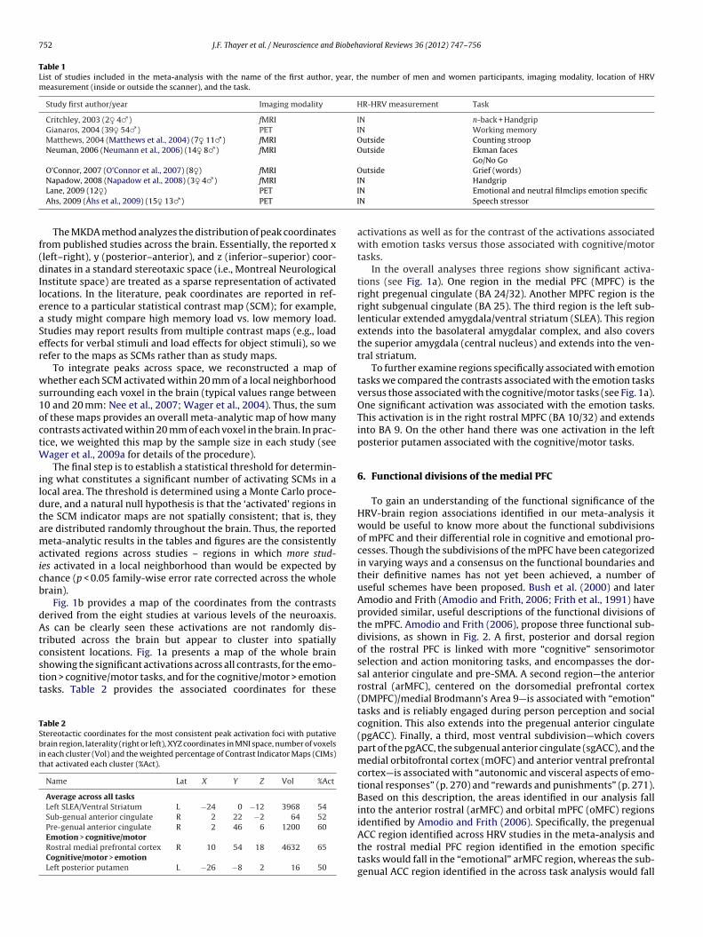
752 J.F. Thayer et al. / Neuroscience and Biobehavioral Reviews 36 (2012) 747–756
Table 1List of studies included in the meta-analysis with the name of the first author, year, the number of men and women participants, imaging modality, location of HRVmeasurement (inside or outside the scanner), and the task.
Study first author/year Imaging modality HR-HRV measurement Task
Critchley, 2003 (2♀ 4♂) fMRI IN n-back + HandgripGianaros, 2004 (39♀ 54♂) PET IN Working memoryMatthews, 2004 (Matthews et al., 2004) (7♀ 11♂) fMRI Outside Counting stroopNeuman, 2006 (Neumann et al., 2006) (14♀ 8♂) fMRI Outside Ekman faces
Go/No GoO’Connor, 2007 (O’Connor et al., 2007) (8♀) fMRI Outside Grief (words)
III
f(dIleaSer
ws1octW
ildtamaicb
dAtcstt
TSbit
Napadow, 2008 (Napadow et al., 2008) (3♀ 4♂) fMRILane, 2009 (12♀) PET
Ahs, 2009 (Åhs et al., 2009) (15♀ 13♂) PET
The MKDA method analyzes the distribution of peak coordinatesrom published studies across the brain. Essentially, the reported xleft–right), y (posterior–anterior), and z (inferior–superior) coor-inates in a standard stereotaxic space (i.e., Montreal Neurological
nstitute space) are treated as a sparse representation of activatedocations. In the literature, peak coordinates are reported in ref-rence to a particular statistical contrast map (SCM); for example,
study might compare high memory load vs. low memory load.tudies may report results from multiple contrast maps (e.g., loadffects for verbal stimuli and load effects for object stimuli), so weefer to the maps as SCMs rather than as study maps.
To integrate peaks across space, we reconstructed a map ofhether each SCM activated within 20 mm of a local neighborhood
urrounding each voxel in the brain (typical values range between0 and 20 mm: Nee et al., 2007; Wager et al., 2004). Thus, the sumf these maps provides an overall meta-analytic map of how manyontrasts activated within 20 mm of each voxel in the brain. In prac-ice, we weighted this map by the sample size in each study (see
ager et al., 2009a for details of the procedure).The final step is to establish a statistical threshold for determin-
ng what constitutes a significant number of activating SCMs in aocal area. The threshold is determined using a Monte Carlo proce-ure, and a natural null hypothesis is that the ‘activated’ regions inhe SCM indicator maps are not spatially consistent; that is, theyre distributed randomly throughout the brain. Thus, the reportedeta-analytic results in the tables and figures are the consistently
ctivated regions across studies – regions in which more stud-es activated in a local neighborhood than would be expected byhance (p < 0.05 family-wise error rate corrected across the wholerain).
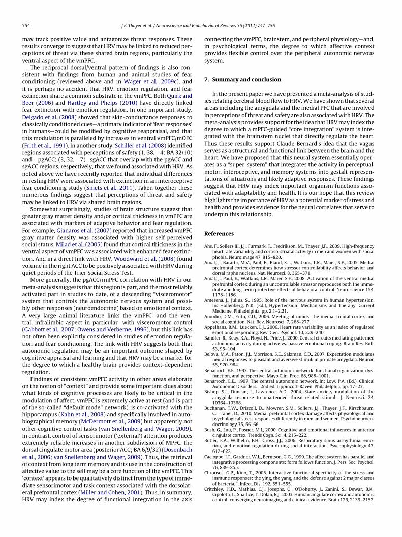
Fig. 1b provides a map of the coordinates from the contrastserived from the eight studies at various levels of the neuroaxis.s can be clearly seen these activations are not randomly dis-
ributed across the brain but appear to cluster into spatiallyonsistent locations. Fig. 1a presents a map of the whole brain
howing the significant activations across all contrasts, for the emo-ion > cognitive/motor tasks, and for the cognitive/motor > emotionasks. Table 2 provides the associated coordinates for theseable 2tereotactic coordinates for the most consistent peak activation foci with putativerain region, laterality (right or left), XYZ coordinates in MNI space, number of voxels
n each cluster (Vol) and the weighted percentage of Contrast Indicator Maps (CIMs)hat activated each cluster (%Act).
Name Lat X Y Z Vol %Act
Average across all tasksLeft SLEA/Ventral Striatum L −24 0 −12 3968 54Sub-genual anterior cingulate R 2 22 −2 64 52Pre-genual anterior cingulate R 2 46 6 1200 60Emotion > cognitive/motorRostral medial prefrontal cortex R 10 54 18 4632 65Cognitive/motor > emotionLeft posterior putamen L −26 −8 2 16 50
N HandgripN Emotional and neutral filmclips emotion specificN Speech stressor
activations as well as for the contrast of the activations associatedwith emotion tasks versus those associated with cognitive/motortasks.
In the overall analyses three regions show significant activa-tions (see Fig. 1a). One region in the medial PFC (MPFC) is theright pregenual cingulate (BA 24/32). Another MPFC region is theright subgenual cingulate (BA 25). The third region is the left sub-lenticular extended amygdala/ventral striatum (SLEA). This regionextends into the basolateral amygdalar complex, and also coversthe superior amygdala (central nucleus) and extends into the ven-tral striatum.
To further examine regions specifically associated with emotiontasks we compared the contrasts associated with the emotion tasksversus those associated with the cognitive/motor tasks (see Fig. 1a).One significant activation was associated with the emotion tasks.This activation is in the right rostral MPFC (BA 10/32) and extendsinto BA 9. On the other hand there was one activation in the leftposterior putamen associated with the cognitive/motor tasks.
6. Functional divisions of the medial PFC
To gain an understanding of the functional significance of theHRV-brain region associations identified in our meta-analysis itwould be useful to know more about the functional subdivisionsof mPFC and their differential role in cognitive and emotional pro-cesses. Though the subdivisions of the mPFC have been categorizedin varying ways and a consensus on the functional boundaries andtheir definitive names has not yet been achieved, a number ofuseful schemes have been proposed. Bush et al. (2000) and laterAmodio and Frith (Amodio and Frith, 2006; Frith et al., 1991) haveprovided similar, useful descriptions of the functional divisions ofthe mPFC. Amodio and Frith (2006), propose three functional sub-divisions, as shown in Fig. 2. A first, posterior and dorsal regionof the rostral PFC is linked with more “cognitive” sensorimotorselection and action monitoring tasks, and encompasses the dor-sal anterior cingulate and pre-SMA. A second region—the anteriorrostral (arMFC), centered on the dorsomedial prefrontal cortex(DMPFC)/medial Brodmann’s Area 9—is associated with “emotion”tasks and is reliably engaged during person perception and socialcognition. This also extends into the pregenual anterior cingulate(pgACC). Finally, a third, most ventral subdivision—which coverspart of the pgACC, the subgenual anterior cingulate (sgACC), and themedial orbitofrontal cortex (mOFC) and anterior ventral prefrontalcortex—is associated with “autonomic and visceral aspects of emo-tional responses” (p. 270) and “rewards and punishments” (p. 271).Based on this description, the areas identified in our analysis fallinto the anterior rostral (arMFC) and orbital mPFC (oMFC) regionsidentified by Amodio and Frith (2006). Specifically, the pregenual
ACC region identified across HRV studies in the meta-analysis andthe rostral medial PFC region identified in the emotion specifictasks would fall in the “emotional” arMFC region, whereas the sub-genual ACC region identified in the across task analysis would fall
J.F. Thayer et al. / Neuroscience and Biobehavioral Reviews 36 (2012) 747–756 753
) map
ilp
tprfpotBptai2is
Fig. 1. (a) map of the whole brain showing significant activations, (b
n the oMFC region. Thus the areas of PFC associated with HRV areocated in regions previously associated with “emotions” and thehysiological aspects of emotional responses.
As discussed above, vmPFC has also been associated with emo-ion regulation and context-dependent reduction of fear behavior,roviding a link between our meta-analytic findings and successfulegulation of negative emotion. For example, Wager et al. (2008b)ound that both vmPFC and dmPFC activity during cognitive reap-raisal of aversive images was associated with greater reductionf negative emotion, as mediated by increases in the ventral stria-um/nucleus accumbens. Given that other studies (for exampleutler et al., 2006) found HRV associated with successful reap-raisal, these findings converge to suggest that HRV may be linkedo successful emotion regulation via these shared brain regions. Innother study of individual differences, van Reekum et al. (2007)
dentified an area associated with psychological well-being (−3,9, −2) that overlaps with the sgACC region associated with HRVn the meta-analysis. Van Reekum et al. (van Reekum et al., 2007)uggested that this region was associated with positive well-being,
Fig. 2. Subdivisions of the me
of the coordinates of the contrasts at various levels of the neuroaxis.
down regulation of the amygdala, and goal-directed positiveaffect.
Similarly, Wager et al. (2009c) identified a region of vmPFCthat was deactivated during social-evaluative threat (SET), com-plementing the increases found with positive reappraisal. Themagnitude of deactivation predicted increased heart rate responsesto the stressor both within and between participants. The effect wasreplicated in a second study (Wager et al., 2009b), which also foundthat vmPFC deactivation predicted threat-induced increases in theperiaqueductal gray, a mediator of physiological and behavioralresponses to threat (Bandler et al., 2000; Davis, 1992). Relatedly,Buchanan et al. (2010) have reported that persons with damage tovmPFC perceived a SET task as more threatening compared to thosewith damage to another region or no brain damage. Interestingly,both of these studies showed more dorsal parts of the MPFC that
showed increases in response to SET (consistent with many previ-ous findings; e.g. (Critchley et al., 2003), suggesting that there maybe valence specificity in the vmPFC: more dorsal regions may medi-ate threat responses, whereas more ventral regions in the mOFCdial prefrontal cortex.
7 iobeh
mrcv
scieBfDcit(rasnifnm
gaFgsvtvq
masbAt(ntactr
owmohboIedeoa‘deH
54 J.F. Thayer et al. / Neuroscience and B
ay track positive value and antagonize threat responses. Theseesults converge to suggest that HRV may be linked to reduced per-eptions of threat via these shared brain regions, particularly theentral aspect of the vmPFC.
The reciprocal dorsal/ventral pattern of findings is also con-istent with findings from human and animal studies of fearonditioning (reviewed above and in Wager et al., 2009c), andt is perhaps no accident that HRV, emotion regulation, and fearxtinction share a common substrate in the vmPFC. Both Quirk andeer (2006) and Hartley and Phelps (2010) have directly linked
ear extinction with emotion regulation. In one important study,elgado et al. (2008) showed that skin-conductance responses tolassically conditioned cues—a primary indicator of ‘fear responses’n humans—could be modified by cognitive reappraisal, and thathis modulation is paralleled by increases in ventral vmPFC/mOFCFrith et al., 1991). In another study, Schiller et al. (2008) identifiedegions associated with perceptions of safety (1, 38, −4: BA 32/10)nd —pgACC; (3, 32, −7)—sgACC that overlap with the pgACC andgACC regions, respectively, that we found associated with HRV. Asoted above we have recently reported that individual differences
n resting HRV were associated with extinction in an interoceptiveear conditioning study (Smets et al., 2011). Taken together theseumerous findings suggest that perceptions of threat and safetyay be linked to HRV via shared brain regions.Somewhat surprisingly, studies of brain structure suggest that
reater gray matter density and/or cortical thickness in vmPFC aressociated with markers of adaptive behavior and fear regulation.or example, Gianaros et al. (2007) reported that increased vmPFCray matter density was associated with higher self-perceivedocial status. Milad et al. (2005) found that cortical thickness in theentral aspect of vmPFC was associated with enhanced fear extinc-ion. And in a direct link with HRV, Woodward et al. (2008) foundolume in the right ACC to be positively associated with HRV duringuiet periods of the Trier Social Stress Test.
More generally, the pgACC/rmPFC correlation with HRV in oureta-analysis suggests that this region is part, and the most reliably
ctivated part in studies to date, of a descending “visceromotor”ystem that controls the autonomic nervous system and possi-ly other responses (neuroendocrine) based on emotional context.
very large animal literature links the vmPFC—and the ven-ral, infralimbic aspect in particular—with visceromotor controlGabbott et al., 2007; Owens and Verberne, 1996), but this link hasot often been explicitly considered in studies of emotion regula-ion and fear conditioning. The link with HRV suggests both thatutonomic regulation may be an important outcome shaped byognitive appraisal and learning and that HRV may be a marker forhe degree to which a healthy brain provides context-dependentegulation.
Findings of consistent vmPFC activity in other areas elaboraten the notion of “context” and provide some important clues abouthat kinds of cognitive processes are likely to be critical in theodulation of affect. vmPFC is extremely active at rest (and is part
f the so-called “default mode” network), is co-activated with theippocampus (Kahn et al., 2008) and specifically involved in auto-iographical memory (McDermott et al., 2009) but apparently notther cognitive control tasks (van Snellenberg and Wager, 2009).n contrast, control of sensorimotor (‘external’) attention producesxtremely reliable increases in another subdivision of MPFC, theorsal cingulate motor area (posterior ACC; BA 6/9/32) (Dosenbacht al., 2006; van Snellenberg and Wager, 2009). Thus, the retrievalf context from long term memory and its use in the construction offfective value to the self may be a core function of the vmPFC. This
context’ appears to be qualitatively distinct from the type of imme-iate sensorimotor and task context associated with the dorsolat-ral prefrontal cortex (Miller and Cohen, 2001). Thus, in summary,RV may index the degree of functional integration in the axis
avioral Reviews 36 (2012) 747–756
connecting the vmPFC, brainstem, and peripheral physiology—and,in psychological terms, the degree to which affective contextprovides flexible control over the peripheral autonomic nervoussystem.
7. Summary and conclusion
In the present paper we have presented a meta-analysis of stud-ies relating cerebral blood flow to HRV. We have shown that severalareas including the amygdala and the medial PFC that are involvedin perceptions of threat and safety are also associated with HRV. Themeta-analysis provides support for the idea that HRV may index thedegree to which a mPFC-guided “core integration” system is inte-grated with the brainstem nuclei that directly regulate the heart.Thus these results support Claude Bernard’s idea that the vagusserves as a structural and functional link between the brain and theheart. We have proposed that this neural system essentially oper-ates as a “super-system” that integrates the activity in perceptual,motor, interoceptive, and memory systems into gestalt represen-tations of situations and likely adaptive responses. These findingssuggest that HRV may index important organism functions asso-ciated with adaptability and health. It is our hope that this reviewhighlights the importance of HRV as a potential marker of stress andhealth and provides evidence for the neural correlates that serve tounderpin this relationship.
References
Åhs, F., Sollers III, J.J., Furmark, T., Fredrikson, M., Thayer, J.F., 2009. High-frequencyheart rate variability and cortico-striatal activity in men and women with socialphobia. Neuroimage 47, 815–820.
Amat, J., Baratta, M.V., Paul, E., Bland, S.T., Watkins, L.R., Maier, S.F., 2005. Medialprefrontal cortex determines how stressor controllability affects behavior anddorsal raphe nucleus. Nat. Neurosci. 8, 365–371.
Amat, J., Paul, E., Watkins, L.R., Maier, S.F., 2008. Activation of the ventral medialprefrontal cortex during an uncontrollable stressor reproduces both the imme-diate and long-term protective effects of behavioral control. Neuroscience 154,1178–1186.
Amerena, J., Julius, S., 1995. Role of the nervous system in human hypertension.In: Hollenberg, N.K. (Ed.), Hypertension: Mechanisms and Therapy. CurrentMedicine, Philadelphia, pp. 2.1–2.21.
Amodio, D.M., Frith, C.D., 2006. Meeting of minds: the medial frontal cortex andsocial cognition. Nat. Rev. Neurosci. 7, 268–277.
Appelhans, B.M., Luecken, L.J., 2006. Heart rate variability as an index of regulatedemotional responding. Rev. Gen. Psychol. 10, 229–240.
Bandler, R., Keay, K.A., Floyd, N., Price, J., 2000. Central circuits mediating patternedautonomic activity during active vs. passive emotional coping. Brain Res. Bull.53, 95–104.
Belova, M.A., Paton, J.J., Morrison, S.E., Salzman, C.D., 2007. Expectation modulatesneural responses to pleasant and aversive stimuli in primate amygdala. Neuron55, 970–984.
Benarroch, E.E., 1993. The central autonomic network: functional organization, dys-function, and perspective. Mayo Clin. Proc. 68, 988–1001.
Benarroch, E.E., 1997. The central autonomic network. In: Low, P.A. (Ed.), ClinicalAutonomic Disorders. , 2nd ed. Lippincott-Raven, Philadelphia, pp. 17–23.
Bishop, S.J., Duncan, J., Lawrence, A.D., 2004. State anxiety modulation of theamygdala response to unattended threat-related stimuli. J. Neurosci. 24,10364–10368.
Buchanan, T.W., Driscoll, D., Mowrer, S.M., Sollers, J.J., Thayer, J.F., Kirschbaum,C., Tranel, D., 2010. Medial prefrontal cortex damage affects physiological andpsychological stress responses differently in men and women. Psychoneuroen-docrinology 35, 56–66.
Bush, G., Luu, P., Posner, M.I., 2000. Cognitive and emotional influences in anteriorcingulate cortex. Trends Cogn. Sci. 4, 215–222.
Butler, E.A., Wilhelm, F.H., Gross, J.J., 2006. Respiratory sinus arrhythmia, emo-tion, and emotion regulation during social interaction. Psychophysiology 43,612–622.
Cacioppo, J.T., Gardner, W.L., Berntson, G.G., 1999. The affect system has parallel andintegrative processing components: form follows function. J. Pers. Soc. Psychol.76, 839–855.
Chrousos, G.P., Kino, T., 2005. Interactive functional specificity of the stress and
immune responses: the ying, the yang, and the defense against 2 major classesof bacteria. J. Infect. Dis. 192, 551–555.Critchley, H.D., Mathias, C.J., Josephs, O., O’Doherty, J., Zanini, S., Dewar, B.K.,Cipolotti, L., Shallice, T., Dolan, R.J., 2003. Human cingulate cortex and autonomiccontrol: converging neuroimaging and clinical evidence. Brain 126, 2139–2152.

iobeh
C
D
D
D
D
E
E
FF
G
G
H
H
H
H
I
J
J
J
K
K
K
K
L
L
L
L
M
M
M
M
M
M
J.F. Thayer et al. / Neuroscience and B
unningham, W.A., Van Bavel, J.J., Johnsen, I.R., 2008. Affective flexibility: evaluativeprocessing goals shape amygdala activity. Psychol. Sci.: J. Am. Psychol. Soc./APS19, 152–160.
avidson, R.J., 2000. Cognitive neuroscience needs affective neuroscience (and viceversa). Brain Cogn. 42, 89–92.
avis, M., 1992. The role of the amygdala in fear and anxiety. Annu. Rev. Neurosci.15, 353–375.
elgado, M.R., Nearing, K.I., Ledoux, J.E., Phelps, E.A., 2008. Neural circuitry underly-ing the regulation of conditioned fear and its relation to extinction. Neuron 59,829–838.
osenbach, N.U., Visscher, K.M., Palmer, E.D., Miezin, F.M., Wenger, K.K., Kang, H.C.,Burgund, E.D., Grimes, A.L., Schlaggar, B.L., Petersen, S.E., 2006. A core systemfor the implementation of task sets. Neuron 50, 799–812.
ippert, F., Veit, R., Weiskopf, N., Erb, M., Birbaumer, N., Anders, S., 2007. Regulationof emotional responses elicited by threat-related stimuli. Hum. Brain Mapp. 28,409–423.
tkin, A., Wager, T.D., 2007. Functional neuroimaging of anxiety: a meta-analysis ofemotional processing in PTSD, Social anxiety disorder, and specific phobia. Am.J. Psychiatry 164, 1476–1488.
rijda, N.H., 1986. The Emotions. Ed. de la Maison des Sciences de l’Homme.rith, C.D., Friston, K.J., Liddle, P.F., Frackowiak, R.S., 1991. A PET study of word
finding. Neuropsychologia 29, 1137–1148.abbott, P.L., Warner, T., Busby, S.J., 2007. Catecholaminergic neurons in medullary
nuclei are among the post-synaptic targets of descending projections frominfralimbic area 25 of the rat medial prefrontal cortex. Neuroscience 144,623–635.
ianaros, P.J., Horenstein, J.A., Cohen, S., Matthews, K.A., Brown, S.M., Flory, J.D.,Critchley, H.D., Manuck, S.B., Hariri, A.R., 2007. Perigenual anterior cingulatemorphology covaries with perceived social standing. Soc. Cogn. Affect. Neurosci.2, 161–173.
are, T.A., Camerer, C.F., Knoepfle, D.T., Rangel, A., 2010. Value computations inventral medial prefrontal cortex during charitable decision making incorporateinput from regions involved in social cognition. J. Neurosci. 30, 583–590.
artley, C.A., Phelps, E.A., 2010. Changing fear. The neurocircuitry of emotion regu-lation. Neuropsychopharmacology 35, 136–146.
erry, C., Bach, D.R., Esposito, F., Di Salle, F., Perrig, W.J., Scheffler, K., Luthi, A.,Seifritz, E., 2007. Processing of temporal unpredictability in human and animalamygdala. J. Neurosci. 27, 5958–5966.
olland, P.C., Gallagher, M., 2004. Amygdala–frontal interactions and rewardexpectancy. Curr. Opin. Neurobiol. 14, 148–155.
ngjaldsson, J.T., Laberg, J.C., Thayer, J.F., 2003. Reduced heart rate variability inchronic alcohol abuse: relationship with negative mood, chronic thought sup-pression, and compulsive drinking. Biol. Psychiatry 54, 1427–1436.
ackson, J.H., 1884. The Croonian Lectures on evolution and dissolution of the ner-vous system. Br. Med. J., 703–707.
ohnson, A.W., Gallagher, M., Holland, P.C., 2009. The basolateral amygdala is criticalto the expression of Pavlovian and instrumental outcome-specific reinforcerdevaluation effects. J. Neurosci. 29, 696.
ose, A.D., Collison, D., 1970. The normal range and determinants of the intrinsicheart rate in man. Cardiovasc. Res. 4, 160–167.
ahn, I., Andrews-Hanna, J.R., Vincent, J.L., Snyder, A.Z., Buckner, R.L., 2008. Distinctcortical anatomy linked to subregions of the medial temporal lobe revealed byintrinsic functional connectivity. J. Neurophysiol. 100, 129–139.
eay, K.A., Bandler, R., 2001. Parallel circuits mediating distinct emotionalcoping reactions to different types of stress. Neurosci. Biobehav. Rev. 25,669–678.
elley, W.M., Macrae, C.N., Wyland, C.L., Caglar, S., Inati, S., Heatherton, T.F., 2002.Finding the self? An event-related fMRI study. J. Cogn. Neurosci. 14, 785–794.
ober, H., Barrett, L.F., Joseph, J., Bliss-Moreau, E., Lindquist, K., Wager, T.D., 2008.Functional grouping and cortical–subcortical interactions in emotion: a meta-analysis of neuroimaging studies. Neuroimage 42, 998–1031.
ane, R.D., McRae, K., Reiman, E.M., Chen, K., Ahern, G.L., Thayer, J.F., 2009.Neural correlates of heart rate variability during emotion. Neuroimage 44,213–222.
ane, R.D., Wager, T.D., 2009. The new field of brain–body medicine: what have welearned and where are we headed? Neuroimage 47, 1135–1140.
eDoux, J., 1996. Emotional networks and motor control: a fearful view. Prog. BrainRes. 107, 437–446.
issek, S., Powers, A.S., McClure, E.B., Phelps, E.A., Woldehawariat, G., Grillon, C., Pine,D.S., 2005. Classical fear conditioning in the anxiety disorders: a meta-analysis.Behav. Res. Ther. 43, 1391–1424.
aier, S.F., Amat, J., Baratta, M.V., Paul, E., Watkins, L.R., 2006. Behavioral control, themedial prefrontal cortex, and resilience. Dialogues Clin. Neurosci. 8, 397–406.
atthews, S.C., Paulus, M.P., Simmons, A.N., Nelesen, R.A., Dimsdale, J.E., 2004. Func-tional subdivisions within anterior cingulate cortex and their relationship toautonomic nervous system function. Neuroimage 22, 1151–1156.
cClure, S.M., Li, J., Tomlin, D., Cypert, K.S., Montague, L.M., Montague, P.R., 2004.Neural correlates of behavioral preference for culturally familiar drinks. Neuron44, 379–387.
cDermott, K.B., Szpunar, K.K., Christ, S.E., 2009. Laboratory-based and auto-biographical retrieval tasks differ substantially in their neural substrates.
Neuropsychologia 47, 2290–2298.cEwen, B.S., 1998. Stress, adaptation, and disease. Allostasis and allostatic load.Ann. N. Y. Acad. Sci. 840, 33–44.
cEwen, B.S., 2001. From molecules to mind. Stress, individual differences, and thesocial environment. Ann. N. Y. Acad. Sci. 935, 42–49.
avioral Reviews 36 (2012) 747–756 755
McEwen, B.S., Sapolsky, R.M., 1995. Stress and cognitive function. Curr. Opin. Neu-robiol. 5, 205–216.
Megginson, L.C., 1963. Lessons from Europe for American business. Soc. Sci. Q 44,3–13.
Melzig, C.A., Weike, A.I., Hamm, A.O., Thayer, J.F., 2009. Individual differences in fear-potentiated startle as a function of resting heart rate variability: implicationsfor panic disorder. Int. J. Psychophysiol.: Offic. J. Int. Organ. Psychophysiol. 71,109–117.
Milad, M.R., Quinn, B.T., Pitman, R.K., Orr, S.P., Fischl, B., Rauch, S.L., 2005. Thickness ofventromedial prefrontal cortex in humans is correlated with extinction memory.Proc. Natl. Acad. Sci. U.S.A. 102, 10706–10711.
Milad, M.R., Quirk, G.J., 2002. Neurons in medial prefrontal cortex signal memoryfor fear extinction. Nature 420, 70–74.
Milad, M.R., Vidal-Gonzalez, I., Quirk, G.J., 2004. Electrical stimulation of medial pre-frontal cortex reduces conditioned fear in a temporally specific manner. Behav.Neurosci. 118, 389–394.
Milad, M.R., Wright, C.I., Orr, S.P., Pitman, R.K., Quirk, G.J., Rauch, S.L., 2007. Recallof fear extinction in humans activates the ventromedial prefrontal cortex andhippocampus in concert. Biol. Psychiatry 62, 446–454.
Miller, E.K., Cohen, J.D., 2001. An integrative theory of prefrontal cortex function.Annu. Rev. Neurosci. 24, 167–202.
Napadow, V., Dhond, R., Conti, G., Makris, N., Brown, E.N., Barbieri, R., 2008. Braincorrelates of autonomic modulation: combining heart rate variability with fMRI.Neuroimage 42, 169–177.
Nee, D.E., Wager, T.D., Jonides, J., 2007. Interference resolution: insights from a meta-analysis of neuroimaging tasks. Cogn. Affect. Behav. Neurosci. 7, 1–17.
Neumann, S.A., Brown, S.M., Ferrell, R.E., Flory, J.D., Manuck, S.B., Hariri, A.R.,2006. Human choline transporter gene variation is associated with corti-colimbic reactivity and autonomic–cholinergic function. Biol. Psychiatry 60,1155–1162.
Northoff, G., Heinzel, A., de Greck, M., Bermpohl, F., Dobrowolny, H., Panksepp, J.,2006. Self-referential processing in our brain—a meta-analysis of imaging stud-ies on the self. Neuroimage 31, 440–457.
Nugent, A.C., Bain, E.E., Thayer, J.F., Sollers III, J.J., Drevets, W.C., 2011. Sex differ-ences in the neural correlates of autonomic arousal: a pilot PET study. Int. J.Psychophysiol. 80, 182–191.
O’Connor, M.-F., Gündel, H., McRae, K., Lane, R.D., 2007. Baseline vagal tone predictsBOLD response during elicitation of grief. Neuropsychopharmacol.: Offic. Publ.Am. Coll. Neuropsychopharmacol. 32, 2184–2189.
Owens, N.C., Verberne, A.J., 1996. An electrophysiological study of the medial pre-frontal cortical projection to the nucleus of the solitary tract in rat. Exp. BrainRes. 110, 55–61.
Paton, J.J., Belova, M.A., Morrison, S.E., Salzman, C.D., 2006. The primate amygdalarepresents the positive and negative value of visual stimuli during learning.Nature 439, 865–870.
Plassmann, H., O’Doherty, J., Shiv, B., Rangel, A., 2008. Marketing actions can mod-ulate neural representations of experienced pleasantness. Proc. Natl. Acad. Sci.U.S.A. 105, 1050.
Quirk, G.J., Beer, J.S., 2006. Prefrontal involvement in the regulation of emotion:convergence of rat and human studies. Curr. Opin. Neurobiol. 16, 723–727.
Ruiz-Padial, E., Sollers, J.J., Vila, J., Thayer, J.F., 2003. The rhythm of the heart in theblink of an eye: emotion-modulated startle magnitude covaries with heart ratevariability. Psychophysiology 40, 306–313.
Ruiz-Padial, E., Vila, J., Thayer, J.F., 2011. The effect of conscious and non-consciouspresentation of biologically relevant emotion pictures on emotion modulatedstartle and phasic heart rate. Int. J. Psychophysiol. 79, 341–346.
Saper, C.B., 2002. The central autonomic nervous system: conscious visceralperception and autonomic pattern generation. Annu. Rev. Neurosci. 25,433–469.
Sapolsky, R.M., 1996. Why stress is bad for your brain. Science 273, 749–750.Schiller, D., Levy, I., Niv, Y., LeDoux, J.E., Phelps, E.A., 2008. From fear to safety and
back: reversal of fear in the human brain. J. Neurosci. 28, 11517–11525.Saul, P.J., 1990. Beat-to beat variations of heart rate reflect modulation of cardiac
autonomic outflow. News Physiol. Sci. 5, 32–37.Schoenbaum, G., Roesch, M.R., Stalnaker, T.A., Takahashi, Y.K., 2009. A new per-
spective on the role of the orbitofrontal cortex in adaptive behaviour. Nat. Rev.Neurosci. 10, 885–892.
Seeman, T.E., McEwen, B.S., Rowe, J.W., Singer, B.H., 2001. Allostatic load as a markerof cumulative biological risk: MacArthur studies of successful aging. Proc. Natl.Acad. Sci. U.S.A. 98, 4770–4775.
Shook, N., Pena, P., Fazio, R.H., Sollers, J.J., Thayer, J.F., 2007a. Friend or foe: heart ratevarability and the negativity bias in learning about novel objects. Psychophysi-ology 44, S39.
Shook, N.J., Fazio, R.H., Vasey, M.W., 2007b. Negativity bias in attitude learning: apossible indicator of vulnerability to emotional disorders? J. Behav. Ther. Exp.Psychiatry 38, 144–155.
Smets, E., Pappens, M., Thayer, J.F., van den Bergh, O., van Diest, I., 2011. Interindi-vidual differences in inhibitory control predict extinction of interoceptive fear.Psychophysiology 4, 8.
Smith, T.W., Cribbet, M.R., Nealey-Moore, J.B., Uchino, B.N., Williams, P.G., Macken-zie, J., Thayer, J.F., 2011. Matters of the variable heart: Respiratory sinus
arrhythmia response to marital interaction and associations with marital qual-ity. Journal of Personality and Social Psychology 100, 103–119.Summerfield, C., Egner, T., Greene, M., Koechlin, E., Mangels, J., Hirsch, J., 2006.Predictive codes for forthcoming perception in the frontal cortex. Science 314,1311.

7 iobeh
T
T
T
T
T
T
T
T
T
T
T
T
T
T
U
W
to masked fearful eye whites. Science 306, 2061.Whalen, P.J., Phelps, E.A., 2009. The Human Amygdala. The Guilford Press.
56 J.F. Thayer et al. / Neuroscience and B
hayer, J.F., 2006. On the importance of inhibition: central and peripheral manifesta-tions of nonlinear inhibitory processes in neural systems. Dose–Response: Publ.Int. Hormesis Soc. 4, 2–21.
hayer, J.F., Brosschot, J.F., 2005. Psychosomatics and psychopathology: looking upand down from the brain. Psychoneuroendocrinology 30, 1050–1058.
hayer, J.F., Friedman, B.H., 2002. Stop that! Inhibition, sensitization, and their neu-rovisceral concomitants. Scand. J. Psychol. 43, 123–130.
hayer, J.F., Lane, R.D., 2000. A model of neurovisceral integration in emotion regu-lation and dysregulation. J. Affect. Disord. 61, 201–216.
hayer, J.F., Lane, R.D., 2007. The role of vagal function in the risk for cardiovasculardisease and mortality. Biol. Psychol. 74, 224–242.
hayer, J.F., Lane, R.D., 2009. Claude Bernard and the heart–brain connection: furtherelaboration of a model of neurovisceral integration. Neurosci. Biobehav. Rev. 33,81–88.
hayer, J.F., Loerbroks, A., Sternberg, E.M., 2011. Inflammation and cardiorespiratorycontrol: The role of the vagus nerve. Respir. Physiol. Neurobiol. 178, 387–394.
hayer, J.F., Sollers, J.J., Labiner, D.M., Weinand, M., Herring, A.M., Lane, R.D., Ahern,G.L., 2009. Age related differences in prefrontal control of heart rate in humans:A pharmacological blockade study. Int. J. Psychophysiol. 72, 81–88.
hayer, J.F., Sternberg, E.M., 2009. Neural concomitants of immunity. Focus on thevagus nerve. Neuroimage 47, 908–910.
hayer, J.F., Siegle, G.J., 2002. Neurovisceral integration in cardiac and emotionalregulation. IEEE Eng. Med. Biol. Mag.: Quart. Mag. Eng. Med. Biol. Soc. 21, 24–29.
hayer, J.F., Sternberg, E., 2006. Beyond heart rate variability: vagal regulation ofallostatic systems. Ann. N. Y. Acad. Sci. 1088, 361–372.
hayer, J.F., Hansen, A.L., Johnsen, B.H., 2010a. The non-invasive assessment of auto-nomic influences on the heart using impedance cardiography and heart ratevariability. In: Steptoe, A. (Ed.), Handbook of Behavioral Medicine: Methods andApplications. Springer, New York.
hayer, J.F., Yamamoto, S.S., Brosschot, J.F., 2010b. The relationship of autonomicimbalance, heart rate variability and cardiovascular disease risk factors. Int. J.Cardiol. 141, 122–131.
hayer, J.F., Fischer, J.E. Heart rate variability, overnight urinary norepinephrine,and plasma cholesterol in apparently healthy human adults, Int. J. Cardiol.doi:10.1016/j.ijcard.2011.05.058, in press.
rry, H.L., van Reekum, C.M., Johnstone, T., Kalin, N.H., Thurow, M.E., Schaefer, H.S.,Jackson, C.A., Frye, C.J., Greischar, L.L., Alexander, A.L., Davidson, R.J., 2006. Amyg-dala and ventromedial prefrontal cortex are inversely coupled during regulationof negative affect and predict the diurnal pattern of cortisol secretion among
older adults. J. Neurosci. 26, 4415–4425.ager, T.D., Barrett, L.F., Bliss-Moreau, E., Lindquist, K., Duncan, S., Kober, H., Joseph,J., Davidson, M., Mize, J., 2008a. The neuroimaging of emotion. In: Lewis, M.,Haviland-Jones, J.M., Barrett, L.F. (Eds.), Handbook of Emotions. , 3rd ed. GuilfordPress, New York, pp. 249–271.
avioral Reviews 36 (2012) 747–756
Wager, T.D., Davidson, M.L., Hughes, B.L., Lindquist, M.A., Ochsner, K.N., 2008b.Prefrontal–subcortical pathways mediating successful emotion regulation. Neu-ron 59, 1037–1050.
Wager, T.D., Hughes, B., Davidson, M., Lindquist, M.L., Ochsner, K.N., 2008c.Prefrontal–subcortical pathways mediating successful emotion regulation. Neu-ron 59, 1037–1050.
Wager, T.D., Jonides, J., Reading, S., 2004. Neuroimaging studies of shifting attention:a meta-analysis. Neuroimage 22, 1679–1693.
Wager, T.D., Lindquist, M., Kaplan, L., 2007. Meta-analysis of functional neu-roimaging data: current and future directions. Soc. Cogn. Affect. Neurosci. 2,150–158.
Wager, T.D., Lindquist, M.A., Nichols, T.E., Kober, H., Van Snellenberg, J.X., 2009a.Evaluating the consistency and specificity of neuroimaging data using meta-analysis. Neuroimage 45, S210-221.
Wager, T.D., van Ast, V.A., Hughes, B.L., Davidson, M.L., Lindquist, M.A., Ochsner,K.N., 2009b. Brain mediators of cardiovascular responses to social threat. PartII: prefrontal–subcortical pathways and relationship with anxiety. Neuroimage47, 836–851.
Wager, T.D., Waugh, C.E., Lindquist, M., Noll, D.C., Fredrickson, B.L., Taylor, S.F., 2009c.Brain mediators of cardiovascular responses to social threat. Part I: reciprocaldorsal and ventral sub-regions of the medial prefrontal cortex and heart-ratereactivity. Neuroimage 47, 821–835.
van Reekum, C.M., Urry, H.L., Johnstone, T., Thurow, M.E., Frye, C.J., Jackson,C.A., Schaefer, H.S., Alexander, A.L., Davidson, R.J., 2007. Individual differ-ences in amygdala and ventromedial prefrontal cortex activity are associatedwith evaluation speed and psychological well-being. J. Cogn. Neurosci. 19,237–248.
van Snellenberg, J.X., Wager, T.D., 2009. Cognitive and motivational functions ofthe human prefrontal cortex. In: Christensen, A., Goldberg, E., Bougakov, D.(Eds.), Luria’s Legacy in the 21st Century. Oxford University Press, Oxford, pp.30–61.
Weber, C.S., Thayer, J.F., Rudat, M., Wirtz, P.H., Zimmermann-Viehoff, F., Thomas,A., Perschel, F.H., Arck, P.C., Deter, H.C., 2010. Low vagal tone is associated withimpaired post stress recovery of cardiovascular, endocrine, and immune mark-ers. Eur. J. Appl. Physiol. 109, 201–211.
Whalen, P.J., Kagan, J., Cook, R.G., Davis, F.C., Kim, H., Polis, S., McLaren, D.G.,Somerville, L.H., McLean, A.A., Maxwell, J.S., 2004. Human amygdala responsivity
Woodward, S.H., Kaloupek, D.G., Schaer, M., Martinez, C., Eliez, S., 2008. Rightanterior cingulate cortical volume covaries with respiratory sinus arrhythmiamagnitude in combat veterans. J. Rehabil. Res. Dev. 45, 451–463.